G-protein-coupled receptor kinase activity is
increased in hypertension.
R Gros, … , C M Tan, R D Feldman
J Clin Invest.
1997;
99(9)
:2087-2093.
https://doi.org/10.1172/JCI119381
.
Impaired vascular beta-adrenergic responsiveness may play an important role in the
development and/or maintenance of hypertension. This defect has been associated with an
alteration in receptor/guanine nucleotide regulatory protein (G-protein) interactions.
However, the locus of this defect remains unclear. G-Protein-coupled receptor kinases
(GRKs) phosphorylate serine/threonine residues on G-protein-linked receptors in an
agonist-dependent manner. GRK activation mediates reduced receptor responsiveness and
impaired receptor/G-protein coupling. To determine whether the impairment in
beta-adrenergic response in human hypertension might be associated with altered GRK activity,
we studied lymphocytes from younger hypertensive subjects as compared with older and
younger normotensive subjects. We assessed GRK activity by rhodopsin phosphorylation
and GRK expression by immunoblot. GRK activity was significantly increased in
lymphocytes from younger hypertensive subjects and paralleled an increase in GRK-2 (beta
ARK-1) protein expression. In contrast, no alterations in cAMP-dependent kinase (A-kinase)
activity or GRK-5/6 expression were noted. GRK activity was not increased in lymphocytes
from older normotensive subjects who demonstrated a similar impairment in
beta-adrenergic-mediated adenylyl cyclase activation. These studies indicate that GRK activity is
selectively increased in lymphocytes from hypertensive subjects. The increase in GRK
activity may underlie the reduction in beta-adrenergic responsiveness characteristic of the
hypertensive state.
Research Article
Find the latest version:
J. Clin. Invest.
© The American Society for Clinical Investigation, Inc. 0021-9738/97/05/2087/07 $2.00
Volume 99, Number 9, May 1997, 2087–2093
Rapid Publication
G-Protein–coupled Receptor Kinase Activity Is Increased in Hypertension
Robert Gros,* Jeffrey L. Benovic,‡ Christopher M. Tan,* and Ross D. Feldman*
*Department of Medicine, Department of Physiology, and Department of Pharmacology and Toxicology, University of Western Ontario, London, Ontario, Canada; and‡Department of Biochemistry and Molecular Pharmacology, Kimmel Cancer Institute, Thomas Jefferson
University, Philadelphia, Pennsylvania 19107-5541
Abstract
Impaired vascular b-adrenergic responsiveness may play an important role in the development and/or maintenance of hypertension. This defect has been associated with an alter-ation in receptor/guanine nucleotide regulatory protein (G-protein) interactions. However, the locus of this defect re-mains unclear. G-Protein–coupled receptor kinases (GRKs) phosphorylate serine/threonine residues on G-protein–linked receptors in an agonist-dependent manner. GRK activation mediates reduced receptor responsiveness and impaired re-ceptor/G-protein coupling. To determine whether the im-pairment in b-adrenergic response in human hypertension might be associated with altered GRK activity, we studied lymphocytes from younger hypertensive subjects as com-pared with older and younger normotensive subjects. We as-sessed GRK activity by rhodopsin phosphorylation and GRK expression by immunoblot. GRK activity was significantly increased in lymphocytes from younger hypertensive sub-jects and paralleled an increase in GRK-2 (bARK-1) pro-tein expression. In contrast, no alterations in cAMP-depen-dent kinase (A-kinase) activity or GRK-5/6 expression were noted. GRK activity was not increased in lymphocytes from older normotensive subjects who demonstrated a similar impairment in b-adrenergic–mediated adenylyl cyclase ac-tivation.
These studies indicate that GRK activity is selectively creased in lymphocytes from hypertensive subjects. The in-crease in GRK activity may underlie the reduction in b -adren-ergic responsiveness characteristic of the hypertensive state. (J. Clin. Invest. 1997. 99:2087–2093.) Key words:
adreno-ceptors • aging • cyclic AMP–dependent protein kinase •
adenylyl cyclase
Introduction
The increase in vascular resistance characteristic of hyperten-sion is mediated by alterations in both structural (hypertrophy/ hyperplasia) and functional determinants (e.g., vascular
signal-ing mechanisms). We and others have suggested that the func-tional defect leading to increased vascular resistance reflects an imbalance between vasoconstrictor and vasodilator mecha-nisms. We have focused on the hypothesis that an impairment in receptor-mediated vasodilation may contribute.
An impairment in b-adrenergic–mediated vasodilation has been demonstrated both in human hypertension (1) (primarily in younger, Caucasian borderline hypertensive subjects) and in animal models of hypertension (2, 3). Further, the defect has been shown to be selective and reversible (1, 2). In animal models, it has been shown that the impairment in b -adrener-gic–mediated vasodilation parallels a decrease in vascular
b-adrenergic–stimulated adenylyl cyclase activity (3). Impair-ment in human b-adrenergic responsiveness as assessed by impaired adenylyl cyclase activity has also been demonstrated in human hypertension (4) (although this finding has not been universal and may depend on other factors including age [5], gender [6], race [7], and sodium intake [1, 2]). These studies have used the human lymphocyte as a model for human vascu-lar b-adrenergic receptor complex. This has been based on the demonstration by ourselves and others that regulation of the lymphocyte b-adrenergic receptor complex parallels the regu-lation of functional vascular b-adrenergic responsiveness (2, 5, 8).
Activation of the b-adrenergic receptor transmembrane signaling mechanisms requires the interaction of three primary protein components, the receptor, adenylyl cyclase enzyme, and the guanine nucleotide regulatory protein (G-protein)1
complex linking receptor activation with adenylyl cyclase acti-vation. Studies by ourselves and others have suggested that the impairment in lymphocyte b-adrenergic responsiveness in younger Caucasian male borderline/mild hypertensive subjects is due to an alteration in receptor/G-protein coupling (2, 4, 9). The mechanism underlying this defect remains unclear.
The efficiency with which G-protein–linked receptors (which include b-adrenoceptors) interact with G-proteins is depen-dent, in part, on the phosphorylation state of the receptor. Increased serine/threonine receptor phosphorylation is asso-ciated with reduced b-adrenergic responsiveness (10–12).
b-adrenoceptor phosphorylation is mediated by cyclic AMP– dependent protein kinase (A-kinase) activity (10–12) and by members of the G-protein–coupled receptor kinase (GRK) family which include the b-adrenergic receptor kinases (bARK) (10–12).
Alterations in GRK activity linked to impaired receptor/
Address correspondence to Dr. Ross D. Feldman, Room 6L13B, London Health Sciences Center, University Campus, 339 Winder-mere Rd., P.O. Box 5339, London, Ontario, Canada N6A 5A5. Phone: 519-663-3929; FAX: 519-663-3232; E-mail: [email protected]
Received for publication 22 October 1996 and accepted in revised form 13 March 1997.
G-protein coupling and reduced adenylyl cyclase activation have been described in GRK overexpression systems (13, 14). Additionally, increased GRK expression and activity have been demonstrated in both human and animal models of con-gestive heart failure and have been linked with the reduced myocardial b-adrenergic responsiveness characteristic of that disease (15, 16). Whether alterations in GRK activity may have a regulatory role in other human diseases is unknown.
Based on these uncertainties, we have assessed GRK activ-ity and expression in human lymphocytes from hypertensive subjects under conditions where we have previously docu-mented reduced lymphocyte b-adrenergic adenylyl cyclase ac-tivation. We demonstrate that hypertensive subjects demon-strate a significant enhancement in both GRK activity and GRK-2 (bARK-1) expression.
Methods
Subject protocol. Younger (n5 16) and older (n5 8) normotensive subjects and younger borderline/mildly hypertensive (n5 8) subjects were studied. All subjects studied were male Caucasians. Younger normotensive subjects were between the ages of 19 and 38 yr, healthy, and not taking any medications on a regular basis or any medications for at least 1 mo before the study. Older normotensive subjects were between the ages of 47 and 70 yr, healthy, and not taking any medica-tions on a regular basis or any medicamedica-tions for at least 1 mo before the study. Hypertensive subjects were between the ages of 20 and 36 yr, otherwise healthy, and had neither renal nor cardiovascular compli-cations. Hypertensive subjects had not taken any antihypertensive medications (or any other medications) for at least 1 mo before the study. The criteria for classifying blood pressure status of subjects were as described previously (4). The borderline or mildly hyperten-sive subjects had pressures . 140/90 mmHg on at least 20% of day-time automatic ambulatory blood pressure readings (model 90207; Spacelabs, Redmond, WA). All subjects were instructed to maintain a high sodium intake for 3 d before study. Compliance with dietary instruction was assessed by urinary sodium determinations based on overnight urine collections. The protocol was approved by the Hu-man Subjects Review Committee, University of Western Ontario.
On the morning of the study subjects were admitted to the Vascu-lar Clinical Investigation Unit, London Health Sciences Center, Uni-versity of Western Ontario and maintained in a supine position. An intravenous catheter was inserted in an antecubital vein and 20 min later a 120-ml blood sample was obtained.
Sample preparation. Mononuclear leukocytes were separated from EDTA anticoagulated whole blood by the method of Böyum (17) and as described previously (18).
For the preparation of cytosolic fractions, mononuclear leuko-cytes were resuspended in ice-cold lysis buffer (20 mM Tris-HCl, 2 mM EDTA, pH 7.5, at room temperature, 100 mg/ml PMSF, 10 mg/ml benzamidine, and 5 mg/ml pepstatin A). Cells were subjected to nitro-gen cavitation (Parr Bomb, 600 psi, 15 min at 48C), followed by cen-trifugation at 45,000 g for 30 min at 48C. The supernatants were col-lected and frozen in liquid nitrogen. Additionally, whole cell samples were pelleted and frozen at 2808C.
For the preparation of permeable cells for assessment of adenylyl cyclase activity, cells were washed in Hanks’ balanced salt solution with 33 mmol/liter Hepes, 0.5 mmol/liter EDTA, and 1 mmol/liter mag-nesium sulfate (pH 7.4 at 48C, buffer A). Cells were permeabilized as previously described (4) with the addition of digitonin (10 mg/ml) in buffer A and incubated for 15 min at 48C. Cells were centrifuged at 400 g for 10 min, resuspended in buffer A, washed as above, and re-suspended at a concentration of 2 3 107 cells/ml in buffer A for assay
of adenylyl cyclase activity.
Assessment of G-protein–coupled receptor kinase activity. GRK enzymatic activity was assessed via determination of the extent of
light-dependent phosphorylation of rhodopsin according to our pre-viously published methods (19). Briefly, rod outer segment mem-branes were prepared from dark-adapted bovine retinas (Rockville Meat Company, Rockville, MD), via stepwise sucrose gradient centri-fugation, and then treated with 5 M urea to inactivate endogenous ki-nase activity. The resulting preparation contains z 95% rhodopsin (19).
To assess the extent of GRK-dependent phosphorylation, 20 mg of lymphocyte cytosolic protein was incubated with 5 mg of the rod outer segment preparation in a buffer containing 20 mM Tris-HCl, pH 7.5 (at room temperature), 2 mM EDTA, 5 mM MgCl2, 130 mM
Na3VO4, 80 mM ATP with [g-32P]ATP (20–50 mCi) in a final reaction
volume of 55 ml. The reactions were carried out at 308C for 30 min in the presence or absence of light. The reactions were terminated by the addition of 10 vol of an ice-cold solution of 100 mM sodium phos-phate, 5 mM EDTA buffer, pH 7, followed by centrifugation at 13,000 g for 3 min (at room temperature). The pellets were solubi-lized in 80 ml of a solution consisting of 125 mM Tris-HCl, pH 6.8, 20% glycerol, 40 g/liter SDS, 100 g/liter 2-mercaptoethanol, and 0.25 g/liter bromophenol blue and sonicated for 15 min. Proteins were sep-arated using SDS-PAGE. A 4% (wt/vol) polyacrylamide stacking gel with a 12% running gel was used for all studies. Gels were stained with 1 g/liter Coomassie brilliant blue R-250, 50% methanol, 10% acetic acid for 30–50 min, followed by destaining in 50% methanol, 10% acetic acid for 4–5 h. The excised portion of gel containing the rhodopsin band (z 38 kD) was quantitated via liquid scintillation
spectroscopy. The extent of GRK-mediated phosphorylation was de-termined as the difference between light- and dark-dependent phos-phorylation, each determined in triplicate.
Assessment of A-kinase activity. A-kinase activity was determined in cytosolic lymphocyte preparations. A-kinase activity was assessed by 32P-phosphorylation of a synthetic substrate (Kemptide; Sigma
Chemical Co., St. Louis, MO) according to our previously published techniques (18). Briefly, 20 mg of cytosolic lymphocyte preparation was incubated for 20 min at 308C with 0.3 mM Kemptide, 1 mg/ml bo-vine serum albumin, 0.5 mM ascorbic acid, 6 mM MgCl2, 140 mM
ATP, and 1–2 mCi [g-32P]ATP in the presence or absence of 100 mM
cAMP. Phosphorylated substrate was separated by adherence to phosphocellulose paper (Whatman P81 chromatography paper). Af-ter five washes in phosphoric acid (75 mM), radioactivity of the phos-phocellulose paper was assessed by liquid scintillation spectroscopy. Extent of A-kinase–mediated peptide phosphorylation was deter-mined as the average of duplicate determinations.
Assessment of GRK and arrestin expression. Assessment of GRK and arrestin protein expression was determined by immunoblotting. GRK-2 protein expression was determined using a 1:100 dilution of a mouse monoclonal antibody 3A10, raised against purified recombi-nant bovine GRK-2. The GRK-2 antibody recognizes a domain in the COOH-terminal region of bovine GRK-2 that is identical in se-quence to human GRK-2 (Marr, R.S., and J.L. Benovic, unpublished observations). GRK-5 and GRK-6 protein expression was determined using a rabbit polyclonal antibody which was generated against the COOH-terminal 102 amino acids of GRK-5, and recognizes both GRK-5 and GRK-6 (20). The GRK-5/6 antibody was used at a dilu-tion of 1:2,000. Protein expression for arrestins was determined using the arrestin mouse monoclonal antibody F4C1 (21), generously pro-vided by Dr. Larry Donosco (Wills Eye Hospital, Philadelphia, PA). This antibody recognizes an epitope found in all arrestins and was used to determine b-arrestin and arrestin-3 protein expression. The antiarrestin antibody was used at a dilution of 1:2,000.
semi-dry transfer cell; Bio-Rad Laboratories). Membranes were soaked overnight in blocking buffer solution of 1% bovine serum albumin in Tris-buffered saline which contained 20 mM Tris-HCl (pH 7.4), 500 mM NaCl, and 0.1% Tween 20. Membranes were incubated with antibod-ies (anti–GRK-2 or anti–GRK-5/6 or antiarrestin) for 3 h at room temperature. Membranes were washed once with blocking buffer, followed by three washes with Tris-buffered saline and then incu-bated for 1 h at room temperature with either peroxidase-conjugated goat anti–mouse IgG (Sigma Chemical Co.) at a 1:1,000 dilution (for both anti–GRK-2 and antiarrestin antibodies) or peroxidase-conju-gated goat anti–rabbit IgG (Jackson ImmunoResearch Laboratories, Inc., West Grove, PA) at a dilution of 1:7,500 (for anti–GRK-5/6 anti-body). Immunoreactivity was detected with enhanced chemilumines-cence detection system (ECL; Amersham Corp., Arlington Heights, IL). Autoradiographs were assessed densitometrically using NIH Im-age 1.6 software.
Assays of adenylyl cyclase activity in permeabilized lymphocytes. Assays of adenylyl cyclase activity are performed on permeabilized cell preparations according to our previously published methods (4). Permeabilized cells resuspended in buffer A were added in an ali-quot of 40 ml to give a final incubation volume of 100 ml with 1 mCi [a-32P]ATP, 0.3 mmol/liter ATP, 2 mmol/liter MgSO
4, 0.1 mmol/liter
cAMP (used in lieu of a phosphodiesterase inhibitor), 5 mM phos-phoenolpyruvate, 40 mg/ml pyruvate kinase, and 20 mg/ml myoki-nase. Incubations were carried out at 378C for 10 min and terminated by addition of 1 ml of a solution containing 100 mg ATP, 50 mg cAMP, and 15,000 cpm [3H]cAMP. Cells were pelleted by
centrifuga-tion at 300 g for 5 min. cAMP was isolated from the supernatant by sequential Dowex and alumina chromatography and was corrected for recovery with [3H]cAMP as the internal standard. Adenylyl
cy-clase activity was linear with time and cell number over the ranges used.
b-adrenoceptor stimulation of adenylyl cyclase activity was as-sessed by isoproterenol (100 mmol/liter) in the presence of GTP (100 mmol/liter). Maximal catalytic activity was assessed with forskolin (10 mmol/liter). Isoproterenol and forskolin-stimulated activities were expressed relative to GTP-stimulated activity. This proportional method of expression was selected prospectively and is consistent with that used in our previous studies comparing stimulated levels of adenylyl cyclase activity in subject groups (4, 8).
Data analysis. Analysis was by ANOVA followed by group t test comparisons where appropriate. A value of P, 0.05 on a two-tailed test was used as a minimum level of significance.
Results
Both systolic and diastolic blood pressures were significantly higher in the borderline/mildly hypertensive subjects com-pared with the younger and older normotensive subjects. No
other differences were apparent in body mass index or sodium excretion between younger and older normotensive and younger hypertensive subjects (Table I).
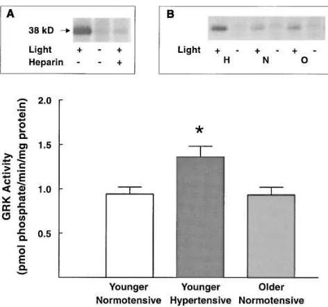
GRK activity in human lymphocytes. Incubation of rod outer segments with lymphocyte cytosolic fractions resulted in the phosphorylation of a 38-kD band, consistent with the labeling of rhodopsin (19). Light exposure resulted in a greater than threefold increase in rhodopsin phosphorylation. The light-dependent phosphorylation of rhodopsin was completely in-hibited by the addition of 10 mg/ml of heparin (Fig. 1, inset A). The addition of protein kinase A inhibitor (PKI 10 mg/ml) or cAMP (1 mM) did not significantly alter cytosolic GRK-medi-ated phosphorylation of rhodopsin (data not shown). GRK ac-tivity was restricted predominantly to the cytosolic fraction (8164% [n5 4] of total cellular activity).
In cytosolic fractions from younger hypertensive subjects GRK activity was significantly increased (4569%) as com-pared with activity in samples from young normotensive sub-jects or compared with GRK activity in samples from older normotensive subjects (Fig. 1, inset B).
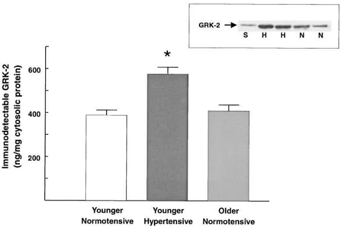
[image:4.612.316.552.398.619.2]GRK-2 expression assessed by immunodetection. Quantita-tive Western blotting was performed to determine whether the increase in GRK-mediated phosphorylation of rod outer seg-ments in lymphocytes from hypertensive subjects represented an increase in immunodetectable GRK-2. On Western blots of cytosolic lymphocyte preparations, the anti–GRK-2-specific 3A10 antibody recognized a peptide with a molecular mass of 80 kD, which migrated identically with recombinant GRK-2
Figure 1. Assessment of GRK activity in lymphocyte cytosolic frac-tions from younger and older normotensive subjects and younger hypertensive subjects. The data represent mean6SEM. *P, 0.01 vs. younger normotensive control subjects. (Inset A) Assessment of GRK activity in lymphocyte cytosolic fraction. Autoradiograph de-picting light-dependent phosphorylation of rhodopsin (z 38 kD).
Lane 1 represents activity in the presence of light, lane 2 in the ab-sence of light. Lane 3 depicts the effect of heparin on light-dependent rhodopsin phosphorylation. (Inset B) Assessment of GRK activity in lymphocytes from younger hypertensive (H) and from younger (N) and older (O) normotensive subjects; representative autoradiograph.
Table I. Clinical Data for Subjects
Younger normotensive
Younger hypertensive
Older normotensive
(n 5 16) (n 5 8) (n 5 8)
Age (yr) 2661 3062 5363* Sodium excretion
(meq/24 h) 138612 153617 138624 Body mass index (kg/m2) 23.360.6 24.460.7 24.061.4
Daytime systolic pressure
(mmHg) 11961 13362* 11962 Daytime diastolic pressure
(mmHg) 7361 8763* 7561
[image:4.612.56.298.580.724.2](Fig. 2, inset). Immunodetectable GRK-2 was increased by 4066% in cytosolic fractions from hypertensive subjects as compared with normotensive subjects (P , 0.01) (Fig. 2). Also, a comparable increase (3067%) in GRK-2 expression in hypertensive subjects was detected when whole cell pellets were assessed (data not shown). In contrast, immunodetect-able GRK-2 protein concentrations were not altered in sam-ples from older normotensive subjects (Fig. 2).
To determine the relationship between alterations in GRK-2 expression and b-adrenergic–mediated response, we assessed isoproterenol-stimulated adenylyl cyclase activity in permeabilized lymphocytes in a subset of the younger normo-tensive (n 5 8) and younger hypertensive (n 5 4) subjects studied under identical conditions as those described above. Isoproterenol-stimulated adenylyl cyclase activity was signifi-cantly reduced in lymphocytes from hypertensive subjects as compared with normotensive controls (normotensives 5 8664% above GTP-stimulated levels; hypertensives 5 6467%, P,
0.02). Isoproterenol-stimulated adenylyl cyclase activity was significantly inversely correlated with GRK-2 protein expres-sion (Fig. 3); i.e., those subjects with higher levels of GRK-2 protein expression demonstrated a lesser extent of b -adrener-gic–mediated adenylyl cyclase activation. In contrast, forsko-lin-stimulated adenylyl cyclase activity and GRK-2 expression were not correlated (data not shown).
A-kinase activity. To determine whether the increase in GRK-mediated activity and GRK-2 expression in cytosolic preparations from hypertensive subjects was related to a gen-eralized increase in cytosolic kinase activity, we examined the activity of A-kinase. Basal A-kinase activity (1,322637 pmol phosphate/min/mg cytosolic protein) was significantly increased by cAMP (to 2,8736104 pmol phosphate/min/mg cytosolic protein). In contrast to the increased GRK activity seen in lymphocyte cytosolic fractions from hypertensive subjects, nei-ther basal nor cAMP-stimulated A-kinase activities were al-tered (Fig. 4). Notably, basal A-kinase activity was marginally but significantly reduced in samples from older subjects. How-ever, cAMP-stimulated A-kinase activity was not significantly different between older and younger subjects (Fig. 4).
Assessment of GRK-5/6 expression by immunodetection.
To determine whether the increase in GRK-2 in hypertensive subjects might be associated with a generalized increase in the expression of other GRKs, we assessed immunodetectable GRK-5/6 in lymphocytes. The anti–GRK-5/6 antisera predom-inantly labeled a 68-kD lymphocyte peptide which comigrated with recombinant GRK-5, consistent with labeling of lympho-cyte GRK-5 (Fig. 5, inset). (It should be noted that GRK-6 mi-grates essentially identically to GRK-5 on SDS-PAGE [20]. Therefore, in the absence of a more specific antibody, a contri-bution of GRK-6 to the immunodetectable labeling with the antisera used cannot be ruled out.) In contrast to the increase in GRK-2 expression in samples from hypertensive subjects, immunodetectable GRK-5/6 levels were not significantly
[image:5.612.57.393.58.285.2]al-Figure 2. Assessment of GRK-2 expres-sion in cytosolic fractions from younger hy-pertensive and normotensive subjects. The data represent the mean6SEM. *P, 0.01 vs. younger normotensive control subjects. (Inset) A representative autoradiograph of a Western blot depicting labeling of 5 ng recombinant GRK-2 standard (S), and im-munodetectable GRK-2 in cytosolic frac-tions from younger hypertensive (H) sub-jects and younger normotensive (N) subjects.
[image:5.612.314.552.496.691.2]tered in either whole cell pellets (Fig. 5) or in cytosolic frac-tions (10169% of the expression in younger normotensive subjects). In whole cell pellets, GRK-5/6 expression was not significantly altered in older subjects. However, cytosolic frac-tions from older subjects demonstrated a marginal but statisti-cally significant reduction in GRK-5/6 expression (7867% of the expression in younger normotensive subjects, P, 0.05).
Assessment of b-arrestin expression by immunodetection.
We next determined whether the increase in immunodetect-able GRK-2 in samples from hypertensive subjects was associ-ated with increased immunodetectable b-arrestin levels. The antiarrestin antibody recognized two proteins migrating at z 55 and z 52 kD, consistent with labeling of b-arrestin and arrestin-3, respectively (Fig. 6). Immunodetectable levels of the arrestins were not significantly different in samples from either younger hypertensive or older normotensive subjects (hypertensive subjects: 9866% of expression in younger
nor-motensive subjects; older subjects: 9165% of expression in younger normotensive subjects).
Discussion
An impairment in b-adrenergic–stimulated adenylyl cyclase activity has been demonstrated in lymphocytes from young borderline (primarily Caucasian) hypertensive subjects (4). These studies demonstrate that this defect in signal trans-duction is associated with increased expression of GRK-2 (bARK-1). No alterations in other kinases were demon-strated. Specifically, cytosolic A-kinase activity was not signifi-cantly increased nor was the expression of GRK-5/6.
Impairment in receptor/G-protein interactions has been described previously in hypertensive subjects (4). To date, studies have linked impaired b-adrenergic–stimulated adenylyl cyclase activity with alterations in receptor affinity for agonists (2, 8, 9). This has been conventionally believed to represent an alteration in efficiency of interaction between the receptor and G-protein complex (9). However, neither receptor density (22) nor G-protein expression (4) has been demonstrated to be re-duced. An alteration in receptor phosphorylation leading to impaired receptor G-protein coupling would be a potential ex-planation for the reduction in receptor/G-protein coupling in lymphocytes from hypertensive subjects. However, in the ab-sence of the ability to assess the extent of receptor serine-thre-onine phosphorylation in vivo, this hypothesis remained un-proven. Studies to date have not demonstrated increased A-kinase activity in hypertension (23) (which could then lead to increased receptor phosphorylation and impaired receptor/ G-protein coupling).
An increase in GRK function and expression leading to in-creased receptor phosphorylation would provide an alterna-tive mechanism for receptor/G-protein uncoupling in hyper-tension. The correlation between increased GRK activity and impaired receptor G-protein coupling has been demonstrated in vitro and in vivo. Increased GRK activity associated with
bARK overexpression systems has been demonstrated to par-allel an impairment of b-adrenergic stimulation of adenylyl
[image:6.612.57.296.55.207.2]cy-Figure 4. Assessment of A-kinase activity in lymphocyte cytosolic fractions from younger normotensive (white bars), older normoten-sive (light shaded bars), and younger hypertensive (dark shaded bars) subjects. A-kinase activity is expressed as picomoles of phosphate per minute per milligram of cytosolic protein. The data represent mean activity6SEM. *P, 0.05 vs. younger normotensive subjects.
[image:6.612.58.370.516.738.2]clase (14), although a reduction in basal adenylyl cyclase activ-ity with bARK overexpression has also been reported (13). Further, bARK inhibitor–expressing mutants have been asso-ciated with increased b-adrenergic–mediated responses (13) and attenuation of b-adrenergic receptor desensitization (24). As discussed above, pathologically, an increase in GRK activ-ity has been directly linked to the impairment in b1
-adrener-gic–stimulated myocardial effects in congestive heart failure in humans (15). Therefore, these studies would generally support the hypothesis that increased GRK activity could mediate an increase in receptor phosphorylation and hence the uncou-pling between the receptor and G-protein characteristic of the human hypertensive state.
The observed increase in GRK activity appears to be selec-tively associated with an increase in bARK-1 (GRK-2) expres-sion. No alterations in GRK-5/6 expression were demon-strated. Further, no alterations in A-kinase activity were apparent in cytosolic fractions in hypertensive versus normo-tensive controls. The lack of alterations in A-kinase activity has several implications. First, A-kinase activation is the other dominant mechanism mediating receptor serine-threonine phosphorylation leading to impaired activity (10–12). Further, the lack of any difference in either basal or stimulated A-kinase activity in cytosolic fractions from younger normotensive and hypertensive subjects would rule out a nonspecific alteration in kinase activity or a blood pressure–related difference in com-position of the cytosolic fractions between hypertensive and normotensive subjects as the explanation for the alteration in GRK activity.
It is notable that the increase in GRK activity and expres-sion was unique to the hypertensive subjects, and was not com-mon to older normotensive subjects. Initial studies by us and others had suggested a common impairment of b-adrenergic– mediated adenylyl cyclase activation in lymphocytes from older and hypertensive subjects (5, 8). More recently, we have suggested that the regulation of the cholera- and pertussis toxin–mediated labeling of G-proteins differs between older and hypertensive subjects (4). The current studies extend these findings to further differentiate the altered pattern of regula-tion of b-adrenergic responsiveness in aging versus hyperten-sion.
If generalized, the increase in GRK activity (paralleling the impairment in b-adrenergic–stimulated adenylyl cyclase activ-ity) could be the explanation for the impairment in vascular b2
-adrenergic responsiveness characteristic of borderline hyper-tensive subjects. As discussed above, previous studies by us and others have demonstrated that regulation of lymphocyte
b-adrenergic–stimulated adenylyl cyclase activity parallels the regulation of vascular b-adrenergic responsiveness in hyper-tension. However, hypertension-related, lymphocyte-specific explanations for the altered GRK activity should be consid-ered, including alterations in lymphocyte activation profiles and in lymphocyte subset distribution.
Lymphocyte activation increases GRK activity (25). How-ever, three lines of evidence would suggest that a selective in-crease in lymphocyte activation in hypertensive subjects is not the explanation for the increase in GRK function. First, other studies have indicated that basal and proliferative responses are not increased (and may be suppressed) in lymphocytes of hypertensive subjects (26). Second, in contrast to the decrease in basal levels of adenylyl cyclase activity seen with lympho-cyte activation (27), our previous studies of hypertensive sub-jects (studied under identical conditions) have not detected any decrease in basal or forskolin-stimulated levels of adenylyl cyclase activity (4). Third, lymphocyte activation increases A-kinase activity (28). Thus, if the increase in GRK activity in hypertensives was related to lymphocyte activation, one would anticipate a parallel increase in both basal adenylyl cyclase ac-tivity and A-kinase acac-tivity. The divergence between increased GRK activity and unaltered basal levels of adenylyl cyclase and A-kinase activity in hypertensives would argue against a lymphocyte-specific explanation for these findings.
An alteration in the distribution of lymphocyte subpopula-tions in hypertensive subjects (if those subsets differed in GRK-2 expression) might also explain our findings. However, no alterations in lymphocyte subpopulations have been de-tected in human hypertensive subjects (29).
[image:7.612.57.336.62.265.2]It should be pointed out that increased GRK activity may not be the unique defect leading to the impairment in b -adren-ergic receptor activation characteristic of the hypertensive state. Recent studies from our laboratory have demonstrated functional G-protein alterations in hypertension which could
not be explained by increased GRK activity (4). It is notable that in congestive heart failure a similar pattern of alterations in receptor/G-protein coupling related to increased GRK activ-ity and altered G-protein function has been described (15, 30).
The mechanism of GRK-2 regulation in hypertensive sub-jects is unclear. In congestive heart failure, increased GRK-2 expression could be linked to the increase in sympathoadrenal activity (31–33). However, that linkage in hypertension would be somewhat more tenuous. First, increased sympathetic activ-ity has not been universally seen in hypertensive populations (34). Second, our previous studies have demonstrated that the defect in b-adrenergic responsiveness in hypertensive subjects is corrected after dietary salt restriction, a maneuver which is associated with increased sympathetic activity (8). Thus, the pattern of dynamic regulation of b-adrenergic responsiveness in hypertension would not be consistent with the classical mod-els of agonist-induced desensitization. Hence, although the mechanism of GRK-2 regulation in hypertension remains un-known (and is the focus of ongoing study in our laboratory), a linkage with increased sympathetic activity is less likely.
In summary, the present studies have demonstrated an in-crease in GRK activity and function in lymphocytes from young borderline Caucasian hypertensive subjects. The gener-alizability of these findings, both to other populations of hy-pertensive subjects, and to the vasculature of hyhy-pertensive hu-mans and animals, has yet to be demonstrated. However, these findings suggest that an alteration in GRK function could un-derlie the defect in b2-adrenergic responsiveness, contributing
to the impairment in vascular function characteristic of the hy-pertensive state.
Acknowledgments
These studies were supported by grants-in-aid from the Medical Re-search Council of Canada and from the National Institutes of Health (GM44944). Dr. Feldman is supported by a Career Investigator Award from the Heart and Stroke Foundation of Ontario. Dr. Ben-ovic is an Established Investigator of the American Heart Associa-tion.
References
1. Feldman, R.D. 1990. Defective venous b-adrenergic response in border-line hypertensive subjects is corrected by a low sodium diet. J. Clin. Invest. 85: 647–652.
2. Naslund, T., D.J. Silberstein, W.J. Merrell, J.H. Nadeau, and A.J.J. Wood. 1990. Low sodium intake corrects abnormality in b-receptor-mediated arterial vasodilation in patients with hypertension: correlation with b-receptor function in vitro. Clin. Pharmacol. Ther. 48:87–95.
3. Feldman, R.D. 1987. Beta-adrenergic receptor alterations in hyperten-sion: physiological and molecular correlates. Can. J. Physiol. Pharmacol. 65: 1666–1672.
4. Feldman, R.D., C.M. Tan, and J. Chorazyczewski. 1995. G-Protein alter-ations in hypertension and aging. Hypertension (Dallas). 26:725–732.
5. Feldman, R.D. 1992. A low sodium diet corrects the defect in b -adrener-gic response in older subjects. Circulation. 85:612–618.
6. Mills, P.J., M.G. Ziegler, R.A. Nelesen, and B.P. Kennedy. 1996. The ef-fects of the menstrual cycle, race, and gender on adrenergic receptors and ago-nists. Clin. Pharmacol. Ther. 60:99–104.
7. Venter, C.P., S. Daya, P.H. Joubert, and W.J. Strydom. 1985. Ethnic dif-ferences in human lymphocytic cyclic AMP production after isoprenaline stim-ulation and propranolol blockade. Br. J. Clin. Pharmacol. 19:187–190.
8. Feldman, R.D., W.J. Lawton, and W.L. McArdle. 1987. Low sodium diet corrects the defect in lymphocyte b-adrenergic responsiveness in hypertensive subjects. J. Clin. Invest. 79:290–294.
9. Feldman, R., L.E. Limbird, J. Nadeau, D. Robertson, and A.J.J. Wood. 1984. Leukocyte beta receptor alterations in hypertensive subjects. J. Clin. In-vest. 73:648–653.
10. Hausdorff, W.P., M.G. Caron, and R.J. Lefkowitz. 1990. Turning off the signal: desensitization of b-adrenergic receptor function. FASEB (Fed. Am. Soc. Exp. Biol.) J. 4:2881–2889.
11. Lefkowitz, R.J., W.P. Hausdorff, and M.G. Caron. 1990. Role of phos-phorylation in desensitization of the b-adrenoceptor. TIPS (Trends Pharmacol. Sci.). 11:190–194.
12. Barnes, P.J. 1995. State of the art. Beta-adrenergic receptors and their regulation. Am. J. Resp. Crit. Care Med. 152:838–860.
13. Koch, W.J., H.A. Rockman, P. Sarmama, R. Hamilton, R.A. Bond, C.A. Milano, and R.J. Lefkowitz. 1995. Cardiac function in mice overexpressing the b-adrenergic receptor kinase or a bARK inhibitor. Science (Wash. DC).
268:1350–1353.
14. Pippig, S., S. Andexingert, K. Daniel, M. Puzicha, M.G. Caron, R.J. Lefkowitz, and M.J. Lohse. 1993. Overexpression of b-arrestin and b -adrener-gic receptor kinase augment desensitization of b2-adrenergic receptors. J. Biol.
Chem. 268:3201–3208.
15. Ungerer, M., M. Bohm, J.S. Elce, E. Erdmann, and M.J. Lohse. 1993. Expression of b-arrestins and b-adrenergic receptor kinases in the failing hu-man heart. Circulation. 87:454–463.
16. Ping, P., A. Li, C. Bentley, and H.K. Hammond. 1995. Increased G-pro-tein receptor kinase activity and expression during development of heart fail-ure. Circulation. 92:57a. (Abstr.)
17. Böyum, A. 1968. Isolation of mononuclear cells and granulocytes from human blood. Scan. J. Clin. Lab. Invest. 21(Suppl. 97):77–89.
18. Feldman, R.D. 1993. Insulin-mediated sensitization of adenylylcyclase activation. Br. J. Pharmacol. 110:1640–1644.
19. Benovic, J.L., F. Mayor, Jr., C. Staniszewsi, R.J. Lefkowitz, and M.G. Caron. 1987. Purification and characterization of the b-adrenergic receptor ki-nase. J. Biol. Chem. 262:9026–9032.
20. Loudon, R.P., B. Perussia, and J.L. Benovic. 1996. Differentially regu-lated expression of the G protein-coupled receptor kinases, bARK and GRK6, during myelomonocytic cell development in vitro.Blood. 88:4547–4557.
21. Donosa, L.A., D.S. Gregerson, L. Smith, S. Robertson, V. Knospe, T. Vrabec, and C.M. Kalsow. 1990. S-antigen: preparation and characterization of site-specific monoclonal antibodies. Curr. Eye Res. 9:343–355.
22. Brodde, O.-E., A.E. Daul, N. O’Hara, and A.M. Khalifa. 1985. Proper-ties of a- and b-adrenoceptors in circulating blood cells of patients with essen-tial hypertension. J. Cardiovasc. Pharmacol. 7:S162–S167.
23. Silver, P.J., R.J. Michalak, and S.M. Kocmund. 1985. Role of cyclic AMP protein kinase in decreased arterial cyclic AMP responsiveness in hyper-tension. J. Pharmacol. Exp. Ther. 232:595–601.
24. Kong, G., R. Penn, and J.L. Benovic. 1994. A b-adrenergic receptor ki-nase dominant negative mutant attenuates desensitization of the b2-adrenergic receptor. J. Biol. Chem. 269:13084–13087.
25. De Blasi, A., G. Parruti, and M. Sallese. 1995. Regulation of G protein– coupled receptor kinase subtypes in activated T lymphocytes. J. Clin. Invest. 95: 203–210.
26. Shasha, S.M., U. Shasha, B. Kristal, M. Barzilai, O. Steinberger, and T. Shkolnik. 1991. Proliferative response of T lymphocytes to mitogenic lectins in essential hypertension. Nephron. 58:413–417.
27. Paietta, E., and J.D. Schwarzmeier. 1983. Beta-adrenergic responsive-ness of human peripheral lymphocytes after mitogenic transformation with phytohemagglutinin. Biochem. Pharmacol. 32:3085–3089.
28. Laxminarayana, D., and G.M. Kammer. 1996. Activation of type l pro-tein kinase A during receptor-mediated human T lymphocyte activation. J. Im-munol. 156:497–506.
29. Mills, P.J., C.C. Berry, J.E. Dimsdale, M.G. Ziegler, R.A. Nelesen, and B.P. Kennedy. 1995. Lymphocyte subset redistribution in response to acute ex-perimental stress: effects of gender, ethnicity, hypertension, and the sympa-thetic nervous system. Brain Behav. Immun. 9:61–69.
30. Stiles, G.L. 1991. Adrenergic receptor responsiveness and congestive heart failure. Am. J. Cardiol. 67:13C–17C.
31. Kiuchi, K., R.P. Shannon, K. Komamura, D.J. Cohen, C. Bianchi, C.J. Homcy, S.F. Vatner, and D.E. Vatner. 1993. Myocardial b-adrenergic receptor function during the development of pacing-induced heart failure. J. Clin. Invest.
91:907–914.
32. Colucci, W.S., J.P. Ribeiro, M.B. Rocco, R.J. Quigg, M.A. Creager, J.D. Marsh, D.F. Gauthier, and L.H. Hartley. 1989. Impaired chronotrophic re-sponse to exercise in patients with congestive heart failure. Role of postsynaptic
b-adrenergic desensitization. Circulation. 80:314–323.
33. Maisel, A.S., C. Phillips, M.C. Michel, M.G. Ziegler, and S.M. Carter. 1989. Regulation of cardiac b-adrenergic receptors by captopril. Implications for congestive heart failure. Circulation. 80:669–675.