A novel form of integrin dysfunction involving
bb
1,
bb
2, and
bb
3 integrins
Alison McDowall, … , Nigel Klein, Nancy Hogg
J Clin Invest.
2003;
111(1)
:51-60.
https://doi.org/10.1172/JCI14076
.
The adhesion receptors known as integrins perform key functions for hematopoietic cells.
The platelet integrin
a
IIb
b
3 is critical in hemostasis, and the
b
1 and
b
2 integrins on
leukocytes have many roles in cell-mediated immunity. Mutations in the
b
2 subunit lead to
integrin nonexpression and to an immune deficiency, leukocyte adhesion deficiency-1.
Mutations in either the
a
or
b
subunit of
a
IIb
b
3 usually lead to integrin nonexpression and a
bleeding tendency termed Glanzmann thrombasthenia. Here we describe a unique patient
with clinical features of both Glanzmann thrombasthenia and leukocyte adhesion
deficiency-1. The patient has normal expression of
b
1,
b
2, and
b
3 integrins, but all are
dysfunctional. The key findings are that “inside-out” signaling pathways leading to integrin
activation are defective and that this is associated with abnormal integrin clustering. The
integrins themselves are intact and capable of function following extracellular stimulation. T
cell motility is normal, as are the expression levels and electrophoretic characteristics of all
cytoskeletal and signaling proteins tested, except PKC-
a
, which has enhanced expression
in the patient’s cells. To our knowledge, this is the first description of a dysfunction affecting
three classes of integrins. We propose that it is caused by a lesion in an intracellular factor
or signaling pathway essential for integrin activation in hematopoietic cells and results in
lack of regulation of clustering, an essential component […]
Article
Vascular biology
Find the latest version:
Introduction
The integrins are a widely expressed family of nonco-valently linked αand βsubunits that mediate cell-cell and cell-extracellular matrix interactions. The inte-grins on circulating leukocytes and platelets bind min-imally to their ligands, but their adhesive capacity can be increased by stimulation of intracellular signaling pathways, for example by chemokines and bacterial peptides, triggering of the T cell receptor complex, or artificially, with phorbol esters. This mechanism of
integrin activation has been termed inside-out signal-ing. It is also possible to activate integrins with diva-lent cations such as Mg2+and Mn2+or with special
anti-integrin mAb’s. These agonists act directly on the integrin ectodomain and stimulate adhesion to ligand (reviewed in ref. 1).
Inside-out signaling induces two major forms of alteration to integrins that enable efficient ligand bind-ing: integrins can undergo conformational change leading to higher affinity receptors and can diffuse lat-erally in the membrane to form higher avidity clusters. In leukocytes, signals leading to clustering of integrins seem to predominate, and the clustering depends on cytoskeletal reorganization and the protease calpain (2, 3). In platelets, inside-out signaling primarily leads to affinity alteration of integrin αIIbβ3, but clustering is also part of the activation process (4, 5). Once activat-ed (and ligand bound), the integrin signals back into the cell, and this is termed outside-in signaling.
Active β1 and β2 integrins play a crucial role in the extravasation of leukocytes from the circulation to sites of injury or infection and in the homing of lymphocytes to tissues, particularly the secondary lymphoid organs. Specifically, integrins α4β1 and LFA-1 mediate firm adhesion of the leukocytes and transendothelial migra-tion. Neutrophils and monocytes are the first cells to be
A novel form of integrin dysfunction
involving
β
1,
β
2, and
β
3 integrins
Alison McDowall,
1David Inwald,
2Birgit Leitinger,
1Alison Jones,
3Ri Liesner,
4Nigel Klein,
2and Nancy Hogg
11Leukocyte Adhesion Laboratory, Cancer Research UK London Research Institute, Lincoln’s Inn Fields Laboratories, London, United Kingdom
2Immunobiology Unit, Institute of Child Health, London, United Kingdom 3Department of Clinical Immunology, and
4Department of Haematology, Great Ormond Street Hospital for Children NHS Trust, Great Ormond Street, London, United Kingdom
The adhesion receptors known as integrins perform key functions for hematopoietic cells. The platelet integrin αIIbβ3 is critical in hemostasis, and the β1 and β2 integrins on leukocytes have many roles in cell-mediated immunity. Mutations in the β2 subunit lead to integrin nonexpression and to an immune deficiency, leukocyte adhesion deficiency-1. Mutations in either the αor βsubunit of
αIIbβ3usually lead to integrin nonexpression and a bleeding tendency termed Glanzmann throm-basthenia. Here we describe a unique patient with clinical features of both Glanzmann thrombas-thenia and leukocyte adhesion deficiency-1. The patient has normal expression of β1, β2, and β3 inte-grins, but all are dysfunctional. The key findings are that “inside-out” signaling pathways leading to integrin activation are defective and that this is associated with abnormal integrin clustering. The integrins themselves are intact and capable of function following extracellular stimulation. T cell motility is normal, as are the expression levels and electrophoretic characteristics of all cytoskeletal and signaling proteins tested, except PKC-α, which has enhanced expression in the patient’s cells. To our knowledge, this is the first description of a dysfunction affecting three classes of integrins. We propose that it is caused by a lesion in an intracellular factor or signaling pathway essential for inte-grin activation in hematopoietic cells and results in lack of regulation of clustering, an essential com-ponent of integrin-mediated adhesion.
J. Clin. Invest.111:51–60 (2003). doi:10.1172/JCI200314076.
Received for publication on August 27, 2001, and accepted in revised form on October 8, 2002.
Address correspondence to: Nancy Hogg, Leukocyte Adhesion Laboratory, Cancer Research UK London Research Institute, Lincoln’s Inn Fields Laboratories, London WC2A 3PX, United Kingdom. Phone: 44-207-269-3255; Fax: 44-207-269-3093; E-mail: [email protected].
Birgit Leitinger’s present address is: Sackler Institute for Muscular Skeletal Research, Department of Medicine, University College London, London, United Kingdom.
Conflict of interest: The authors have declared that no conflict of interest exists.
Nonstandard abbreviations used: leukocyte adhesion deficiency-1 (LAD-1); Glanzmann thrombasthenia (GT); phorbol-12,13-dibutyrate (PdBu); platelet-rich plasma (PRP); thrombin receptor agonist peptide (TRAP); platelet-poor plasma (PPP); integrin-linked kinase (ILK).
recruited to inflammatory sites where, making use of β2 integrins (CD11/CD18), LFA-1, Mac-1, and p150,95, they phagocytose and undergo respiratory burst in response to bacterial infection. The platelet integrin αIIbβ3 is stimulated to bind fibrinogen by agonists, such as thrombin, ADP, and thrombospondin, released at sites of vascular injury. This event is pivotal to the aggregation of platelets that is required for formation of the platelet plug and clotting of blood.
In humans, two inherited autosomal recessive dis-eases result from germline mutations in the genes encoding integrins specific to cells of hematopoietic origin. These disorders are leukocyte adhesion defi-ciency-1 (LAD-1) and Glanzmann thrombasthenia (GT) (6, 7). The hallmark of these disorders is lack of expression of the affected integrin. LAD-1 is caused by mutations in the β2 subunit of the leukocyte integrins. The lack of β2 integrin function results in elevated numbers of circulating neutrophils because these cells fail to adhere to or migrate across the endothelium. LAD-1 patients are susceptible to recurring, life-threat-ening, bacterial infections, which are typically evident in soft tissue. Severely affected people often die of infec-tion in childhood or early adulthood unless bone mar-row transplantation is successfully accomplished. The bleeding disorder GT is usually caused by mutations in either the αor βsubunit of the platelet integrin αIIbβ3 (CD41/CD61). The platelets are unable to bind to fib-rinogen and thus fail to aggregate and form a primary hemostatic plug in response to agonists. GT patients suffer easy bruising, mucocutaneous bleeding, and, occasionally, gastrointestinal and intracranial bleeding. Here we describe an unusual patient with clinical fea-tures of both LAD-1 and GT. The results suggest that the patient has a novel form of integrin dysfunction in which the β1, β2, and β3 integrins are expressed on the cell surface at normal levels but cannot be stimulated to bind ligand by intracellular signaling pathways.
Methods
The following Ab’s were gifts: KIM 185 (β2 activating) from M. Robinson (Celltech Group PLC, Slough, Unit-ed Kingdom); HUTS21 (β1 activation reporter) from C. Cabañas (CSIC-Universidad Complutense, Madrid, Spain); IB4 (β2 function blocking) from S.K. Law (Uni-versity of Oxford, Oxford, United Kingdom); 2E7 (β2 blotting) from C. Gahmberg (University of Helsinki, Helsinki, Finland); LIBS6 (αIIbβ3 activating) from M. Ginsberg (The Scripps Research Institute, La Jolla, Cal-ifornia, USA); anti-Vav from V. Tybulewicz (National Institute for Medical Research, London, United King-dom); anti–PKC-ζfrom P. Parker (Cancer Research UK, London, United Kingdom). The following mAb’s were produced at Cancer Research UK: 38 (αL function blocking and blotting); ICRF44 (αM); 3.9 (αX); 24 (β2 activation reporter); 7.2R (α4 blotting); UCHT-1 (CD3). The hybridoma cell lines producing TS1/18 (β2), P5D2 (β1), and TS2/16 (β1 activating) were obtained from American Type Culture Collection (Rockville,
Mary-land, USA). HP2/1 (α4 function blocking) and PM6/13 (β3 blotting) were purchased from Serotec Ltd. (Oxford, United Kingdom); SAM-1 (α5 function block-ing) was purchased from Eurogenetics UK Ltd. (Hamp-ton, United Kingdom); SZ22 (αIIb blotting) was pur-chased from Immunotech (Marseilles, France); anti-fibrinogen, 5B12 (αIIb), and Y2/51 (β3) were pur-chased from DAKO Ltd., (Ely, United Kingdom) in FITC-conjugated form; G25.2 (αL nonfunction block-ing), FITC-conjugated PAC-1 (αIIb activation reporter), and Abs against Rac-1, Cdc42, Rap-1, SLAP-130, ILK, and PKC-α, -β, -δ, and -θ were purchased from Becton Dickinson UK Ltd. (Oxford, United Kingdom); B3B11 (β1 blotting) and anti-filamin were purchased from Chemicon International Ltd. (Harrow, United King-dom). Ab’s against talin, α-actinin, vinculin, ezrin, pax-illin, and actin were purchased from Sigma-Aldrich (Poole, United Kingdom). Ab’s against RhoA and Rac-2 were obtained from Autogen Bioclear UK Ltd. (Calne, United Kingdom. The reagents FMLP, phorbol-12,13-dibutyrate (PdBu), ionomycin, thapsigargin, and 2′,7′ -bis-(carboxyethyl)-5(6′)-carboxyfluorescein acetoxy-methyl ester (BCECF-AM) were obtained from CN Biosciences UK Ltd. (Nottingham, United Kingdom). ICAM-1Fc and ICAM-3Fc were produced as described previously (8), VCAM-1Fc was a gift from M. Robinson (Celltech Group PLC). ADP, fibronectin, and fibrino-gen were purchased from Sigma-Aldrich and thrombin receptor agonist peptide (TRAP) was purchased from Peninsula Laboratories (St. Helen’s, United Kingdom).
Preparation of platelets, neutrophils, T cells and Epstein-Barr virus–transformed B cells. Blood samples were col-lected following approval by the Great Ormond Street/Institute of Child Health Ethics Committee and the parents’ written informed consent. Platelet-rich plasma (PRP) was prepared by collecting blood into 0.38% sodium citrate followed by centrifugation at 150
gfor 10 minutes at room temperature. To prepare washed platelets, PRP was spun at 800 g for 5 minutes, and the pellet was washed with HBSS without cations and resuspended in RPMI-1640. All steps were per-formed in the presence of 30 ng/ml prostacyclin (Glaxo-SmithKline, Uxbridge, United Kingdom) to prevent platelet activation.
Neutrophils and T cells were purified from EDTA- or heparin-anticoagulated venous blood. Neutrophils and PBMCs were fractionated, and T cells were cultured as described previously (8).
Epstein-Barr virus–transformed (EBV-transformed) B lymphoblastoid cells were derived from patient FM and a control donor by the Research Cell Services, Can-cer Research UK, using standard procedures. The cells were maintained in RPMI-1640 with 10% FCS.
Flow cytometry. Leukocytes (5 ×10⁵) were incubated
at 37°C. Bound mAb was detected by incubation with FITC-conjugated goat anti-mouse IgG (Sigma-Aldrich) for 20 minutes on ice.
For platelets, 5 µl of PRP was added to 50 µl of platelet buffer (10 mM HEPES, 145 mM NaCl, 5 mM KCl, 1 mM MgSO4, pH7.4) (9) containing
fluo-rochrome conjugated mAb and agonists and incubat-ed at 37°C for 20 minutes in an adaptation of a previ-ously described method (10). Experiments looking at PAC-1 and fibrinogen binding were also performed in the presence of 20 µg/ml eptifibatide (integrilin) (Schering-Plough Ltd., Welwyn Garden City, United Kingdom), an αIIbβ3 antagonist.
For soluble ligand binding, ICAM-1Fc and VCAM-1Fc at 300 and 2 µg/ml, respectively (saturating levels), were incubated with T cells using methodology as described previously (11).
Aggregometry.For aggregometry, the residual blood following PRP collection was centrifuged at 1,200 gfor 15 minutes at room temperature to obtain platelet-poor plasma (PPP). PRP with a final platelet count of 250 ×109/l was prepared by diluting PRP with PPP.
Aggregometry was performed at 37°C with a stir speed of 900 rpm. Platelet agonists were added to the cuvettes and light transmission recorded.
SDS-PAGE and Western blotting of detergent soluble cell extracts. T cells were suspended at 5 × 10⁷/ml and
platelets at 109/ml in ice-cold lysis buffer (50 mM Tris
pH 8 containing 150 mM NaCl, 2 mM MgCl2, 2 mM
EGTA, 1 µg/ml aprotonin, 20 µg/ml PMSF, and 1% Tri-ton X-100), and lysed for 20 minutes on ice. The lysate was microfuged for 15 minutes to remove insoluble material. Proteins were separated by SDS-PAGE. After transfer to nitrocellulose membrane and incubation with primary Ab’s, the bound Ab was detected with HRP-conjugated sheep anti-mouse Ig (Amersham Bio-sciences UK Ltd., Chalfont St. Giles, United Kingdom) or HRP-conjugated goat anti-rabbit Ig (DAKO Ltd.) and enhanced chemiluminescence Western blotting detection reagents (Amersham Biosciences UK Ltd.). Quantification of bands at subsaturating levels was performed using NIH Image 1.60 Software.
Adhesion assays. T cell adhesion to immobilized ICAM-1Fc, ICAM-3Fc, VCAM-1Fc (all at 5 µg/ml) or fibronectin (20 µg/ml), and EBV-transformed B cell binding to immobilized ICAM-1 (5 µg/ml) or fibronectin (20 µg/ml) were performed using similar methods, as described previously (8). Neutrophil adhesion to immobilized fibrinogen (0.5 mg/ml) was performed as described previously (8).
Microscopy. For video microscopy, 35 mm glass-bot-tom microwell dishes (Mattek Corp, Ashland, Massa-chusetts, USA) were coated overnight with 10 µg/ml ICAM-1Fc, then blocked with 2.5% BSA/PBS. One mil-liliter of T cells (4 × 10⁵/ml in HEPES buffer) was
allowed to settle for 4 minutes before addition of 500 µl of HEPES buffer containing 15 mM MgCl2/3 mM
EGTA. Images were taken at 5 second intervals for 20 minutes using a Nikon Diaphot 300 microscope and
AQM2001Kinetic Acquisition Manager (Kinetic
Imag-ing Ltd., Bromborough, United KImag-ingdom). Cells were tracked using Motion Analysis software (Kinetic Imag-ing Ltd.) and the data analyzed usImag-ing a Mathematica notebook (Wolfram Research Europe Ltd., Long Han-borough, United Kingdom) developed by Daniel Zicha (Cancer Research UK).
Samples were prepared for confocal microscopy as described previously (12), except that 2 ×10⁶cells
were used.
Results
Patient FM was born at term by elective Caesarean sec-tion to nonconsanguineous Maltese parents. She had two female siblings, of whom one is 7 years old and well, whereas the other died within hours of birth with widespread bruising and bleeding. Despite her atrau-matic delivery, patient FM was noted to have extensive bruising and petechiae within hours of birth. She had an antenatal intraventricular hemorrhage and later required insertion of a ventriculo-peritoneal shunt for posthemorrhagic hydrocephalus. A platelet count and routine clotting screen performed at this point were in the normal range. The umbilical cord separated nor-mally at 1 week of age. At three months of age she was referred for investigation of prolonged bleeding fol-lowing minor trauma.
The platelet count, thrombin time, activated partial thromboplastin time, prothrombin time, and fibrino-gen levels were all within the normal range. However, platelet aggregation was absent in response to ADP, collagen, and arachidonic acid. In contrast, platelets from both parents aggregated normally in response to these agonists. Flow cytometry revealed that the patient’s platelets had normal expression of GPIb and αIIbβ3. These results suggested a diagnosis of type 2 GT with dysfunctional, rather than absent, platelet αIIbβ3. She has been managed with tranexamic acid and platelet transfusions as required.
From 5 months of age the patient developed recurrent bacterial infections and at 11 months she developed leg ulcers and was commenced on prophylactic antibiotics. She was found to have leukocytosis (38.4 ×10⁹/l; normal
range 5 ×10⁹–15 ×109/l), suggestive of a leukocyte
adhe-sion defect, but had normal expresadhe-sion of the leukocyte integrin αL, αM, αX, and β2 subunits and the selectin ligand sialyl Lewis x. She had normal humoral immune responses to tetanus toxoid and Haemophilus influenza, neutrophil phagocytosis of Staphylococcus aureus, and oxidative burst. Her lymphocyte count, however, was high (15.7 ×10⁹/l; normal range of 1.5 ×10⁹–4 ×10⁹/l).
Her T cell mitogenic activity to phytohemagglutinin was reduced by 50% compared with healthy age-matched controls. The patient is now 3 years old and has under-gone a successful bone marrow transplant. There is no family history of either LAD-1– or GT-type disorders.
the cell surface expression of the major integrins on platelets, neutrophils and T cells was analyzed by flow cytometry. The overlapping profiles in Figure 1 show that the expression of the αIIb and β3 subunits on platelets, the αL, αM, αX, and β2 subunits on neu-trophils, and the αL, β2, α4, α5, and β1 subunits on T cells was similar for the patient and a control donor.
To determine whether the patient’s integrins were abnormally processed or posttranslationally modified, cell lysates were subjected to SDS-PAGE and Western blotting for the relevant integrin subunits. No differ-ences were detected in the electrophoretic characteris-tics of the αIIb or β3 integrin subunits of platelet lysates (Figure 2a) or the αL, β2, α4, or β1 subunits of T cell lysates (Figure 2b) prepared from the patient and a control donor. Therefore, the patient’s integrins resembled normal controls in both expression and bio-chemical characteristics.
Functional analysis of αIIbβ3 on the patient’s platelets.
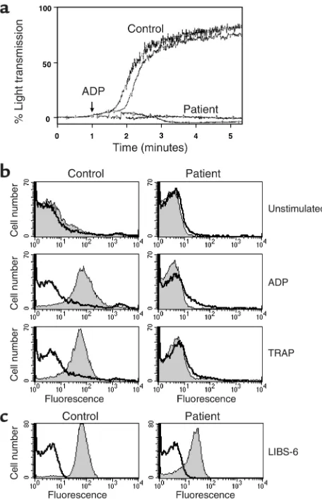
Although the patient had normal cell surface expression of the three classes of integrins tested, it was possible that her symptoms were due to the inability of these inte-grins to function normally. The function of the platelet integrin αIIbβ3 was assessed by aggregometry. Although control platelets responded to 5 µM ADP as expected, the patient’s platelets did not aggregate (Figure 3a). We next assessed the ability of two standard platelet
ago-nists, ADP and TRAP, which signal through two distinct platelet receptors, to cause platelets to bind soluble fib-rinogen (Figure 3b). Both stimuli induced binding of control platelets to fibrinogen, and this was inhibited by the αIIbβ3 antagonist, eptifibatide; however, the patient’s platelets failed to bind soluble fibrinogen under any circumstances of inside-out stimulation. These ago-nists induced upregulation of α-granule contents, such as P-selectin, indicating platelet activation was normal (data not shown). Another way to activate integrins is to use mAb’s, such as LIBS-6, which stimulate αIIbβ3 by direct activation of the ectodomain (termed outside-in signaling) (13). LIBS-6 induces expression of the αIIbβ3 activation epitope recognized by the mAb PAC-1 (14). Here LIBS-6 induces the PAC-1 epitope on both patient and control platelets (Figure 3c).
Functional analysis of Mac-1 on the patient’s neutrophils.
The function of the β2 integrin Mac-1 was examined by inducing neutrophil binding to immobilized fibrino-gen. A variety of activating stimuli were used, which tested both inside-out and outside-in signaling to inte-grins. All treatments caused control neutrophils to bind to fibrinogen in a β2 integrin-dependent manner (Figure 4). In contrast, the patient’s neutrophils failed to bind in response to either FMLP or the phorbol ester PdBu (inside-out signaling), but did bind to fibrinogen following exposure to the β2 activating mAb KIM 185. Therefore, the ability of Mac-1 on the patient’s neu-trophils to bind fibrinogen was impaired in response to typical stimulants of inside-out signal transduction. However, the patient’s neutrophils were able to mobi-lize intracellular Ca2+in response to FMLP and the Ca2+
mobilizing agent thapsigargin, and FMLP induced sim-ilar levels of L-selectin shedding and Mac-1 upregula-tion in patient and control neutrophils (data not shown). Therefore, the patient’s neutrophils are responsive to FMLP in non-integrin–dependent ways.
[image:5.576.59.293.55.315.2]Functional analysis of LFA-1 on the patient’s T cells.The function of the β2 integrin LFA-1 was examined on T cells by inducing adhesion to immobilized ligands. PdBu, the Ca2+mobilizer ionomycin, or CD3 mAb Figure 1
[image:5.576.305.541.545.667.2]Comparison of integrin expression on platelets, T cells, and neu-trophils from patient and control. Expression of (a) αIIb and β3 subunits on platelets (n= 2). (b) αL, αM, αX, and β2 subunits on neutrophils. (c) αL, β2, α4, α5, and β1 subunits on T cells from control (black line) and patient (gray region). Background binding is indicated (dotted line) and is identical for control and patient. Representative histograms (n= 3) are shown.
Figure 2
Electrophoretic characteristics of integrin subunits from patient and control platelets and T cells. (a) Platelet lysates from a control donor (C) and the patient (P) blotted for αIIb and β3 subunits. (b) T cell lysates from a control donor (C) and the patient (P) blotted for αL,
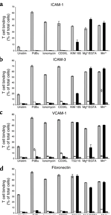
UCHT-1, which cross links the T cell receptor/CD3 complex, were used to test LFA-1 activation by inside-out signaling, whereas the β2-activating mAb KIM 185, Mg2+/EGTA, or Mn2+were used to directly activate
LFA-1. All the stimuli induced LFA-1–mediated bind-ing of control T cells to both ICAM-1 and ICAM-3 (Figure 5, a and b). However, only KIM 185, Mg2+/EGTA, and Mn2+ induced adhesion of the
patient’s T cells. None of the stimuli that act through intracellular signaling pathways induced LFA-1–medi-ated adhesion of the patient’s T cells to either ligand (Figure 5, a and b).
Functional analysis of α4β1 and α5β1 on the patient’s T cells.T cells express β1 as well as β2 integrins, with α4β1 and α5β1 being involved in many immune processes in association with the β2 integrins. When adhesion to the α4β1 ligand VCAM-1 (Figure 5c) or the α4β1/α5β1 lig-and fibronectin (Figure 5d) was assessed, all the stimuli tested induced adhesion of control T cells, whereas the patient’s T cells only adhered when stimulated by Mg2+/EGTA, Mn2+, or the β1-activating mAb TS2/16.
Thus β1 and β2 integrins on the patient’s T cells were able to bind their ligands when stimulated directly through the ectodomain, but failed to bind when inside-out stimuli were used. These results suggested a possible lesion in an intracellular signaling pathway.
Functional analysis of β1 and β2 integrins on the patient’s B cells.To assess whether the defect in inside-out stimu-lation of integrin-mediated adhesion also affected B cells, we used EBV-transformed B lymphoblastoid cells derived from the patient’s blood and a control donor’s blood. Both these cell lines were able to adhere to ICAM-1 (Figure 6a) or fibronectin (Figure 6b) when stimulated with Mn2+, but the patient’s cells failed to
adhere when stimulated with PdBu.
The state of integrin affinity and avidity on the patient’s T cells. We next investigated the activation state of the integrins on the T cells. The β2 integrin LFA-1, when in higher affinity form, is recognized by mAb 24 (15) and binds soluble ICAM-1 with increased affinity (11). Exposure to Mn2+ of control and patient T cells
[image:6.576.60.288.54.407.2]induced equivalent levels of both mAb 24 and soluble ICAM-1 binding (Figure 7a), indicating that the capac-ity for LFA-1 to adopt a higher-affincapac-ity form was intact when stimulated from outside the cell. Similarly,β1 integrins from patient and control T cells could be induced to express the β1 activation epitope, recog-nized by mAb HUTS 21 (16), and to bind soluble VCAM-1 (α4β1 only) (Figure 7a). Therefore, the lack of β1 and β2 integrin function on the patient’s T cells could not be explained by an inability to assume a high-er affinity conformation.
Figure 3
[image:6.576.309.534.513.649.2]Comparison of integrin αIIbβ3 function in patient and control platelets. (a) Platelet aggregation in response to 5 µM ADP (single experiment performed in duplicate). (b) Binding of FITC-conjugated antifibrinogen to platelets in the presence (black line) or absence (gray region) of 20 µg/ml eptifibatide. Platelets were either unstim-ulated or stimunstim-ulated for 20 minutes with 10 µM ADP or 1 µM TRAP. Data are representative of four separate experiments. (c) Binding of mAb PAC-1 to platelets stimulated with 10 µg/ml β3 integrin mAb LIBS-6 in the presence (black line) and absence (gray region) of epti-fibatide. Data are representative of two separate experiments.
Figure 4
When β2 integrins on leukocytes are triggered through intracellular pathways, they become laterally mobile and cluster (2, 17). T cell adhesion to ICAM-1 is dependent on this clustered form of LFA-1 (2, 3). When viewed by confocal microscopy, control T cells exhibit-ed increasexhibit-ed LFA-1 clustering following exposure to PdBu or the Ca2+mobilizer thapsigargin, but not when
exposed to Mg2+/EGTA (Figure 7b), as reported
previ-ously (2). In contrast, LFA-1 was already in a clustered state on the patient’s T cells and additional stimulation with PdBu and thapsigargin caused no further increase (Figure 7b). Preliminary evidence indicated parallel findings for β1 integrins on control and patient T cells using both α4 and β1 mAbs (n= 2; data not shown). Other abundant cell surface membrane proteins such as CD2, CD4, CD8, CD55, and MHC class I were not clustered on the patient’s cells (data not shown). These findings suggest a disruption of signaling pathways
causing dysregulation of integrin clustering or lateral mobility on the patient’s T cells.
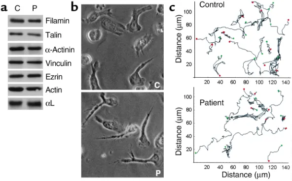
Analysis of the cytoskeleton. For leukocytes and platelets, the link between integrins and the cytoskeleton is criti-cal for their function, and the cytoskeleton is also involved in the process of integrin clustering. To address whether associations with the cytoskeleton are defective in the patient’s T cells, lysates were blotted for some of the most commonly reported integrin-associated cytoskeletal proteins. Filamin, talin, α-actinin, vinculin, ezrin, paxillin (not shown), and actin (Figure 8a) were all present at equivalent levels in the control and patient’s T cells and migrated as expected on SDS-PAGE. The findings indicate that none of these cytoskeletal pro-teins in the patient’s cells had been cleaved or subjected to altered posttranslational modification.
When T cells are stimulated through LFA-1 they polarize and migrate on immobilized ICAM-1 (18), suggesting that activated LFA-1 can signal remodeling of the cytoskeleton. Therefore, to determine whether a component of the patient’s T cell cytoskeletal network was dysfunctional, Mg2+/EGTA-treated T cells were
[image:7.576.61.294.49.498.2]adhered to ICAM-1 and their ability to polarize and migrate assessed. Both the patient and control T cells polarized (Figure 8b) and migrated (Figure 8c) on ICAM-1 in a comparable manner. The average speed of control T cells was calculated to be 12.7 ± 6.3 µm/min
Figure 5
Adhesion of patient and control T cells to LFA-1 ligands ICAM-1 and ICAM-3, to α4β1 ligand VCAM-1, and to α4β1/α5β1 ligand fibronectin. The binding of control (light gray bars) and patient (black bars) T cells to plates coated with (a) ICAM-1, (b) ICAM-3, (c) VCAM-1, and (d) fibronectin when stimulated with 50 nM PdBu, 1
[image:7.576.309.537.544.658.2]µM ionomycin, 10 µg/ml UCHT-1, 10 µg/ml KIM 185 or TS2/16, 5 mM MgCl2/1 mM EGTA, or 0.5 mM MnCl2. The presence of αL mAb 38 at 10 µg/ml in aand b, α4 mAb HP2/1 at 10 µg/ml in c, and α4 mAb HP2/1 plus α5 mAb SAM-1 both at 10 µg/ml in dinhibits adhe-sion of control (white bars) and patient (dark gray bars) cells. Data (mean of triplicates ± SD) from one representative experiment (n= 3) are shown. Unstim, unstimulated.
Figure 6
and of patient T cells was 12.4 ± 5.9 µm/min. These data provide evidence that both the cytoskeleton and the adapter proteins linking the cytoskeleton and inte-grins function normally in the patient’s cells.
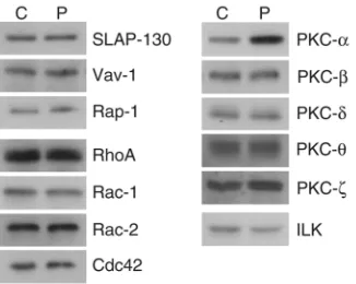
Expression of GTPases, PKCs, and other adhesion-related molecules. Inside-out signaling pathways leading to integrin activation are poorly characterized. However, in an attempt to discover the nature of the signaling lesion giving rise to the lack of integrin function in the patient, we decided to assess the expression of var-ious signaling molecules that have been associated with adhesion of leukocytes. The adaptor protein SLAP-130 (19, 20), the guanine nucleotide exchange factor Vav-1 (21), and the GTPase Rap-1 (22) have been shown recently to have a role in LFA-1 clustering and adhesion. The GTPases RhoA, Rac-1, and Cdc42 are involved in integrin-mediated cell migration (23) and mutations in Rac-2 give rise to
neutrophil defects similar to the patient’s abnormalities (24). PKCs have been implicated in several aspects of leukocyte adhesion (18,
25), and the patient’s leukocytes do not adhere in response to activation of PKC with phorbol ester. Finally, the integrin-linked kinase (ILK) is involved in adhesion of several classes of integrin (26). The expression and electrophoretic characteristics of 12 of the 13 proteins tested were identical in patient and control T cells (Figure 9). Expression of PKC-α, how-ever, was elevated 2.5-fold in the patient’s T cells.
Discussion
[image:8.576.65.532.58.200.2]We report a patient with a novel form of inherited inte-grin dysfunction in which the β1 and β2 integrins are expressed on the leukocyte cell surface at normal levels, but cannot be stimulated to bind ligand by intracellu-lar signaling pathways. The defect also affects the inte-grin αIIbβ3, because the patient’s platelets cannot be induced to bind soluble fibrinogen or to aggregate by
Figure 7
Comparison of the affinity and avidity state of integrins. (a) Control (black line) and patient (gray region) T cells incubated with mAbs 24 (β2 integrin activation reporter) or HUTS 21 (β1 integrin activation reporter) at 25 µg/ml, or ICAM-1Fc (300 µg/ml) or VCAM-1Fc (2 µg/ml) for 20 minutes at 37°C in the presence of 0.5 mM MnCl2; control (dotted line) and patient (dashed line) T cells incubated with mAbs or soluble ligand as above for 20 minutes at 4°C in the presence of 1 mM EDTA. Data are from one representative experiment (n= 3). (b) T cells were either unstimulated or treated with 5 mM Mg2+/1 mM EGTA, 50 nM PdBu, or 5 µM thapsigargin then labeled with LFA-1 mAb G25.2 and analyzed by confocal microscopy. A false color scheme is employed, which depicts the intensity of the fluorescent signal from blue (low) to yellow (high) (2). One optical section is shown at midheight of the cells. Data are representative of four experiments. The total fluorescent sig-nal was quantified and averaged over four experiments as follows: no treatment, control 61.5 ± 5.0, patient 98.8 ± 4.9; Mg2+/EGTA, control 59.5 ± 3.5, patient 105.5 ± 6.8; PdBu, control 78.8 ± 2.9, patient 101.0 ± 1.8; thapsigargin, control 81.0 ± 3.4, patient 96.5 ± 6.6.
Figure 8
[image:8.576.241.537.549.733.2]platelet agonists. However, all three classes of integrins can be activated directly either with divalent cations Mg2+/Mn2+or activating mAbs.
Characteristically LAD-1 patients lacking β2 inte-grins have elevated numbers of circulating neutrophils and fail to clear bacterial infections because these cells have restricted ability to traffic into infected tissue. The patient described here also has an abnormally elevated number of circulating lymphocytes, a feature that is not evident in classical LAD-1. This is probably due to the lack of function of both the β1 and β2 families of integrins, resulting in impaired responses of both myeloid cells and lymphocytes to inflammatory sig-nals. In experiments with knockout mice, lack of β2 integrin LFA-1 can be partially compensated for by the β1 integrins, but blocking function of both classes of integrins further increases the numbers of circulating neutrophils and lymphocytes and substantially impairs inflammatory responses (27).
To date, four patients with nonclassical forms of LAD-1 have been described. The leukocytes in two cases expressed approximately 60% of the normal lev-els of β2 integrins, but had no β2 integrin function (8, 28). These LAD-1 variant cases are related to classical LAD-1, having a mutation in one allele that prevents expression and a mutation in the other allele that allows expression but not ligand binding. Similar GT patients have been described who have αIIbβ3 expres-sion but no function (29).
There are two reported LAD-1 variant cases in which no mutations in the integrin βsubunit genes have been detected. The first case had the clinical indicators of LAD-1 and the β2 integrins were expressed but non-functional (30). After some years, defects in β3 integrin function also became apparent; but, in contrast to our patient, β1 integrins were not affected. For the second patient, there were problems with β2 and β1 integrin function, and a bleeding problem then developed that was not explored experimentally (31). A speculation is that the genetic lesions in these two patients and patient FM are individual, but may be related and
potentially highlight a specific common pathway ded-icated to integrin activation.
Because the activities of at least three integrin families are affected in patient FM and the integrins can func-tion when activated directly from outside the cell, it is unlikely that the dysfunction is due to mutations in the integrin subunits themselves. It is more probable that the faulty gene encodes a protein that is critical for inte-grin function. Moreover, the expression of the affected gene product is predicted to be confined to hematopoi-etic cells or have redundant function in other cell types. The fact that murine β1 integrin knockouts are embry-onic lethal (32) implies that the patient would not have survived if β1 integrin functioning was universally affected. The results demonstrating that other leuko-cyte and platelet functions are relatively normal provide evidence that the lesion is confined to a component of a key pathway dedicated to integrin activation.
Further insight into the nature of the patient’s lesion has come from confocal microscopy, which revealed that LFA-1 and the β1 integrins on the patient’s T cells are constitutively clustered and that the state of clus-tering does not change when the cells are activated. Inte-grin clustering is believed to be dynamic, but much is unknown about the sequence of events regulating this process. It has been proposed that integrins on resting cells are tethered to the cytoskeleton in an unclustered form and that activation of the cell removes the cytoskeletal constraint allowing the integrin to move and form clusters in the membrane, mediate firm lig-and binding, lig-and potentially reassociate with the cytoskeleton (2, 3, 17). The observation that the patient has constitutively clustered integrins, yet these integrins do not function, implies that dynamic clustering is required for inside-out stimulated adhesion and that, in the patient’s cells, integrin mobility may be restricted. Although exposure to low concentrations of cytocha-lasin D promotes adhesion of naive T cells by removing the cytoskeletal tethering of the integrins (3), this pro-cedure did not alter the adhesion capabilities of the patient’s T cells (data not shown). It is interesting that outside-in signaling through LFA-1 was sufficient for T cell adhesion, polarization, and migration, suggesting that the state of clustering is irrelevant for these adhe-sion-dependent activities, or, alternatively, that it can be altered by signaling directly through LFA-1.
[image:9.576.91.254.53.183.2]Although the platelet integrin αIIbβ3 is activated primarily through signaling that leads to a change in affinity (reviewed in ref. 5), avidity regulation also plays a role in the activation of this integrin (4). In the patient’s platelets, inside-out agonists, such as ADP and thrombin, failed to induce binding of soluble fib-rinogen, suggesting that this pathway is not func-tioning. The relationship between affinity and avidi-ty regulation remains to be resolved, but the evidence gathered from the unusual patient described here suggests that, at least in platelets, both integrin acti-vation pathways may be defective or, alternatively, that they are interdependent.
Figure 9
As far as LFA-1 clustering is concerned, the adapter protein SLAP-130 (also known as Fyb and ADAP) and the Rac-1 guanine nucleotide exchange factor,Vav-1, recently have been reported to be involved (19–21). Deletion of either protein in murine T cells prevents LFA-1 clustering and adhesion to ICAM-1 and the β1 integrin ligand fibronectin in response to CD3 engage-ment. These cells, however, do adhere when stimulated with phorbol ester, which is not the case for the patient’s T cells. Caution is necessary when extrapolat-ing results obtained with mouse cells to human stud-ies, but the above findings, as well as the fact that the expression of SLAP-130 and Vav-1 in the patient’s T cells is normal, suggests that it is unlikely that these proteins are defective in this patient. Additionally, SLAP-130 is not expressed in B cells, whereas adhesion of the patient’s EBV-transformed B lymphoblastoid cells is defective. Expression of an active form of the GTPase Rap-1 in thymocytes also leads to constitutive clustering of LFA-1 (22), and Rap-1 is reported to lie in the signaling pathway leading to LFA-1, Mac-1, and αIIbβ3 activation (reviewed in ref. 33). Rap-1, however, is not hematopoietic cell specific, is expressed at nor-mal levels in the patient, and, as with SLAP-130 and Vav-1, the effects on clustering and adhesion differ from the patient’s problem, which consists of consti-tutively clustered nonfunctioning integrins.
Other Rho family GTPases, RhoA, Rac, and Cdc42, have been directly implicated in adhesion and are involved in changes in the F-actin cytoskeleton that are important for cell migration (23). Of interest is anoth-er patient with clinical features of LAD-1, whanoth-ere the lesion was found to be a mutation in the gene encod-ing the hematopoietic cell-specific small GTPase Rac-2, preventing GTP binding (24). However, the patient described here has elevated numbers of lymphocytes and an intact superoxide burst, distinguishing her from the Rac-2 defective patient for whom the described dysfunctions are restricted to neutrophils. We found no difference in the expression or electro-phoretic characteristics of Rac-1, Rac-2, RhoA, or Cdc42, or with T cell migration, suggesting that our patient’s problem does not lie with these proteins.
PKC isozymes have been implicated in adhesion processes (34). In our study, phorbol ester was unable to activate β1 or β2 integrin–mediated adhesion, suggest-ing that the lesion could be either in a PKC subtype or downstream of such a kinase. Consistent with this phe-notype, phorbol ester–sensitive Jurkat cells mutant in ERK-1 have been generated, in which β1 and β2 integrin function is lacking (25). The patient’s cells, however, have normal ERK and p38 MAP kinase expression (data not shown). PKC-β1 has been suggested to have a role in LFA-1–mediated adhesion stimulated from outside (18), a pathway that is functional in the patient’s cells. We found T cells to express normal amounts of PKC-β, -δ, -θ, and -ζ, but 2.5-fold increased levels of PKC-α, and it is of interest to speculate about the meaning of this increase. The use of the Ca2+mobilizer, thapsigargin, is
one of the inside-out signaling protocols that failed to induce adhesion of the patient’s cells. As this form of adhesion is not sensitive to the broadly based PKC inhibitor Ro 31-8220 (2), it seems that the patient’s adhesion lesion is evident in at least one model of β1 and β2-induced adhesion that is not PKC dependent. It is therefore unlikely that the increased PKC-αlevel is the cause of the lesion, but is probably a consequence of it. PKC-αhas been implicated in various adhesion phenomena. It is physically associated with β1 inte-grins and involved in membrane trafficking by con-trolling integrin internalization (35). Overexpression of a number of PKC isozymes, including PKC-α, cause adhesion of Jurkat T cells (36). Taken together, these results suggest that the overexpression of PKC-α observed in the patient’s T cells may well be a down-stream effect of the clustered state of the integrins. In any event, this observation provides a valuable clue to the aberrant molecular events in the patient’s leuko-cytes and will require further investigation.
patient’s adhesion defect lies in the PI3K pathway. In addition, the p110δsubunit of PI3K is predominant-ly expressed in leukocytes, and recent characterization of mice expressing an inactive form of p110δshow CD3-stimulated α4β1/α5β1- and LFA-1–mediated adhesion to be normal (41).
In summary, a defect in the activation of three classes of integrin on leukocytes and platelets has been described. A key observation is that the β2 and β1 leuko-cyte integrins are constitutively clustered, and it is spec-ulated that the lack of regulation of this clustering leads to a defect in the ability of the integrins to function cor-rectly and, in turn, to the LAD-1- and GT-like symptoms of this unique patient. It is hoped that study of this patient will yield further key insights into the common features that lead to integrin activity on leukocytes.
Acknowledgments
We are grateful to our colleagues Kairbaan Hodivala, Matthew Robinson, and Madelon Bracke (Cancer Research UK) for their helpful comments, Dave Fergu-son for his assistance in compiling the manuscript, and to Darren Harvey, Research Cell Services, Cancer Research UK, for preparation of the EBV-transformed B cells. D. Inwald was supported by a Medical Research Council Clinical Training Fellowship and B. Leitinger was funded by the Celltech Group PLC. Research at the Institute of Child Health and Great Ormond Street Hospital for Children NHS Trust benefits from research and development funding received from the National Health Service Executive, United Kingdom.
1. Stewart, M., and Hogg, N. 1996. Regulation of leukocyte integrin func-tion: affinity vs. avidity. J. Cell. Biochem.61:554–561.
2. Stewart, M.P., McDowall, A., and Hogg, N. 1998. LFA-1-mediated adhe-sion is regulated by cytoskeletal restraint and by a Ca2+-dependent pro-tease, calpain. J. Cell Biol.140:699–707.
3. van Kooyk, Y., van Vliet, S.J., and Figdor, C.G. 1999. The actin cytoskele-ton regulates LFA-1 ligand binding through avidity rather than affinity changes. J. Biol. Chem.274:26869–26877.
4. Hato, T., Pampori, N., and Shattil, S.J. 1998. Complementary roles for receptor clustering and conformational change in the adhesive and sig-naling functions of integrin αIIbβ3. J. Cell. Biol.141:1685–1695. 5. Parise, L.V. 1999. Integrin αIIbβ3 signaling in platelet adhesion and
aggregation. Curr. Opin. Cell Biol.11:597–601.
6. Hogg, N., and Bates, P.A. 2000. Genetic analysis of integrin function in man: LAD-1 and other syndromes. Matrix Biol.19:211–222.
7. Inwald, D., Davies, E.G., and Klein, N. 2001. Demystified...adhesion mol-ecule deficiencies. Mol. Pathol.54:1–7.
8. Hogg, N., et al. 1999. A novel leukocyte adhesion deficiency caused by expressed but nonfunctional β2 integrins Mac-1 and LFA-1. J. Clin. Invest.
103:97–106.
9. Warkentin, T.E., Powling, M.J., and Hardisty, R.M. 1990. Measurement of fibrinogen binding to platelets in whole blood by flow cytometry: a micromethod for the detection of platelet activation. Br. J. Haematol.
76:387–394.
10. Ginsberg, M.H., et al. 1990. Analysis of platelet aggregation disorders based on flow cytometric analysis of membrane glycoprotein IIb-IIIa with conformation-specific monoclonal antibodies. Blood.76:2017–2023. 11. Stewart, M.P., Cabanas, C., and Hogg, N. 1996. T cell adhesion to
inter-cellular adhesion molecule-1 (ICAM-1) is controlled by cell spreading and the activation of integrin LFA-1. J. Immunol.156:1810–1817.
12. Leitinger, B., and Hogg, N. 2000. Effects of I domain deletion on the func-tion of the β2 integrin lymphocyte function-associated antigen-1. Mol. Biol. Cell.11:677–690.
13. Frelinger, A.L., 3rd, Du, X.P., Plow, E.F., and Ginsberg, M.H. 1991. Mon-oclonal antibodies to ligand-occupied conformers of integrin αIIbβ3 (gly-coprotein IIb-IIIa) alter receptor affinity, specificity, and function. J. Biol. Chem.266:17106–17111.
14. Shattil, S.J., Hoxie, J.A., Cunningham, M., and Brass, L.F. 1985. Changes
in the platelet membrane glycoprotein IIb.IIIa complex during platelet activation. J. Biol. Chem.260:11107–11114.
15. Dransfield, I., Cabanas, C., Craig, A., and Hogg, N. 1992. Divalent cation regulation of the function of the leukocyte integrin LFA-1. J. Cell Biol.
116:219–226.
16. Luque, A., et al. 1996. Activated conformations of very late activation integrins detected by a group of antibodies (HUTS) specific for a novel regulatory region (355-425) of the common β1 chain. J. Biol. Chem.
271:11067–11075.
17. Kucik, D.F., Dustin, M.L., Miller, J.M., and Brown, E.J. 1996. Adhesion-activating phorbol ester increases the mobility of leukocyte integrin LFA-1 in cultured lymphocytes. J. Clin. Invest.97:2139–2144.
18. Volkov, Y., Long, A., McGrath, S., Ni Eidhin, D., and Kelleher, D. 2001. Crucial importance of PKC-βI in LFA-1-mediated locomotion of acti-vated T cells. Nat. Immunol.2:508–514.
19. Griffiths, E.K., et al. 2001. Positive regulation of T cell activation and integrin adhesion by the adapter Fyb/Slap. Science.293:2260–2263. 20. Peterson, E.J., et al. 2001. Coupling of the TCR to integrin activation by
Slap-130/Fyb. Science.293:2263–2265.
21. Krawczyk, C., et al. 2002. Vav1 controls integrin clustering and MHC/peptide-specific cell adhesion to antigen-presenting cells. Immu-nity.16:331–343.
22. Sebzda, E., Bracke, M., Tugal, T., Hogg, N., and Cantrell, D.A. 2002. Rap1A positively regulates T cells via integrin activation rather than inhibiting lymphocyte signaling. Nat. Immunol.3:251–258.
23. Ridley, A.J. 2001. Rho GTPases and cell migration. J. Cell Sci.
114:2713–2722.
24. Ambruso, D.R., et al. 2000. Human neutrophil immunodeficiency syn-drome is associated with an inhibitory Rac2 mutation. Proc. Natl. Acad. Sci. USA.97:4654–4659.
25. Mobley, J.L., Ennis, E., and Shimizu, Y. 1996. Isolation and characteri-zation of cell lines with genetically distinct mutations downstream of protein kinase C that result in defective activation-dependent regulation of T cell integrin function. J. Immunol.156:948–956.
26. Wu, C., and Dedhar, S. 2001. Integrin-linked kinase (ILK) and its inter-actors: a new paradigm for the coupling of extracellular matrix to actin cytoskeleton and signaling complexes. J. Cell Biol.155:505–510. 27. Henderson, R.B., et al. 2001. The use of lymphocyte function-associated
antigen (LFA)-1-deficient mice to determine the role of LFA-1, Mac-1, and α4 integrin in the inflammatory response of neutrophils. J. Exp. Med.
194:219–226.
28. Mathew, E.C., Shaw, J.M., Bonilla, F.A., Law, S.K., and Wright, D.A. 2000. A novel point mutation in CD18 causing the expression of dysfunctional CD11/CD18 leucocyte integrins in a patient with leucocyte adhesion deficiency (LAD). Clin. Exp. Immunol.121:133–138.
29. Loftus, J.C., et al. 1990. A β3 integrin mutation abolishes ligand binding and alters divalent cation-dependent conformation. Science.
249:915–918.
30. Kuijpers, T.W., et al. 1997. Leukocyte adhesion deficiency type 1 (LAD-1)/ variant. A novel immunodeficiency syndrome characterized by dysfunc-tional β2 integrins. J. Clin. Invest.100:1725–1733.
31. Harris, E.S., et al. 2001. A novel syndrome of variant leukocyte adhesion deficiency involving defects in adhesion mediated by β1 and β2 inte-grins. Blood.97:767–776.
32. Fassler, R., and Meyer, M. 1995. Consequences of lack of β1 integrin gene expression in mice. Genes Dev.9:1896–1908.
33. Bos, J.L., de Rooij, J., and Reedquist, K.A. 2001. Rap1 signaling: adhering to new models. Nat. Rev. Mol. Cell Biol.2:369–377.
34. Kolanus, W., and Zeitlmann, L. 1998. Regulation of integrin function by inside-out signaling mechanisms. Curr. Top. Microbiol. Immunol.
231:33–49.
35. Ng, T., et al. 1999. PKCalpha regulates beta1 integrin-dependent cell motility through association and control of integrin traffic. EMBO J.
18:3909–3923.
36. Katagiri, K., et al. 2000. Rap1 is a potent activation signal for leukocyte function-associated antigen 1 distinct from protein kinase C and phos-phatidylinositol-3-OH kinase. Mol. Cell. Biol.20:1956–1969.
37. Liu, S., Calderwood, D.A., and Ginsberg, M.H. 2000. Integrin cytoplas-mic domain-binding proteins. J. Cell Sci.113:3563–3571.
38. Sampath, R., Gallagher, P.J., and Pavalko, F.M. 1998. Cytoskeletal inter-actions with the leukocyte integrin β2 cytoplasmic tail. Activation-dependent regulation of associations with talin and α-actinin. J. Biol. Chem.273:33588–33594.
39. Schoenwaelder, S.M., Yuan, Y., Cooray, P., Salem, H.H., and Jackson, S.P. 1997. Calpain cleavage of focal adhesion proteins regulates the cytoskele-tal attachment of integrin αIIbβ3 (platelet glycoprotein IIb/IIIa) and the cellular retraction of fibrin clots. J. Biol. Chem.272:1694–1702. 40. Constantin, G., et al. 2000. Chemokines trigger immediate β2 integrin
affinity and mobility changes: differential regulation and roles in lym-phocyte arrest under flow. Immunity.13:759–769.
![2 {[4 (Phenyldiazenyl)phenyl]iminomethyl}phenol](data:image/gif;base64,R0lGODlhAQABAIAAAP///wAAACH5BAEAAAAALAAAAAABAAEAAAICRAEAOw==)