
Autocrine interaction between IL-5 and IL-1
bb
mediates altered responsiveness of atopic
asthmatic sensitized airway smooth muscle
Hakon Hakonarson, … , Sing Chuang, Michael M. Grunstein
J Clin Invest.
1999;
104(5)
:657-667.
https://doi.org/10.1172/JCI7137
.
T-helper type 2 (Th2) cytokines have been implicated in the pathogenesis of the pulmonary
inflammatory response and altered bronchial responsiveness in allergic asthma. To
elucidate the mechanism of Th2-dependent mediation of altered airway responsiveness in
the atopic asthmatic state, the expression and actions of specific cytokines were examined
in isolated rabbit and human airway smooth muscle (ASM) tissues and cultured cells
passively sensitized with sera from atopic asthmatic patients or nonatopic/nonasthmatic
(control) subjects. Relative to control tissues, the atopic asthmatic sensitized ASM exhibited
significantly enhanced maximal isometric contractility to acetylcholine and attenuated
relaxation responses to isoproterenol. These proasthmatic changes in agonist
responsiveness were ablated by pretreating the atopic sensitized tissues with either an IL-5
receptor blocking antibody 5ra) or the human recombinant IL-1 receptor antagonist
(IL-1ra), whereas an IL-4 neutralizing antibody had no effect. Moreover, relative to controls,
atopic asthmatic sensitized ASM cells demonstrated an initial, early (after 3 hours of
incubation) increased mRNA expression and protein release of IL-5. This was followed
(after 6 hours of incubation) by an enhanced mRNA expression and release of IL-1
b
protein,
an effect that was inhibited in sensitized cells pretreated with IL-5ra. Extended studies
demonstrated that naive ASM exposed to exogenously administered IL-5 exhibited an
induced upregulated mRNA expression and protein release of IL-1
b
associated with
proasthmatic-like changes in ASM constrictor and relaxant […]
Article
Introduction
Exaggerated agonist-mediated bronchoconstriction, impaired β-adrenoceptor–mediated airway relaxation, and airway inflammation, the latter principally involv-ing infiltration of the airways with eosinophils, lym-phocytes, and mast cells, are all characteristic features of the pathobiology of bronchial asthma (1–4). Where-as the mechanism(s) underlying these inflammation-associated changes in airway responsiveness remains largely unknown, atopy has been identified as a princi-pal causative factor of asthma, as reflected by relatively enhanced serum IgE levels and predominant T-helper type 2 (Th2) cytokine responses following exposure to common allergens. Accordingly, the Th2 cytokines, principally including IL-4 and IL-5, have been implicat-ed in mimplicat-ediating various proinflammatory humoral and cellular responses in atopic asthma. These include IL-4–directed immunoglobulin isotope switching to IgE
synthesis and IL-5–mediated eosinophil differentiation, recruitment, and activation (5–9). Thus, the combined actions of these Th2 cytokines have been mechanisti-cally linked to IgE-coupled proinflammatory changes in airway responsiveness that characterize the atopic asthmatic phenotype. Indeed, in extended support of this concept, there is substantial evidence that IL-5 acts as a central mediator in the induction of altered airway responsiveness in asthma (10–12).
Whereas the elaboration of Th2 cytokines is princi-pally attributed to the activation of mononuclear leukocytes, most notably CD4+/Th2 lymphocytes,
recent reports have demonstrated that certain non-bone marrow–derived resident tissue cells, including epithelial cells, keratinocytes, and other cell types, also have the capacity to express and respond to spe-cific Th2 cytokines (13–16). In light of this evidence, together with new information demonstrating that
Autocrine interaction between IL-5 and IL-1
β
mediates altered responsiveness of atopic asthmatic
sensitized airway smooth muscle
Hakon Hakonarson, Neil Maskeri, Carrie Carter, Sing Chuang, and Michael M. Grunstein
Division of Pulmonary Medicine, Joseph Stokes, Jr. Research Institute, The Children’s Hospital of Philadelphia, University of Pennsylvania School of Medicine, Philadelphia, Pennsylvania 19104, USA
Address correspondence to: Michael M. Grunstein, Division of Pulmonary Medicine, The Children’s Hospital of Philadelphia, University of Pennsylvania School of Medicine, 34th Street and Civic Center Boulevard, Philadelphia,
Pennsylvania 19104, USA. Phone: (215) 590-3497; Fax: (215) 590-1397; E-mail: [email protected].
Received for publication April 20, 1999, and accepted in revised form July 22, 1999.
T-helper type 2 (Th2) cytokines have been implicated in the pathogenesis of the pulmonary inflam-matory response and altered bronchial responsiveness in allergic asthma. To elucidate the mechanism of Th2-dependent mediation of altered airway responsiveness in the atopic asthmatic state, the expres-sion and actions of specific cytokines were examined in isolated rabbit and human airway smooth muscle (ASM) tissues and cultured cells passively sensitized with sera from atopic asthmatic patients or nonatopic/nonasthmatic (control) subjects. Relative to control tissues, the atopic asthmatic sensi-tized ASM exhibited significantly enhanced maximal isometric contractility to acetylcholine and atten-uated relaxation responses to isoproterenol. These proasthmatic changes in agonist responsiveness were ablated by pretreating the atopic sensitized tissues with either an IL-5 receptor blocking antibody (IL-5ra) or the human recombinant IL-1 receptor antagonist (IL-1ra), whereas an IL-4 neutralizing antibody had no effect. Moreover, relative to controls, atopic asthmatic sensitized ASM cells demon-strated an initial, early (after 3 hours of incubation) increased mRNA expression and protein release of IL-5. This was followed (after 6 hours of incubation) by an enhanced mRNA expression and release of IL-1βprotein, an effect that was inhibited in sensitized cells pretreated with IL-5ra. Extended stud-ies demonstrated that naive ASM exposed to exogenously administered IL-5 exhibited an induced upregulated mRNA expression and protein release of IL-1βassociated with proasthmatic-like changes in ASM constrictor and relaxant responsiveness, and that these effects were ablated in tissues pre-treated with IL-1ra. Taken together, these observations provide new evidence that (a) the Th2 cytokine IL-5 and the pleiotropic proinflammatory cytokine IL-1βare endogenously released by atopic asth-matic sensitized ASM and mechanistically interact to mediate the proasthasth-matic perturbations in ASM responsiveness; and (b) the nature of this interaction is given by an initial endogenous release of IL-5, which then acts to induce the autologous release of IL-1βby the sensitized ASM itself, resulting in its autocrine manifestation of the proasthmatic phenotype.
the airway smooth muscle (ASM) itself can be induced to autologously express certain cytokines in the atopic asthmatic sensitized state, including IL-1β (17) and specific Th1 and Th2 cytokines (18), the present study examined the potential role and mech-anisms of action of certain Th2 cytokines in regulat-ing ASM responsiveness in the atopic asthmatic sen-sitized state. The results provide new evidence demonstrating that (a) the Th2 cytokine IL-5 and the pleiotropic proinflammatory cytokine IL-1β are endogenously released by atopic asthmatic sensitized ASM, and these cytokines mechanistically interact in mediating the altered constrictor and relaxant responsiveness in the sensitized ASM; and (b) the lat-ter inlat-teraction is characlat-terized by the initial release of IL-5, which then acts to upregulate the mRNA expression and endogenous release of IL-1βprotein by the sensitized ASM itself, resulting in an autocrine manifestation of the proasthmatic phenotype of altered ASM responsiveness.
Methods
Animals. Twenty-six adult New Zealand white rabbits were used in this study, which was approved by the Biosafety and Animal Research Committee of the Joseph Stokes Research Institute at The Children’s Hospital of Philadelphia. The animals had no signs of respiratory disease for several weeks before the study. Preparation and sensitization of rabbit and human ASM tis-sue. Following general anesthesia with xylazine (10 mg/kg) and ketamine (50 mg/kg), rabbits were sacri-ficed with an overdose of pentobarbital (130 mg/kg). As described previously (18), the tracheae were removed using open thoracotomy; the loose connective tissue and epithelium were scraped and removed; and the tra-cheae were divided into 8 ring segments 6–8 mm long. Each alternate ring was incubated for 24 hours at room temperature in either (a) human serum containing more than 1,000 IU/mL IgE, obtained from allergic patients with moderate to severe asthma who are 4–5/6+ radioallergosorbent test–positive (RAST-posi-tive; specific IgE concentration of >17.5 Phadebas RAST units [PRU]/mL) to Dermatophagoides pteronys-simus, Dermatophagoides farinae, and ragweed, and who have a positive skin test to these antigens; or (b) human serum from nonatopic nonasthmatic (control) indi-viduals with normal serum IgE levels (<70 IU/mL) and negative skin test reactivity to D. pteronyssimus, D. fari-nae, and ragweed (17, 18). In parallel experiments, ASM segments were incubated in either control serum or atopic asthmatic serum in the absence and presence of maximum effective concentrations of either an anti–IL-5 receptor neutralizing antibody (IL-anti–IL-5ra), an IL-4 neu-tralizing antibody, or the human recombinant IL-1 receptor antagonist (IL-1ra). In addition, in separate experiments, ASM tissues were incubated in serum-free medium (SFM) with maximum effective concentra-tions of IL-5 (5 ng/mL) in the absence and presence of IL-1ra (140 ng/mL). All the tissues studied were aerat-ed with a continuous supplemental O2mixture (95%
O2/5% CO2) during the incubation phase.
In separate experiments, ASM tissue was isolated from 4 nonatopic nonasthmatic subjects who underwent lung resection for removal of a peripheral lung carcino-ma. Tumor-free ASM tissue was carefully cleaned of its epithelium and loose connective tissue under a micro-scope, divided into several strips, and each alternate ASM strip was then incubated in SFM alone or in SFM in the presence of a maximum effective concentration of IL-5 (5 ng/mL) for varying time points. The tissues’ mRNA and protein were subsequently isolated for analysis of cytokine expression (see below).
[image:3.612.90.280.54.376.2]Preparation of cultured human ASM cells. Cultured human ASM cells were obtained from Clonetics Corp. (San Diego, California, USA). The ASM cells were derived from 2 male donors, 16 and 21 years of age, who had no evidence of lung disease. The cells were carefully characterized by the manufacturer with spe-cific markers to confirm their selective smooth muscle phenotype and to exclude any contamination with
Figure 1
other cell types. The cells were grown in smooth mus-cle basal medium (SmBM) supplemented with 5% FBS, insulin (5 ng/mL), EGF (10 ng/mL; human recombi-nant), FGF (2 ng/mL; human recombirecombi-nant), gen-tamycin (50 ng/mL), and amphotericin-B (50 ng/mL). The experimental protocol involved growing the cells to confluence in the above medium. Thereafter, the cells were starved in unsupplemented SmBM for 24 hours, at which time the cells were treated for 0, 3, 6, and 24 hours with either human control serum, human atopic asthmatic serum, SFM alone, or SFM in the presence of a maximum effective concentration of IL-5. The cells were then examined for mRNA and pro-tein expression of IL-5 and IL-1β, as described below. Pharmacodynamic studies. Following incubation of the rabbit ASM tissue preparations in control and atopic asthmatic serum, each airway segment was suspended longitudinally between stainless steel triangular sup-ports in siliconized Harvard 20-mL organ baths. The lower support was secured to the base of the organ bath, and the upper support was attached with a gold chain to a Grass FT.03C force transducer from which isometric tension was continuously displayed on a multichannel recorder. Care was taken to place the membranous portion of the trachea between the sup-ports in order to maximize the recorded tension gener-ated by the contracting trachealis muscle. The tissues were bathed in modified Krebs-Ringer solution con-taining (in mM) 125 NaCl, 14 NaHCO3, 4 KCl, 2.25
CaCl2H2O, 1.46 MgSO4H2O, 1.2 NaH2PO4H2O, and 11
glucose. The baths were aerated with 5% CO2in O2; a
pH of 7.35–7.40 was maintained, and the organ bath temperature was held at 37°C. Passive resting tension of each ASM segment was set at 1.5–2.0 g after the tis-sue had been passively stretched to a tension of 8 g in order to optimize its resting length for contraction (Lo), as described previously by our laboratory (19). The tissues were allowed to equilibrate in the organ baths for 45 minutes, at which time each tissue was primed with a 1-minute exposure to 10-4 M acetylcholine
(ACh). Cholinergic contractility was subsequently assessed in the ASM segments by cumulative adminis-tration of ACh in final bath concenadminis-trations ranging from 10–10to 10–3M. Thereafter, in separate studies,
relaxation dose-response curves to isoproterenol (10–10–10–4M) were conducted in tissues
half-maxi-mally contracted with ACh. The relaxant responses to isoproterenol were analyzed in terms of percent maxi-mal relaxation (Rmax) from the initial level of active
cholinergic contraction, and sensitivity to the relaxing agent was determined as the negative logarithm of the dose of the relaxing agent producing 50% of Rmax(pD50;
i.e., geometric mean ED50).
Determination of IL-5 and IL-1βmRNA expression. Total RNA was isolated from the above tissue and cell prepa-rations using the modified guanidinium thiocyanate phenol-chloroform extraction method to include pro-teinase K (in 5% SDS) for digestion of protein in the initial RNA pellet, as previously described by our
lab-oratory (17, 20). The concentration of each RNA sam-ple was determined spectrophotometrically. This pro-cedure consistently produced yields of 15–25µg of intact RNA from each T-75 flask of HBSM cells and from each tissue specimen under study. To analyze for mRNA expression of IL-5 and IL-1β, we used an RT-PCR protocol that included human-specific primers for these molecules, as well as for the constitutively expressed ribosomal protein (RP)L7. cDNA was syn-thesized from total RNA isolated from human ASM cells passively sensitized with human control or atopic asthmatic serum in the absence and presence of IL-5ra or IL-1ra, or in SFM with maximum effective
concen-Figure 2
Comparison of relaxation dose-response relationships to isopro-terenol in paired human control serum–treated (open circles) and human atopic asthmatic serum–treated rabbit ASM segments half-maximally contracted with their respective ED50doses of ACh in the
[image:4.612.312.535.55.443.2]trations of IL-5. cDNA from activated PBMCs served as positive controls for the IL-5 and IL-1βexpression analyses. The cDNA was primed with oligo-dT 12-18 and extended with Superscript II reverse transcriptase (GIBCO BRL, Gaithersburg, Maryland, USA). The PCR was used to amplify the specific products from each cDNA reaction, based on the published sequences of the human IL-5, IL-1β,and RPL7 genes (21–23), and included the following primer sets. For IL-5: 5′-primer 5′-GTATGCCATCCCCACAGAAA-3′, 3′-primer 5′-TACAGACATTCACAGCCACC-3′ (prod-uct is 433 bp); for IL-1β: 5′-primer 5′ -AGATGAAGT-GCTCCTTCCAG-3′, 3′-primer 5′ -CAACACGCAGG-ACAGGTACAG-3′ (product is 471 bp); RPL7: 5′-primer 5′-AAGAGGCTCTCATTTTCCTGGCTG-3′, 3′-primer 5′-TCCGTTCCTCCCCATAATGTTCC-3′ (product is 157 bp).
The cycling profile used was as follows: denaturation at 95° C for 1 minute; annealing at 52–55°C for 1.0 minute; and extension at 72°C for 1.0 minute with 34 cycles for the IL-5 and IL-1βgenes and 26 cycles for the RPL7 gene. The number of cycles were determined to be in the linear range of the PCR products. The PCR reactions for the human IL-5, IL-1β,and RPL7 primers were performed using equivalent amounts of cDNA
prepared from 2.5 µg of total RNA. Equal aliquots of each PCR reaction were then run on a 1.2% agarose gel and subsequently transferred to a Zeta-probe mem-brane, where it remained overnight in 0.4 N NaOH. Following capillary transfer, the DNA was immobilized by ultraviolet cross-linking using a Stratalinker UV Crosslinker 2400 at 120,000 µJ/cm2(Stratagene, La
Jolla, California, USA). Prehybridization in a hybridiza-tion oven was conducted for 2–3 hours at 42°C in 50% formaldehyde, 7% (wt/vol) SDS, 0.25 M NaCl, 0.12 M Na2HPO4(pH 7.2), and 1 mM EDTA. Hybridization
was for 20 hours at 42°C in the same solution. The IL-5, IL-1β,and RPL7 DNA levels were assayed by South-ern blot analysis using 32P-labeled probes, prepared by
pooling several RT-PCR reactions for the individual IL-5, IL-1β,and RPL7 PCR fragments and purifying them from a 1.2% agarose gel using the QIAEX II agarose gel extraction kit (QIAGEN Inc., Valencia, California, USA). The individual PCR products were subsequently sequenced for confirmation. Washes were as follows: 1
×15 minutes in 2×SSC, 0.1% SDS and 1 ×15 minutes in 0.1×SSC, 0.1% SDS (both at room temperature); and 2 ×15 minutes at 50°C in 0.1 ×SSC, 0.1% SDS. South-ern blots were quantitated by direct measurements of radioactivity in each band using a PhosphorImager (Molecular Dynamics, Sunnyvale, California, USA).
ELISA measurements of IL-5 and IL-1βproteins. IL-5 and IL-1βprotein levels were measured initially in the con-trol and atopic asthmatic sera. Thereafter, the protein levels were assayed in the culture media of ASM cells that were exposed for 0, 3, 6, and 24 hours to either the control serum or atopic asthmatic serum. IL-1βprotein levels were also assayed in the culture media from human ASM cells at the same time points following treatment with SFM alone or with SFM containing a maximum effective concentration of IL-5. The IL-5 and IL-1βprotein levels were quantitatively assessed using an enzyme-specific immunoassay, as described previ-ously by our laboratory (17). The latter assay was per-formed using a double-antibody sandwich strategy in which an acetylcholine-esterase (AChE), Fab-conjugat-ed IL-5– or IL-1β–specific secondary antibody is tar-geted to a first cytokine-captured antibody. The enzy-matic activity of the AChE was measured spectrophotometrically, and, relative to a linear stan-dard curve (range: 0–250 pg/mL), the results were used to quantify the amount of the targeted IL-5 and IL-1β present in the cell culture media.
Determination of IL-1βprotein expression by Western blot analysis. Expression of IL-1βprotein was assayed by Western blot analysis of membrane protein lysate sam-ples isolated from cultured human ASM cells and ASM tissues following 24-hour treatment with either SFM alone or SFM containing IL-5 (5 ng/mL). The protein lysate samples were homogenized and pre-pared in 40 volumes of 50 mM Tris-HC1, 150 mM NaCl, 1 mM EDTA (pH 7.4) containing 1 mM phenyl-methylsulfonyl fluoride, 5 µg/mL aprotinin, and 5
[image:5.612.60.294.51.259.2]µg/mL leupeptin. Nuclei and large particulates were
Figure 3
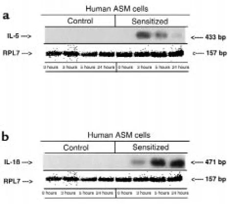
Comparison of expression of IL-5 (a) and IL-1β(b) mRNAs using RT-PCR and Southern blot analysis in human ASM cells after 0-, 3-, 6-, and 24-hour exposure to human control serum and human atopic asthmatic serum. Expression of RPL7 mRNA was used to control for gel loading. The blots were probed with human specific IL-5, IL-1β, and RPL7 32P-labeled cDNA probes (see Methods). Whereas mRNA
removed by centrifugation at 100 gfor 5 minutes. The supernatant was then salvaged, and the protein con-centration was measured using the Lowry assay. Equiv-alent amounts (30–50 µg) of membrane protein were fractionated in 9–11% SDS-polyacrylamide gels, fol-lowed by transfer to nitrocellulose membranes. The membranes were then blotted overnight at room tem-perature in 25 mM Tris-HCl (pH 7.5), 150 mM NaCl, and 0.05% Tergitol NP-40 containing 5% nonfat milk, as described previously by our laboratory (18). The pri-mary mouse anti-human IL-1β antibody used was diluted 1:250 and incubated for 1–2 hours at room temperature. The primary and secondary antibody incubations and washes were done in 25 mM Tris-HCl (pH 7.5), 150 mM NaCl, and 0.05% NP-40 containing 0.50 % nonfat milk. The IL-1β protein levels were detected using enhanced chemiluminescence after a 1-hour incubation with a 1:1,000 dilution of an mouse horseradish peroxidase–linked secondary anti-body and subsequent exposure to autoradiography film. The protein levels expressed were then quanti-tated using laser densitometry (Bio-Rad Laboratories Inc., Hercules, California, USA).
Statistical analysis. Statistical analysis was performed by means of the 2-tailed paired Student’s ttest. Pvalues less than 0.05 were considered to be significant.
Reagents. The human airway tissue was provided by the Cooperative Human Tissue Network, which is funded by the National Cancer Institute. The human ASM cells and SmBM medium were obtained from Clonetics Corp. The IL-5, IL-1β,and RPL7 primers were obtained from Integrated DNA Technologies Inc. (Coralville, Iowa, USA). The IL-1 receptor antagonist, human IL-5 receptor and IL-4 neutralizing anti-bodies, IL-5 and IL-1βELISA kits, mouse anti-human IL-1βprimary antibody, and anti-mouse secondary antibody used in protein assay studies were purchased from R&D Systems Inc. (Minneapolis, Minnesota, USA). ACh and isoproterenol were purchased from Sigma Chemical Co. (St. Louis, Missouri). All drug con-centrations are expressed as final bath concon-centrations. Isoproterenol and ACh were made fresh for each exper-iment and were dissolved in normal saline to prepare stock solutions (10–4M for both).
Results
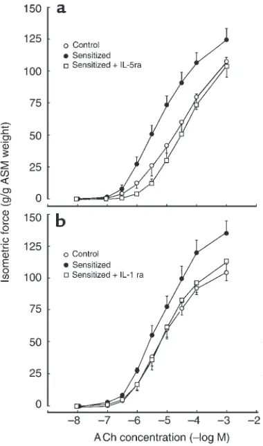
Roles of IL-5 and IL-1βin regulating ASM responsiveness in the atopic asthmatic sensitized state. Agonist-induced con-strictor and relaxation responses were separately examined in isolated rabbit tracheal ASM segments that were incubated for 24 hours in either human atopic asthmatic serum or serum from non-atopic/nonasthmatic (i.e., control) individuals, both in the absence and presence of either a monoclonal neutralizing antibody to IL-4 (IL-4Nab), an IL-5 receptor blocking antibody (IL-5ra), or the endoge-nous IL-1 receptor antagonist (IL-1ra) (see Methods). As shown in Figure 1a, relative to control serum–treated tissues (open circles), the maximal
con-strictor responses (Tmax) and sensitivities (pD50; i.e.,
–log ED50values) to exogenously administered ACh
were significantly enhanced in ASM passively sensi-tized with atopic asthmatic serum (filled circles). Accordingly, the mean ± SE Tmaxvalues amounted to
112.1 ± 5.2 and 124.5 ± 9.1 g/g tracheal smooth mus-cle (TSM) in the control and atopic sensitized tissues, respectively (P< 0.01); and the corresponding mean pD50values amounted to 4.83 ± 0.04 and 5.39 ± 0.06
–log M, respectively (P< 0.01). These induced aug-mented constrictor responses to ACh in atopic sensi-tized ASM were unaffected by pretreatment of the sensitized tissues with IL-4Nab (data not shown), and in these tissues the mean ± SE Tmaxand ED50values
remained elevated at 127.3 ± 7.1 g/g TSM and 5.42 ± 0.06 –log M, respectively. In contrast, the heightened constrictor responses to ACh were largely prevented in atopic asthmatic sensitized tissues that were pre-treated with a maximally effective concentration (i.e., 50 ng/mL) of IL-5ra (Figure 1a; open squares), where-in the mean ± SE Tmaxand pD50values amounted to
[image:6.612.309.536.409.617.2]110.1 ± 11.9 g/g TSM and 4.93 ± 0.05 –log M, respec-tively. The latter determinations were not significant-ly different from those obtained in the control serum–treated ASM tissues. Similarly, in comparable experiments, consistent with our earlier observations (17), we found that pretreatment of atopic asthmatic sensitized ASM with IL-1ra (140 ng/mL) also ablated the tissues’ heightened constrictor responsiveness to
Figure 4
ACh (Figure 1b). Accordingly, the Tmaxvalues in the
atopic sensitized vs. control tissues averaged 138.1 ± 9.9 vs. 101.2 ± 6.9 g/g TSM, respectively (P< 0.01); the corresponding pD50values averaged 5.72 ± 0.05 and
5.19 ± 0.06 –log M, respectively (P< 0.05). In the pres-ence of IL-1ra, the Tmaxand pD50values in the
sensi-tized ASM were similar to those obtained in the con-trol serum–treated tissues and averaged 104.8 ± 5.7 g/g TSM and 5.17 ± 0.04 –log M, respectively. In con-trast to these observations in atopic sensitized tissues, IL-4Nab, IL-5ra, and IL-1ra had no appreciable effect on ASM constrictor responsiveness to ACh in control serum–exposed tissues (data not shown).
In separate studies, during comparable levels of initial ACh-induced contractions in control and atopic asth-matic sensitized ASM segments averaging approxi-mately 40% of Tmax, administration of the β-adrenergic
receptor agonist, isoproterenol, elicited cumulative dose-dependent relaxation of the precontracted tissues. As shown in Figure 2a, relative to control serum–treat-ed ASM (open circles), the maximal relaxation respons-es (Rmaxvalues) and sensitivities (pD50values) to
iso-proterenol were significantly attenuated in the corresponding atopic asthmatic sensitized tissues (filled circles). Accordingly, the mean ± SE Rmaxvalue for
iso-proterenol amounted to 40.1 ± 6.2% in the atopic sensi-tized ASM, compared with 55.5 ± 6.3% in the control serum–treated ASM (P< 0.005); the corresponding pD50
values averaged 6.11 ± 0.06 and 6.47 ± 0.07 –log M, respectively (P < 0.05). These attenuated relaxation
responses to isoproterenol were unaffected in sensitized tissues pretreated with IL-4Nab, wherein the Rmaxand
pD50values averaged 38.1 ± 7.0% and 6.09 ± 0.07 –log M,
respectively (data not shown). In contrast, the impaired relaxation responses to isoproterenol were prevented in atopic asthmatic sensitized tissues that were pretreated with IL-5ra (Figure 2a; open squares), wherein the mean ± SE Rmaxand pD50values averaged 55.4 ± 5.2 % and 6.40
± 0.06 –log M, respectively. Similarly, in comparable experiments, the attenuated isoproterenol-induced relaxation responses were also ablated in atopic asth-matic sensitized ASM segments that were pretreated with IL-1ra (Figure 2b). Contrasting these observations obtained in atopic asthmatic sensitized ASM, in tissues incubated with control serum, we found that pretreat-ment with IL-4Nab, IL-5ra, and IL-1ra did not affect the tissues’ subsequent relaxation responsiveness to iso-proterenol (data not shown).
[image:7.612.58.290.52.268.2]Interactive roles of IL-5 and IL-1βin atopic asthmatic sensi-tized ASM. Taken together, the above observations sug-gested that IL-5 and IL-1βmay share a common or inter-active mechanism in mediating the proasthmatic phenotypic changes in ASM responsiveness in the atopic sensitized state. In addressing this issue, we initially con-ducted a series of experiments that examined whether cultured human ASM cells express the mRNAs and pro-teins for the above cytokines, and further assessed whether the expressions of IL-5 and IL-1βwere altered in the atopic asthmatic sensitized state. For the mRNA analyses using RT-PCR and human IL-5– and IL-1β –spe-cific primers, cDNA was reverse-transcribed from total isolated RNA primed with oligo-dT or with random hexamers, and Southern blots were subsequently pre-pared and probed with the human cDNA probes specif-ic for the human IL-5 and IL-1βgenes (see Methods). In addition, a 157-bp ribosomal protein L7 (RPL7) probe was also used as a control for gel loading, and the signals for the individual PCR products were quantitated on a PhosphorImager. As depicted in Figure 3, exposure of cultured human ASM cells to atopic asthmatic sensitiz-ing serum induced upregulated expression of both the IL-5 and IL-1βmRNA signals (Figure 3, a and b, respec-tively). Accordingly, in contrast to control serum–treat-ed cells, wherein the intensities of the IL-5 and IL-1β mRNA signals were low or barely detectable, the mRNAs for both these cytokines were markedly induced in the atopic asthmatic sensitized cells, whereas the correspon-ding constitutively expressed RPL-7 signal remained essentially unaltered. In further examining Figure 3, a and b, it can be seen that although both the 5 and IL-1βmRNA signals were upregulated in the atopic sensi-tized ASM cells, the temporal patterns of enhanced mRNA expression of these cytokines were notably dif-ferent. Thus, whereas IL-5 mRNA expression was clearly increased as early as 3 hours in the atopic sensitized cells, and the signal was subsequently attenuated by 24 hours, the corresponding IL-1βmRNA signal was more dis-tinctly increased relatively later, at 6 hours, and remained enhanced thereafter at 24 hours in the sensitized cells.
Figure 5
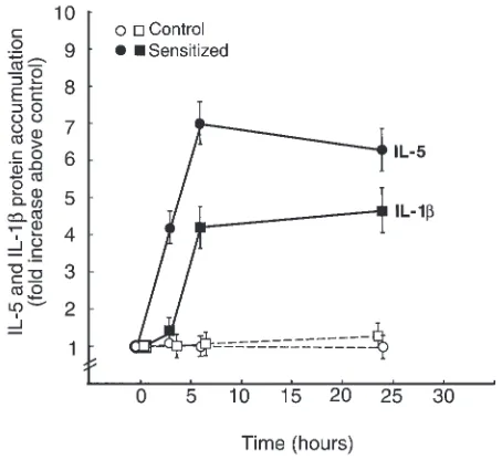
Given the above findings, in complementary studies using cytokine-specific RIAs (see Methods), we exam-ined whether the above temporal changes in IL-5 and IL-1βmRNA expression are also reflected by corre-sponding changes in the release of these cytokine pro-teins into the culture media of human ASM cells exposed to control and atopic asthmatic serum. The basal concentrations of IL-5 protein in the control and atopic asthmatic sera averaged 3.01 ± 0.37 and 4.34 ± 1.84 pq/mL, respectively; the concentrations of IL-1β averaged 10.15 ± 2.72 and 12.14 ± 1.95 pg/mL, respec-tively. The concentrations of these cytokines were then assayed in the culture medium within approximately 30 minutes of exposure of the cells to the sera (base-line), and thereafter, following 3, 6, and 24 hours of serum incubation. The basal levels of IL-5 and IL-1β protein in the culture media averaged 1.65 ± 0.28 and 8.35 ± 3.7 pg/mL, respectively. As shown in Figure 4, these levels did not significantly change in cells exposed to control serum (open symbols) for up to 24 hours. In contrast, the IL-5 (filled circles) and IL-β(filled squares) protein levels progressively increased in the culture media of the atopic asthmatic sensitized cells, reaching maximal levels of approximately 7- and 4-fold above their respective basal levels, and remained elevated at 24 hours. In comparing the temporal patterns of IL-5 and IL-1βprotein release, it will be seen that, apart from a relatively enhanced maximal proportional increase in IL-5 relative to IL-1β, the atopic sensitized cells showed an earlier release of IL-5 protein (at 3 hours), whereas the release of IL-1βprotein was large-ly delayed until 6 hours after exposure to the sensitiz-ing serum. Of note, these temporal differences in IL-5 and IL-1βprotein release by the sensitized ASM cells parallel the different temporal patterns of enhanced mRNA expression of these cytokines (Figure 3).
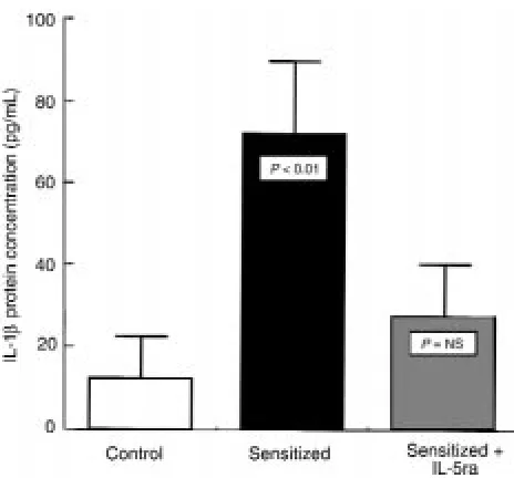
In light of the above observations, and given the sub-stantial evidence in earlier independent studies that implicated a role for either IL-5 (10–12) or IL-1β(17, 24–26) in the induction of altered airway responsive-ness following antigen challenge in various animal models, we next examined whether the above changes in IL-5 and IL-1βexpression in the atopic asthmatic sensitized state are causally and specifically related. Accordingly, in studies using cultured human ASM cells, levels of IL-1βprotein accumulation in the cell culture media were assayed after 24-hour exposure of the cells to control serum or atopic asthmatic serum in the absence and presence of IL-5ra and IL-4Nab. As shown in Figure 5, relative to control serum–treated cells, atopic asthmatic serum–sensitized cells demon-strated a significantly enhanced release of IL-1βprotein into the cell culture medium. Moreover, it will be noted that the enhanced IL-1βrelease was largely ablated in the atopic sensitized cells that were pretreated with IL-5ra (50 ng/mL). Contrasting this effect of IL-IL-5ra, pre-treatment of atopic sensitized cells with IL-4Nab had no significant effect on the augmented release of IL-1β protein (data not shown).
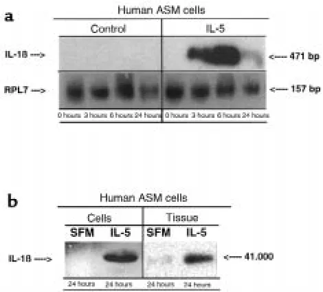
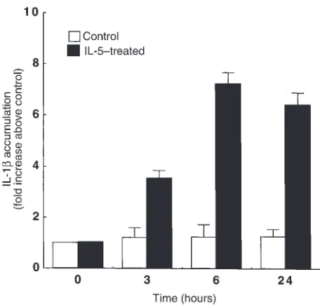
[image:8.612.305.540.52.264.2]In a complementary series of experiments, we next investigated the effects of exogenous IL-5 adminis-tration on IL-1βmRNA and protein expression, both in human cultured ASM cells and human ASM tissue preparations. As shown in Figure 6a, in contrast to control cells maintained in SFM alone, cells treated with IL-5 (5 ng/mL) exhibited upregulated IL-1β mRNA expression at 3 and 6 hours, and this aug-mented induction of IL-1β mRNA was largely resolved by 24 hours. As exemplified by the Western blots depicted in Figure 6b, relative to the essentially undetectable levels of IL-1βprotein in the membrane lysate samples of human ASM tissues maintained for 24 hours in SFM alone, expression of IL-1βprotein was markedly induced in the tissue preparations exposed to IL-5. Finally, in concert with these obser-vations, we also found that IL-5 administration to cultured ASM cells induced a progressive increase in their elaboration of IL-1βprotein into the cell culture medium for up to 24 hours (Figure 7; filled bars), whereas IL-1βrelease was unaltered in cells exposed to SFM alone (Figure 7; open bars). Thus, collective-ly, these results support the notion of a regulatory role for IL-5 in inducing the upregulated IL-1β mRNA and protein release in the atopic asthmatic sensitized state.
Figure 6
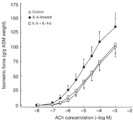
In view of the aforementioned findings, together with the above pharmacodynamic evidence implicating roles for both IL-5 and IL-1βin mediating the changes in ASM responsiveness in atopic asthmatic sensitized tis-sues (Figures 1 and 2), we subsequently examined whether the proasthmatic phenotypic changes in ASM responsiveness obtained in the atopic asthmatic sensi-tized state could be simulated by treating naive ASM with exogenously administered IL-5, and whether any observed IL-5–mediated perturbations in ASM respon-siveness are specifically modulated by pretreating the tis-sues with IL-1ra. Accordingly, in separate paired experi-ments, constrictor and relaxant dose-response relationships to ACh and isoproterenol, respectively, were compared in ASM exposed for 24 hours to vehicle alone (i.e., control) or to IL-5 in the absence and presence of IL-1ra or IL-4Nab (see Methods). As shown in Figure 8, relative to their respective controls (open circles), the Tmaxresponses to ACh were significantly enhanced in
tis-sues treated with a maximally effective concentration of IL-5 (5 ng/mL; filled circles), wherein the mean ± SE Tmax
responses amounted to 135.1 ± 24.1 vs. 100.1 ± 16.8 g/g ASM weight in the control ASM (P< 0.05). Similarly, the pD50values for ACh were significantly enhanced in
tis-sues treated with IL-5, wherein the mean ± SE pD50
value amounted to 5.11 ± 0.05 vs. 4.83 ± 0.06 –log M obtained in the control tissues (P< 0.05). In contrast, as further depicted in Figure 8, in tissues pretreated with IL-1ra, the IL-5–induced changes in ASM constrictor responsiveness to ACh were largely prevented (open squares), and there were no differences between their Tmaxand pD50values and those obtained in
correspon-ding control tissues. Moreover, in contrast to its preven-tative action in IL-5–treated ASM, we found that admin-istration of IL-1ra had no effect on either the Tmaxor
pD50values obtained in control tissues (data not shown).
Finally, pretreatment of control or IL-5–exposed ASM with IL-4Nab had no appreciable effect on the tissues’ constrictor responses to ACh (data not shown), a find-ing consistent with our recent study demonstratfind-ing that cultured ASM cells do not express IL-4 protein in either the control or atopic sensitized state (18).
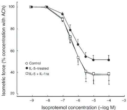
In parallel with the above observations, during simi-lar initial sustained levels of ACh-induced contractions (i.e., ∼45% Tmax), the subsequent relaxation responses to
cumulative administration of isoproterenol were markedly reduced in IL-5–treated tissues compared with control tissues (Figure 9), wherein the mean ± SE Rmax responses amounted to 48.8 ± 4.4 vs. 61.9 ± 9.0%,
respectively (P < 0.01). Similarly, the corresponding pD50values were also significantly different and
aver-aged 6.21 ± 0.06 vs. 6.55 ± 0.07 –log M in the IL-5–treat-ed vs. control tissues, respectively (P< 0.05). Moreover, in accordance with the above observations, whereas IL-4Nab had no effect, pretreatment with IL-1ra prevent-ed the attenuatprevent-ed ASM relaxant responsiveness to iso-proterenol in IL-5–exposed tissues, and there were no differences between their Rmaxvalues and pD50values
and those obtained in control tissues (Figure 9). Final-ly, in contrast to its effect in IL-5–treated ASM, admin-istration of IL-1ra to control tissues had no effect on either their Rmax or pD50responses to isoproterenol
(data not shown). Taken together, these and the above observations suggest that the nature of the mechanistic interplay between IL-5 and IL-1β in eliciting the proasthmatic perturbations in ASM responsiveness in the atopic asthmatic sensitized state is given by IL-5–induced upregulated expression of IL-1β,whereby, as previously described (17, 26), the latter cytokine appears to act in an autocrine fashion to mediate the proasth-matic changes in ASM responsiveness (see Discussion).
Discussion
In recent years, substantial evidence has been accumu-lated that implicates a predominant CD4+/Th2 profile
[image:9.612.61.289.53.270.2]of cytokine release in the pathobiology of allergic asth-ma. In this regard, it has been demonstrated that, rela-tive to nonallergic/nonasthmatic subjects, children and adults with atopic asthma show increased levels of the Th2 cytokines IL-4 and IL-5 in both their serum and bronchoalveolar lavage (BAL) fluid samples (27–32). Moreover, it has been reported that mononuclear cells isolated from serum or BAL fluid samples from atopic asthmatic individuals also demonstrate a relatively enhanced expression of the Th2 phenotype when the cells are stimulated with antigen (27, 30–33). Finally, more recent data based on genetic analyses of families with allergic asthma have shown a significant direct correlation between altered airway constrictor respon-siveness and IL-4 and IL-5 cytokine levels in serum or BAL fluid samples (27, 33, 34).
Figure 7
The roles of the Th2 cytokines in both generating and perpetuating the pulmonary inflammatory response in allergic asthma have been reasonably well character-ized. Thus, IL-4 has been found to direct immunoglob-ulin isotype switching to IgE synthesis, whereas IL-5 appears largely responsible for the maturation, differ-entiation, recruitment, and effector functions of eosinophils (5–9). Additionally, IL-5 has been impli-cated in the development of airway constrictor hyper-responsiveness in various animal models of airway sen-sitization. Accordingly, recent studies have demonstrated that (a) treatment with IL-5 induces air-way constrictor hyperresponsiveness (10–12); (b) cer-tain strains of genetically IL-5–deficient mice fail to develop airway eosinophilia and constrictor hyperre-sponsiveness after allergen challenge in the sensitized state (35); and (c) transgenic mice that constitutively overexpress IL-5 exhibit airway inflammatory changes and constrictor hyperresponsiveness, both of which are pathognomonic of human allergic asthma (36). Notwithstanding this evidence, the mechanism(s) by which IL-5 reportedly elicits the proasthmatic pheno-type of altered airway responsiveness remains to be identified. In view of the latter, together with more recent information demonstrating that the ASM itself can be induced to autologously express certain proin-flammatory cytokines (notably, IL-1β) in the atopic asthmatic sensitized state (17), the present study exam-ined the role and mechanism of action of IL-5 in direct-ly regulating ASM responsiveness in the atopic asth-matic sensitized state. Collectively, the results herein provide new evidence demonstrating that (a) IL-5 and IL-1βare endogenously released and mechanistically interact in mediating the proasthmatic-like changes in constrictor and relaxant responsiveness in atopic asth-matic sensitized ASM; and (b) the nature of this inter-action is characterized by the initial release of IL-5 by the sensitized ASM, which then induces upregulated expression of IL-1β, the latter resulting in an autocrine manifestation of the changes in ASM responsiveness. To our knowledge, this study is the first to demon-strate that the autologous sequential release of the proinflammatory cytokines IL-5 and IL-1βby atopic asthmatic sensitized ASM results in its autocrine man-ifestation of altered ASM responsiveness. In addressing the collection of evidence supporting this central find-ing, certain issues pertaining to the present observa-tions are worthy of consideration. Among these, it is rel-evant to note that our observed changes in constrictor and relaxant agonist responsiveness in the atopic asth-matic sensitized ASM tissues (Figures 1 and 2) closely mimicked the perturbations in airway function that characterize the in vivo asthmatic condition, including enhanced bronchoconstrictor responsiveness and impaired β-adrenoceptor–mediated airway relaxation (17, 19). Moreover, to the extent that these effects were found to be largely ablated in atopic sensitized ASM that were pretreated with receptor antagonists to either IL-5 or IL-1β, these results suggest that both IL-5 and
[image:10.612.307.535.428.634.2]IL-1βare released by the sensitized ASM and that these cytokines act similarly in ultimately mediating the observed changes in ASM responsiveness. In general, both of these considerations appear to concur with the collection of related information based on other pub-lished reports. Accordingly, previous studies have demonstrated that, apart from certain leukocytes, vari-ous other non-bone marrow–derived resident tissue cells (e.g., epithelial cells, keratinocytes, etc.) can also elaborate specific cytokines, including Th2-like cytokines (13–16). In this connection, our present obser-vations extend this earlier evidence by demonstrating that ASM cells can also be induced in the atopic sensi-tized state to autologously express mRNAs for IL-5 and IL-1β(Figure 3) and to elaborate these cytokine proteins into the cell culture media (Figure 4). Moreover, regard-ing the potential roles of IL-5 and IL-1β, previous stud-ies that were based on the use of specific neutralizing antibodies or receptor antagonists in in vivo animal models of allergic asthma have independently reported a role for both IL-1β(17, 24–26) and IL-5 (10–12) in mediating the observed pulmonary inflammatory responses and altered airway constrictor responsiveness following antigen challenge in the allergen-sensitized state. A priori, these earlier findings seem to be in con-flict, because they implicate a role for IL-5 and IL-1βin mediating the proasthmatic phenotype of altered air-way responsiveness. Our present observations, however, appear to reconcile this disparity, because they provide evidence in support of mutually inclusive roles for IL-5
Figure 8
and IL-1β in mediating the perturbations in ASM responsiveness in the atopic sensitized state (see below). In addressing the potential mechanism by which IL-5 and IL-1βinteract in mediating the observed changes in ASM responsiveness in the atopic sensitized state, our observations in cultured human ASM cells demon-strated that their mRNA expression of both 5 and IL-1β, as well as the release of these cytokine proteins, is upregulated in the atopic sensitized state. Moreover, based on the additional findings demonstrating that the upregulated mRNA expression and release of IL-5 preceded (i.e., by ∼3 hours) the enhanced mRNA expres-sion and release of IL-1β(Figures 3 and 4), the consid-eration was raised that the initial stimulated release of IL-5 provoked the subsequent elaboration and action of IL-1β. Indeed, in support of this concept, our extended observations demonstrated that (a) the enhanced release of IL-1βprotein by atopic asthmatic sensitized ASM cells was inhibited when the cells were pretreated with IL-5ra (Figure 5); (b) exogenous administration of IL-5 induced an upregulation of IL-1βmRNA and pro-tein expression in cultured human ASM cells (Figure 6a) and ASM tissue (Figure 6b), as well as an increased elab-oration of IL-1βprotein into the ASM culture medium (Figure 7); and (c) the proasthmatic-like perturbations in agonist constrictor and relaxant responsiveness obtained after the administration of exogenous IL-5 to naive ASM were prevented in tissues pretreated with IL-1ra (Figures 8 and 9, respectively). Taken together, these findings support the notion that our observed effects of IL-5 were largely attributed to its induced autocrine release of IL-1βand the resultant action of the latter
cytokine. This concept is, in part, consistent with our previous findings based on studies using the same experimental model described herein. In these studies, we found that the effects on ASM responsiveness of pas-sive sensitization of rabbit ASM with atopic asthmatic serum were attributed to the presence of serum IgE and its coupled activation of the low-affinity IgE receptor FceRII (20). Furthermore, under these conditions, the changes in ASM responsiveness in the atopic sensitized state were found to be mediated by the induced autocrine release and action of IL-1β(17). Additionally, we also identified that the effect of IL-1β on ASM responsiveness was largely due to its induced upregu-lated expression and action of Giprotein, specifically
Giα2and Giα3(26), which inhibit intracellular cAMP
accumulation (19). Thus, given these earlier findings, when examined in light of the present extended obser-vations, the collection of evidence implicates the pres-ence of an IgE/FceRII-initiated cooperative network of sequential autocrine signaling that involves the release of IL-5 that, in turn, induces the release of IL-1βby the ASM itself, a process that ultimately results in Gi
-medi-ated perturbations in ASM agonist responsiveness in the atopic asthmatic sensitized state.
To the extent that the present findings pertain to in vitro experimental conditions, the potential role of the above-implicated ASM autocrine cytokine interaction in inducing comparable autologous changes in ASM responsiveness in vivo remains to be identified. Our results, however, may have an important bearing on the possible mechanistic events that relate to the in vivo state. In this connection, it is relevant to note that in a recent study using an in vivo murine model of antigen-mediated chronic asthma (37), treatment with an anti–IL-5 mAb (TRFK-5) several days after antigen chal-lenge was found to reverse the existing antigen-induced airway eosinophilia, whereas the antigen-induced airway hyperresponsiveness to methacholine was unaffected by the subsequent administration of TRFK-5. Thus, these data provided evidence that, once established following antigen challenge, the enhanced airway constrictor hyperresponsiveness becomes insensitive to subsequent blockade of IL-5, despite the reversal of airway eosinophilia (37). These in vivo findings, when examined in light of our present observations, provoke the con-sideration of a potential mechanism by which the man-ifestation of an IL-5–independent state of altered airway hyperresponsiveness may occur subsequent to initial antigen challenge. Accordingly, our results demonstrate that (a) altered ASM responsiveness in the atopic asth-matic sensitized state is attributable to the activation of an autocrine mechanism that is intrinsic to the sensi-tized ASM itself; and (b) this mechanism is characterized by an early induction of IL-5 release that promotes the subsequent release of the pleiotropic proinflammatory cytokine IL-1β, which then elicits the observed changes in ASM responsiveness. Taken together, these observa-tions support the notion that, following the initial release of IL-5 by the sensitized ASM, the tissue’s
subse-Figure 9
Comparison of relaxation dose-response relationships to isopro-terenol in paired rabbit ASM segments half-maximally contracted with their respective ED50doses of ACh after treatment with SFM
[image:11.612.76.288.55.239.2]quent manifestation of the proasthmatic phenotype rep-resents a phenomenon that is dependent on the second-ary induced release and autocrine action of IL-1βand, at this point in the sequence of events, is largely independ-ent of the primary initiating action of IL-5. The presence and contribution of this potential mechanism in the in vivo state, however, remains to be established.
In conclusion, the results of the present study provide new evidence demonstrating that both the Th2 cytokine 5 and the pleiotropic proinflammatory cytokine IL-1β are endogenously released by atopic asthmatic serum–sensitized ASM, and that these cytokines act cooperatively in mediating the changes in ASM respon-siveness in the sensitized state. Furthermore, the results demonstrate that the nature of the latter cytokine inter-action is given by the sequential autocrine release of IL-5 and IL-1βby the sensitized ASM, wherein the initial endogenous elaboration of IL-5 induces the subsequent elaboration and autocrine action of IL-1β. Thus, apart from the conventional concepts related to the roles of various inflammatory cells in the pathogenesis of asth-ma, the present findings identify a potentially impor-tant mechanism by which the resident ASM itself may autologously regulate its own state of altered respon-siveness in the atopic asthmatic condition.
Acknowledgments
We thank J. Grunstein for excellent technical assistance and M. Brown for typing the manuscript. H. Hakonar-son was supported as a Parker B. Francis Fellow in Pul-monary Research. This work was supported in part by National Heart, Lung, and Blood Institute grants HL-31467 and HL-58245 and by an Institutional Develop-mental Fund Award from the Joseph Stokes, Jr. Research Institute of the Children’s Hospital of Philadelphia.
1. Bai, T.R. 1990. Abnormalities in airway smooth muscle in fatal asthma.
Am. Rev. Respir. Dis.141:552–557.
2. Goldie, R.G., Spina, D., Henry, P.J., Lulich, K.M., and Paterson, J.W. 1986. In vitro responsiveness of human asthmatic bronchus to carbachol, his-tamine, beta-adrenoceptor agonists and theophylline. Br. J. Clin. Phar-macol.22:669–676.
3. Bousquet, J., et al. 1990. Eosinophilic inflammation in asthma. N. Engl. J. Med.323:1033–1039.
4. Djukanovic, R., et al. 1990. Mucosal inflammation in asthma. Am. Rev. Respir. Dis.142:434–457.
5. Koning, H., Neijens, H.J., Baert, M.R., Oranje, A.P., and Savelkoul, H.F. 1997. T cells subsets and cytokines in allergic and non-allergic children. II. Analysis and IL-5 and IL-10 mRNA expression and protein produc-tion. Cytokine.9:427–436.
6. Gascan, H., et al. 1991. Regulation of human IgE synthesis. Clin. Exp. Allergy.21(Suppl. 1):162–166.
7. Romagnani, S. 1995. Biology of human TH1 and TH2 cells. J. Clin. Immunol.15:121–129.
8. Tavernier, J.H., et al. 1996. In Hematopoetic cell growth factors and their recep-tors.A.D. Whetton and J. Gordon, editors. Plenum Press. New York, NY. 321–361.
9. Shimoda, K., et al. 1996. Lack of IL-4-induced Th2 response and IgE class switching in mice with disrupted Stat6 gene. Nature.380:630–633. 10. Foster, P.S., Hogan, S.P., Ramsay, A.J., Matthaei, K.I., and Young, I.G. 1996. Interleukin 5 deficiency abolishes eosinophilia, airways hyperreac-tivity, and lung damage in a mouse asthma model. J. Exp. Med.
183:195–201.
11. Hall, L.R., Mehlotra, R.K., Higgins, A.W., Haxhiu, M.A., and Pearlman, E. 1998. An essential role for interleukin-5 and eosinophils in helminth-induced airway hyperresponsiveness. Infect. Immun.66:4425–4430. 12. Till, S.J., et al. 1998. Allergen-induced proliferation and interleukin-5
production by bronchoalveolar lavage and blood T cells after segmental allergen challenge. Am. J. Respir. Crit. Care Med.158:404–411. 13. Stadnyk, A.W. 1994. Cytokine production by epithelial cells. FASEB J.
8:1041–1047.
14. Luger, T.A., et al. 1990. Cytokines and the skin. Curr. Probl. Dermatol.
19:35–49.
15. Jordana, M., Sarnstrand, B., Sime, P.J., and Ramis, I. 1994. Immune-inflammatory functions of fibroblasts. Eur. Respir. J.7:2212–2222. 16. Barker, J.N., Mitra, R.S., Griffiths, C.E., Dixit, V.M., and Nickoloff, B.J.
1991. Keratinocytes as initiators of inflammation. Lancet.337:211–214. 17. Hakonarson, H., Herrick, D.J., Serrano, P.G., and Grunstein, M.M. 1997. Autocrine role of interleukin 1beta in altered responsiveness of atopic asthmatic sensitized airway smooth muscle. J. Clin. Invest.99:117–124. 18. Hakonarson, H., Maskeri, N., Carter, C., and Grunstein, M.M. 1999. Reg-ulation of TH1- and TH2-type cytokine expression and action in atopic asthmatic sensitized airway smooth muscle. J. Clin. Invest.103:1077–1087. 19. Hakonarson, H., Herrick, D.J., and Grunstein, M.M. 1995. Mechanism of impaired beta-adrenoceptor responsiveness in atopic sensitized air-way smooth muscle. Am. J. Physiol.269:L645–L652.
20. Hakonarson, H., and Grunstein, M.M. 1998. Autologously up-regulat-ed Fc receptor expression and action in airway smooth muscle mup-regulat-ediates its altered responsiveness in the atopic asthmatic sensitized state. Proc. Natl. Acad. Sci. USA.95:5257–5262.
21. Campbell, H.D., et al. 1987. Molecular cloning, nucleotide sequence, and expression of the gene encoding human eosinophil differentiation fac-tor (interleukin 5). Proc. Natl. Acad. Sci. USA.84:6629–6633.
22. Conlon, P.J., III, Cosman, D.J., Grabstein, D.J., Mopp, T.P., Kronham, S.R., Larsen, A.D., March, C.J., Price, V.L., and Current, D.P., inventors. Gene encoding biologicallyactive human interleukin 1. US patent 5,122,459. Issued June 16, 1992.
23. Seshadri, T., Uzman, J.A., Oshima, J., and Campisi, J. 1993. Identification of a transcript that is down-regulated in senescent human fibroblasts. Cloning, sequence analysis, and regulation of the human L7 ribosomal protein gene. J. Biol. Chem.268:18474–18480.
24. Watson, M.L., Smith, D., Bourne, A.D., Thompson, R.C., and Westwick, J. 1992. Cytokines contribute to airway dysfunction in antigen-chal-lenged guinea pigs: inhibition of airway hyperreactivity, pulmonary eosinophil accumulation, and tumor necrosis factor generation by pre-treatment with an interleukin-1 receptor antagonist. Am. J. Respir. Cell Mol. Biol. 8:365–369.
25. Wills-Karp, et al. 1993. Organ culture with proinflammatory cytokines reproduces impairment of the beta-adrenoceptor-mediated relaxation in tracheas of a guinea pig antigen model. Am. J. Respir. Cell Mol. Biol.
8:153–159.
26. Hakonarson, H., Herrick, D.J., Serrano, P.G., and Grunstein, M.M. 1996. Mechanism of cytokine-induced modulation of beta-adrenoceptor responsiveness in airway smooth muscle. J. Clin. Invest. 97:2593–2600. 27. Robinson, D.S., et al. 1992. Predominant TH2-like bronchoalveolar T-lymphocyte population in atopic asthma. N. Engl. J. Med.326:298–304. 28. Walker, C., et al. 1992. Allergic and nonallergic asthmatics have distinct patterns of T-cell activation and cytokine production in peripheral blood and bronchoalveolar lavage. Am. Rev. Respir. Dis.146:109–115. 29. Ackerman, V., et al. 1994. Detection of cytokines and their cell sources
in bronchial biopsy specimens from asthmatic patients. Relationship to atopic status, symptoms, and level of airway hyperresponsiveness. Chest.
105:687–696.
30. Hamid, Q., et al. 1991. Expression of mRNA for interleukin-5 in mucos-al bronchimucos-al biopsies from asthma. J. Clin. Invest.87:1541–1546. 31. Ying, S., Durham, S.R., Corrigan, C.J., Hamid, Q., and Kay, A.B. 1995.
Phe-notype of cells expressing mRNA for TH2-type (interleukin 4 and inter-leukin 5) and TH1-type (interinter-leukin 2 and interferon gamma) cytokines in bronchoalveolar lavage and bronchial biopsies from atopic asthmatic and normal control subjects. Am. J. Respir. Cell Mol. Biol.12:477–487. 32. Robinson, D., et al. 1993. Prednisolone treatment in asthma is
associat-ed with modulation of bronchoalveolar lavage cell interleukin-4, inter-leukin-5, and interferon-gamma cytokine gene expression. Am. Rev. Respir. Dis.148:401–406.
33. Umetsu, D.T., and DeKruyff, R.H. 1997. Th1 and Th2 CD4+ cells in the pathogenesis of allergic diseases [review]. Proc. Soc. Exp. Biol. Med.215:11–20. 34. Zangrilli, J.G., et al. 1995. sVCAM-1 levels after segmental antigen chal-lenge correlate with eosinophil influx, IL-4 and IL-5 production, and the late phase response. Am. J. Respir. Crit. Care Med.151:1346–1353. 35. Foster, P.S., Hogan, S.P., Ramsay, A.J., Matthaei, K.I., and Young, I.G. 1996.
Interleukin 5 deficiency abolishes eosinophilia, airways hyperreactivity, and lung damage in a mouse asthma model. J. Exp. Med.183:195–201. 36. Lee, J.J., et al. 1997. Interleukin-5 expression in the lung epithelium of
transgenic mice leads to pulmonary changes pathognomonic of asthma.
J. Exp. Med.185:2143–2156.

![2,5 Dimethyl 3 (3 methylthiophen 2 yl)perhydropyrrolo[3,4 d]isoxazole 4,6 dione](data:image/gif;base64,R0lGODlhAQABAIAAAP///wAAACH5BAEAAAAALAAAAAABAAEAAAICRAEAOw==)