0095-1137/11/$12.00 doi:10.1128/JCM.00275-11
Copyright © 2011, American Society for Microbiology. All Rights Reserved.
Multilocus Sequence Typing of
Bartonella henselae
in the United
Kingdom Indicates that Only a Few, Uncommon Sequence
Types Are Associated with Zoonotic Disease
䌤
†
Gemma L. Chaloner,
1* Timothy G. Harrison,
2Karen P. Coyne,
1David M. Aanensen,
3and Richard J. Birtles
1,4Department for Infection Biology, Institute for Infection and Global Health and School of Veterinary Science, University of
Liverpool, South Wirral CH64 7TE, United Kingdom1; Respiratory and Systemic Infection Laboratory (RSIL),
Health Protection Agency Centre for Infections, London NW9 5HT, United Kingdom2; Department of
Infectious Disease Epidemiology, Imperial College London, London W2 1PG,
United Kingdom3; and School of Environment and Life Sciences,
University of Salford, Salford M5 4WT, United Kingdom4
Received 8 February 2011/Returned for modification 18 March 2011/Accepted 30 March 2011
Bartonella henselaeis one of the most common zoonotic agents acquired from companion animals (cats) in industrialized countries. Nonetheless, although the prevalence of infections in cats is high, the number of
human cases reported is relatively low. One hypothesis for this discrepancy is thatB. henselaestrains vary in
their zoonotic potential. To test this hypothesis, we employed structured sampling to explore the population
structure ofB. henselaein the United Kingdom and to determine the distribution of strains associated with
zoonotic disease within this structure. A total of 118B. henselaestrains were delineated into 12 sequence types
(STs) using multilocus sequence typing. We observed that most (85%) of the zoonosis-associated strains belonged to only three genotypes, i.e., ST2, ST5, and ST8. Conversely, most (74%) of the feline isolates belonged to ST4, ST6, and ST7. The difference in host association of ST2, ST5, and ST8 (zoonosis associated) and ST6
(feline) was statistically significant (P< 0.05), indicating that a few, uncommon STs were responsible for the
majority of symptomatic human infections.
Bartonella henselaeinfections, manifesting most frequently
as cat scratch disease (9), are one of the most common zoo-noses acquired from companion animals in industrialized countries (15–17). In the reservoir host, the domestic cat, the prevalence of B. henselae bacteremia can exceed 40% (8), although it is usually somewhat lower in more temperate re-gions such as the United Kingdom, where the prevalence was found to be about 9% (6). Although the isolation ofB. henselae
from the blood of infected cats is straightforward, obtaining isolates from humans is difficult, and hence almost all human cases are diagnosed using serological or PCR-based ap-proaches (1, 10, 11, 15).
Genetic diversity among B. henselae strains has been as-sessed using different genotypic methods (11, 25, 30), and all have delineated multiple genotypes (2–5, 7, 10, 15, 18, 20, 21). A study in the Netherlands, based on comparison of 16S rRNA gene sequences, provided the first evidence that B. henselae
strains possessing a particular genotype were more frequently associated with zoonosis than others (5). Further support for this observation has resulted from the use of multilocus se-quence typing (MLST) (1, 15, 20). The first application of MLST toB. henselaeinvolved 37 feline and human isolates and
identified seven sequence types (STs) that formed three deep-rooted lineages, termed clonal complexes (CCs), within the species (15). The study revealed that human isolates were significantly overrepresented in one particular ST, ST1 (15). A subsequent survey of 182B. henselae isolates acquired from archives around the world also found that ST1 was significantly associated with human infection and that, broadly, the geo-graphical distribution of STs was not homogenous (1). How-ever, in both MLST studies, the sampling of strains was not coordinated either geographically or temporally. Furthermore, the number of human-associatedB. henselaeisolates examined was low.
We built on these earlier studies by (i) rigorously testing for the existence of temporal and national geographic determi-nants on the population structure ofB. henselae, (ii) develop-ing and employdevelop-ing a non-culture-based MLST protocol to fa-cilitate genotyping ofB. henselaestrains infecting symptomatic humans and thus extending exploration of their diversity in relation to the natural (feline-infecting) B. henselae popula-tion, and (iii) introducing a online database for the storage and sharing ofB. henselaeMLST data.
MATERIALS AND METHODS
Collection of cat blood samples and recovery and identification ofB. henselae
isolates.Feline blood samples were provided by six veterinary hospitals and clinics (Table 1), selected primarily on the basis of their geographic spread across England. Samples were not collected specifically for this project but rather were residual samples from cats undergoing other tests. Cats were included in the study if at least 0.5 ml of blood remained once all the required veterinary tests had been performed. Samples that shared clear epidemiological links, such as
* Corresponding author. Mailing address: The University of Liver-pool, Leahurst Campus, Chester High Road, Neston, South Wirral CH64 7TE, United Kingdom. Phone: 44 151 794 6017. Fax: 44 151 794 6005. E-mail: [email protected].
† Supplemental material for this article may be found at http://jcm .asm.org/.
䌤Published ahead of print on 6 April 2011.
2132
on May 16, 2020 by guest
http://jcm.asm.org/
repeated collections from the same animal or samples from animals living at the same address, were excluded from the study.
Primary isolates were obtained by freezing samples at⫺20°C for 24 h and then plating onto Columbia agar plates (Oxoid, United Kingdom) containing 10% defibrinated horse blood and incubating the plates at 35°C in a 5% CO2 atmo-sphere for 4 weeks. When putative colonies of bartonellae were observed, sweeps through them were subcultured onto fresh medium. DNA extracts were prepared by boiling suspensions of bacteria in sterile distilled water for 10 min.Bartonella DNA was detected using the second round of a seminested PCR assay targeting theBartonella16S-23S rRNA intergenic spacer region (ISR) (28) and charac-terized by sequencing of the PCR product in both directions using the primers used for amplification (28).
Collection of clinical specimens from humans and detection of Bartonella
DNA.The Health Protection Agency (HPA) offers a serology-based service for diagnosis of suspectedBartonellainfections in humans. In November 2005 we instigated a follow-up study of all serology-positive patients. Sera were examined for the presence ofB. henselaeandBartonella quintanaantibodies using Barto-nellaIgG and IgM indirect immunofluorescent-antibody test kits (Focus Diag-nostics, Cypress, CA). The manufacturer’s criteria were adopted for the inter-pretation of serological results but with slight modification (13). Thus, an IgM titer of⬎20 was considered evidence of current or recent infection, an IgG titer ofⱖ256 was considered presumptive evidence of recent infection, a single IgG titer of 128 was considered evidence of infection at an undetermined time, and stable or falling IgG titers from 128 in two sera taken more than 10 days apart was considered suggestive of past infection. For patients who fulfilled these criteria, demographic and clinical data were extracted from the specimen request forms and a report was sent to the requesting laboratory asking for clinical
material suitable for PCR-based confirmatory diagnostics. DNA was extracted from clinical material using a QIAamp DNA minikit (Qiagen, Hilden, Germany) at the HPA Centre for Infections in London. Extracts were typically prepared in batches of between 1 and 3 samples. A water-only negative extraction control was concurrently prepared with each batch. Extracts and extraction controls were sent to the University of Liverpool for PCR analysis. The presence of DNA from Bartonellaspecies was determined using the seminested version of the ISR-targeting PCR (28) and sequencing, as described above. A positive control, comprisingBartonella bacilliformisDNA, and a water-only negative control were concurrently tested with each batch of samples/extraction controls. Reaction mixes for the first and second stages of the assay were each prepared in dedicated laboratories, designed for purpose, in which no other activities took place and which were remote from laboratories where amplification products were pre-pared for sequencing.
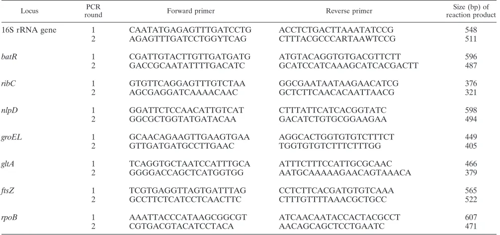
MLST.Sequence data were obtained for the eight genetic loci described in the previously definedB. henselaeMLST scheme (Table 2) (1, 15). For feline iso-lates, these data were obtained using previously described PCRs (15). However, when DNA extracts prepared fromB. henselae-infected human tissues were incorporated into these assays, they failed to yield discernible amplification products. To circumvent this problem, an approach using nested PCRs was developed. For each MLST locus, additional oligonucleotide primers targeting sequences close to, but outside, those targeted by the previously described primers were designed (Table 2). These new primers were used in first-round PCRs, and then products of these assays were incorporated as templates into second-round PCRs that employed the previously described primers. Each first-round reaction mixture contained 12.5l 2⫻PCR master mix (Abgene, Epsom, Surrey, United Kingdom), 0.5l of a 20-pmoll⫺1
[image:2.585.42.541.83.172.2]solution of both forward and
TABLE 1. Origins of cat blood samples and prevalence ofBartonellaspp. in these samples
Establishment Location No. of blood
samples
No. of isolates (% prevalence; 95% confidence interval)
All B. henselae B. clarridgeiae
RSPCA Greater Manchester Animal Hospital Manchester 190 29 (15.3; 10.6–21.4) 28 (14.7; 10.2–20.8)a 1 (0.5; 0.03–3.5)
Bristol RSPCA Clinic Bristol 193 20 (10.4; 6.6–15.7) 18 (9.3; 5.8–14.6) 2 (1.0; 0.18–4.1) Birmingham RSPCA Animal Hospital Birmingham 313 25 (8.0; 5.3–11.7) 25 (8.0; 5.3–11.7)a 0 (0; 0–1.5)
Bury & Oldham RSPCA Branch Oldham 65 3 (4.6; 1.2–13.8) 2 (3.1; 0.5–11.6) 1 (1.5; 0.08–9.4) Cats Protection National Cat Centre Sussex 568 16 (2.8; 1.7–4.6) 15 (2.6; 1.5–4.4) 1 (0.2; 0.01–0.11) University of Liverpool Wirral 453 12 (2.6; 1.4–4.7) 10 (2.2; 1.1–4.1) 2 (0.4; 0.08–1.8)
aIncludes 2 different STs that were obtained from the same cat.
TABLE 2. Oligonucleotide primers used in theB. henselaenested MLST scheme
Locus PCR
round Forward primer Reverse primer
Size (bp) of reaction product
16S rRNA gene 1 CAATATGAGAGTTTGATCCTG ACCTCTGACTTAAATATCCG 548
2 AGAGTTTGATCCTGGYTCAG CTTTACGCCCARTAAWTCCG 511
batR 1 CGATTGTACTTGTTGATGATG ATGTACAGGTGTGACGTTCTT 596
2 GACCGCAATATTTTGACATC GCATCCATCAAAGCATCACGACTT 487
ribC 1 GTGTTCAGGAGTTTGTCTAA GGCGAATAATAAGAACATCG 376
2 AGCGAGGATCAAAACAAC GCTCTTCAACACAATTAACG 321
nlpD 1 GGATTCTCCAACATTGTCAT CTTTATTCATCACGGTATC 598
2 GGCGCTGGTATGATACAA GACATCTGTGCGGAAGAA 494
groEL 1 GCAACAGAAGTTGAAGTGAA AGGCACTGGTGTGTCTTTCT 449
2 GTTGATGATGCCTTGAAC TGGTGTGTCTTTCTTTGG 405
gltA 1 TCAGGTGCTAATCCATTTGCA ATTTCTTTCCATTGCGCAAC 466
2 GGGGACCAGCTCATGGTGG AATGCAAAAAGAACAGTAAACA 379
ftsZ 1 TCGTGAGGTTAGTGATTTAG CCTCTTCACGATGTGTCAAA 565
2 GCCTTCTCATCCTCAACTTC CTTTGTTTTAAACGCTGCC 522
rpoB 1 AAATTACCCATAAGCGGCGT ATCAACAATACCACTACGCCT 607
2 CGTGACGTACATCCTACA AACAGCAGCTCCTGAATC 471
on May 16, 2020 by guest
http://jcm.asm.org/
[image:2.585.41.544.489.726.2]reverse primers, 10.5l water, and 1l DNA extract. Reaction mixtures were exposed to a thermal program of 96°C for 5 min followed by 40 cycles of 96°C for 10 s, 55°C for 10 s, and 72°C for 50 s, with a final extension step at 72°C for 10 min. The second-round reaction mixture comprised 12.5l 2⫻PCR master mix, 0.5l of a 20-pmoll⫺1
solution of both forward and reverse primers, 9.5l water, and 2l of the first-round reaction mix. The thermal program described above was used. MLST typing of human-associatedB. henselae strains was performed on anad hocbasis during the course of the study period, most often individually. To reduce the risk of cross-contamination, no positive controls were used for nested MLST PCRs; hence, only water-only negative controls were used. Reaction mixes for each stage of the MLST assays were prepared in dedicated laboratories, as detailed above for the ISR-targeting assay.
The nucleotide sequences of MLST amplification products were determined as described above and were analyzed and verified using Chromas Pro V1.4.1 (Technelysium Pty Ltd., Queensland, Australia). Alignment of verified se-quences was carried out using MEGA V4.0 (27). Alleles and STs were assigned in accordance with published data (1, 15, 20, 22, 29). New allelic combinations were assigned to new STs in their order of detection. All MLST data generated during this study and from previous work (1, 15), together with details of the provenance of theB. henselaestrains from which these data were derived, were uploaded onto a newly created publicly available internet database hosted on the MLST website (http://bhenselae.mlst.net). The juxtapositions of STs within the B. henselaepopulation structure were examined using eBURST (http://eburst .mlst.net) to analyze differences in allelic profiles (12, 26) and phylogeny inferred from alignments of concatenated sequences. Phylogenetic inferences were car-ried out using MEGA V4.0 (27) and included maximum-likelihood, parsimony, and neighbor-joining algorithms.
Statistical analysis.The existence of significant geographical or seasonal vari-ation of STs was assessed using Pearson’s chi-square test for associvari-ation in STATA V10. Values were considered to be statistically significant if thePvalue wasⱕ0.05 (5%). The relative frequencies of the feline- and human-associated strains within each ST were compared with the overall frequency of isolates in this study using Fisher’s exact test, with values considered to be statistically significant if thePvalue wasⱕ0.05 (5%).
RESULTS
Prevalence ofBartonellaspp. in cats in the United Kingdom
and MLST of feline isolates.Isolates were obtained from 103
of 1,782 feline blood samples (5.8%) (Table 1).B. henselaewas cultured from 96 samples (5.4%) andBartonella clarridgeiae
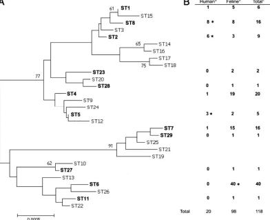
from seven (0.4%). Complete MLST profiles were obtained for isolates recovered from 94 of the 96 samples. However, for isolates obtained from two samples, unequivocal sequence data could not be obtained. Thus, five single-colony picks were taken from cultures of these samples, and each pick was indi-vidually subjected to MLST. All picks yielded unequivocal sequence data, but different picks from the same cat yielded different STs, indicating coinfection; cat RSPCA210 was coin-fected with ST5 and ST7, and cat BHAM76 was coincoin-fected with ST6 and ST8. In total, 12 STs were encountered, three of which were new (designated ST27 to ST29 in order of detec-tion). All the new STs were rare, and theB. henselae popula-tion was dominated by four STs, i.e., ST4, ST6, ST7, and ST8, which represented 19%, 41%, 15%, and 8% of isolates, respec-tively (Fig. 1).
Geographical and temporal correlates forB. henselaeSTs.
ST6 was the most commonB. henselaeST found in English cats (Fig. 1), being encountered at all six clinics involved in this study and during all seasons throughout the study period (see Fig. S1 and S2 in the supplemental material). Other common STs were also widely distributed, spatially and temporally (see Fig. S1 and S2 in the supplemental material). There was no statistical support for the geographical clustering of any STs (2 ⫽13.8,P⫽0.54) or significant seasonal variation in the incidence of specific STs (2⫽9.9,P⫽0.40).
To determine how the population structure ofB. henselaein domestic cats (i.e., the natural reservoir) in England fits with that on a wider geographical scale, data from previous MLST studies (1, 15, 20, 22, 29) were collated and divided into three categories, i.e., United Kingdom, continental Europe, and the rest of the world (Table 3). The distributions of the common STs in these three categories were not random; ST1 and ST5 were significantly more common in countries outside Europe than in England, and ST5 and ST7 was significantly more common in continental Europe than in England. Conversely, ST4 and ST6 have been significantly more frequently isolated from English cats than from cats living elsewhere (Table 3).
Identification and MLST analysis of zoonotic B. henselae
strains. Appropriate clinical material (fresh, frozen lymph
node biopsy specimens or aspirates, or heart valves) was re-ceived from 51 patients with serological evidence ofBartonella
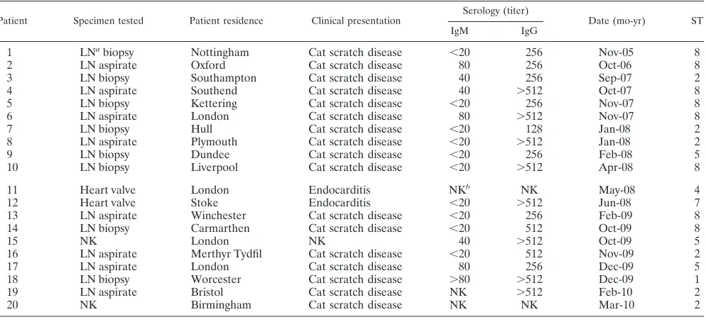
infection. Bartonella DNA was detected in tissues from 32 patients (63%). Sequencing of ISR amplification products re-vealed that 20 patients were infected withB. henselae(Table 4) and that the remaining 12 patients were infected withB. quin-tana. The nested MLST PCR protocol successfully amplified all MLST loci from the 20 patients with symptomaticB. hense-lae infections. ST8 was the most frequent ST encountered (eight patients) and ST2 was also common (six patients), while ST5 was found in three patients and ST1, ST4, and ST7 were each encountered in one patient (Fig. 1; Table 4).
Relationship between ST and host species. Although ST6
was the most predominant ST among isolates recovered from cats in our study, it was not encountered among human-asso-ciated strains (P ⬍ 0.001). However, ST2 (P ⬍ 0.001), ST5 (P⬍0.05), and ST8 (P⬍0.001) were significantly more com-mon acom-mong zoonotic strains than acom-mong feline isolates.
Analysis of B. henselaepopulation structure. Phylogenetic
analysis indicated that the 29B. henselaeSTs encountered to date, including the three new STs reported in this study, formed well-supported, distinct clusters on three deep-rooted lineages (Fig. 1). The existence of these clusters and lineages was supported by all three approaches to phylogenetic infer-ence taken (data not shown). The proposed branching orders within each cluster were not robust, with low bootstrap support and inconsistency between algorithms. The three new STs were not specifically related to one another (Fig. 1). Comparison of allelic profiles of STs using eBURST resulted in the three new STs being assigned to one of the three previously described CCs within the species (1, 15, 20, 22, 29) (see Fig. S3 in the supplemental material).
DISCUSSION
B. henselaeis a successful parasite of domestic cats; surveys
carried out worldwide have consistently demonstrated the presence of the organism in feline blood, almost always at a prevalence of greater than 5% and often exceeding 20% (8). Cat ownership is common around the globe; for example, there are an estimated 8 million cats in the United Kingdom, living in 43% of the nation’s households (23, 24), and this popularity creates a common and widespread natural reservoir for B.
henselae. However, given the scale, and hence the potential
public health threat, of this reservoir, the reported incidence of
B. henselaezoonoses is remarkably low (the HPA diagnoses
on May 16, 2020 by guest
http://jcm.asm.org/
only about 125 patients per year in the United Kingdom (14). There are several possible explanations for this discrepancy; for example, transmission ofB. henselaeto humans may be an extremely inefficient process, or transmission itself may be ef-ficient but the virulence ofB. henselaemay be such that symp-tomatic infections are rare. However, a further explanation may be thatB. henselaestrains vary in their ability to provoke disease. This explanation has support from previous studies in which certain genotypes were found to be overrepresented among human-associated strains (1, 15, 29). However, in all these studies, the validity of the association observed was somewhat compromised by the opportunistic manner in which strains were collected. Furthermore, the paucity of human isolates resulted in few being included in these studies. We have aimed to circumvent both these shortfalls. First, we at-tempted to sample the diversity ofB. henselaestrains circulat-ing in humans and domestic cats in a scirculat-ingle country durcirculat-ing the same time period. Second, rather than genotype human-asso-ciated B. henselaeisolates, we applied MLST to extracts of clinical material containingB. henselaeDNA. This strategy has previously been applied for the genotyping of
human-associ-atedB. henselaestrains with multispacer typing (MST), which
is an approach similar to MLST (19), and has also been very recently used in conjunction with MLST itself (29).
We observed rich genetic diversity among the B. henselae
strains circulating in the United Kingdom, delineating 12 dif-ferent STs in the 118 strains studied. This figure is akin to that reported recently in an MLST-based study ofB. henselae di-versity in Germany (22). In that work, 39 feline isolates were characterized into 17 STs distributed in all three of the previ-ously defined CCs of the species (22). However, such diversity was not observed in a recent study in Japan, in which only three STs, all belonging to CC1, were detected in 55 isolates char-acterized (29). These observations suggest that B. henselae
diversity is more pronounced in some parts of the world than in others. Furthermore, the frequency with which certain STs were encountered in the United Kingdom appears to be dif-ferent from that worldwide and in mainland Europe, adding weight to previous work in which a geographic correlation ofB.
henselae STs was first demonstrated and extending this by
revealing significant variation on a regional (i.e., United King-dom versus mainland Europe) as well as on a global scale. We also attempted to identify temporal variation in ST incidence but could find no evidence for this.
FIG. 1. (A) Dendrogram showing phylogenetic relationships among the 29B. henselaeMLST STs encountered to date. STs in bold were encountered in this study. The dendrogram was inferred from a 3,398-bp alignment of concatenated sequence data derived from the eight MLST loci and assessed using a neighbor-joining approach. The strength of the proposed branching order was assessed using bootstrapping (1,000 replications), and the results of this analysis are expressed as a percentage at each node. Only scores of over 60% are indicated. (B) Frequency with which each ST was encountered in cats or humans in the study. For each ST encountered, the statistical significance of the difference in the frequency of encounter in cats and humans was calculated. An asterisk indicates a significant overrepresentation of that ST within a host type as examined by Fisher’s exact test.
on May 16, 2020 by guest
http://jcm.asm.org/
[image:4.585.96.488.74.393.2]We revealed a significant correlation between specific STs and human disease in the United Kingdom. We found that ST2, ST5, and ST8 were significantly associated with zoonosis. Although the finding of a correlation between a specific ST and human disease was in accordance with earlier work (1, 15), previous studies found that human-associated strains were overrepresented in ST1, as opposed to ST2, ST5, and ST8. Why these three STs, and not ST1, present the greatest public health risk in the United Kingdom is unclear; however, given that geography is a determinant of B. henselae population structure, it is not an unsurprising observation. It is noteworthy that the vast majority of human-associated isolates included in earlier MLST studies were not from Western Europe (1, 15, 20, 22, 29), and the worldwide zoonotic importance of ST1 was determined using univariant analysis without considering geo-graphic influence. Thus, whereasB. henselaestrains belonging to ST1 may be more likely to be associated with zoonosis in Australia, Japan, and maybe the United States, this association does not necessarily hold elsewhere, as demonstrated in our study. Further application of MLST on a national or regional scale will be necessary to resolve this.
TheB. henselaepopulation structure defined by MLST
ap-pears to be generally congruent with that defined using MST (18), and using both approaches, MLST CC1/MST cluster 1 contains significantly more human-associated strains than other clusters (1, 15, 19). However, a recent study using MST to characterize human-associated strains predominantly from France found that many belonged to MST cluster 3, which corresponds to MLST CC2 (19). Furthermore, the distribution of the genotypes of human-associated strains was not signifi-cantly different from that of the genotypes of feline isolates. These observations are surprising given that they are discrep-ant with our work and previous MLST-based studies (1, 15), and explaining the discrepancy is difficult. Perhaps MST geno-TABLE 3. Correlation between ST and geographical origin among
felineB. henselaeisolates for which MLST data are currently availablea
ST
No. of isolates from:
United Kingdom
Elsewhere in Europe
Elsewhere in world
1 5 12 74b
2 3 0 2
3 0 0 1
4 25 1b 11b
5 2 33b
48b
6 46 13b 31b
7 20 50b 55
8 9 7 8
9 0 1 4
10 2 0 2
11 1 1 1
12 0 0 1
13 0 1 1
14 0 2 2
15 0 0 1
16 0 1 1
17 0 1 1
18 0 1 1
19 0 2 2
20 0 1 1
21 0 1 1
22 0 1 1
23 2 1 1
24 0 1 1
25 0 1 1
26 0 3 3
27 1 0 0
28 1 0 0
29 1 0 0
Total 118 135 256
a
Isolates are broken down into those from the United Kingdom, the rest of Europe, and the rest of the world (including Europe). The numbers of isolates assigned to each ST were calculated from previous MLST studies (1, 15, 20, 22, 29).
b
[image:5.585.43.283.100.422.2]Significant difference as calculated by Fisher’s exact test.
TABLE 4. Clinical, diagnostic, and MLST data relating to the 20 patients included in this study
Patient Specimen tested Patient residence Clinical presentation Serology (titer) Date (mo-yr) ST
IgM IgG
1 LNabiopsy Nottingham Cat scratch disease ⬍20 256 Nov-05 8
2 LN aspirate Oxford Cat scratch disease 80 256 Oct-06 8
3 LN biopsy Southampton Cat scratch disease 40 256 Sep-07 2
4 LN aspirate Southend Cat scratch disease 40 ⬎512 Oct-07 8
5 LN biopsy Kettering Cat scratch disease ⬍20 256 Nov-07 8
6 LN aspirate London Cat scratch disease 80 ⬎512 Nov-07 8
7 LN biopsy Hull Cat scratch disease ⬍20 128 Jan-08 2
8 LN aspirate Plymouth Cat scratch disease ⬍20 ⬎512 Jan-08 2
9 LN biopsy Dundee Cat scratch disease ⬍20 256 Feb-08 5
10 LN biopsy Liverpool Cat scratch disease ⬍20 ⬎512 Apr-08 8
11 Heart valve London Endocarditis NKb NK May-08 4
12 Heart valve Stoke Endocarditis ⬍20 ⬎512 Jun-08 7
13 LN aspirate Winchester Cat scratch disease ⬍20 256 Feb-09 8
14 LN biopsy Carmarthen Cat scratch disease ⬍20 512 Oct-09 8
15 NK London NK 40 ⬎512 Oct-09 5
16 LN aspirate Merthyr Tydfil Cat scratch disease ⬍20 512 Nov-09 2
17 LN aspirate London Cat scratch disease 80 256 Dec-09 5
18 LN biopsy Worcester Cat scratch disease ⬎80 ⬎512 Dec-09 1
19 LN aspirate Bristol Cat scratch disease NK ⬎512 Feb-10 2
20 NK Birmingham Cat scratch disease NK NK Mar-10 2
a
LN, lymph node.
b
NK, not known.
on May 16, 2020 by guest
http://jcm.asm.org/
[image:5.585.45.542.485.712.2]type 5, which among the genotypes within MST cluster 3 is the one by far the most frequently associated with zoonosis, is rare or absent in the United Kingdom. Clearly, combined MLST-and MST-based analysis ofB. henselaestrains from the United Kingdom and France would be useful in resolving this uncer-tainty.
Why specific STs are more frequently associated with symp-tomatic human infections than others is uncertain; clearly, the ability to infect humans holds no selective advantage for B.
henselae, as humans are accidental hosts, and thus the zoonotic
importance of these STs must be an unfortunate side effect of some other adaptation. Indeed, whether these STs share com-mon determinants of zoonotic relevance or whether each pos-sesses unique traits that coincidently result in more frequent human disease is unknown. Given this uncertainty, identifying the genetic basis of the specific zoonotic threat posed by these STs is likely to be extremely difficult. Nonetheless, the use of comparative genomics may, at least, offer a means to identify ST-specific genetic “motifs” and could help identify “viru-lence” factors associated only with thoseB. henselaeSTs which are most frequently associated with zoonosis.
ACKNOWLEDGMENTS
This work was funded in part by a BBSRC Doctoral Training Award to G.L.C.
We thank the RSPCA Greater Manchester Animal Hospital, the Birmingham RSPCA Animal Hospital, the Cats Protection’s National Cat Adoption Centre, the Bury & Oldham RSPCA Branch, the Bristol RSPCA Clinic, and the University of Liverpool Small Animal Hospital for submitting excess blood samples. We also thank Heather Ford, Teresa Stocki, and Agatha Opoku-Boateng of the HPA, who carried out the serology and DNA extractions on the human clinical samples. None of the authors of this paper has a financial or personal rela-tionship with other people or organizations that could inappropriately influence or bias the content of the paper.
REFERENCES
1.Arvand, M., E. Feil, M. Giladi, H. Boulouis, and J. Viezens.2007. Multi-locus sequence typing ofBartonella henselaeisolates from three continents reveals hypervirulent and feline-associated clones. PLoS One2:e1346.
2.Arvand, M., A. J. Klose, D. Schwartz-Porsche, H. Hahn, and C. Wendt.2001. Genetic variability and prevalence ofBartonella henselaein cats in Berlin, Germany, and analysis of its genetic relatedness to a strain from Berlin that is pathogenic for humans. J. Clin. Microbiol.39:743–746.
3.Arvand, M., and J. Viezens.2007. Evaluation of pulsed-field gel electropho-resis and multi-locus sequence typing for the analysis of clonal relatedness amongBartonella henselaeisolates. Int. J. Med. Microbiol.297:255–262. 4.Berghoff, J., J. Viezens, L. Guptill, M. Fabbi, and M. Arvand.2007.
Barto-nella henselaeexists as a mosaic of different genetic variants in the infected host. Microbiology153:2045–2051.
5.Bergmans, A. M. C., J. F. P. Schellekens, J. D. A. vanEmbden, and L. M. Schouls.1996. Predominance of two Bartonella henselaevariants among cat-scratch disease patients in the Netherlands. J. Clin. Microbiol.34:254– 260.
6.Birtles, R. J., G. Laycock, M. J. Kenny, S. E. Shaw, and M. J. Day.2002. Prevalence ofBartonellaspecies causing bacteraemia in domesticated and companion animals in the United Kingdom. Vet. Rec.151:225–229.
7.Bouchouicha, R., et al.2009. Molecular epidemiology of feline and human Bartonella henselaeisolates. Emerg. Infect. Dis.15:813–816.
8.Boulouis, H. J., C. C. Chang, J. B. Henn, R. W. Kasten, and B. B. Chomel.
2005. Factors associated with the rapid emergence of zoonoticBartonella infections. Vet. Res.36:383–410.
9.Chomel, B. B., and R. W. Kasten.2010. Bartonellosis, an increasingly rec-ognized zoonosis. J. Appl. Microbiol.109:743–750.
10.Dillon, B., et al.2002. Limited diversity among human isolates ofBartonella henselae. J. Clin. Microbiol.40:4691–4699.
11.Drancourt, M., et al.1996. New serotype ofBartonella henselaein endocar-ditis and cat-scratch disease. Lancet347:441–443.
12.Feil, E. J., B. C. Li, D. M. Aanensen, W. P. Hanage, and B. G. Spratt.2004. eBURST: inferring patterns of evolutionary descent among clusters of re-lated bacterial genotypes from multilocus sequence typing data. J. Bacteriol.
186:1518–1530.
13.Harrison, T. G., and N. Doshi.1999. Serological evidence ofBartonellaspp. infection in the UK. Epidemiol. Infect.123:233–240.
14.Health Protection Agency.2010. Bartonella data. http://www.hpa.org.uk/web /HPAweb&HPAwebStandard/HPAweb_C/1195733754721. Health Protec-tion Agency, London, United Kingdom.
15.Iredell, J., et al.2003. Characterization of the natural population of Barto-nella henselaeby multilocus sequence typing. J. Clin. Microbiol.41:5071– 5079.
16.Jackson, L. A., B. A. Perkins, and J. D. Wenger.1993. Cat-scratch disease in the United-States—an analysis of 3 national databases. Am. J. Public Health
83:1707–1711.
17.Kaplan, S., et al.2002. Cat-scratch disease in children—Texas, September 2000-August 2001. JAMA287:2647–2649. [Reprinted from MMWR Morb. Mortal. Wkly. Rep.51:212–214, 2002.]
18.Li, W. J., et al.2006. Multispacer typing to study the genotypic distribution ofBartonella henselaepopulations. J. Clin. Microbiol.44:2499–2506. 19.Li, W. J., D. Raoult, and P. E. Fournier.2007. Genetic diversity ofBartonella
henselaein human infection detected with multispacer typing. Emerg. Infect. Dis.13:1178–1183.
20.Lindroos, H., et al.2006. Genome rearrangements, deletions, and amplifi-cations in the natural population ofBartonella henselae. J. Bacteriol.188:
7426–7439.
21.Maruyama, S., et al.2001. Genomic diversity ofBartonella henselaeisolates from domestic cats from Japan, the U. S. A. and France by pulsed-field gel electrophoresis. Vet. Microbiol.79:337–349.
22.Mietze, A., et al.21 September 2010. Combined MLST and AFLP typing of Bartonella henselaeisolated from cats reveals new sequence types and sug-gests clonal evolution. Vet. Microbiol. doi:10.1016/j.vetmic.2010.08.012. 23.Murray, J. K., W. J. Browne, M. A. Roberts, A. Whitmarsh, and T. J.
Gruffydd-Jones.2010. Number and ownership profiles of cats and dogs in the UK. Vet. Rec.166:163–168.
24.Pet Food Manufacturers’ Association.2010. http://www.pfma.org.uk/overall /pet-population-figures-.htm. Pet Food Manufacturers’ Association, London, United Kingdom.
25.Sander, A., M. Ruess, S. Bereswill, M. Schuppler, and B. Steinbrueckner.
1998. Comparison of different DNA fingerprinting techniques for molecular typing ofBartonella henselaeisolates. J. Clin. Microbiol.36:2973–2981. 26.Spratt, B. G., W. P. Hanage, B. Li, D. M. Aanensen, and E. J. Feil.2004.
Displaying the relatedness among isolates of bacterial species—the eBURST approach. FEMS Microbiol. Lett.241:129–134.
27.Tamura, K., J. Dudley, M. Nei, and S. Kumar.2007. MEGA4: Molecular evolutionary genetics analysis (MEGA) software version 4.0. Mol. Biol. Evol.
24:1596–1599.
28.Telfer, S., et al.2005. Disruption of a host-parasite system following the introduction of an exotic host species. Parasitology130:661–668. 29.Yanagihara, M., et al.2010. Molecular typing ofBartonella henselaeDNA
extracted from human clinical specimens and cat isolates in Japan. FEMS Immunol. Med. Microbiol.60:44–48.
30.Zeaiter, Z., P. E. Fournier, and D. Raoult.2002. Genomic variation of Bartonella henselaestrains detected in lymph nodes of patients with cat scratch disease. J. Clin. Microbiol.40:1023–1030.