Immunoassay for Detection of
Plasmodium
Species in Settings Where
Malaria Is Endemic
P. F. Mens,aH. M. de Bes,aP. Sondo,bN. Laochan,c,dL. Keereecharoen,c,dA. van Amerongen,eJ. Flint,fJ. R. S. Sak,c,dS. Proux,c,d
H. Tinto,band H. D. F. H. Schalliga
Koninklijk Instituut voor de Tropen/Royal Tropical Institute, Amsterdam, Netherlandsa; Institut de Recherche en Science de la Sante, Bobo-Dioulasso, Burkina Fasob; Shoklo Malaria Research Unit, Mae Sot, Thailandc; Mahidol-Oxford Tropical Medicine Research Unit, Mahidol University, Bangkok, Thailandd; Wageningen UR Food and Biobased Research, Biomolecular Sensing and Diagnostics, Wageningen, Netherlandse; and Forsite Diagnostics, Ltd., Sand Hutton, York, United Kingdomf
Declining malaria transmission and known difficulties with current diagnostic tools for malaria, such as microscopy and rapid
diagnostic tests (RDTs) in particular at low parasite densities, still warrant the search for sensitive diagnostic tests. Molecular
tests need substantial simplification before implementation in clinical settings in countries where malaria is endemic. Direct
blood PCR (db-PCR), circumventing DNA extraction, to detect
Plasmodium
was developed and adapted to be visualized by
nu-cleic acid lateral flow immunoassay (NALFIA). The assay was evaluated in the laboratory against samples from confirmed
Suda-nese patients (
n
ⴝ
51), returning travelers (
n
ⴝ
214), samples from the Dutch Blood Bank (
n
ⴝ
100), and in the field in Burkina
Faso (
n
ⴝ
283) and Thailand (
n
ⴝ
381) on suspected malaria cases and compared to RDT and microscopy. The sensitivity and
specificity of db-PCR-NALFIA compared to the initial diagnosis in the laboratory were 94.4% (95% confidence interval [CI]
ⴝ
0.909 to 0.969) and 97.4% (95% CI
ⴝ
0.909 to 0.969), respectively. In Burkina Faso, the sensitivity was 94.8% (95% CI
ⴝ
0.88.7 to
97.9%), and the specificity was 82.4% (95% CI
ⴝ
75.4 to 87.7%) compared to microscopy and 93.3% (95% CI
ⴝ
87.4 to 96.7%)
and 91.4% (95% CI
ⴝ
85.2 to 95.3%) compared to RDT. In Thailand, the sensitivity and specificity were 93.4% (CI
ⴝ
86.4 to
97.1%) and 90.9 (95% CI
ⴝ
86.7 to 93.9%), respectively, compared to microscopy and 95.6% (95% CI
ⴝ
88.5 to 98.6%) and
87.1% (95% CI
ⴝ
82.5 to 90.6) compared to RDT. db-PCR-NALFIA is highly sensitive and specific for easy and rapid detection of
Plasmodium
parasites and can be easily used in countries where malaria is endemic. The inability of the device to discriminate
Plasmodium
species requires further investigation.
P
roper and fast diagnosis followed by appropriate treatment is
essential for the management of malaria (
20
) and is
funda-mental for the control and eradication of the disease.
Neverthe-less, in many areas particularly, in resource-poor settings, the
di-agnosis of malaria is still based on clinical symptoms without
laboratory confirmation. Laboratory examination of blood
spec-imens to support malaria diagnosis is mainly based on microscopy
or rapid diagnostic tests (RDTs) (
18
). However, these tests require
experienced staff (microscopy) or have a low detection limit
(RDTs) (
5
). Molecular techniques such as PCR to detect
Plasmo-dium
infections have demonstrated high sensitivity and specificity
(
14
,
15
) and have the ability to quantify parasitemia when used in
a quantitative real-time PCR format. Therefore, molecular
tech-nologies are frequently used in malaria studies and in
well-equipped laboratories as the “gold standard” (
15
). However, the
implementation of molecular techniques in resource-poor
set-tings is hindered by the requirement for DNA isolation, as well as
careful handling of the clinical specimen in order to avoid sample
contamination. In addition, the analysis of amplicons obtained
from conventional PCR formats is elaborate and often requires
either a toxic and environmentally hazardous ethidium bromide
gel for visualization. An alternative is expensive real-time PCR
equipment for analysis (
3
,
6
).
Notwithstanding the above, a more sensitive tool for the
detec-tion of low-level parasitemia is needed not only for general
ma-laria case management but especially in areas where mama-laria
inci-dence is decreasing. In these areas, close vigilance of patients and
asymptomatic carriers harboring a low level of parasites is
essen-tial for immediate treatment and subsequent case detection in
order to avoid a resurgence of malaria.
Several attempts have been made to simplify molecular tools
such as the amplification of DNA in an isothermal manner (
7
),
adding the specimen without complete DNA isolation (
4
), or by
adding colorimetric substances that allow the PCR result to be
interpreted visually (
8
). In the present study, a direct blood PCR
(db-PCR) combined with a rapid readout system, nucleic acid
lateral flow immunoassay (NALFIA), is described. The direct
blood approach circumvents preamplification handling such as
DNA extraction. The full blood sample can be directly added to
the PCR mixture and subsequently amplify the target DNA of
Plasmodium
in less than 1 h.
Thereafter, the product can be visualized with NALFIA, which
is a rapid immunochromatographic test to detect labeled
ampli-con products on a nitrocellulose stick coated with specific
anti-bodies (
12
). The amplicons are labeled via specific primers that
contain a biotin molecule and a hapten. This complex is detected
Received8 June 2012Returned for modification9 July 2012
Accepted29 July 2012
Published ahead of print22 August 2012 Address correspondence to P. F. Mens, [email protected]. P.F.M. and H.M.D.B. contributed equally to this article.
Copyright © 2012, American Society for Microbiology. All Rights Reserved.
doi:10.1128/JCM.01426-12
on May 16, 2020 by guest
http://jcm.asm.org/
by direct interaction with a colloidal, neutravidin-labeled carbon
particle. The detection test is a simple, straightforward, and safe
one-step procedure in which the results are visible within 10 min.
This methodology was extensively tested in a laboratory setting
and subsequently evaluated for its sensitivity and specificity
com-pared to RDT and expert microscopy in Nanoro, Burkina Faso,
and Mae Sot, Thailand—two areas of high endemicity for
P.
fal-ciparum
and
P. falciparum
/
P. vivax
, respectively.
MATERIALS AND METHODS
db-PCR.The db-PCR is based on a combination of Phusion db-PCR buffer (Finnzymes, Espoo, Finland) and Phire Hotstart II DNA polymer-ase (Finnzymes). The dB-PCR requires two primer pairs, one pair for the amplification of pan-Plasmodiumand the second pair for amplification of the human housekeeping gene glyceraldehyde 3-phosphate dehydroge-nase (GAPDH). The GAPDH gene is used as an amplification control, as well as an internal running control for the NALFIA (see below for the interpretation of the results). In order to prevent extra handling steps, an internal amplification control from another source was not added. Each primer pair contains a digoxigenin- or Texas Red-labeled primer and a primer labeled with biotin. The dB-PCR comprises of 10l of Phusion blood direct buffer, 250 nMPlasmodium18S forward primer (digoxige-nin-5=-TCAGATACCGTCGTAATCTTA-3=), 250 nMPlasmodium18S reverse primer (biotin-5=-AACTTTCTCGCTTGCGCG-3=), 88 nM GAPDH forward (biotin-5=-TGCACCACCAACTGCTTAGC-3=), and 88 nM GAPDH reverse primer (Texas Red-5=-GGCATGGACTGTGGTCAT GAG-3=), 0.4l of Phire Hotstart II DNA polymerase, water, and 2.5l of EDTA blood, with a final volume of 25l. The cycling conditions con-sisted of an initial activation step of 30 s at 98°C, followed by 10 cycles of 5 s at 98°C, 15 s at 60°C and 30 s at 72°C, than 28 cycles of 5 s at 98°C, 15 s at 58°C, and 30 s at 72°C, and a final step of 1 min at 72°C (MyCycler; Bio-Rad). For each experiment a blood sample negative forPlasmodium, a blood sample containingP. falciparumand a nontemplate control of only water were used as control samples.
NALFIA.A HiFlow 135 nitrocellulose membrane (25 by 5 mm per strip) (Millipore, Amsterdam, Netherlands) was used to fabricate the NALFIA sticks for this assay. On the nitrocellulose, 0.8 mg/ml (0.8l/cm) of anti-Texas Red rabbit IgG fraction (Invitrogen, Paisley, United King-dom) and 0.2 mg/ml (0.8l/cm) of anti-digoxigenin polyclonal antibody (Roche Diagnostics, Mannheim Germany) were sprayed within 9 mm of one another. A Surewick G041 glass fiber sample pad (Millipore, Billerica, MA) was used to spray 2.5l of neutravidin-labeled carbon suspension (three parts neutravidin-labeled carbon/cm and two parts sodium tet-raborate buffer solution containing 6.25% sucrose and 6.25% bovine se-rum albumin [BSA]) and attached to the NALFIA stick. The strips were air dried at 37°C, packed in plastic housing, and sealed in air-tight bags con-taining silica until further use.
Detection of PCR fragments by NALFIA and gel electrophoresis.
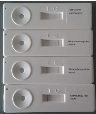
After PCR, 5l of PCR product and 70l of running buffer (0.1 M borate buffer [pH 8.8], 1% BSA, and 1% [wt/vol] sodium azide) were added on the sample pad of the NALFIA and, after 10 min, the results were read. The NALFIA was considered valid but negative forPlasmodiumif a black line at the GAPDH position was visible (control line). If two lines (control and test line) were visible, the NALFIA was considered to be positive for Plas-modium. If no lines were present or only a single test line was present, the NALFIA was considered to be a test failure. Examples of the different interpretation possibilities can be found inFig. 1.
During laboratory evaluation NALFIA analysis was compared to visu-alization of DNA bands in ethidium bromide-stained 3% agarose gels in which a pan-Plasmodiumfragment of 180 bp and human GAPDH frag-ment of 224 bp could be observed.
Laboratory evaluation db-PCR-NALFIA.The analytical sensitivity of the dB-PCR-NALFIA was assessed in triplicate by using a 5%P. falcipa-rumNF54 ring stage culture (in 5% hematocrit) and aP. falciparum-positive patient blood sample with 12% parasitemia. The cultured
para-sites were diluted 10-fold with Plasmodium-negative donor blood to obtain malaria parasites dilutions ranging from 0.55 to 5.10⫺8% in a 45%
hematocrit. The patient sample was diluted to aPlasmodiumdensity of 12 to 5.10⫺4%.
The specificity of the test was determined by analyzing malaria-posi-tive and -negamalaria-posi-tive EDTA blood samples (n⫽365) from travelers return-ing from areas of malaria endemicity, from Sudanese patients with a con-firmedP. falciparuminfection, and samples provided by the Dutch Blood Bank, which excludes donations from patients who have traveled in the past 9 months to areas of malaria endemicity and can therefore be con-sidered negative for malaria. The samples from returning travelers were provided by the London School of Hygiene and Tropical Medicine (Lon-don, United Kingdom), and the samples from Sudanese patients (n⫽51) were provided by the Wad Medani Teaching Hospital in Sudan. These blood samples were analyzed by an experienced technician by microscopy and/orPlasmodium-specific nested PCR according to the method of Snounou et al. (17). The malaria-positive samples (n⫽250) contained the following species:P. falciparum(n⫽65),P. vivax(n⫽33),P. malariae (n⫽48),P. ovale(n⫽92),P. falciparum-P. malariaemixed infection (n⫽ 4),P. falciparum-P. vivaxmixed infection (n⫽5), andP. falciparum-P. ovalemixed infection (n⫽3). Samples of malaria-suspected patients (n⫽ 15) that were found to be negative by expert microscopy and PCR served together with the Dutch blood bank samples (n⫽100) as negative con-trols.
Field evaluation of db-PCR-NALFIA study site and patient descrip-tion.Two prospective studies were conducted: one in the Health and Social Promotion Centers of Nanoro Health District (Nanoro and Nazo-anga), Boulkiemdé Province, Burkina Faso, and the other in Wang Pa and Mae Khon Ken, Mae Sot district, Tak Province, Thailand. Malaria in Nanoro is holoendemic, and transmission is perennial, with a seasonal peak during the rainy season that usually lasts from June to October (21). P. falciparumwas the main infecting species. That study was performed in
FIG 1Examples of different test outcomes of the db-PCR-NALFIA. Control and test lines are absent. (A) This is a test failure but can also be seen when only water is amplified (negative control). (B) A positive control line is visible and a test line is absent, indicating that the test is valid but that no parasite DNA is detected. (C) Both a positive control and a test line are visible, indicating a valid test positive forPlasmodium. (D) Only the test line is visible. This test should be considered a test failure.
on May 16, 2020 by guest
http://jcm.asm.org/
[image:2.585.320.524.66.307.2]conjunction with the ongoing study “Pharmacovigilance for Artemisinin-Based Combination Treatments in Africa” (protocol A70283). After col-lection, samples were transported to the Clinical Research Unit of Nanoro for further analysis and processing.
Mae Sot is located on the Thai-Myanmar border. In this hill-forested region, with low year-round transmission and two seasonal peaks, the predominant species causing malaria areP. vivax, and during the rainy season,P. falciparum(16). Patients were recruited in the malaria clinics of Wang Pa and Mae Khon Ken located around Mae Sot. Samples were transported to the Shoklo Malaria Research Unit (SMRU) in Mae Sot for further processing.
Patients presenting in outpatient clinics with a clinical suspicion of uncomplicated malaria and an axillary temperatureⱖ37.5°C or a history of fever in the past 24 h were enrolled. In Burkina Faso patients of all ages were enrolled, whereas in Thailand patients older than 3 years were en-rolled. For both field evaluations ethical approval was obtained from local ethical review boards.
Laboratory procedure field evaluation.From each participant, 200l of finger prick blood was collected in EDTA tubes (Stastedt, Numbrecht, Germany) for the preparation of thin and thick Giemsa-stained micros-copy slides, a histidine-rich protein II- andPlasmodiumlactate dehydro-genase-based RDT (SD Bioline Malaria Antigen Pf/Pan; Standard Diag-nostics, Inc., Kyonggi-do, Korea), and the db-PCR-NALFIA. Microscopy was performed according to international and good clinical and labora-tory practices guidelines by local expert microscopists (19,22). In Burkina Faso, parasites were counted against 200 leukocytes, with parasite-nega-tive results based on screening of 100 microscopic fields at⫻1,000 mag-nification. In the case of lower parasitemia (⬍10 parasites/200 leuko-cytes), parasites were counted against 500 leukocytes. Slides were examined by two readers and, in the case of discordant results, by a third reader. Discordant results were defined as a difference between the two readers in (i)Plasmodiumspecies, (ii) positive and negative, (iii) with parasitemia⬎400/l; if the higher count divided by the lower count was ⬎2 or⬎4) with parasitemia atⱕ400/l and if the higher reading density was⬎1 log10higher than the lowest reading. In Thailand, parasites were counted against 500 leukocytes at⫻1,000 magnification. If only one par-asite was found after counting 500 leukocytes, then counting continued until a second parasite was observed with a limit of counting 4,000 leuko-cytes. Also, here all slides were read by two individual microscopists, and discordant results were read by a third reader. For both settings, a leuko-cyte count of 8,000/l was assumed to calculate the parasite density perl, and the final result was the geometric mean of the readings. In both set-tings, peripheral blood was applied to the SD Bioline Malaria Antigen Pf/Pan RDT according to the manufacturer’s instructions. The db-PCR-NALFIA was performed as described above by local operators. All test operators were blinded from each other’s test result. APlasmodium spe-cies differentiation PCR was performed as described by Snounou et al. (17) on specimens that yielded discordant results and on a selection of microscopy-negative samples from Thailand. For the differentiation PCR, DNA was extracted from blood using a DNA minikit (Qiagen, Germany) according to the manufacturer’s instructions. Samples from Burkina Faso were not available for species differentiation PCR analysis.
Data analysis.All data from the field evaluations were collected on separate case record forms and subsequently entered into Microsoft Excel. Calculations on sensitivity, specificity, and agreement between the mi-croscopy, RDT, and db-PCR-NALFIA results were done using Epi Info version 6.04 (Centers for Disease Control and Prevention, Atlanta, GA). The kappa () value (calculated with a 95% confidence interval [CI]) expresses the agreement beyond chance (2). Avalue of 0.21 to 0.60 is moderate, avalue of 0.61 to 0.80 is good, and avalue of⬎0.80 is an almost-perfect agreement beyond chance.
RESULTS
Analytical performance of the db-PCR-NALFIA.
The lower
de-tection limit of the developed db-PCR-NALFIA was determined
to be 1 parasite/
l and 8 parasites/
l when tested on cultured
parasites and patient blood, respectively. db-PCR-NALFIA was
able to detect a 10-fold-higher dilution than the ethidium
bro-mide staining of agarose gels (data not shown).
In total, 365 samples were analyzed by db-PCR-NALFIA and
compared to their initial laboratory diagnosis. Of the 115 samples
considered to be negative in their initial diagnosis, 112 were also
found to be negative by db-PCR-NALFIA. The three positive
sam-ples belonged to the group of Dutch Blood Bank donors. Fourteen
samples that were initially identified as positive for
Plasmodium
were identified as negative by db-PCR-NALFIA. Of these, seven
samples contained
P. falciparum
, one sample contained
P. vivax
,
three samples were microscopically identified as
P. ovale
, and two
were microscopically identified as
P. malariae.
In addition, one
P.
falciparum
-
P. vivax
mixed infection was missed by the assay. The
sensitivity and specificity db-PCR-NALFIA were, respectively,
0.944 (95% CI
⫽
0.909 to 0.969) and 0.974 (95% CI
⫽
0.926 to
0.995) with an agreement of 0.953 (95% CI
⫽
0.926 to 0.973) and
a Cohen’s kappa value of 0.894 (95% CI
⫽
0.846 to 0.944).
All PCR products were also analyzed using gel-electrophoresis.
Thirteen of the fourteen samples that were found to be negative by
db-PCR-NALFIA but positive for
Plasmodium
in their initial
di-agnosis were determined to be negative by gel analysis as well.
However, in total 11 samples identified as
Plasmodium
positive at
initial diagnosis were found to be positive by db-PCR-NALFIA
but negative by gel analysis. One sample containing
P. malariae
was determined to be positive by gel electrophoresis and negative
by db-PCR-NALFIA. The three samples from Dutch Blood Bank
donors that were determined to be positive with db-PCR-NALFIA
were negative by gel electrophoresis.
Evaluation of the db-PCR-NALFIA in areas of malaria
ende-micity.
The evaluation of the db-PCR-NALFIA in Burkina Faso
took place shortly after the rainy season from November 2010 to
May 2011. In total, 283 malaria-suspected patients were included
in the study. A total of 54% of the participants were female, and
46% male, with a mean age of 4.1 years (range, 5 months to 56
years).
During the first 2 months recruitment was focused on
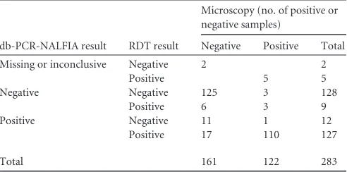
micros-copy positive cases. An overview of the results of the different tests
can be found in
Table 1
. In total, 122 microscopy positive
partic-ipants were included, of which four cases were
P. falciparum
-
P.
malariae
coinfections and one was a
P. falciparum
-
P. ovale
coin-fection. The parasitemia ranged from 66 to 235,417 parasites/
l
(geometric mean
⫽
11,519 parasites/
l). Of the 122
microscopy-TABLE 1 Number of samples that were positive or negative in
respective diagnostic tests in Burkina Faso
db-PCR-NALFIA result RDT result
Microscopy (no. of positive or negative samples)
Negative Positive Total
Missing or inconclusive Negative 2 2
Positive 5 5
Negative Negative 125 3 128
Positive 6 3 9
Positive Negative 11 1 12
Positive 17 110 127
Total 161 122 283
on May 16, 2020 by guest
http://jcm.asm.org/
[image:3.585.299.545.87.209.2]positive samples, 118 were also determined to be positive by RDT.
The four samples determined to be negative by RDT had
micro-scopic parasitemia levels ranging from 336 to 2,664 parasites/
l.
However, 23 of the microscopy-negative samples were positive by
RDT. The agreement between microscopy and RDT was 90%.
When db-PCR-NALFIA is compared to microscopy, only 276
samples could be analyzed since four samples were not available
for db-PCR-NALFIA analysis, and three samples were
inconclu-sive. Of the 276 remaining samples 28 microscopy-negative
sam-ples were determined to be positive with db-PCR-NALFIA and six
microscopy-positive samples were determined to be negative with
the PCR method. Of these six microscopy positive samples, three
were also negative by RDT. Of the 28 microscopy-negative
sam-ples that were determined to be positive by db-PCR-NALFIA, 17
were also positive by RDT (
Table 1
). The agreement, predictive
values, sensitivity, and specificity of all tests compared to each
other are shown in
Table 2
.
In Thailand, the evaluation of the db-PCR-NALFIA was
per-formed from December 2011 to January 2012. In total, 381
pa-tients were included. The mean age of the participants was 20.6
years (range, 4 to 63 years), 33% were female and 67% were male.
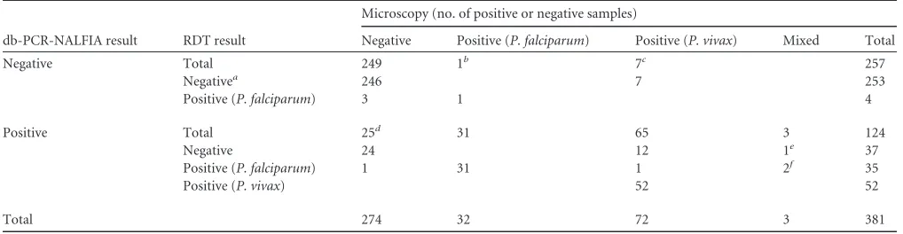
In total, 107 participants had a positive microscopy slide (
Table 3
):
32 patients had
P. falciparum
infections, 72 had
P. vivax
infections,
and 3 mixed infections were observed, one of which was
P.
vivax
-
P. malariae
. The mean parasitemias of
P. falciparum
and
P.
vivax
were 5,185 and 730 parasites/
l, respectively. RDT detected
all
P. falciparum
cases. However, 19 samples that were determined
to be
P. vivax
positive by microscopy were negative by RDT (
Table
3
), resulting in a lower overall sensitivity than that observed in the
patients from Burkina Faso (
Table 2
). Three of the four patients
considered negative for malaria by microscopy but positive by
RDT had a history of malaria in the last 2 months prior to
enroll-ment. The overall agreement between microscopy and RDT was
92%. When microscopy and db-PCR-NALFIA were compared, 25
of the microscopy-negative samples were found to be positive; 3 of
which these patients were reported to have had a history of malaria
in the previous 2 months, and 1 of them was also positive by RDT.
Seven of the
P. vivax
-positive samples determined by microscopy
were negative by db-PCR-NALFIA (
Table 3
). Four had a
para-sitemia of
⬍
2 parasites/
l, and the other three had 4, 10, and 32
parasites/
l, respectively. The agreement, predictive values,
sen-sitivity, and specificity of all tests compared to each other are
sum-marized in
Table 2
. All specimens that yielded discordant results,
as well as 65 microscopy- and db-PCR-NALFIA-negative samples
from Thailand, were tested by the species differentiation PCR.
This showed that of the 25
db-PCR-NALFIA-positive/micros-copy-negative samples, 13 were positive with the species
differen-tiation PCR (12
P. vivax
and 1
P. falciparum
). The remaining 12
were negative, including the samples from the three patients with
[image:4.585.39.546.77.183.2]a history of malaria. Six of the seven
P. vivax
samples that were
TABLE 2 Sensitivity, specificity, and agreement of different diagnostic tests in Burkina Faso and Thailanda
Location and comparison (n) Sensitivity (95% CI) Specificity (95% CI) PPV (95% CI) NPV (95% CI) value
Burkina Faso
SD Bioline vs microscopy (283) 0.967 (0.913–0.989) 0.857 (0.791–0.905) 0.837 (0.763–0.891) 0.972 (0.925–0.991) 0.81 Microscopy vs dB-PCR-NALFIA (276) 0.799 (0.720–0.860) 0.956 (0.903–0.982) 0.949 (0.887–0.979) 0.824 (0.754–0.878) 0.75 SD Bioline vs dB-PCR-NALFIA (276) 0.914 (0.851–0.953) 0.934 (0.875–0.968) 0.934 (0.875–0.968) 0.914 (0.851–0.953) 0.85
Thailandb
SD Bioline vs microscopy (377) 0.813 (0.724–0.879) 0.985 (0.960–0.995) 0.956 (0.885–0.986) 0.930 (0.892–0.956) 0.83 Microscopy vs dB-PCR-NALFIA (381) 0.798 (0.715–0.863) 0.973 (0.942–0.988) 0. 934 (0.864–0.971) 0.909 (0.867–0.939) 0.80 SD Bioline vs dB-PCR-NALFIA (377) 0.702 (0.612–0.779) 0.984 (0.958–0.995) 0.956 (0.885–0.986) 0.871 (0.825–0.906) 0.74 a
PPV, positive predictive value; NPV, negative predictive value.n, Number of samples.
bAll calculations are before species differentiation PCR correction and with the results ofP. falciparumandP. vivaxdetection combined.
TABLE 3Number of samples that were positive or negative in respective diagnostic tests in Thailand
db-PCR-NALFIA result RDT result
Microscopy (no. of positive or negative samples)
Negative Positive (P. falciparum) Positive (P. vivax) Mixed Total
Negative Total 249 1b 7c 257
Negativea 246 7 253
Positive (P. falciparum) 3 1 4
Positive Total 25d 31 65 3 124
Negative 24 12 1e 37
Positive (P. falciparum) 1 31 1 2f 35
Positive (P. vivax) 52 52
Total 274 32 72 3 381
a
Includes four tests that were in conclusive.
bContainingPlasmodium falciparumgametocytes only from a patient with history of malaria 1 week preceding testing.
c
All positive forP. vivax, with confirmative PCR according to Snounou et al. (17).
dIncludes three samples from patients with a history of malaria in the week preceding testing. Thirteen of these samples were also positive forPlasmodiumwith confirmative PCR.
e
Mixed infection ofP. vivaxandP. malariae. fMixed infection ofP. falciparumandP. vivax.
on May 16, 2020 by guest
http://jcm.asm.org/
[image:4.585.39.548.539.671.2]positive by microscopy and negative by db-PCR-NALFIA were
also positive with the differentiation PCR. Of the 65 samples that
were determined to be negative by microscopy, RDT, and
db-PCR-NALFIA, 12 were positive with the species differentiation
PCR (3
P. falciparum
and 9
P. vivax
).
When the microscopy-positive samples were stratified by
par-asitemia, it was shown that db-PCR-NALFIA and RDT had
simi-lar sensitivities for the samples from Burkina Faso and that
db-PCR-NALFIA was more sensitive than RDT for the samples from
Thailand (
Table 4
). This difference was due to the presence of
P.
vivax
in samples from Thailand, for which db-PCR-NALFIA was
substantially more sensitive than RDT.
DISCUSSION
We describe here a sensitive and robust method,
db-PCR-NALFIA, for the detection of
Plasmodium
species directly from
blood samples. The standard PCR technology has been
simpli-fied by circumventing DNA extraction prior to amplification
and adding an easy visual detection of the products using a
lateral flow immunoassay. Most PCR protocols still require
some specimen processing steps such as boiling or chemical
treatment of samples or postamplification analysis that
re-quires equipment or instrumentation (
4
). The sensitivity and
specificity of the db-PCR-NALFIA was good in both in
tory setting and for field evaluations. However, in the
labora-tory evaluation, 14 samples initially identified as
Plasmodium
positive were found to be negative with db-PCR-NALFIA. This
could be due to DNA degradation since these samples were
stored for several years under suboptimal storage conditions in
a refrigerator.
The db-PCR-NALFIA is similar in concept to the simple RDT,
except that the lateral flow immunoassay is preceded by a PCR.
This makes the technology more complicated and so requires
some additional training. However, we have shown that
db-PCR-NALFIA is much more sensitive than RDT, especially for the
de-tection of
P. vivax.
There were discrepancies in results between the
db-PCR-NALFIA and microscopy and RDT. Although in both
field settings there were false-negative db-PCR-NALFIA samples
that were determined to be positive by microscopy and/or RDT,
there were a greater number of false-positive db-PCR-NALFIA
samples that were negative with the other methods. In both
sce-narios, this could be due to a very low number of parasites in
samples (
⬍
2 parasites/
l), resulting in sampling error.
Unfortu-nately, no sample material was available from Burkina Faso to
perform the additional species differentiation PCR, but analysis of
the discrepant samples from Thailand resolved a number of the
apparent db-PCR-NALFIA false-positive results as in fact true
positives and also confirmed the false-negative results. Remaining
discordant samples could be due to sampling error. In 80% of the
discordant db-PCR-NALFIA-positive samples,
P. vivax
DNA was
found.
Although the field technicians were experienced in PCR
tech-nology, the db-PCR-NALFIA was performed in Burkina Faso by a
technician after very little training in the protocol, and in Thailand
the assay was performed without any prior training. This shows
that the db-PCR-NALFIA can be performed by technicians with
no or little training in the protocol itself.
One of the drawbacks of the current technology is the
require-ment for a reliable source of electricity for cold storage of the PCR
reagents and the operation of the PCR thermal cycler. This may
limit the current technology to areas where some laboratory
in-frastructure is available (
18
,
9
). Regional or national malaria
con-trol programs could decentralize their testing since this is an
op-erator-friendly technology in which a large number of people can
be screened with a high sensitivity in a short time. This may be
especially interesting in areas with moderate to low transmission,
since the probability of having malaria is less in these areas and
therefore sensitive and accurate diagnosis is essential (
11
). In the
light of eradication programs, this technology may be useful for
the detection of asymptomatic carriers that usually harbor low
parasitemia. The availability of thermal cyclers that operate on
solar-powered batteries (
23
) and room temperature-stable PCR
reagents would expand the availability of this technology in
re-source-poor settings.
Another drawback of the technology is that, as with all antigen
detection and molecular methods, db-PCR-NALFIA cannot
dif-ferentiate between asexual (responsible for disease) and sexual
(gametocytes, responsible for transmission) stages. This could
po-tentially lead to the misdiagnosis of patients that harbor
gameto-cytes but are suffering from a different disease as well. Clinical
signs and ruling out of other conditions should thus always be
considered when applying these tests.
The current db-PCR-NALFIA is highly sensitive for the
detec-tion of
P. vivax
, as well as
P. falciparum
, but the current format
does not differentiate the
Plasmodium
species. The sensitivity of
other human
Plasmodium
species— e.g.,
P. ovale
,
P. knowlesi
, and
P. malaria
— could not be assessed here. For areas where there are
high rates of transmission of
P. vivax
or other
Plasmodium
species,
[image:5.585.42.545.78.196.2]additional species-specific detection lines should be added to the
db-PCR-NALFIA detection device. Since currently available
TABLE 4Sensitivities of SD-Bioline RDT and db-PCR-NALFIA in Burkina Faso and Thailand versus microscopy-determined levels of parasitemia
Parasitemia level (count/l)
% Sensitivity (no. of positive samples/total no. of samples tested)a
Burkina Faso Thailand
SD Bioline
RDT db-PCR-NALFIA
SD Bioline
RDT db-PCR-NALFIA
⬎50,000 100 (27/27) 95.8 (23/24) 100 (3/3) 100 (3/3)
5,000 to 50,000 100 (59/59) 98.8 (57/58) 100 (49/49) 100 (49/49)
500 to 5,000 86.2(25/29) 92.9 (26/28) 100 (22/22) 100 (22/22)
100 to 500 80.0(4/5) 80.0(4/5) 81.8(9/11) 100 (11/11)
50 to 100 100 (4/4) 75.0(3/4) 66.7(2/3) 100 (3/3)
⬍50 10.5(2/19) 57.9(11/19)
aSensitivities that are remarkably low are indicated in boldface.
on May 16, 2020 by guest
http://jcm.asm.org/
RDTs have a low sensitivity for the detection of
P. vivax
(
1
,
10
), in
remote field settings microscopy is still the only relatively easy,
although very time-consuming, field-deployable method for the
detection of
P. vivax
(
10
).
The current db-PCR-NALFIA consists of a combination of
components from two commercially available kits, the buffer
component of the Phusion direct blood PCR kit and the separately
available Phire Hotstart II DNA polymerase. Unfortunately, it was
not possible to purchase the reagents separately from the complete
kits, adding to the cost of the current technology and making it
unnecessarily more expensive than conventional PCR. However,
the ability to add blood directly to the PCR without any form of
processing saves reagent costs. Ideally, all components would be
combined in a single kit to make the assay affordable and
conve-nient, which are prerequisites if large-scale implementation is
de-sired (
13
). Because of the large-scale production, the price of the
NALFIA detection device was
⬍
$0.50. Finally, it would be useful
to provide standardized quality control material in order for
lab-oratories to harmonize results among regions or countries (
14
,
9
).
This should always be accompanied by ongoing quality assurance,
including training and continuing education of laboratory
scien-tists (
9
,
13
).
In conclusion, the db-PCR-NALFIA is a relatively easy-to-use
method that is robust, sensitive, and specific and could have great
potential in locales where malaria is endemic, especially in areas
where there is low transmission of malaria, and thus a very
sensi-tive technology is warranted.
ACKNOWLEDGMENTS
This project was financially supported by the EU funded FP7 project MALACTRES (Multidrug Resistance in Malaria under Combination Therapy: Assessment of Specific Markers and Development of Innovative, Rapid and Simple Diagnostics) under grant 201889.
We thank all collaborators of the MALACTRES project for their fruit-ful discussions on the project and initial evaluation of the developed pro-tocols. We want to specifically thank Colin Sutherland of the London School of Hygiene and Tropical Medicine (United Kingdom) and Senior Lecturer of the Malaria Reference Laboratory (United Kingdom) and Bakri Nour from Wad Medani Teaching Hospital (Sudan) for kindly pro-viding blood samples for the laboratory evaluation. We thank Marc Tahita (Unité de Recherche Clinique de Nanoro) for his supervision and support of the Burkina Faso trial and his staff and patients in Nanoro, Burkina Faso, for the possibility of doing this study in conjunction with the ongo-ing Pharmacovigilance study. We are also grateful to the staff and patients of the Mae Khon Ken and Wang Pa clinics and the Shoklo Malaria Re-search Unit (Thailand), part of the Mahidol-Oxford Tropical Medicine Research Unit (MORU) supported by the Wellcome Trust (United King-dom), for their willingness to participate in this trial.
REFERENCES
1.Alam MS, et al.2011. Real-time PCR assay and rapid diagnostic tests for the diagnosis of clinically suspected malaria patients in Bangladesh. Malar J.10:175.
2.Altman DG.1991. Practical statistics for medical research, p. 611. Chap-man & Hall, London, United Kingdom.
3.Fabre R, Berry A, Morassin B, Magnaval JF.2004. Comparative assess-ment of conventional PCR with multiplex real-time PCR using SYBR Green I detection for the molecular diagnosis of imported malaria. Para-sitol.128:15–21.
4.Fuehrer HP, et al.2011. Novel nested direct PCR technique for malaria diagnosis using filter paper samples. J. Clin. Microbiol.49:1628 –1630. 5.Hawkes M, Kain KC.2007. Advances in malaria diagnosis. Expert Rev.
Anti-Infect. Ther.3:485– 495.
6.Kamau E, et al.2011. Development of a highly sensitive genus-specific quantitative reverse transcriptase real-time PCR assay for detection and quantitation of plasmodium by amplifying RNA and DNA of the 18S rRNA genes. J. Clin. Microbiol.49:2946 –2953.
7.Lucchi NW, et al.2010. Real-time fluorescence loop mediated isothermal amplification for the diagnosis of malaria. PLoS One5:e13733. doi: 10.1371/journal.pone.0013733.
8.Luo L, et al.2011. Visual detection of high-risk human papillomavirus genotypes 16, 18, 45, 52, and 58 by loop-mediated isothermal amplifica-tion with hydroxynaphthol blue dye. J. Clin. Microbiol.49:3545–3550.
9.malERA Consultative Group on Diagnoses and Diagnostics.2011. A
research agenda for malaria eradication: diagnoses and diagnostics. PLoS Med.8:1000396. doi:10.1371/journal.pmed.1000396.
10. McMorrow ML, Aidoo M, Kachur SP.2011. Malaria rapid diagnostic
tests in elimination settings— can they find the last parasite? Clin. Micro-biol. Infect.17:1624 –1631.
11. Mens P, et al.2007. Is molecular biology the best alternative for diagnosis of malaria to microscopy? A comparison between microscopy, antigen detection and molecular tests in rural Kenya and urban Tanzania. Trop. Med. Int. Health12:238 –244.
12. Mens PF, van Amerongen A, Sawa P, Kager PA, Schallig HDFH.2008.
Molecular diagnosis of malaria for the field: development of a novel, 1-step nucleic acid lateral flow immunoassay for the detection of all 4 humanPlasmodiumspp. and its evaluation Mbita, Kenya. Diagn. Micro-biol. Infect. Dis.61:421– 427.
13. Peeling RW, Mabey D.2010. Point-of-care tests for diagnosing infections in the developing world Clin. Microbiol. Infect.16:1026 –1029. 14. Proux S, et al.2011. Considerations on the use of nucleic acid-based
amplification for malaria parasite detection. Malar. J.10:323.
15. Rantala AM, et al.2010. Comparison of real-time PCR and microscopy for malaria parasite detection in Malawian pregnant women. Malar. J.
9:269.
16. SMRU-Shoklo Malaria Research Unit. 2012. Malaria on the
Myanmar border. SMRU-Shoklo Malaria Research Unit, Mae-Sot, Thai-land.http://www.shoklo-unit.com/MTF/tb_border.php.
17. Snounou G, Viriyakosol S, Jarra W, Thaithong S, Brown KN.1993.
Identification of the four human malaria parasite species in field samples by the polymerase chain reaction and detection of a high prevalence of mixed infections. Molecular Biochem. Parasitol.58:283–292.
18. Snounou G.2007., Rapid, Sensitive and cheap molecular diagnostics of malaria: is microscopy on the way out? Future Microbiol.2:447– 480. 19. World Health Organization.1991. Basic laboratory methods in medical
parasitology. World Health Organization, Geneva, Switzerland. 20. World Health Organization.2010. Guidelines for the treatment of
ma-laria, 2nd ed. World Health Organization, Geneva, Switzerland. 21. World Health Organization.2010. World malaria report: technical
re-port. World Health Organization, Geneva, Switzerland.
22. World Health Organization.2010. Basic malaria microscopy. I. Learner’s guide, 2nd ed. World Health Organization, Geneva, Switzerland. 23. Yashon RK, Cummings MR.2012. Human genetics and society, 2nd ed,
p 189. Brooks/Cole Cencage Learning, Independence, KY.