Available Online at www.ijpret.com
185
INTERNATIONAL JOURNAL OF PURE AND
APPLIED RESEARCH IN ENGINEERING AND
TECHNOLOGY
A PATH FOR HORIZING YOUR INNOVATIVE WORK
CANCER DETECTION USING NANOSENSOR
PRACHI V. YAWALE1, PROF. S. M. MAHALLE2
1. ME Student, Department of Electronics & Telecommunication, IBSS, Amravati, Maharashtra, India.
2. Assistant Professor, Department of Electronics & Telecommunication, IBSS, Amravati , Maharashtra, India.
Accepted Date: 27/02/2014 ; Published Date: 01/05/2014
\
Abstract:Early detection of cancer is one of the most hope full approaches to reduce cancer
death rates. Cancer biomarkers are molecule that can indicate the biological states of the disease, which increase in concentration during the onset of the disease . Sensitive and accurate detection of cancer biomarkers in human fluid samples could offer seminal contributions to early diagnosis. Biosensor technology has the potential to provide fast and accurate detection, reliable imaging of cancer cells, and monitoring of angiogenesis and cancer metastasis, and the ability to determine the effectiveness of anticancer chemotherapy agents. A variety of sensors based on different nanostructured materials are highly sensitive and selective for the detection of cancer biomarker . A variety of nanostructured materials including carbon nanotubes, silicon nanowires, gold nanoparticals and quantum dots , in fabrication of sensors.
Keywords: Biosensor, Biomarker, Cancer detection.
Corresponding Author: MS. PRACHI V. YAWALE
Access Online On:
www.ijpret.com
How to Cite This Article:
Prachi Yawale, IJPRET, 2014; Volume 2 (9): 185-193
Available Online at www.ijpret.com
186
INTRODUCTION
Cancer is the second leading cause of death. Cancer can take over 200 distinct forms, including lung, prostate, breast, ovarian, hematologic, skin, and colon cancer, and leukemia, and both environmental factors (eg. tobacco smoke, alcohol, radiation, and chemicals) and genetic factors (eg, inherited mutations and autoimmune dysfunction) are associated with an increased risk of developing cancer.
Cancer is defined as abnormal and uncontrolled cell growth. Unregulated cell growth leads to the formation of tumor mass that over time becomes independent of normal homeostatic checks and balances. As the cancer progresses, the tumor begins to spread beyond the site of origin and metastasize to other body organs and systems, at which point, the cancer is essentially incurable.
The two major tumor genesis mechanisms are activation of oncogenes and inactivation of tumor suppressor genes (TSGs). Activation of oncogenes occurs through mutation or duplication of a normal gene (a proto-oncogene) involved in the regulation of cell growth, proliferation, and/or differentiation. This typically results in constitutive activation or excess levels of a normal gene product, leading to the deregulation of cell growth, increased cell division, and tumor formation. Perhaps more so than any other type of oncogene, growth factor receptors have been investigated as potential cancer biomarkers. The human epidermal growth factor receptor Her-2, for example, is amplified in ∼33% of all breast cancers, and cancers with amplified Her-2[6] tend to grow and spread more aggressively. TSGs are involved in the regulation of inappropriate cell growth and proliferation by slowing or stopping cell division. Three of the most well-studied TSGs in cancer are retinoblastoma protein (Rb), BRCA1/2, and p53.3 The Rb is a master regulator of cell division, and mutation of Rb plays a role in many cancers. Point mutations and deletions are the most common causes of inactivation of the Rb1 gene. BCRA1 gene mutations account for about 50% of heredi- tary breast cancers and 80%–90% of hereditary breast and ovarian cancers. Another major concern with the loss of p53 is that it can serve as a mechanism of chemotherapeutic drug resistance. The development of biosensors that can detect the presence of mutations in p53, Rb, and BRCA1 is of great importance in terms of being better able to determine cancer susceptibility as well as more accurate prognosis and treatment regimens.
II. CANCER BIOMARKERS
Available Online at www.ijpret.com
187
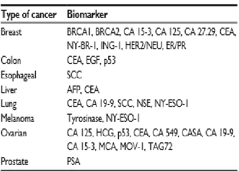
condition or disease. A biomarker may be used to see how well the body responds to a treatment for a disease or condition” Cancer biomarkers are potentially one of the most valuable tools for early cancer detection, accurate pretreatment staging, determining the response of cancer to chemotherapy treatment, and monitoring disease progression. Biomarkers are typically detected in human fluids such as blood, serum, urine, or cerebral spinal fluid, but they can also be present in or on tumor cells. A partial list of tumor biomarkers is presented in Table.
Table 1: Common Biomarker utilized for cancer
Prostate-specific antigen
Prostate-specific antigen (PSA) was one of the first tumor biomarkers to be identified and put into routine clinical use for screening and diagnosis of prostate cancer. Studies have shown that above-normal PSA levels correlate directly with prostate cancer. A normal level of PSA is 4.0 ng/mL.
Cancer antigen 125
Available Online at www.ijpret.com
188
CA 15-3
CA 15-3 is an important biomarker analyzed in breast cancer patients. Other biomarkers that are linked to breast cancer are carcinoembryonic antigen (CEA), BRCA1, BRCA2, and CA 15-3 is used clinically most often to monitor patient therapy in cases of advanced breast cancer.
Cancer-testis Antigens
Cancer-testis (CT) antigens are a unique class of cancer biomarker. They are highly expressed in many tumors, but not in normal cells. Thus, they have been heavily pursued as potential immunogenic targets for cancer immune therapies (ie, cancer vaccines), and auto antibodies to CT antigens have been pursued as cancer biomarkers. One of the advantages of CT auto antibodies as cancer biomarkers is that they are present in serum.
RCAS1 (eBAG-9)
Receptor-binding cancer antigen expressed on SiSo cells (RCAS1)[4] has been shown to be over expressed in nearly 100% (98.4%) of gastric carcinomas and correlates closely with gastric tumor progression. In terms of cancer biomarkers, RCAS1 expression appears to be highly indicative of cancer progression and perhaps prognosis. As with most of the cancer biomarkers discussed here, RCAS is over expressed by many different types of tumors and should be factored in with other parameters when establishing treatment regimens.
III. REPRESENTATION OF BIOSENSOR
A sensor that integrates a biological element with a physiochemical transducer to produce an electronic signal proportional to a single analyte which is then conveyed to a detector.
Information such as whether or not the analyte is present and at what level is transduced into an electrical signal that can be amplified, displayed, and analyzed. Examples of analytes include proteins (antigen, antibody, and enzyme), nucleic acid, or other biological or metabolic component (ie, glucose).In terms of cancer, the analyte being detected by the biosensor is a tumor biomarker.
Available Online at www.ijpret.com
189
the transducer then converts the biological signal to an electrical output. The basic structure and function of a biosensor is shown in Figure.[1]
I. Biosensor recognition element
The recognition element is a critical component of the biosensor. Examples of recognition elements are receptor proteins, antigens, antibodies, enzymes, and nucleic acids
Figure [1]. Schematic representation of a biosensor. .
Receptor recognition elements
Cell surface receptors are targets for drug delivery and are useful for monitoring the effectiveness of cancer therapeutics. Activation or inactivation of receptor molecules triggers diverse signaling events within a cell. Altering the function of a receptor can result in opening or closing of ion channels and changes to membrane permeability, activation of adenyl cyclase and second messengers, and activation of small G proteins, kinases, phosphatases, and transcription factors.
Antigen/antibody recognition elements
Available Online at www.ijpret.com
190
recognition. The recognition element for PSA biosensors, some of the most widely used biosensors in clinical use for the detection of prostate cancer, is an anti-PSA[4] antibody. Anti-PSA recognition elements have been linked to microcantilever-based transducers and to surface plasmon resonance-based sensors, in which changes in vibrational frequency upon antigen binding to antibody are used to detect PSA.
Enzymes
Allosteric enzymes show great potential as recognition elements. In most cases, the regulatory subunit takes on the role of the recognition element and the catalytic site becomes the transducer. One of the most advanced sensors of this class, and in general, is the glucose sensor, which uses glucose oxidase as the recognition element.
Nucleic acids
Aptamers are oligonucleotides that are selected from among a pool of thousands of different sequences for very high binding affinities for their targets. As a result, they are rapidly emerging as biosensor recognition elements. A combinatorial chemistry-based technology termed SELEX (systematic evolution of ligands by exponential enrichment)[2] has been developed to generate nucleic acid ligands from a library of DNA and RNA oligonucleotides. Biosensors built on this type of system have proven to be useful in the discovery of new biomarkers that are critical to early cancer diagnosis.
II Biosensor transducer
Electrochemical biosensors
Amperometric and potentiometric transducers are the most commonly used electrochemical transducers. In amperometric transducers, the potential between the two electrodes is set and the current produced by the oxidation or reduction of electroactive species is measured and correlated to the concentration of the analyte of interest. Most electrodes are made of metals like platinum, gold, silver, or carbon-based materials that are inert at the potentials at which the electrochemical reaction takes place.
Available Online at www.ijpret.com
191
Mass-based biosensors
Piezoelectric and acoustic wave biosensors make up the class of mass-based biosensors. In terms of cancer detection, piezoelectric biosensors are more commonly used. Piezoelectric sensors are based on changes in the mass of quartz crystals when potential energy is applied to them. This change in mass generates a frequency, which can be converted into a signal. Immunosensors and microcantilever sensors that use piezoelectric technology have proven useful in the identification of cancer biomarkers.
Calorimetric biosensors
Calorimetric biosensors are less common than other biosensors for cancer diagnostics, but the introduction of nanotechnology to the field of biosensors has increased the range of applications for these types of biosensors. Calorimetric biosensors measure exothermic reactions. Many enzymatic reactions generate heat, and changes in heat can be used to measure analyte concentration. The reaction is monitored by measuring enthalpy changes, which indirectly provide information about substrate concentration. Calorimetric biosensors are not capabilities have been demonstrated.
Carbon Nanotubes (CNTs)
CNTs, which were first discovered in 1991, are made up of graphene sheets rolled up into the shape of seamless tubular structure. CNTs can be structurally classified into two categories: single-wall CNTs[4], which consist of one layer of cylinder graphene and multi-wall CNTs, which contain multiple layers of concentric graphene cylinders. The detection of cancer biomarkers using CNT-based sensors shows high sensitivity and selectivity, as well as real-time response capabilities.
Available Online at www.ijpret.com
192
Back-gated chemical Field-Effect Transistor (FET)
CNTs configured with FETs are a three electrode system. The conductance between the source and drain electrodes is modulated by the gate electrode. Prostate-Specific Antigen (PSA), a 28-kDa glycoprotein produced by the prostate gland and also a serine protease enzyme, has been used as a biomarker for the diagnosis and prognosis of prostate cancer. In serum, PSA has been found either in a free form (fPSA) or as a complex with protease inhibitor alpha 1-antichymotrypsin (PSA-ACT complex). Total PSA (tPSA) refers to the sum of all detectable free and complexed PSA forms. CNT-FETs have been modified with linkers and spacers of different ratios.
Figure[3]. Schematic diagrams of a CNT-FET
PSA-ACT complex antibodies were then immobilized on the CNT surface via the linkers. Results showed that CNT-FETs modified with only linkers could not detect. target proteins unless more than 500 ng mL 1 of PSAACT complex solution was injected, while those modified with linkers and spacers of a 1-to-3 ratio could detect 1.0 ng mL 1 of PSA-ACT complex without any pretreatment. This study implies that modifying the CNT surface with a spacer could offer a useful strategy of improving the sensitivity of the device.
IV. CONCLUSION
Available Online at www.ijpret.com
193
biosensor technology has the potential to provide fast, accurate results, while maintaining cost effectiveness.
REFERENCES
1. Tothill IE. Biosensors for cancer markers diagnosis. Semin Cell Dev Biol. 2009.
2. Kubokawa M, Nakashima M, Yao T, et al. Aberrant intracellular localization of RCAS1 is associated with tumor progression of gastric cancer. Int J Oncol. 2001.
3. Wong SC, Chan CM, Ma BB, et al. Advanced proteomic technologies for cancer biomarker discovery.
4. Grodzinski P, Silver M, Molnar LK. Nanotechnology for cancer diagnostics: promises and challenges. Expert Rev Mol Diagn. 2006.
5. Asphahani F, Zhang M. Cellular impedance biosensors for drug screening and toxin detection. Analyst. 2007.
6. Bharali DJ, Khalil M, Gurbuz M, Simone TM, Mousa SA. Nanoparticles and cancer therapy: a concise review with emphasis on dendrimers. Int J Nanomedicine. 2009.
7. Wu G, Ji H, Hansen K, et al. Origin of nanomechanical cantilever motion generated from biomolecular interactions. Proc Natl Acad Sci U S A. 2001.
![Figure [1]. Schematic representation of a biosensor. .](https://thumb-us.123doks.com/thumbv2/123dok_us/8749700.1748217/5.595.71.281.225.448/figure-schematic-representation-biosensor.webp)
