Original Article
Comparative Morphologic and Morphometric Study on the
Developmental Aspects of In Vitro and In Vivo Reared
Echino-coccus granulosus
Sensu Stricto Using Differential Interference
Contrast (DIC)/Nomarski and Phase Contrast Microscopy
Seyedeh Faezeh SADJJADI 1, *Mina MOTAMEDI 1, Tahereh MOHAMMADZADEH 2,3, *Seyed Mahmoud SADJJADI 4,5
1. Department of Biology, Faculty of Sciences, Shahid Bahonar University of Kerman, Kerman, Iran 2. Health Research Center, Life Style Institute, Baqiyatallah University of Medical Sciences, Tehran, Iran 3. Department of Parasitology, Faculty of Medicine, Baqiyatallah University of Medical Sciences, Tehran, Iran 4. Department of Parasitology and Mycology, School of Medicine,Shiraz University of Medical Sciences, Shiraz, Iran
5. Basic Sciences in Infectious Diseases Research Center, Shiraz University of Medical Sciences, Shiraz, Iran
Received 10 Jan 2019 Accepted 16 Mar 2019
Abstract
Background: Echinococcus granulosus is a zoonotic parasite with worldwide distribution. The
pre-sent study focused on comparative morphologic and morphometric observations on the devel-opmental aspects of whole body, more special the reproductive structures of in vitro reared adult worms (RAW) and in vivo reared adult worms in definitive host (AWIDH) using differential interference contrast (DIC)/Nomarski, phase contrast and routine optical microscopy.
Methods: A total number of 10 in vitro and 10 in vivo reared adult worms of E. granulosus sensu
stricto, G1 strain were selected. The worms were processed by Formaldehyde-Alcohol-Azocarmine-Lactophenol (FAAL). The details of morphological factors and reproductive struc-tures of each worm including 25 biometrical parameters were studied by routine optical, phase contrast and Nomarski microscopy. The details of the samples were photographed, measured and analyzed. The fine structures of the parasite including the details of cirrus sac and develop-mental stages in different strobila were more obvious observing by Nomarski microscopy.
Results: The morphometric characters in the RAW and AWIDH showed that length of
imma-ture proglottid, length of maimma-ture proglottid, length of suckers are larger in RAW than AWIDH worms with statistical difference. Characters in E. granulosus of RAW and AWIDH showed that total number of segments, number of mature segments and the total number of testes were greater in RAW than AWIDH worms; while only the number of mature segments was statistical-ly different is two groups.
Conclusion: Application of DIC/Nomarski and phase contrast microscopy together with
mor-phometric criteria are useful means for comparing the developmental aspects of in vitro and in vivo reared adults of E. granulosus.
Keywords:
Echinococcus granulosus sensu stricto;
G1 strain; DIC/Nomarski microscopy;
Phase contrast; Morphology; Morphometry
*Correspondence
Email:
[email protected] [email protected]
Iranian Society of Parasitology http://isp.tums.ac.ir
Iran J Parasitol
Open access Journal at http://ijpa.tums.ac.ir Tehran University of Medical
Introduction
chinococcus granulosus is a small zoonotic
tapeworm with worldwide distribution (1, 2). Its length is approximately 2 to 7 mm with typically three segments and rarely more than five proglottids. The adult cestodes live in the small intestine of canids where they produce infective eggs which may infect ungu-lates (sheep, goats, cattle, pigs, horses, etc.) and also humans (3-5). Eating infected offal of the intermediate hosts by the definitive host will complete its life cycle.
So, for prevention of the disease in infected areas, boiling offal containing hydatid cysts for 30 minutes will be very helpful (6). E.
granu-losus sensu lato (s.l.) consists of independent
species including E. granulosus sensu stricto (s.s.), E. equinus, E. ortleppi, E. canadensis and E.
felidis (7). E. granulosus (s.s.), E. ortleppi, and E. canadensis have been reported in Iran, so far (2,
8, 9).
Although major advances have been made in research on Echinococcus and echinococcosis, many questions remain, particularly in the are-as of developmental biology and host-parare-asite relationships (3). In this regard, detailed study of adult worms will help a better understand-ing of the developmental biology of this para-site which could be applicable for treatment and vaccine developments. This is usually done by routine optical microscopy. However, use of routine optical microscopy alone could not be enough for a detailed study of the morphological structure of this parasite. Dif-ferential Interference Contrast (DIC) micros-copy, also known as Nomarski Interference Contrast (NIC) or Nomarski microscopy which is applicable in biology more special in developmental biology has been used for the present study (10). Imaging modalities, such as differential interference contrast (DIC) croscopy (10, 11) and phase-contrast (PC) mi-croscopy (11), which can capture images of transparent objects are of a significant im-portance (12).
Many aspects of adult worms of E. granulosus have been studied (5, 13-17). However, the morphologic details of in vitro reared adult worms (RAW) and in vivo reared adult worms from the definitive host have not been com-pared by Nomarski and phase contrast mi-croscopy, so far. Therefore, the present study was designed to compare the morphologic and morphometric criteria of whole body es-pecially developmental aspects of in vitro and in vivo reared E. granulosus s.s. adult worms using differential interference contrast (DIC)/ Nomarski and phase contrast and routine op-tical microscopy.
Materials and Methods
In vitro adult worms
In vitro reared adult worms were obtained under aseptic conditions by culturing a num-ber of protoscoleces in culture media. In this regard, sheep hydatid cysts collected in abat-toir were dissected for the collection of proto-scoleces (PSCs) followed by washing and cul-tivation in a diphasic S.10E.H medium using a CO2 incubator. Briefly, the surface of the hy-datid cysts was washed by 70% ethanol and opened in order to remove Hydatid cyst fluid (HCF) and protoscoleces (PSC) under aseptic conditions. HCF containing PSCs was collect-ed from cysts and transferrcollect-ed into sterile tubes while their PSC were sedimented after 30 min. PSCs were washed 3 times with sterile PBS. The protoscoleces were separated and culti-vated under laminar flow hood. The medium was S.10E.H, consisted of 2 parts: liquid phase and solid phase using the same procedures described before with some modifications (13, 15, 18).
(19). A number of protoscoleces were also used for molecular works.
Preparation of samples for routine, phase contrast and DIC/Nomarski microscopy
A total of 10 samples of in vitro reared adult worms were used for microscopic studies. A total of 10 samples of in vivo adult worms from an experimentally infected dog were also used for the present study. Partial cox1 se-quence was used to finding their strain which revealed them as E. granulosus sensu stricto, G1 strain (20). Routine optical, phase contrast and DIC/Nomarski microscopy were used for the study on the morphological details of each parasite. The detail of each worm was studied separately by descriptive and numerical meth-ods. All aspects of the worms were photo-graphed by a digital camera and prepared for measurement and further analysis.
Image analysis and statistical analysis A total of 25 biometrical parameters were observed and measured (Table 1-3).
Image-J software base on micron unit was used for image measurements and analysis. Data which obtained from Image-J software were collected in an EXCEL sheet and ana-lyzed by IBM SPSS Statistics 25. The t-test
was used for statistical differences (P-value<0.05).
Results
The culture of protoscoleces in CMRL me-dia ended with 10 reared adult E. granulosus sensu stricto which were fixed, transparented and stained with FAAL. The adult worm sam-ples isolated from a dog experimentally infect-ed with G1 strain of E. granulosus were also cleared, fixed and stained with FAAL (Fig. 1). Nomarski microscopy showed suitable details of the samples reproductive structures (Fig. 2-3).
Descriptive results
Among reared adult worms and adult worms isolated from the definitive host (10 worms from each group), all the worms possess a scolex with two rows of hooks including large and small ones.
Each hook has a hand attached to a blade. A narrow neck attached to the immature pro-glottid.
The immature proglottid lacked reproduc-tive structures (Fig. 4).
Fig. 1: Adult Echinococcus granulosus. (A) Reared in culture media. (B) Isolated from definitive host. Scale bars:
300 µm
Both phase contrast and Nomarski micros-copy showed the hooks, scolex, eggs, and de-tails of reproductive structures with specific
Fig. 2: Reproductive structures in mature proglottid in adult worm isolated from definitive host. (A, C) Nomarski microscopy (B) Phase contrast microscopy (D) Optical microscopy. Scale bars: 100 µm
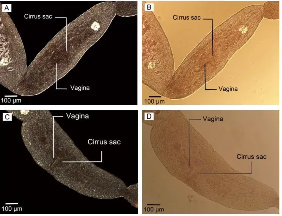
Fig. 3: Nomarski microscopy in adult worm isolated from final host. (A) Eggs, cirrus sac and vagina in gravid
proglottid. (B) Cirrus sac and Cirrus. Scale bars: (A) 100 µm, (B) 50 µm
Fig. 4: Optical microscopy. (A) Adult worm reared in culture media with narrow neck. (B) Immature
Fig. 5: Scolex. (A) Nomaski microscopy. (B) Phase contrast microscopy. (C) Optical microscopy. Scale bars: 100 µm
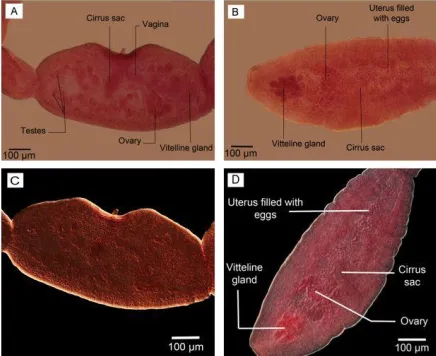
The details of the reproductive system with specific contrast are shown in phase contrast microscopy photos (Fig 6. A, B) as well as Nomarski microscopy (Fig. 6. C, D).
The contrast of eggs and cirrus sac were sharper in Nomarski microscopy observation (Fig. 3). The gravid proglottids in reared adult worms were somewhat like the mature seg-ment containing all reproductive structures with uterus a little bigger without eggs. No egg/s was observed in the uterus of adult worms reared in culture media. However, their
cirrus sac, vagina, ovary, vitelline gland, and a number of testes were seen clearly; more ob-vious using phase contrast microscopy (Fig. 6, A). On the other hand, the gravid segment in the adult worms isolated from the final host had a uterus full of eggs which were observed as ovoid-shaped with more obvious structures DIC microscopy observation (Fig. 7). Com-paring two types of worms, the maximum number of segments equal to 6 was observed in adult worm reared in culture media (Fig. 8).
Fig. 6: The details of reproductive system in adult worms. (A, B) Phase contrast microscopy; (A) reared in
Fig. 7: Uterus filled with eggs in gravid segment in adult worm isolated from definitive host. (A) Nomarski microscopy. (B) Phase contrast microscopy. Scale bars: 100 µm
Fig. 8: Adult worm reared in culture media (A) 6 segments. (B) 3 segments. Scale bars: 300 µm
Numerical results
The results of the morphometric study in the adult worms of E. granulosus reared in cul-ture media and adult worms of E. granulosus isolated from the definitive host are present in three parts including general characteristics, reproductive system and measurable charac-ters which have been shown in Tables 1-3.
The details of morphometric measurements of the reproductive structures in the adult E.
granulosus worms reared in culture media and
adult E. granulosus worms isolated from the definitive host are shown in Table 2.
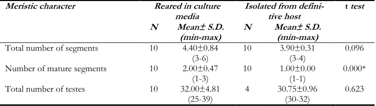
The results of measurable characters like the total number of segments, the number of ma-ture segments and the total number of testes are shown in Table 3. According to this table,
the average number of mature segments in RAW was 2.00±0.47 with a range of 1-3 while of AWIDH was 1.00±0.00. Using t-test the difference was significant. The vaginal size was 99.36 in the RAW group and 87.24 in the AWIDH group with a statistically significant difference. Analysis of 12 characters related to the reproductive system between two groups, showed that 5 characters including the length of cirrus sac, the width of the ovary, length of vagina, the width of the vagina, length of the seminal receptacle are a statistically significant difference between two groups (P-value ≤0.05).
Table 1: Morphometric characters of Adult Echinococcusgranulosus worm reared in culture media and isolated from
defini-tive host. Significant differences indicate by star. The measurements are in µm. S.D. refers to standard deviation. Min refers to minimum and max to maximum
Morphometric characters Reared in culture media Isolated from definitive host t test
N Mean± S.D.
(min-max) N Mean± S.D. (min-max)
Body length 10 3138.18±695.64
(1931.86-4584.39)
10 2403.52±908.09
(12.96.89-4307.12)
0.057
Maximum width 10 423.54±63.86
(331.50-529.18)
10 470.22±70.94
(395.07-589.18)
0.139
Length of immature proglottid 10 361.93±90.97
(218.18-546.78) 10 237.14±111.05 (78.02-414.91) 0.013*
Width of immature proglottid 10 216.77±52.68
(141.47-297.39) 10 (115.51-308.83) 211.16±63.14 0.832
Length of mature proglottid 10 886.45±177.68
(713.99-1314.96)
9 553.31±287.32
(260.36-1020.48)
0.007*
Width of mature proglottid 10 356.98±61.9
(248.10-447.45) 9 (169.35-389.91) 293.42±79.08 0.066
Length of gravid proglottid 9 1174.37±393.50
(187.37-1519.03) 10 (611.30-2105.68) 1198.10±444.60 0.904
Width of gravid proglottid 9 430.52±63.07
(339.47-529.18)
10 470.22±70.94
(395.07-589.18)
0.217
Width of scolex 10 252.06±28.96
(200.53-298.43) 10 (166.34-309.34) 231.60±35.88 0.178
Length of suckers 10 137.77±23.97
(109.50-193.65) 10 (62.13-142.55) 107.43±21.09 0.008*
Width of suckers 10 111.41±18.35
(92.46-144.96)
10 99.07±11.93
(74.86-117.11)
0.092
Length of neck 10 279.74±53.74
(199.20-347.34) 10 (146.23-546.27) 278.36±121.37 0.974
Minimum width of neck 10 142.34±25.73
(101.23-188.55) 10 (91.44-181.81) 134.28±34.34 0.560
Table 2: Morphometric characters of reproductive structures in the Adult Echinococcus granulosus worm reared in culture media and Adult worm isolated from definitive host. Significant differences indicate by star. The measurements are in µm. S.D. refers to standard deviation; min refers to minimum and max refers to maximum. N is the total examined specimens
Morphometric characters Reared in culture media Isolated from definitive host t test
N Mean± S.D.
(min-max) N Mean± S.D. (min-max)
Length of cirrus sac 10 256.22±29.84
(197.97-309.86)
10 215.60±20.1
(191.55-248-92)
0.002*
Width of cirrus sac 10 69.37±5.98
(57.33-78.52) 10 (49.60-92.69) 66.75±13.82 0.588
Length of ovary 9 239.07±53.56
(175.28-331.50) 3 (224.71-326.83) 287.55±54.98 0.207
Width of ovary 9 93.52±17.34
(69.54-128.56)
3 51.08±7.078
(44.43-58-52)
0.002*
Length of vitelline gland 10 131.66±48.54
(55.91-227.86) 6 (75.53-180.04) 119.20±35.08 0.594
Width of vitelline gland 10 61.65±21.36
(29.64-91.92) 6 (42.52-109.91) 67.40±25.21 0.633
Length of vagina 10 195.44±32.99
(152.27-246.42)
10 245.20±58.88
(156.32-334.91)
0.032*
Width of vagina 10 36.99±7.80
(25.83-50.18) 10 (20.15-38.58) 24.87±5.96 0.001*
Length of seminal receptacle 10 112.49±41.07
(50.30-179.32) 9 (41.22-110.70) 77.43±21.48 0.035*
Width of seminal receptacle 10 68.13±36.27
(35.62-140.28)
9 59.09±17.47
(36.14-88.93)
0.506
Length of egg _ _ 10 27.67±13.84
(11.85-51.61) _
Width of egg _ _ 10 24.19±13.30
Table 3: Descriptive analysis of meristic characters in the Adult Echinococcus granulosus worm reared in culture media and isolated from definitive host. Significant differences indicate by star. S.D. refers to standard
devia-tion; min refers to minimum and max refers to maximum; N is the total examined specimens
Meristic character Reared in culture
media Isolated from defini-tive host t test
N Mean± S.D.
(min-max) N Mean± S.D. (min-max)
Total number of segments 10 4.40±0.84
(3-6) 10 3.90±0.31 (3-4) 0.096
Number of mature segments 10 2.00±0.47
(1-3) 10 1.00±0.00 (1-1) 0.000*
Total number of testes 10 32.00±4.81
(25-39) 4 30.75±0.96 (30-32) 0.623
Discussion
Many aspects in the life cycle of E. granulosus has been studied in details in recent years and major advances have been achieved on
Echino-coccus and echinococcosis. However, many
questions remain, particularly in the areas of developmental biology and host-parasite rela-tionships (3).
In vitro cultivation of E. granulosus has been undertaken for the last few decades in order to study the developmental biology, differentia-tion, host-parasite relationships and using their antigens for vaccination aims (18, 21-23). Moreover, E. granulosus parasite can be consid-ered as a model for studying all the develop-mental stages of the reproductive system in a single adult worm (24). It has been introduced as a new model for parasitologic studies, gene expression studies, stem cell investigation and evolution and developmental biology (25, 26). In vitro cultivation of Echinococcus species re-vealed this method as a suitable differentiation model. In this regard, different morphological stages have been classified and the genes re-sponsible for the main molecular events that lead to structural developments of E.
granu-losus have been investigated (14, 21, 27).
How-ever, they did not care about the details of morphology and morphometric criteria in their reports which are one of the main aims of the present study. Our results showed both morphologic and morphometric differences in the in vitro and in vivo reared adult worms. In
has been used for observing the differences. However, simultaneous application of Nomarski (DIC) and phase contrast micros-copy have not been used, so far. Application of such instruments in the present study has revealed more obvious and measurable results on this important zoonotic helminth. The mean total number of segments in our study was 4.40±0.84 with a range of 3-6 proglottids in the reared worms which is different by the report of Smyth, 1967 who reported that alt-hough in diphasic basic medium (with solid serum) cultured worms underwent segmenta-tion and produced one, two and finally three proglottids (13, 14, 21).
Final maturation resulting in the production of eggs was not achieved as has been reported by others (14, 18, 21, 28). The maximum de-velopment of the female genitalia in a proglot-tid has been observed during in vitro studies and the uterus and vagina have not been tubu-lar but remained as solid cords of cells (21), which are similar to our results.
The failure of the 3-segmented worms to develop completely to maturity in vitro is like-ly to be a nutritive one due to lack of some growth materials or factors in either the solid or liquid phase (21). However, in vitro culture of E. granulosus s.s from protoscolex to the adult stage showed, up to six proglottids in a worm (18).
material. The absence of fertilized eggs in vitro reared worms seems to be due to a varie-ty of factors, rather than just a failure of self-insemination in cultural worms, reflecting pos-sible deficiencies in the culture system (28).
Other studies on in vitro generated E.
granu-losus adult worms have shown similarities with
naturally grown worms, with a difference by missing egg production in vitro grown hel-minths (28). In our study, we used routine op-tical microscopy, phase contrast microscopy, and Nomarski microscopy to study the sam-ples which stained with FAAL, while in a pre-vious study on E. granulosus developmental stages just carmine staining has been used (29).
Comparison of body lengths in RAW and AWIDH groups showed no significant differ-ence. These results are matching with other researches working on mature worms (30, 31). Length of immature proglottid, length of ma-ture proglottid and length of suckers based on Table 1 have shown a significant difference. Several in vitro studies have tried to culture protoscoleces to adult worms with no infor-mation on morphometric criteria (18, 27). In a study, routine light microscopy was used for morphometric measurements of strobila of adult worms from dogs experimentally infect-ed with protoscoleces (29). However, using DIC/ Nomarski and phase contrast micros-copy has not been applied to the mentioned studies. The greater size of the seminal recep-tacle was observed in the RAW group which is probably due to the absence of eggs in this group and the not expanded uterus in the gravid proglottid.
Different works on the morphology and morphometry have been carried on adult worm collected from dogs (29-32). The max-imum number of segments to be 4. In the same report, the total number of testes was measured to be 37.0 ± 3.94 in the sheep origin worms (32). However, another study on the isolated worms from dogs showed that the total number of segments were 2-3 (31), which is different from our study.
A study on goat-dog specimens showed that the number of segments in the gravid worms was either 3 or 4 (33). Different developmen-tal stages of E. granulosus in biphasic culture media has been reported which is lacking the details of developmental criteria of the reared worms (27). A study concluded that adult’s morphology could be genetically determined by the E. granulosus s.l. genotype instead of being influenced by the intermediate host of origin (16). This phenomenon needs to be more investigated by molecular studies. Ob-servations on the development of adult E.
granulosus demonstrated that in addition to
‘normal’ worms, cultures often contained worms that had matured but not segmented (monozoic) and worms with more than one scolex and other malformations (24). This phenomenon was not observed in our study. Previous studies represent anatomical differ-ences in RAW and AWIDH groups (14) which was similar to our study.
Conclusion
Application of DIC/Nomarski and phase contrast microscopy for details of morphology together with morphometry are useful means for comparing the whole worms, more special in a demonstration of developmental aspects of the reproductive system of RAW and AWIDH worms. To our knowledge, this is the first morphometric comparison between details of
RAW and AWIDH worms using
DIC/Nomarski and phase contrast microscopy.
Acknowledgements
This study has been supported by Shahid Bahonar University of Kerman, Iran (Grant No. 1394-5) and Shiraz University of Medical Sciences, Iran (Grant No. 94-01-43-9864).
Conflict of interests
References
1. Deplazes P, Rinaldi L, Alvarez Rojas CA, et al. Global Distribution of Alveolar and Cystic Echinococcosis. Adv Parasitol. 2017; 95:315-493.
2. Rokni, MB. Echinococcosis /hydatidosis in Iran. Iranian J Parasitol: 2009; 4(2): 1-16. 3. Thompson RCA, Deplazes P, Lumbery A.
Preface. Adv Parasitol. 2017; 96: xiii-xiv. 4. Smyth JD and McManus DP. The Physiology
and Biochemistry of Cestodes. Cambridge University Press. 2007.
5. Thompson RC. Biology and Systematics of Echinococcus. Adv Parasitol. 2017; 95:65-109. 6. Li J, Wu C, Wang H, Liu H, Vuitton DA, Wen
H, Zhang W. Boiling sheep liver or lung for 30 minutes is necessary and sufficient to kill Echi-nococcus granulosus protoscoleces in hydatid cysts. Parasite. 2014; 21:64.
7. Ito A. Review of "Echinococcus and Echinococ-cosis, Part A." edited by R. C. Andrew Thomp-son, Alan J. Lymbery and Peter Deplazes. Par-asit Vectors. 2017; 10(1):408.
8. Sadjjadi SM, Mikaeili F, Karamian M, et al. Evidence that the Echinococcus granulosus G6 genotype has an affinity for the brain in hu-mans. Int J Parasitol. 2013; 43(11): 875-877. 9. Ebrahimipour M, Sadjjadi SM, Yousofi Darani
H, Najjari M. Molecular Studies on Cystic Echinococcosis of Camel (Camelus dromedarius) and Report of Echinococcus ortleppi in Iran. Iran J Parasitol. 2017; 12(3):323-331.
10. Murphy DB. Differential interference contrast (DIC) microscopy and modulation contrast microscopy in fundamentals of light microsco-py and digital imaging. Wiley-Liss, New York. 2001. 153.
11. Salmon E and Tran P. High-resolution video-enhanced differential interference contrast (VEDIC) light microscope,” in Video Micros-copy, G. Sluder and D. Wolf, eds. (Academic, New York). 2003.
12. Fard AM, Mahjoubfar A, Goda K, Gossett DR, Di Carlo D, Jalali B. Nomarski serial time-encoded amplified microscopy for high-speed contrast-enhanced imaging of transparent me-dia. Biomed Opt Express. 2011; 2(12):3387-3392.
13. Smyth JD and Davies Z. In vitro culture of the strobilar stage of Echinococcus granulosus (sheep
strain): A review of basic problems and results. Int J Parasitol. 1974; 4: 631-644.
14. Macpherson CN and Smyth JD. In vitro cul-ture of the strobilar stage of Echinococcus granu-losus from protoscoleces of human, camel, cat-tle, sheep and goat origin from Kenya and buf-falo origin from India. Int J Parasitol. 1985; 15(2):137-40.
15. Mohammadzadeh T, Sadjjadi SM, Rahimi HM. Still and Moving Image Evidences for Mating of Echinococcus granulosus Reared in Culture Me-dia. Iran J Parasitol. 2014; 9(1):129-33.
16. Soriano SV, Debiaggi MF, Pierangeli NB, et al. First study about the development of adult Echinococcus canadensis G6 genotype of goat origin in experimentally infected dogs. Vet Par-asitol. 2016; 228:6-12.
17. Koziol U. Evolutionary developmental biology (evo-devo) of cestodes. Exp Parasitol. 2017; 180:84-100.
18. Mohammadzadeh T, Sadjjadi SM, Rahimi HM, Shams S. Establishment of a modified in vitro cultivation of protoscoleces to adult Echinococcus granulosus; an important way for new investiga-tions on hydatidosis. Iran J Parasitol. 2012; 7(1):59-66.
19. Zahabiun F, Sadjjadi SM, Esfandiari F. Devel-opment of a double glass mounting method using formaldehyde alcohol azocarmine lacto-phenol (FAAL) and its evaluation for perma-nent mounting of small nematodes. Iran J Par-asitol. 2015; 10:617-624.
20. Yanagida T, Mohammadzadeh T, Kamhawi S, Nakao M, Sadjjadi SM, Hijjawi N, Abdel-Hafez SK, Sako Y, Okamoto M, Ito A. Genetic pol-ymorphisms of Echinococcus granulosus sensu stricto in the Middle East. Parasitol Int. 2012; 61: 599-603.
21. Smyth JD, Miiller HJ, Howkins AB. Further analysis of the factors controlling strobilization, differentiation, and maturation of Echinococcus granulosus in vitro. Exp Parasitol. 1967; 21:31-41. 22. Smyth JD, Howkins AB, Barton M. Factors
controlling the differentiation of the hydatid organism, Echinococcus granulosus, intocystic or strobilar stages in vitro. Nature. 1966; 211:1374-1377.
Balb/c mice. Iran J Immunol. 2011; 8(4):236-43.
24. Thompson RCA and Jenkins DJ. Echinococcus as a model system: Biology and epidemiology. Int J Parasitol. 2014; 44:865-877.
25. Smyth JD. Parasites as biological models. Para-sitology. 1969; 59(1):73-91.
26. Koziol U and Brehm K. Recent advances in Echinococcus genomics and stem cell research. Vet Parasitol. 2015; 213(3-4): 92-102.
27. Dezaki ES, Yaghoubi MM, Spiliotis M, et al. Comparison of ex vivo harvested and in vitro cultured materials from Echinococcus granulosus by measuring expression levels of five genes puta-tively involved in the development and matura-tion of adult worms. Parasitol Res. 2016; 115(11):4405-4416.
28. Thompson RCA, Deplazes P, Eckert J. Uni-form strobilar development of Echinococcus mul-tilocularis in vitro from protoscolex to immature stages. J Parasitol. 1990; 76(2):240-7.
29. Dubinsky P, Stefancikova A, Turcekova L, Macko Jk, Soltys J. Development and morpho-logical variability of Echinococcus granulosus. Para-sitol Res. 1998; 84(3):221-229.
30. Kumaratilake LM and Thompson RCA. Mor-phological characterization of Australian strains of Echinococcus granulosus. Int J Parasitol. 1984; 14(5):467-77.
31. Hosseini SH and Eslami A. Morphological and developmental characteristics of Echinococcus granulosus derived from sheep, cattle and camels in Iran. J Helminthol. 1998; 72:337-341. 32. Hussain A, Maqbool A, Tanveer A, Anees A.
Studies on morphology of Echinococcus granulosus from different animal-dog origin. Punjab Univ J Zool. 2005; 20(2):151-157.
33. Pandey VS. Observations on the morphology and biology of Echinococcus granulosus (Batsch, 1786) of goat-dog origin. J Helminthol. 1972; 46(3):219-33.