ISSN: 1735-0344 Tanaffos 2014; 13(4): 41-47
Qualification Study of Two Genomic DNA Extraction
Methods in Different Clinical Samples
Alireza Javadi 1, Masoud Shamaei 2, Leila Mohammadi Ziazi 3, Mihan Pourabdollah 3, Atosa Dorudinia 4, Seyed Mohammad Seyedmehdi 4, Shirin Karimi 1
1 Mycobacteriology Research Center, National Research
Institute of Tuberculosis and Lung Diseases (NRITLD), Shahid Beheshti University of Medical Sciences, Tehran, Iran, 2 Clinical Tuberculosis and Epidemiology Research Center, National Research Institute of Tuberculosis and Lung Diseases (NRITLD), Shahid Beheshti University of Medical Sciences, Tehran, Iran, 3 Pediatric Respiratory Diseases Research Center, National Research Institute of Tuberculosis and Lung Diseases (NRITLD), Shahid Beheshti University of Medical Sciences, Tehran, Iran, 4 Chronic Respiratory Diseases Research Center,
National Research Institute of Tuberculosis and Lung Diseases (NRITLD), Shahid Beheshti University of Medical Sciences, Tehran, Iran.
Received: 13 May 2014 Accepted: 7 September 2014
Correspondence to: Mohammadi Ziazi L Address: National Research Institute of Tuberculosis and Lung Diseases (NRITLD), Shahid Beheshti University of Medical Sciences, Tehran, Iran
Email address: [email protected]
Introduction: The purity of genomic DNA (gDNA) extracted from different clinical specimens optimizes sensitivity of polymerase chain reaction (PCR) assays. This study attempted to compare two different DNA extraction techniques namely salting-out and classic phenol-chloroform.
Materials and Methods: Qualification of two different DNA extraction techniques for 634 clinical specimens highly suspected of having mycobacterial infection was performed. Genomic DNA was extracted from 330 clinical samples using phenol-chloroform and 304 by non-toxic salting-out. Qualification of obtained gDNA was done through amplification of internal controls, β-actin and β-globin.
Results: β-actin-positive was detected in 279/330 (84%) and 272/304 (89%) samples by phenol-chloroform technique and salting-out, respectively. PCR inhibitor was found for the gDNA of 13/304 (4%) patient samples were negative by β-actin and β-globin tests via salting-out technique in comparison with gDNAs from 27/330 (8.5%) samples extracted by phenol-chloroform procedure. No statistically significant difference was found between phenol-chloroform technique and salting-out for 385 sputum, 29 bronchoalveolar lavage (BAL), 105 gastric washing, and 38 body fluid (P=0.04) samples. This illustrates that both techniques have the same quality for extracting gDNA. Conclusion: This study discloses salting-out as a non-toxic DNA extraction procedure with a superior time-efficiency and cost-effectiveness in comparison with phenol-chloroform and it can be routinely used in resource-limited laboratory settings.
Key
words: PCR, DNA, Salting-out, Phenol-chloroform
INTRODUCTION
Genomic DNA is a key component for genomic research. After collection of clinical samples, isolation of gDNA is the first step to run molecular diagnostic assays(1). Thus, it is essential to obtain highly pure gDNA from the sample populations using appropriate DNA isolation techniques. This optimizes the sensitivity of the
PCR assays(2). Although there are some techniques for extraction of gDNA, cost-effectiveness, time-efficiency and technical instruments are significant factors to consider when choosing a suitable DNA isolation method, especially when a large number of samples are available. Generally, the most common DNA extraction methods
function based on using organic and non-organic solutions and some centrifuging steps (1). The salting-out technique was initially established by Miller et al (3).
DNA extraction procedures depend on the total volume of clinical samples. However, proteinase K and RNase remove protein groups such as lipids and degrade RNA, respectively (4).
The classic DNA isolation procedure is phenol-chloroform described by Barker in 1998 (5). In this technique, the tissues must be first lysed with a specific solution like SDS. Buffers are mixed with EDTA as a chelating substance. In the next step, phenol and chloroform/isoamyl alcohol denature proteins. The spin down yields an upper aqueous layer containing DNA and an organic layer containing the precipitated proteins. To remove the precipitated proteins, extraction must be continued. High concentration of salt is used, and next, two washes of ethanol precipitate DNA. Then, the sample is re-suspended in a suitable reagent containing EDTA(6).
Although pure gDNA is obtained by this technique, toxicity of phenol and labor-intensity should be carefully considered. Moreover, the presence of phenol minimizes the quantitation of DNA detected by UV absorbance since phenol shows high extinction coefficient at 260 nm. Salting-out is another simple DNA isolation method. In this procedure, cells or tissues are first lysed and treated with proteinase K and RNase. The use of saturated NaCl results in protein precipitations. Next, the samples are centrifuged under distinct conditions and the DNA is purified from the supernatant via washing with ethanol detergent. In this approach, a pure DNA is obtained and non-toxic substances are used during sample processing. Also, it is important to bear in mind that this technique is fast and inexpensive for use in laboratory settings (6).
Although these two methods were introduced many years ago, this study attempts to compare the efficacy of two different standard DNA extraction protocols namely classic phenol-chloroform and non-toxic salting-out. On the other hand, due to the affordability of these two
methods, clinical laboratories in countries with limited resources can use them for DNA extraction. Since salting-out is a non-toxic DNA extraction technique, this study aims to compare it with phenol-chloroform procedure in order to compare the rate of PCR inhibitor and quality of gDNA extracted between the two techniques.
MATERIALS AND METHODS
Study subjects
The current study used 634 clinical samples available for bacillus detection by nucleic acid amplification assays (PCR). All the specimens were obtained from inpatients, suspected for mycobacterial infection. TB suspicion was clinically reported by clinicians according to its symptoms and signs. The patients were recruited from April 2012 to January 2013 in Masih Daneshvari Hospital, a referral tuberculosis center in Tehran, Iran. To do the experiment, 385 sputum, 29 BAL, 105 gastric washing, 38 body fluid and 77 tissue biopsy samples were included in the study. The specimens were separately transferred to falcon tubes and sent to the central laboratory for isolation of gDNA by salting-out and phenol-chloroform techniques in our laboratory for the PCR test.
DNA isolation techniques
the sediment was re-suspended with 50 µl of deionized water (dH2O) for beginning of the PCR processing.
The classic Phenol-chloroform method was done according to the experiment of Barker et al, in 1998 (5). Simply, each specimen was gently poured into a 1.5 ml Eppendorf tube and centrifuged at 12000 rpm for 10 minutes. The supernatants were completely removed and obtained pellets were mixed with 150 µl of lysing buffer followed by incubation at 80o C for 20 minutes. Proteinase K was added and finally the mixture was heated at 56o C for 30 minutes. Next, only 50 µl of neutralizing buffer was added to the mixture. Also, 200 µl of equilibrium phenol was transferred to the tube and thus the mixture was spun down at 5000 rpm for 10 minutes. The upper aqueous layer containing the target DNA was preserved and mixed with 200 µl of chloroform and 20 µl of sodium acetate. The mixture was centrifuged at 12000 rpm for 10 minutes. Then, 120 µl of isopropanol was added to the mixture and incubated overnight. Next, the mixture was centrifuged at 12000 rpm for 10 minutes at 4o C. The supernatant was removed and an aliquot of 200 µl of 70% alcohol was poured into the tube and centrifuged at 12000 rpm for 4 minutes at 4o C. The supernatant was completely discarded and 50 µl of distilled water (DW) was added to the tube.
PCR
A 190-bp segment of IS6110 Mycobacterium tuberculosis
was amplified by specific pair primers TB-F 5´ ATCCTGCGAGCGTAGGCGTCGG 3´ and TB- R 5´ CAGGACCACGATCGCTGATCCGG 3´ designed by our molecular technicians. Also, obtained genomic DNA from the patients was amplified by a 330-bp segment using designed pair primers β-actin-F 5'-TCCTGT GGCATCCACCAAACT-3' and β-actin-R 5'-GAAGCATTTGCGGTGGACCAT-3'. The amplification was performed in 200 µl micro-tubes containing 25 µl PCR reaction including 5 µl of isolated DNA, 1 X buffer (containing 20 mM NH4SO4, 7 Mm Tris-HCl pH=8, and 0.1% Tween 20), 2 mM of MgCl2,0.2 mM of each dNTP, 2
units/µl of Taq DNA polymerase (Fermentas Company, Germany), and 10 pmol/µl of each forward and reverse primers of M. tuberculosis and β-actin. Then, it was cycled in the program temp control system PC-320 thermocyclers with the following condition: denaturation for 2 minutes at 96 °С and 35 cycles for 30 seconds at 96 °С, 1 minute at annealing temperature and 72oC for 30 seconds followed by the extension step at 72o C for 5 minutes. Only 10 µl of the amplified products were loaded on 2% agarose gel for analysis of the PCR products. The IS6110 specific DNA and
β-actin bands corresponding to 190 bp and 330 bp were
detected by a Gel Doc 1000 transilluminator (Bio-Rad). Genomic DNAs were also qualified by β-globin PCR. For β
-globin a 110 bp segment was amplified using a set of
designed pair primers P1 5' ACACAACTGTGTTCACTAGC 3' and 5' CAACTTCATCCACGTTCACC 3' designed by our colleagues. PCR reaction was prepared in a 200µl micro-tube containing 5 µl of patient DNAs, 1 X buffer, 2 mM of MgCl2, 20 pmol/µl of each primer, 1.5 units/µl of Taq DNA polymerase (Fermentas Company, Germany), 20 pmol/µl of each dNTP, and 15 µl of deionized water (dH20). Next, the PCR reaction was subjected to denaturation for 4 minutes at 94o C and for 30 seconds at 94o C followed by annealing phase at 55o C for 30 seconds and completed the thermal cycler cycles by the extension step at 72o C for 5 minutes. The same condition was also used for detection of β-globin bands on the gel as mentioned earlier. The gDNA was qualified by amplification of both human genomes, β-actin and β-globin
corresponding to 330 bp and 110 bp, respectively. β-actin
was first applied to confirm gDNA in the samples. In case of negative β-actin, β-globin as the second human genome target was considered. Thus, PCR inhibitor was reported only if β-globin was negative.
and phenol-chloroform, using chi-square test was reported and p-value less than 0.01 was considered significant.
RESULTS
DNA extraction and PCR yields
Salting-out and phenol-chloroform were routinely used for isolation of genomic DNA (gDNA) from each patient's sample. Genomic DNA qualification was performed using PCR amplification of human genomes β-actin and β-globin
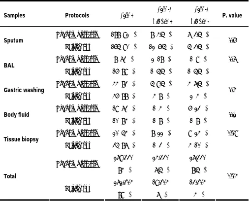
(Figures 1 and 2). In this experiment, salting-out method was considered as a proper DNA isolation technique with a maximum positive result about 89%, in which β-actin was tested only. But, 84% of the samples were β-actin-positive by phenol-chloroform technique (Table 1). Furthermore, gDNA obtained from 13/304 (4%) samples was negatively detected by β-actin and β-globin tests via the sa1lting-out technique, in comparison with 27/330 (8.5%) specimens negatively detected by phenol-chloroform. Based on the results, there was no significant difference between outcomes of salting-out and phenol-chloroform techniques in sputum, BAL, gastric washing, and body fluid samples. This highlights that both techniques have the same quality for extracting gDNA. For tissue biopsy, a difference between two gDNA extraction methods was found indicating that salting-out caused the minimum PCR inhibitor (Table 1). Also, no association was seen (P=0.04) in which total samples were grouped. Hence, in total, both DNA extraction protocols can have the same efficacy for isolation of gDNA from samples.
Figure 1: Analysis of 2% gl electrophoresis. From left, lanes contain marker, positive control, and six clinical samples. A sharp band corresponding to 190 bp shows the presence of Mycobacterium tuberculosis. Genomic DNA was qualified using PCR amplification of β-actin gene. β-actin-positive was reported when 330 bp bands were visualized.
Figure 2: The simple analysis of β-globin test. Lanes bear marker, positive control, and three clinical samples. β-actin tests were negatively reported for those three samples. However, the β-globin gene was amplified. As shown here, three sharp bands (A, B, and C) corresponding to 110 bp, are confirming the amplification of β-globin gene.
Table 1. Number (%) of positive and/or negative internal controls detected via phenol-chloroform and salting-out techniques
Samples Protocols β-actin + β-actin -/
β-globin +
β-actin -/
β-globin - P. value
Phenol-chloroform 188 (92%) 8 (4.5%) 7 (3.5%)
Sputum
Salting-out 165 (90%) 12 (6.5%) 5 (3.5%) 0.6 Phenol-chloroform 8 (73%) 2 (18%) 1 (9%)
BAL
Salting-out 16 (89%) 1 (5.5%) 1 (5.5%) 0.7
Phenol-chloroform 44 (83%) 5 (9.5%) 4 (7.5%)
Gastric washing
Salting-out 46 (88%) 4 (8%) 2 (4%) 0.4
Phenol-chloroform 19 (73%) 1 (4%) 6 (23%)
Body fluid
Salting-out 10 (84%) 1 (8%) 1 (8%) 0.2
Phenol-chloroform 20 (54%) 8 (22%) 9 (24%)
Tissue biopsy
Salting-out 35 (87%) 1 (3%) 4 (10%)
0.09
Phenol-chloroform 279/330 (84%)
24/330
(7.5%)
27/330
(8.5%)
Total
Salting-out 272/304 (89%)
19/304
(7%)
13/304
(4%)
0.04
DISCUSSION
In molecular diagnostic laboratories, the quality of
DNA extraction is the key evaluation criterion(7). A number of different DNA extraction techniques are used in the laboratories each having specific advantages and technical drawbacks. Genomic DNA (gDNA) extraction is
an important procedure for both clinical and experimental purposes and DNA can be isolated from various fresh or frozen clinical specimens including blood, plasma, and paraffin-embedded tissues(8). The current study aimed to
samples via phenol-chloroform and salting-out extraction protocols.
The classic phenol-chloroform is one of the oldest DNA extraction protocols. As clarified earlier, this protocol benefits from the use of sodium dodecyl sulfate (SDS) for lysis of the cell wall, and proteinase K and RNase for digestion of protein and RNA, respectively. This method mostly gives high yields of good-quality DNA (6, 9). Some methods are available for the quantitation of DNA such as UV measurements and PCR. Spectrophotometry is one of the routine methods for analyzing gDNA quantity. Whereas the phenol used in the phenol-chloroform protocol reduces the quantitation of DNA (6). The use of internal controls is another technique for the qualification of gDNA. Thus, we routinely applied β-actin and β-globin
as internal controls.
Another major disadvantage of phenol-chloroform protocol is the use of highly toxic reagents and also more time is required to extract gDNA from the samples in comparison with salting-out that isolates gDNA in less than 1 hour(1, 10). The total time for each DNA extraction protocol is also important especially when a large number of samples need to be tested (1).
For clinical purposes, amplification of specific gene fragments from target DNA can be done using PCR. Cao et al. reported that both the simple boiling and the phenol-chloroform methods are proper methods for PCR amplification of a gene segment less than 256 bp than DNA Mini Kit(11). We also used PCR for amplification of the β-
actin gene (110 bp) sequence by salting-out and
phenol-chloroform methods in the specimens. As illustrated in Table 1, there was no statistically significant difference between the two methods. This confirms that both DNA extraction techniques are suitable for amplification of short size gene fragments and appropriate for β- actin internal control amplification.
Since our institution is a referral TB clinical and research center in Iran, this study evaluated a wide range
of specimens from patients suspected for TB for evaluation of gDNA, compared with a few other studies that only used one type of clinical samples. Thus, the current study can more definitely demonstrate the appropriateness of these two methods for different samples. Therefore, considering the simplicity of salting-out method without using many organic solvents to extract gDNA from various clinical samples, this technique can be used in referral clinical centers in resource-limited countries when a large number of samples need to be tested.
A few studies have assessed PCR extraction sensitivity of phenol-chloroform and salting-out (12-14). We also in another unpublished study confirmed the sensitivity and specificity of PCR for diagnosis of mycobacterial DNA extracted from 620 TB suspected patients using either salting-out or phenol-chloroform methods. Diagnostic sensitivity and specificity of the PCR were 87.7% and 85.6%, respectively. PCR inhibitor was present only in 12 of the samples. The PCR inhibitor is considered when gDNA is not amplified to B-actin pair primers.
Due to the presence of phenol that results in decreased quantitation of DNA detected by UV absorbance(2), this study only qualified DNA extracted by amplification of the internal controls, β-actin and β-globin. In this study, gDNA was extracted from 385 sputum samples, 29 BAL specimens, 105 gastric washing samples, 38 body fluid samples and 77 tissue biopsy specimens obtained from TB suspected patients. β-actin PCR was first negatively reported for 7% and 7.5% of the samples using salting-out and phenol-chloroform extraction procedures, respectively (Table 1). For β-actin-negative PCR samples, β-globin PCR was then applied as the second PCR test. This study demonstrated no significant difference between the two protocols (P=0.04). Thus, salting-out method provides a proper rate of gDNA by amplification of β-actin gene fairly the same as with 279/330 positive cases determined by β
-actin PCR via the phenol-chloroform method.
phenol-chloroform, Chacon-Cortes et al, in 2012 presented the results of DNA extraction techniques by quantity (measurement of concentration of DNA extracted) and quality (260/280 ratio and PCR product) (1). They extracted gDNA from whole blood samples obtained from breast cancer patients using three different gDNA extraction techniques (a traditional salting-out method, a modified salting-out method and a commercial kit). The three methods had no statistically significant differences with the final result, but the time duration for each method showed significant differences. The limitation of the afore-mentioned study was using only whole blood samples of breast cancer patients. Whereas, in the current study, gDNA was extracted from various clinical specimens obtained from TB suspects. Elena et al, in 2006 reported an efficient and easy salting-out procedure for extraction of DNA from formalin-fixed paraffin-embedded tissues(15). The samples were subjected to a DNA extraction method using two different concentrations of ammonium acetate (2 and 4M) and then it was compared with a phenol– chloroform extraction method and the commercially available DNA extraction kit. Qualification of DNA extraction was performed by targeting 268 bp segment of
β-globin gene. The results showed that DNA isolated by all
the examined methods and in all samples was amplified to the gene (15). Therefore, they concluded that salting-out extraction method has a superior yield for isolation of DNA as compared with the phenol–chloroform and the DNA isolation kit.
Considering the disadvantages of phenol-chloroform technique such as high toxicity of phenol, being time-consuming and labor intensity, salting-out can be used as a routine DNA isolation technique in different clinical specimens. Whereas the salting-out method uses only a few Eppendorf tubes for each sample and reduces the use of consumables in the laboratory and the possibility of contamination.
REFERENCES
1. Chacon-Cortes D, Haupt LM, Lea RA, Griffiths LR.
Comparison of genomic DNA extraction techniques from
whole blood samples: a time, cost and quality evaluation
study. Mol Biol Rep 2012; 39 (5): 5961- 6.
2. McOrist AL, Jackson M, Bird AR. A comparison of five
methods for extraction of bacterial DNA from human faecal
samples. J Microbiol Methods 2002; 50 (2): 131- 9.
3. Miller SA, Dykes DD, Polesky HF. A simple salting out
procedure for extracting DNA from human nucleated cells.
Nucleic Acids Res 1988; 16 (3): 1215.
4. Grimberg J, Nawoschik S, Belluscio L, McKee R, Turck A,
Eisenberg A. A simple and efficient non-organic procedure for
the isolation of genomic DNA from blood. Nucleic Acids Res
1989; 17 (20): 8390.
5. Barker. Phenol-Chloroform Isoamyl Alcohol (PCI) DNA
Extraction 1998.
6. Santella RM. Approaches to DNA/RNA Extraction and whole
genome amplification. Cancer Epidemiol Biomarkers Prev
2006; 15 (9): 1585- 7.
7. Watson JD, Baker TA, Bell SP, Gann A, Lecine M, Losick R.
Molecular Biology of the Gene, 5th edition, USA San
Francisco, California: Benjamin Cummings 2004.
8. Srinivasan M, Sedmak D, Jewell S. Effect of fixatives and tissue
processing on the content and integrity of nucleic acids. Am J
Pathol 2002; 161 (6): 1961- 71.
9. Kirschner P, Rosenau J, Springer B, Teschner K, Feldmann K,
Böttger EC. Diagnosis of mycobacterial infections by nucleic
acid amplification: 18-month prospective study. J Clin
Microbiol 1996; 34 (2): 304- 12.
10. Chan PK, Chan DP, To KF, Yu MY, Cheung JL, Cheng AF.
Evaluation of extraction methods from paraffin wax
embedded tissues for PCR amplification of human and viral
DNA. J Clin Pathol 2001; 54 (5): 401- 3.
11. Cao W, Hashibe M, Rao JY, Morgenstern H, Zhang ZF.
Comparison of methods for DNA extraction from
paraffin-embedded tissues and buccal cells. Cancer Detect Prev 2003;
12. Pan S, Gu B, Wang H, Yan Z, Wang P, Pei H, et al.
Comparison of four DNA extraction methods for detecting
Mycobacterium tuberculosis by real-time PCR and its clinical
application in pulmonary tuberculosis. J Thorac Dis 2013; 5
(3): 251- 7.
13. Leung ET, Zheng L, Wong RY, Chan EW, Au TK, Chan RC, et
al. Rapid and simultaneous detection of Mycobacterium
tuberculosis complex and Beijing/W genotype in sputum by
an optimized DNA extraction protocol and a novel multiplex
real-time PCR. J Clin Microbiol 2011; 49 (7): 2509- 15.
14. Chantranuwat C, Assanasen T, Shuangshoti S, Sampatanukul
P. Polymerase chain reaction for detection of Mycobacterium
tuberculosis in papanicolaou-stained fine needle aspirated
smears for diagnosis of cervical tuberculous lymphadenitis.
Southeast Asian J Trop Med Public Health 2006; 37 (5): 940- 7.
15. Rivero ER, Neves AC, Silva-Valenzuela MG, Sousa SO, Nunes
FD. Simple salting-out method for DNA extraction from
formalin-fixed, paraffin-embedded tissues. Pathol Res Pract