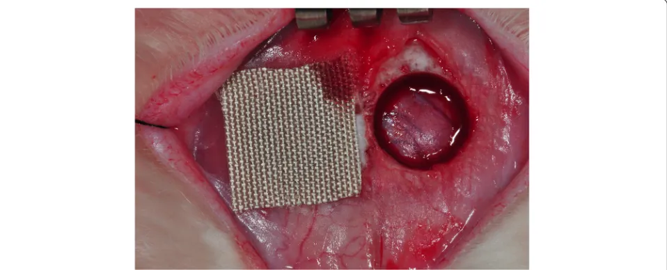
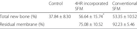
Evaluation of bone formation and membrane degradation in guided bone regeneration using a 4-hexylresorcinol-incorporated silk fabric membrane
Full text
Figure

Related documents
Aim of the current study was to test the interrater reliability of the authorized Italian version of the CAARMS (CAARMS-ITA) in young adult help-seekers consecutively recruit
Most antioxidase activity of the trop- ical maize was higher than temperate maize after three days of treatment, indicating the antioxidant system of tropical
The characterization of tester plants has potential to significantly reduce the number of hybridizations necessary to characterize the genotypes of the S gene by carrying out
• TCP SYN Scan -- “half-open” scan, look for SYN-ACK, then send RESET, target system will not record connection, also faster than TCP connect scan. • TCP FIN, Xmas Tree, Null
Next, we apply the woven principle to Hilbert-Schmidt frames and study the stability of weaving Hilbert-Schmidt frames under perturbations.. Finally, we present sufficient
In this paper, we explore the implementation of standard particle swarm optimization (SPSO) on a swarm of physical mobile robots conducting a source seeking task.. The signal source
