R E S E A R C H
Open Access
Long-term radiographic assessment of
maxillary sinus floor augmentation using
beta-tricalcium phosphate: analysis by
cone-beam computed tomography
Tsuneji Okada
1*, Toru Kanai
1, Noriko Tachikawa
1, Motohiro Munakata
2and Shohei Kasugai
3Abstract
Background:The long-term stability of maxillary sinus floor augmentation withβ-TCP remains largely unknown. We report the long-term assessment of volumetric changes in maxillary sinus floor augmentation withβ-TCP by cone-beam computed tomography (CBCT).
Methods:The subjects included 30 patients who underwent maxillary sinus floor augmentation usingβ-TCP and 58 implant placement for unilateral maxillary defect, simultaneously. Volumetric changes inβ-TCP and the height of peri-implant bone were analyzed by CBCT.
Results:In all patients, the mean volume of the grafted bone decreased from immediately after implant placement to 6 months after implant placement (75.6 % reduction rate); it decreased further at 2.5 years after implant placement (54.9 % reduction rate). The mean of the height from the implant tip to the maxillary sinus floor was 2.00 ± 1.51 mm, 0.73 ± 1.33 mm, and−0.72 ± 1.11 mm immediately, 6 months, and 2.5 years after implant placement, respectively. The implant tip protruded beyond the maxillary sinus floor in approximately 70 % of the implants (41/58 implants) at 2.5 years after surgery. During the observation period, the implant survival rate was 100 %.
Conclusions:The radiographic analysis by CBCT is considerably more advanced than previous radiographic examinations. Although maxillary sinus pneumatization continues to progress≥1 year after maxillary sinus floor augmentation withβ-TCP, it stabilizes 3 years after surgery.
Keywords:Beta-tricalcium phosphate, Maxillary sinus floor augmentation, CBCT analysis
Background
The maxillary sinus gradually expands after birth and becomes fully pneumatized with the eruption of all per-manent teeth. Although the physiological cause and maxillary sinus pneumatization are largely unknown, it is believed that genetics, atmospheric pressure, and hormones are involved in it. This sinus is closely related to the root apex of the premolar and molar teeth, and it is either separated from the teeth by a thin layer of bone or its mucous membrane is in direct contact with the teeth. The pneumatization of the maxillary sinus and the
reduction in the bone wall thickness resulting from the loss of teeth is attributed to atrophy caused by a reduc-tion in stimulareduc-tion to the bone. It is also believed that physiological effects of the mucous membrane of the maxillary sinus as well as factors such as osteoclasts and loss of tooth root resistance to atmospheric pressure in the maxillary sinus are considered to play a role [1–3].
When an implant is placed in the maxillary molar re-gion with atrophy, the maxillary sinus floor is close to the alveolar crest. Thus, some cases may require bone grafting for maxillary sinus floor augmentation. Initially, the gold standard for bone grafting material used to per-form this procedure was autogenous bone. The advan-tages of autogenous bone graft are that it has osteogenic, osteoinductive, and osteoconductive properties. It has * Correspondence:[email protected]
1Clinic for Implant Dentistry, Dental Hospital, Tokyo Medical and Dental University, 1-5-45 Yushima, Bunkyo-ku, Tokyo 113-8549, Japan Full list of author information is available at the end of the article
mass (e.g., the mental region and mandibular ramus in the oral cavity or the iliac crest and tibia outside the oral cavity), and it places great stress on both the practitioner and the patient [4, 5]. In recent years, maxillary sinus floor augmentation has been introduced using various bone grafting materials to decrease invasiveness. In Japan, xenogeneic bone grafts are not permitted for eth-ical reasons; instead, synthetic bone grafting agents that are not derived from animals, such as hydroxyapatite (HA) and β-TCP, have been used. However, unlike non-absorbable materials such as HA, β-TCP has properties to get absorbed in the body and to be replaced by the bone; in addition, its usefulness has been reported in orthopedic surgery and maxillofacial surgery [6, 7]. We have achieved good outcomes using β-TCP as the bone grafting material for maxillary sinus floor augmentation. However, the predictability of the material and the change in absorption of the transplanted material mass remain largely unknown.
Radiographic examinations to determine changes in bone mass in maxillary sinus floor augmentation have been conducted primarily using panoramic radiog-raphy [8–12], which allows for the assessment of the height of the maxillary sinus in only two dimensions. In addition, it does not allow for detailed examination or measurement of the interior of the maxillary sinus without factoring in magnification or distortion. Hounsfield developed X-ray computed tomography in 1982. In 2001, cone-beam computed tomography (CBCT), the features of which included low exposure and high spatial resolution, became available in the clinical setting. Shortcomings of CBCT are that it de-livers inferior spatial resolution of soft tissue com-pared with conventional CT and does not offer the quantification of CT values. However, narrowing the field of view (FOV) decreases the amount of radiation exposure. Moreover, its greatest advantages include isotropic spatial resolution (voxel) and high spatial resolution (superior resolution of bone), which allow for the accurate measurement of the interior of the maxillary sinus.
The purpose of this study was to examine the effect of bone grafting material resorption on volumetric change and the predictability of the long-term pro-gress of sinus floor augmentation using β-TCP by performing radiographic examinations using CBCT.
tion at the same time as implant placement.
Patient selection
Patients were excluded from the study if they met the following criteria:
They were completely edentulous and believed to be affected by postoperative denture wearing.
They required sinus floor augmentation and buccal onlay graft at the same time for narrow bone width or if they required large-scaled bone graft or floor augmentation, such as that for guided bone regener-ation and split crest.
They had insufficient occlusal clearance or
extremely poor tooth crown-to-root ratio, indicated by a Cawood and Howell classification V.
They had a history of diabetes, osteoporosis, bisphosphonate administration, immunological diseases, such as rheumatoid arthritis, chronic sinusitis, or otolaryngological problems, as well as other untreated systemic disease, or were smokers.
The informed consent was obtained from all the pa-tients regarding bone grafting material use, the surgical procedure, as well as the multiple CBCT scans. This study was carried out with the approval of the ethics committee of the School of Dentistry.
Surgical procedure
Maxillary sinus floor augmentation was performed by a skilled surgeon using the technique according Boyne et al. [13]. Patients underwent preoperative examina-tions, after which maxillary sinus floor augmentation and implant placement were performed simultaneously. The grafting material used was OSferion (Olympus Terumo Biomaterials Corp., Japan) composed of β-TCP (Ca3PO4)2 (calcium phosphate). Second-stage surgery was performed 6 months after implant placement, and prosthetic treatment was performed.
Radiographic examination
size at 0.146 mm3 and FOV of φ82.00 mm × 75.1 mm (Fig. 1).
In the present study, we used the superior spatial reso-lution of CBCT to measure changes over time in (a) the volume of the bone graft (BV) and (b) the height of the bone surrounding the implant (BH).
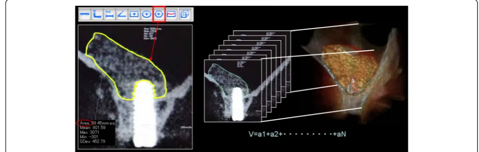
(a) The method of calculating the volume of the implant site is shown below (Fig.2).
The slice thickness (voxel value) was resized (0.146 mm→1 mm) to derive the volume of the grafting agent on the basis of CBCT data. After setting the slice interval to 1 mm and specifying the output range (FOV option), the DICOM server for storing data was selected and registered.
Next, image data of four time axes from prior to surgery, immediately after surgery, 6 months after surgery, and 2.5 years after surgery was
simultaneously imported into a multi-data image analysis system. For confirming that the four images had the same positional relationship, we imple-mented synchronized scrolling after identifying
similar anatomical reference points. The area of the implant site was measured using the polygon tool which was dragged around the perimeter of the target site to measure area. It allows measurements up to two places after the decimal point. It is oper-ated by dragging the cursor only around the bone graft material site and avoiding the implant itself. In cases in which it was impossible to determine the margin between the implanted bone and the existing bone after 1-year examination, the entire bone was measured and the preoperative measurement was subtracted from the postoperative measurement. Then the images were taken at 1-mm increments mesio-distally, and the volume was calculated by adding the entire area of the bone grafting material. BoneVolume (BV) cm3= area cm2×N (number of images)
(b)The method of measuring the height of the bone surrounding the implant (BH) is described below (Fig.3).
The measurements were taken at the long axis of the implant and on the frontal plane of the
Fig. 1Treatment protocol for the present study. Postoperative CBCT was performed a minimum of three times, i.e., immediately, 6 months, and 2.5 years after implant placement
perpendicular frontal plane. In the event that there was grafted bone above the implant, the distance was measured from the point of intersection of the line connecting the buccal and palatal side of the maxillary sinus floor and the medial axis of the implant to the implant tip (+mm). Moreover, in the event that absorption of the grafting material exceeded the implant tip, the distance was measured from the point of intersection of the medial axis of the implant and the line connecting the most apical part of the grafted bone-to-implant contact (BIC) to the implant tip (−mm). In addition, the liner valu-ables (RBH, SW, IL) that may effect the outcomes of BH were measured. The following are the explana-tions of the linear valuables.
Residual bone height (RBH): existing bone height to the maxillary sinus at the implant site
Implant length (IL): the length of the part of the implant that projects into the maxillary sinus
Width of sinus (SW): the width of the maxillary sinus from lateral wall to medial wall at the height of the center of the IL
Statistical analysis
In the present study, the radiographic examinations were statistically analyzed for (a) volumetric changes of the bone graft (BV) and (b) changes in the height surround-ing the implant (BH) over time. Moreover, it was exam-ined whether the liner parameters (RBH, SW, IL), BH immediately after surgery (iBH), and the implant place-ment site affected the changes in BH over time.
Because the time CBCT was performed 2.5 years the sur-gery varied, analysis was performed using the generalized estimating equation (GEE). Thus, the time of CBCT was used as a variable to resolve the problem.
Results
Participants
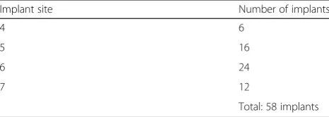
The mean age of the 30 patients was 57.4 (40–75) years, and the male/female ratio was 4:26. The mean postoper-ative observation period was 3 years and 8 months (the maximum period was 5 years and 0 months; the mini-mum period was 2 years and 2 months). Total of 58 im-plants were placed at premolar and molar region. There were no dropouts, such as participants not visiting the clinic, during the observation period (Tables 1, 2, and 3).
Clinical evaluation
Implant osseointegration was achieved in all patients without any complications. During the observation period between January 2009 and January 2014, the im-plant survival rate was 100 %. At the time of second-stage surgery, macroscopic observation of the lateral wall Fig. 3Radiographic examination of the height of the bone
surrounding the implant (BH): Measurement of changes in the height of the implant tip to the bone fixation part over time in the frontal plane: the distance measured from the intersecting point of the long axis of the implant and the maxillary sinus floor to the implant tip: +maxillary side,−alveolar crest side. The liner valuables: residual bone height (RBH), implant length (IL), and width of sinus (SW)
Table 1Age groups of the 30 patients
Age group (years) Number of patients
40–49 7
50–59 10
60–69 10
70–79 3
Mean age 57.4 years Total: 30 patients
43–48 2
49–54 2
55–60 4
Mean 3 years and 8 months Total: 30 patients
Table 3The number of implants according to site
Implant site Number of implants
4 6
5 16
6 24
7 12
of the maxillary sinus revealed differing degrees of β -TCP replaced by new bone (Fig. 4).
Radiographic evaluation
In consideration of the radiation the patient would be exposed to and to increase the benefits to the patients, the fourth CBCT (2.5 years after surgery) was performed at a time when it did not overlap with radiation expos-ure that was conducted as part of complete physical ex-aminations or required tests (periodontal disease of the remaining tooth, endodontic examinations, root fracture, etc.). Therefore, the fourth CBCT was performed at varying times. The distribution of observation time is shown in Table 4.
β-TCP could be differentiated from existing bone on CBCT even 6 months after surgery; however, the distinc-tion became unclear 2.5 years after surgery, indicating that the replacement of the grafted bone by new bone appeared to have been accelerated. The effect of implant artifacts made it difficult to measure changes in the bone density index (gray scale value).
(a) Volumetric changes in the graft: The meanβ-TCP volume decreased from immediately after surgery (1206.9 ± 437 cm3) to 6 months after surgery (912.6 ± 356 cm3). It further decreased 2.5 years after surgery (662 ± 294 cm3), and the reduction in volume was observed in all patients. Percent volume changes 6 months after surgery and 2.5 years after surgery using volume immediately after surgery as the baseline value were 75.6 and 54.9 %, respectively (Table5).
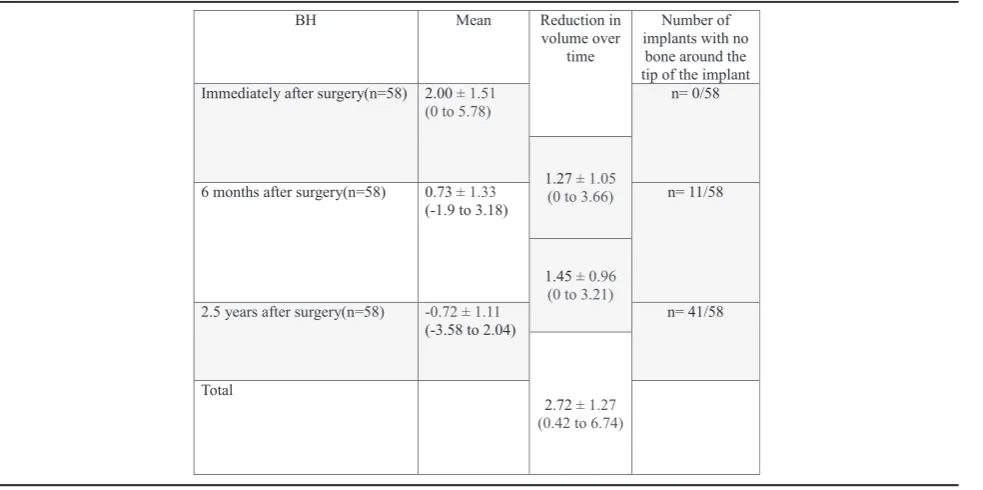
(b)Change in height of bone surrounding the implant (height from the maxillary sinus floor to the implant tip): Immediately after surgery, all patients had grafted bone between the implant tip and the maxillary sinus floor, with a mean of 2.00 ± 1.51 mm. This height decreased to 0.73 ± 1.33 mm at
6 months after surgery and−0.72 ± 1.11 mm at 2.5 years after surgery (Fig.5). 41/58 implants had developed pneumatization 2.5 years after surgery and had no bone around the implant tip. The overall mean reduction in bone height was 2.72 ± 1.27 mm: (i) the mean reduction from immediately after surgery to 6 months afterward was 1.28 ± 1.05 mm and (ii) that from 6 months after surgery to 2.5 years after surgery was 1.44 ± 1.45 mm (Table6, Fig.6).
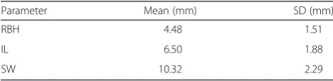
The liner parameters: mean RBH, IL, and SW were 4.48 ± 1.51 mm, 6.50 ± 1.88 mm, and 10.32 ± 2.29 mm with a mean value, respectively (Table 7). In addition, on examining the 10 patients (20 implants) with multiple long-term data of ≥1 year after surgery, the height reduction was found to have progressed to the same ex-tent from approximately 1–2 years after surgery, after which there was only a slight change (Fig. 5).
Statistical analysis
Radiographic evaluation items were statistically ana-lyzed: (a) a significant decrease in graft volume (BV) was observed over time (p< 0.00) and there was no sta-tistically significant effect of age or gender and (b) a Fig. 4Clinical findings of the second surgery on biopsy at 6 months. The degree of residual grafting materials varied depending on the patient.
amost of theβ-TCP remained.bReplacement of theβ-TCP by new bone had progressed
Table 4The distribution of CBCT examination after 2.5 years
CBCT examination (year) Number of patients
0–1 0
1–2 14
2–3 5
3–4 5
4–5 6
Mean 2.5 Total: 30
Table 5Radiographic examination of BV (volumetric changes in
graft bone over time)
BV Mean Reduction rate
Immediately after surgery (n= 30)
1206 ± 437 (477 to 2292) 100 %
6 months after surgery (n= 30)
912 ± 356 (428 to 1760) 75.6 %
2.5 years after surgery (n= 30)
662 ± 294 (204 to 1568) 54.9 %
BVbone volume,n= 30: the number of maxillary sinuses
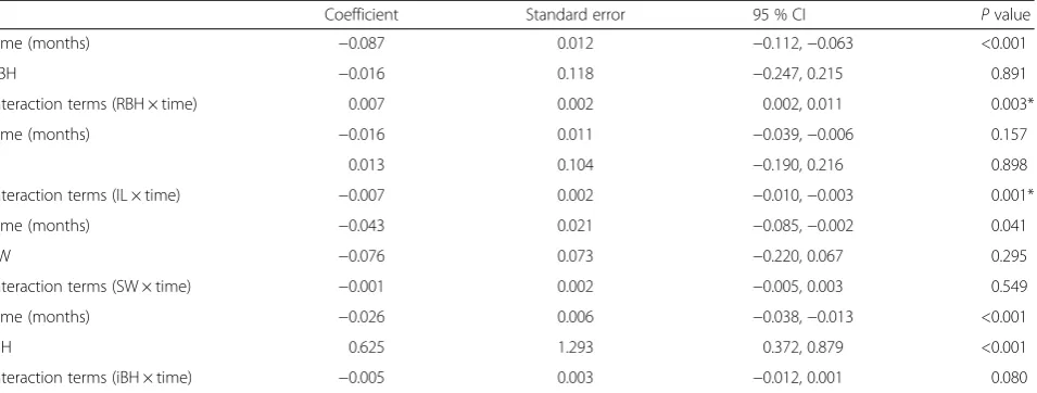
significant decrease in bone height (BH) was observed over time (p< 0.001) and there was no statistically sig-nificant effect of age or gender. We found that this change of BH was affected by RBH (p= 0.003) and IL (p= 0.001); the thicker the RBH, the less the decrease of BH; the longer the IL, the more the decrease of BH over time. We determined that the higher the immedi-ately postoperative bone height (iBH) was, the higher the subsequent total height would be. We also observed that the coefficient for the interactive items tended to
have negative values and that the higher the postopera-tive bone height was, the larger the amount of decrease was. There was no significant difference in the change in bone height according to SW and also no significant difference according to implant site (Wald chi-square value = 1.221,P= 0.748) (Table 8).
Discussion
As grafting materials for maxillary sinus floor augmenta-tion, autogenous bone, which is considered as the gold
Table 6Radiographic examination of BH (changes in bone height surrounding the implant)
BHbone height surrounding the implant,n= 58: number of implants Analysis using generalized estimating equations (GEE) with patient clusters
standard and reported first by Boyne et al. using iliac bone graft, has been used as the first-choice material. In terms of osteogenic, osteoinductive, and osteoconductive properties, autogenous bone is considered ideal; how-ever, the use of autogenous bone places great physical stress on patients because of the need for surgery at the donor site. Synthetic bone substitutes such asβ-TCP are clinically very beneficial because donor site surgery can be avoided. Moreover, in the 1996 Sinus Consensus Conference, these materials were concluded as having highly predictable outcomes. Although there are many related factors, no difference in the success rate has been observed according to the grafting material [14].
Artzi et al. conducted a comparative experiment of bo-vine bones and β-TCP to fill defect areas in dogs. They reported that β-TCP was absorbed within 24 h and re-placed by autogenous bone, whereas with bovine bone, approximately 30 % granules remained [15]. Thus, β -TCP is considered the ideal grafting material.
The permanence of the new bone formed within the maxillary sinus has been primarily evaluated in humans using radiography. The clinical assessment and short-term and long-short-term investigations have also been con-ducted using panoramic radiography and conventional CT [8–12]. Panoramic radiography analysis does not have the shortcomings of radiation risk and expensive CT equipment; therefore, images can generally be ob-tained frequently. However, its limitations include the ability to assess two-dimensional height, and various
magnifications or errors, such as distortion, must also be taken into consideration. In the past, evaluations during implant treatment were primarily performed using pano-ramic radiography and conventional CT. Recently, how-ever, the use of preoperative and postoperative CBCT has gained popularity, and radiographic assessment by CBCT for volumetric changes in graft bone in maxillary sinus floor augmentation has also been reported [16]. According to our radiographic analysis by CBCT of den-tal implantation using β-TCP in the present study, we found that the graft volume decreased over time, both at 6 months after surgery and even at 2.5 years after sur-gery. The result of 54.9 % graft volume change of 2.5 years after surgery is similar to previous reports [17]. Loads were applied to the implant only 6 months after surgery. Nevertheless, the results suggested that it is difficult to control maxillary sinus pneumatization by exerting implant occlusive load. Zijderveld et al. conducted a 5-year radiographic study using panoramic radiography to evaluate periodic changes inβ-TCP [20 patients: 10 pa-tients with autogenous bone (taken from the mental re-gion) and 10 patients withβ-TCP]. They reported that the majority of β-TCP absorption occurred within the first 7.5 months and that very little change was observed after 1.5 years [12]. Furthermore, in 2004, Hatano et al. used a 2:1 mixture of autogenous bone-to-bovine bone and re-ported that the maxillary sinus floor was situated at the same height or lower than the implant tip in most patients [11]. These results are similar to our study that maxillary sinus pneumatization continues to progress≥1 year after surgery; it stabilizes 3 years after surgery and the implant tip protruded beyond the maxillary sinus floor in approxi-mately 70 % implants (41/58 implants) in patients who were followed up 2.5 years after surgery. However, there were no clinical abnormalities, such as maxillary sinusitis. With regard to liner valuables (RBH, IL, SW, iBH) in the present study, the results suggested that it is difficult to
Table 7The radiographic measurements of liner parameters at
immediately after surgery (RBH, IL, SW)
Parameter Mean (mm) SD (mm)
RBH 4.48 1.51
IL 6.50 1.88
SW 10.32 2.29
control maxillary sinus pneumatization. There was no sig-nificant difference in the change in bone height according to SW, and this result is inconsistent with previous reports [18]. There was a limited number of subjects in this study; we believe it is necessary to conduct long-term tests of lar-ger numbers of subjects in the future.
During the observation period, there was no clinical implant failure and the suvival rate for implantation and maxillary sinus floor augmentation was 100 %. Histological examination in a previous report on max-illary sinus augmentation using β-TCP (Cerasorb®), Szabo et al. compared bilateral maxillary sinus aug-mentation in the same patients using β-TCP and iliac bone at 6 months after surgery and reported a new bone ratio of 36.5 % in the β-TCP group (control group: 38.3 % autogenous bone), and in a similar ex-periment, Suba et al. reported a new bone ratio of 32.4 % in the β-TCP group (control group: 34.7 % au-togenous bone), which were similar outcomes to those for the autogenous bone. These histological ex-aminations provided supporting evidence that during the year after grafting, β-TCP is gradually replaced by new bone over time [19–21]. Our clinical and radio-graphic examination of maxillary sinus floor augmen-tation using β-TCP suggests that β-TCP is a very useful synthetic bone substitute with very similar progress to autogenous graft. It is of our interest to further continue our long-term clinical study.
Conclusions
Maxillary sinus graft augmentation usingβ-TCP is clinically effective.
Analysis by CBCT provides superior spatial resolution and allows for extremely accurate
postoperative evaluation of maxillary sinus floor augmentation and bone volume measurements.
On the basis of CBCT examinations, although maxillary sinus pneumatization continues to progress≥1 year after surgery, it stabilizes 3 years after surgery.
Competing interests
Tsuneji Okada, Toru Kanai, Noriko Tachikawa, Motohiro Munakata, and Shohei Kasugai declare that they have no competing interests.
Authors’contributions
TO performed the data acquisition, both of surgical and prosthodontic treatments, the data analysis, statistical analysis, and wrote the manuscript. TK performed the treatment plan, making of pictures, and collecting of references. NT and MM performed the data acquisition and both of surgical and prosthodontic treatments. SK performed the data analysis. All authors read and approved the final manuscript.
Acknowledgements
The bone grafting agent,β-TCP (OSferion), was kindly supplied by Olympus Terumo Biomaterials Corp.
Author details
1Clinic for Implant Dentistry, Dental Hospital, Tokyo Medical and Dental University, 1-5-45 Yushima, Bunkyo-ku, Tokyo 113-8549, Japan.2Oral Implantology Department of Prosthodontic Dentistry for Function of TMJ and Occlusion, Kanagawa Dental University, 82, Inaokachou, Yokosuka-shi, 238-8580 Kanagawa, Japan.3Oral Implantology and Regenerative Dental Medicine, Department of Masticatory Function Rehabilitation, Division of Oral Health Sciences, Graduate School of Medical Dental Sciences, Tokyo Medical and Dental University, 1-5-45 Yushima, Bunkyo-ku, Tokyo 113-8510, Japan.
Received: 25 November 2015 Accepted: 23 March 2016
References
1. Chanavaz M. Maxillary sinus: anatomy, physiology, surgery, and bone grafting related to implantology: eleven years of surgical experience (1979– 1990). J Oral Implantol. 1990;16:199–209.
2. Misch CE. Maxillary sinus lift and elevation with subantral augmentation. In: Misch CE, editor. Contemporary implant dentistry. St. Louis, MO: Mosby; 1993. p. 545–74.
Interaction terms (IL × time) −0.007 0.002 −0.010,−0.003 0.001*
Time (months) −0.043 0.021 −0.085,−0.002 0.041
SW −0.076 0.073 −0.220, 0.067 0.295
Interaction terms (SW × time) −0.001 0.002 −0.005, 0.003 0.549
Time (months) −0.026 0.006 −0.038,−0.013 <0.001
iBH 0.625 1.293 0.372, 0.879 <0.001
Interaction terms (iBH × time) −0.005 0.003 −0.012, 0.001 0.080
3. Sharan A, Madjar D. Maxillary sinus pneumatization following extractions: a radiographic study. Int J Oral Maxillofac Implants. 2007;23:48–56. 4. Uchida Y, Goto M, Katuki T, et al. Measurement of maxillary sinus volume
using computerized tomographic images. Int J Oral Maxillofac Implants. 1998;13:811–8.
5. Krennmair G, Krainhofner M, Maier H, et al. Computerized tomography-assisted calculation of sinus elevation volume. Int J Oral Maxillofac Implants. 2006;21:951–6.
6. Ozawa M, Tanaka K, Morikawa S, et al. Clinical study of the pureβ -tricalciumphosphate—reports of 167 cases. J East Jpn Orthop Traumatol. 2000;12:409–13.
7. Horch H, Sader R, Pautke C, et al. Synthetic, pure-phaseβ-tricalcium phosphate ceramic granules (Cerasorb) for bone regeneration in the reconstructive surgery of the jaws. Int J Oral Maxillofac Surg. 2006;35:708–13.
8. Geurs NC, Chung-Wang I, Shulman LB, et al. Retrospective radiographic analysis of sinus graft and implant placement from the academy of osseointegration consensus conference on sinus graft. Int J Periodontics Restorative Dent. 2001;21:517–23.
9. Hallmann M, Hedin M, Sennerby L, et al. A prospective 1-year clinical and radiographic study of implants placed after maxillary sinus floor elevation with bovine hydroxyapatite and autogenous bone. Int J Oral Maxillofac Implants. 2002;b60:277–84.
10. Ozyuvaci H, Bilgic B, Firatli E. Radiographic and histomorphometric evaluation of maxillary sinus grafting with alloplastic materials. J Periodontol. 2003;74:909–15.
11. Hatano N, Shimizu Y, Ooya K. A clinical long-term radiographic evaluation of graft height changes after maxillary sinus floor elevation with a 2:1 autogenous bone/xenograft mixture and simultaneous placement of dental implants. Clin Oral Impl Res. 2004;15:339–45.
12. Zijderveld SA, Schulten EA, Aartman IA, et al. Long-term changes in graft height after maxillary sinus floor elevation with different grafting materials: radiographic evaluation with a minimum follow-up of 4.5 years. Clin Oral Impl Res. 2009;20:691–700.
13. Boyne PJ, James RA. Grafting of the maxillary sinus floor with autogenous marrow and bone. J Oral Maxillofac Surg. 1980;38:613–6.
14. Jensen OT, Shulman LB, Block MS, et al. Report of the sinus consensus conference of 1996. Int J Oral Maxillofac Implants. 1998;13:11–45. 15. Artzi Z, Weinreb M, Givol N, et al. Biomaterial resorption rate and healing
site morphology of inorganic bovine bone andβ-tricalcium phosphate in the canine: a 24-month longitudinal histologic study and morphometric analysis. Int J Oral Maxillofac Implants. 2004;19:357–68.
16. Umanjec-Korac S, Wu G, Hassan B, et al. A retrospective analysis of the resorption rate of deproteinized bovine bone as maxillary sinus graft material on cone beam computed tomography. Clin Oral Impl Res. 2014;25:781–5. 17. Kim ES, Moon SY, Kim SG, et al. Three-dimensional volumetric analysis after
sinus grafts. Implant Dent. 2013;22:170–4.
18. Zheng X, Teng M, Zhou F, et al. Influence of maxillary sinus width on transcrestal sinus augmentation outcomes: radiographic evaluation based on Cone Beam CT. Clin Implant Dent Relat Res. 2015; Feb 26. Epub ahead of print. 19. Szabo G, Huys L, Coulthard P, et al. A prospective multicenter randomized
clinical trial of autogenous bone versusβ-tricalcium phosphate graft alone for bilateral sinus elevation: histologic and histomorphometric evaluation. Int J Oral Maxillofac Implants. 2005;20:371–81.
20. Suba Z, Takacs D, Matusovits D, et al. Maxillary sinus floor grafting withβ -tricalcium phosphate in humans: density and microarchitecture of the newly formed bone. Clin Oral Impl Res. 2006;17:102–8.
21. Zijderveld SA, Zerbo IR, van den Bergh JPA, et al. Maxillary sinus floor elevation using aβ-tricalcium phosphate (Cerasorb) alone compared to
autogenous bone grafts. Int J Oral Maxillofac Implants. 2005;20:432–40.
Submit your manuscript to a
journal and benefi t from:
7 Convenient online submission
7Rigorous peer review
7 Immediate publication on acceptance
7Open access: articles freely available online
7 High visibility within the fi eld
7Retaining the copyright to your article