*
Cor
T. Se
1SAST Tirum 61340 2Trich Garde 002, In 3Instit Techn ChidaIntr
Wou acco unde haem from time mana with site stren flava main facilit prop The quite trans like s guine chos studi ThisEvaluatio
rresponding
enthil kumar
TRA University, alaisamudram, T 01, India hy Pharma Chemen, Devadhanam ndia
ute of Pharmace nology, Annamal ambaram, India
oduction
nd is a break in ompanied by d erlying normal t matoma, lacerat the moment of depending on th agement is to h minimal pain, d of wound closu ngth is desired. anoids as their c nly due to their
tates rapid heal erties could be v outline of the e e similar to thos sparent, evenly sheets.[1]. Due t ea pig in comp sen for experim ies were done o
work is licens
on of herb
T.Senth
g author:
Thanjavur m P Ltd., VInYY m. Trichy 620
eutical lai University,
n the epithelial in disruption of th issue and may ion or an abras
injury and can he extent of wou heal the wound discomfort, and ure a flexible a . Phytopharmac constituents play r antimicrobial a ling process. M very well used fo epidermal corne se of the human
pink stained pe to higher correla parison to othe mentation. Cont n Guinea pig mo
sed under a C
http
bal ointm
leave
hil kumar*
1, D
A b s
The obj infection use of l containi ointmen and exc was pre coconut nitrofura treated (allopat contract 12.7 da ointmen facilitati healing Keywor tinctoria
ntegrity of the sk he structure an also result from sion. Healing of
continue for var unding. The obje
in the shortest scaring to the p nd fine scar w ceuticals with t y major role in and astringent Medicinal plants or wound healing
ocytes of guine n skin: typically,
entagons formin ation of human s r non-human p trolled wound h odel with incision
reative Comm
p://www.arjo
Origin
ent formu
es for effic
D.Venkappayy
s t r a c t
jective in wound n and fast woun eaves of Wright ing Wrightia tinc nt.Controlled wo cision wounds. S epared by melt
t oil. The woun azone ointment. groups in con hic control and tion to 50% too ays for ointment nt. Increased wo ng wound heali as a safe altern rds:Wound heal a (Roxb.) R. Br.
kin and may be nd function of m a contusion, f wounds starts rying periods of ective in wound t time possible, patient. At the ith high tensile tannins and or wound healing property which with the above g.
ea pig skin was they were flat, ng honeycomb-skin with that of primates it was healing efficacy n and excision
mons Attributio
ournals.org/in
nal Researc
ulated wit
cacy of w
ya
1, Vilambi N
d management nd closure.This htia tinctoria by e ctoria which may ound healing eff Suitable ointmen pour and mixing d healing effec
A better healing trast to the co
Wrightia tincto ok 6.3 days (allo t base. The ten ound breaking st ing, thus provin ative to syntheti ling Exc
wound Apocy and a etheni antiox to che of Wr formu skin c
Mate
Colle
The le Nacha Anato specimExtra
Leave cleaneon 3.0 License
ndex.php/ijpm
ch Article
th
Wright
wound hea
N.R.K.Reddy
2,
is to heal the w investigation is evaluating the w y facilitate in vivo
icacy studies w nt base was sele g technique with
ts of the formu g pattern with co ntrol group. T oria ointment) w pathic control), 6 nsile strength of
trength indicates ng that Wrightia c drug ointments cision model
ds [2]. Wrighti ynaceae. Its lea applied for ecze ic groups in hills xidant and anti ps eck the scientific
ightia tinctoria b lated ointment w urative property
erials and M
ection and aut
eaves of Wrightia allur, Karur distr
my Research men was deposit
action and form
es of Wrightia ed leaves were
e.
m/index
e
htia tinctor
aling.
, R.Manavala
wound in the sh to check the ra ound healing pr o quantification o were done on Gu cted by Preform h incorporation o
lations were co omplete wound c The total epithe with 19 days for 6.5 days (Wrigh f the test was a s increase in co a tinctoria ointme
s.
Incision mo
a tinctoria (Rox aves were soake ema, psoriasis s. As Wrightia t soriatic property c rationale behin by evaluating th
which may faci of the ointment.
Methods
thentication of
ia tinctoria were rict, identified by Centre (PARC ted at PARC.
mulation of pl
tinctoria were minced to sma
ria
(ROXB
n
3hortest time pos ationale behind roperty of formul of skin curative p
uinea pig mode mulation studies. of Wrightia tinct ompared to that
closure was obs elization period r ointment base htia tinctoria ointm
almost the same ollagen strength
ent could be us odel Ointment
xb) R. Br. belo ed in coconut oil and other skin tinctorialeaves y, hence an attem
d the traditional he wound healin litate in vivo qu .
f plant materia
collected from V y Prof. P. Jayara C), Chennai an
ant material
collected and all pieces. The m
B)
R. BR.
ssible, without the traditional lated ointment property of the el with incision The ointment toria extract in of 0.2% w/w erved with the was 14 days e. The wound ment), against e as standard and obviously sed for wound
t Wrightia
ongs to family l for few hours n diseases by
have reported mpt was made use of leaves ng property of uantification of
al
VInYY garden, aman of Plant nd a voucher
PAGE |
229
|
were brought into contact with coconut oil in the ratio 1:5 and leftfor a period of 5 days under sun. The completion of extraction was indicated through the colour of the oil turning to purplish blue. The oil was filtered and the filtrate was used for formulation. The ointment was prepared using 70% of 5% Wrightia tinctoria oil extract. Preformulation studies were conducted and suitable ointment base consisting of 15% bees wax, 10% hard paraffin wax and 5% soft paraffin wax was selected. Butylated hydroxyl toluene was used as preservative. The ointment was prepared by melt pour and mixing technique.
Wound healing activity
The protocol of the study was approved by the local animal ethical committee of IIMT college of medical science. The guinea pigs were kept in standard conditions in the animal house and were used after an acclimatization period of 7 days to get elaborated to the environment. They were provided with food and water ad libitum.
Animal model for wound healing activity
Excision Model
For the excision study,[2]. 3 groups of 6 animals each were anaesthetized with diethyl ether and the hairs on the skin of the back, shaved with sterilized razor blades. A circle of diameter 2 cm was marked on each of the two sides of the skin. Circular incisions were then made on the marked areas of the skin surface and the skin carefully dissected out and the wound was left undressed to open environment. The area was measured immediately by tracing out the wound area using a transparent tracing paper and the squares counted.One group was treated with the Wrightia tinctoria ointment; the second group was treated with allopathy control (0.2% w/w nitrofurazone ointment); and the third group received ointment base (blank control). The test sample was applied once daily and the treatment site was assessed for wound healing on T1, T4, T7, T10, T14, T16 and T19, after surgery on intermitted basis for 19 days. Falling of scar leaving no raw wound behind was taken as an end point of complete epithelization and the days required for this was taken as period of epithelization. This model was used to monitor wound contraction and wound closure time. Wound contraction was calculated as percent contraction on wound area and was monitored planimetrically by tracing the wound margin on graph paper at prementioned duration.
Incision Model
For the incision study [3]. 3 groups of 6 animals each were used and two paravertebral long incisions were made through the skin and cutaneous muscles at a distance of about 1.5 cm from the midline on each side of the depilated back of the guinea pig . Full aseptic measures were not taken and no local or systemic antimicrobials were used throughout the experiment. No ligature was used for stitching. After the incision was made with 5.0 cm cut the parted skin was kept together and stitched with black silk at 1cm intervals using surgical threads (No.000) and a curved needle
(No.11) for stitching after complete haemostasis, by means of interrupted sutures of 1 cm apart. The continuous threads on both wound edges were tightened for good closure of the wound. The wound was left undressed.
One group was treated with the Wrightia tinctoria ointment; the second group was treated with allopathy control (0.2% w/w nitrofurazone ointment); and the third group received ointment base. The test sample was applied once daily sutures were removed on 8th post wounding day and tensile strength was
determined on 10th post wounding day according to the method of
Lee [4].
Tensile strength, the force required to open a healing skin wound, was used to measure healing. The instrument for this measurement is called tensiometer. It consisted of a 6x12 inc board with one post of 4 inch long fixed on each side of the longer ends. The board was placed at the end of a table. A pulley with bearing was mounted on the top of one of the posts. An alligator clamp wit 1 cm width, was tied on the tip of the post without pulley by a piece of fishing line (20-lb test monofilament) so that the clamp could react at the middle of the board. Another alligator clamp was tied on a piece of fishing line with a 1-L polyethylene bottle tied on the other end. Before testing, the animal was anesthetized with ether in an open mask. The sutures of the wound were cut out with a pair of scissors, The animal was then placed on a stack of paper towels on the middle of the board. The amount of the towels could be adjusted so that the wound was on the same level of the tips of the posts. The clamps were then carefully clamped on the skin of the opposite sides of the wound at a distance of 0.5 cm away from the wound. The longer piece of fishing line was placed on the pulley, and the position of the board as adjusted so that the polyethylene bottle was freely hanging in the air. Water was removed at constant rate by siphon from a large reservoir (20-L bottle) until the wound began to open up. The amount of water in the polyethylene bottle was weighed and considered as the tensile strength of the wound.
Statistical Analysis
Mean value and standard deviation were calculated for each tested formulation during each day of observation. The data were analyzed by one way ANOVA and p values were considered significant at p>0.005.
Results
PAGE |
230
|
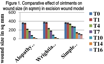
Table no.2 Efficiency of Wrightia tinctoria ointment on woundhealing (in percentage) in excision wound model
Time
Allopathy control
Wrightia tinctoria ointment
Ointment base T1 2.33 μ 0.61 2.15 μ 0.45 1.67 μ 0.37 T4 38.56 μ 0.61 37.46 μ 0.6 7.65 μ 1.16 T7 53.37 μ 0.2 52.5 μ 0.52 23.2 μ 0.63 T10 79.23 μ 0.56 77.24 μ 0.47 25.61 μ 0.88 T14 92.71 μ 0.83 92.4 μ 0.84 61.17 μ 1.39 T16 98.32 μ 0.48 98.22 μ 0.4 68.65 μ 0.8 T19 100 μ 0 100 μ 0 74.2 μ 0.35
It is observed that the wound contracting ability of Wrightia tinctoria ointment was significantly wound healing from the fourth day onwards, which was comparable to that of the reference standard, allopathy ointment. There was also a significant decrease in the odema at the wound site, 3 days after the initial application. A better healing pattern with complete wound closure was observed with the treated groups in contrast to the control
group within. The epithelization period and wound contraction (50%) is shown in Table 3 and figure 3 and 4.
Table no.3 Effect of Wrightia tinctoria ointment on wound epithelization and wound contraction (%) in excision wound model.
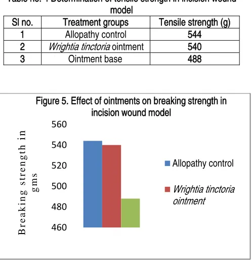
The total epithelization period was 14 days for both the allopathic control and the Wrightia tinctoria ointment with 19 days for ointment base. The wound contraction to 50% took 6.3 days for allopathic control and 6.5 days for Wrightia tinctoria ointment, against 12.7 days for the ointment base. Thus wound healing was observed with all 3 groups with the wound healing rates for allopathy control > Wrightia tinctoria ointment > ointment base. In the incision wound studies, there was a significant increase in tensile strength of the 10-day old wound due to treatment with the Wrightia tinctoria (540g) and the reference standard ointment (544g) when compared with the control group (488g). The measurements of the tensile strength are shown in Table 4 and figure 5.
0 200 400 600
w
o
und
siz
e in sq mm
Figure 1. Comparative effect of ointments on wound size (in sqmm) in excision wound model
T0
T1
T4
T7
T10
T14
T16
0 20 40 60 80 100 120
0 10 20
percentage wound
size
reduction
Time (day)
Figure 2. Wound reduction rates in animals treated with different ointments
Allopathy control
Wrightia tinctoria ointment Ointment base
0 10 20
Period
of
ep
ith
elisatio
n
in
day
s
Figure 3. Effect of ointments on period of epithelisation in excision wound mode
Allopathy control
0 5 10 15
Wound contraction
(50%
)
in day
s
Figure 4. Effect of ointments on wound contraction in excision wound model
Allopathy control
Wrightia tinctoria ointment Ointment base Sl no. Test formulations Epithelization period (days) Wound contraction WC - 50% (days)
1 Allopathy control 14 6.3
2 Wrightia tinctoria
ointment 14 6.5
PAGE |
231
|
Table no: 4 Determination of tensile strength in incision woundmodel
Sl no. Treatment groups Tensile strength (g)
1 Allopathy control 544
2 Wrightia tinctoria ointment 540
3 Ointment base 488
The tensile strength of the Wrightia tinctoria ointment treated group were almost the same to that of the allopathy ointment treated group. Increased wound breaking strength indicates increase in collagen strength and obviously facilitating wound healing.
Discusion
The structural characteristics of the glabrous skin of normal laboratory animals such as mice, rats, guinea pigs, rabbits, dogs and non-human primates differ markedly from those of human skin. For example, these species have skin with a thinner epidermis, relatively flat dermal-epidermal junctions devoid of rete ridges,[5] a loosely organized dermal structure,[6]. and a rudimentary dermal vascular system.[7]. Consequently, the reactivity of their skin to a variety of chemicals is quite different to that of human skin.[8] Experimentation was done on guinea pigs as its skin has got many similarities to that of human skin.[9].
There are four distinct stages involved in wound healing namely inflammatory stage, debridement stage, proliferation stage and maturation/remodeling stage. When an injury occurs, the vascular integrity of the injured area is disrupted leading to extravasations of blood into the surrounding tissue or plasma when the damage is minor. The inflammatory stage is directed at preventing further loss
of blood by platelet adhesion/accumulation at the site leading to coagulation that result to the formation of thrombus. The debridement stage occurs from the third to the sixth day after injury and involves the appearance of neutrophils to clear contaminating organisms. The proliferation or repair stage is characterized by endothelial budding in the nearby blood vessels forming new capillaries that penetrate and nourish the injured tissue. The maturation stage commences from the tenth day to several months depending on wound severity during which the number of capillaries decreases and wound changes from pink to white.[2] Flavanoids show wound healing properties due to their antibacterial and antioxidant properties. They are synthesized by plants in response to microbial infection and are often found effective in vitro as antimicrobial substances against a wide array of microorganisms.[10] As the wound healing process initially involves inflammatory stage and debridement stage, due to its anti-inflammatory and anti microbial property Wrightia tinctoria ointment has comparatively reduced their duration and severity effects on the dermal vessels and facilitated its earlier onset of proliferation and maturation stage, leading to earlier healing.
Conclusion
The data of the present study exhibits significant wound healing by Wrightia tinctoria ointment based on the parameters of evaluation. The activity could be attributed to its antioxidant property exhibited due to its content of free and bound flavonoids and phenolic compounds. Thus Wrightia tinctoria which has antipsoriatic property, through this wound healing evaluation has exhibited a valid quantified measurement of skin curative property.
Authors Contribution
Dr. R. Manavalan conceived of the study and participated in its design and co-ordination. Mr. T. Senthil Kumar carried out the wound healing studies. Dr. Vilambi N.R.K Reddy contributed in the analysis and interpretation of results. Dr. Venkappayya involved in drafting the manuscript and revising itÊs critically for important intellectual content. All authors read and approved the final manuscript.
Acknowledgement
We are grateful to the Vice Chancellor and Research Dean of SASTRA University for their infrastructural facilitation and research motivation.
.
References
[1]. Grove GL. Exfoliative cytological procedures as a nonintrusive method for
dermatological studies. Journal of Investigative Dermatology. 1979; (73): 67-69.
[2]. Esimone CO, Ibezim EC, Chah KF. The wound healing effect of herbal ointments formulated with Napoleona imperialis. 460
480 500 520 540 560
B
reaki
ng st
rengt
h
in
gm
s
Figure 5. Effect of ointments on breaking strength in incision wound model
Allopathy control
PAGE |
232
|
Journal of Pharmaceutical and AlliedSciences. 2000; 1 (3): 294 -299.
[3]. Krishnaveni B, Neeharika V, Venkatesh S, Padmavathy R and Madhava Reddy B. Wound healing activity of Carallia brachiata bark. Indian journal of pharmaceutical science. 2000; 71 (95): 576-578.
[4]. Lee KH. Studies on the mechanism of action of salicylate retardation of wound healing by aspirin. Journal of pharmaceutical science 1968; 57: 1042.
[5]. Mackenzie IC. Ordered structure of the epidermis. Journal of Investigative Dermatology. 1975; (65): 45-51.
[6]. Spearman RIC. The comparative biology of collagenous tissue. In: Jarett A, edition, The physiology and pathophysiology of the skin. London: Academic Press, 1974; PP: 896-899.
[7]. Winkelmannm RK, Scheen SR, Pyka RA and Coventry MB. Cutaneous vascular pattern in studies with injection preparation and alkaline phosphatase reaction. In: Montagna, W, RA Ellis, eds, Blood vessels and circulation. London: Pergamon press, 1961; PP: 15-18.
[8]. Elias PM, Fritsch P, Lampe MA, Williams ML, Brown BE, Nemanic MK and Grayson S. Retinoid effects on epidermal structure, differentiation, and
permeability. Laboratory Investigation, 1981; (44): 531-40.
[9]. Brantley SK, Davidson SF and Das SK. A dose-related curve of wound tensile strength following ultraviolet irradiation in the hairless guinea pig, The American Journal of the Medical Science, 1991; (302):75-81.