ISSN: 0976-3031
Research Article
PERFORMANCE ANALYSIS AND CLASSIFICATION OF HUMAN IN VITRO FERTILIZED (IVF)
EMBRYOS USING VESSELNESS FILTERS AND HOUGH TRANSFORM ALGORITHM
Sujata N Patil
1., Uday Wali
2., Swamy M K
3., Nagaraj S P
4and Nandeshwar Patil
51
Research Scholar, KLE Academy of Higher Education & Research, Belagavi, India
2
Dept. of Electronics & Communication, KLEDrMSS College of Engineering &Technology, Belagavi, India
3
J N Medical College, KLE Academy of Higher Education, Belagavi, India
4
LIVFC & SSFC, Jayanagar, Bangalore, India
5
KLE Dr Prabhakar Kore Hospital & MRC, Belagavi, India
DOI: http://dx.doi.org/10.24327/ijrsr.2018.0901.1476
ARTICLE INFO ABSTRACT
Precise visualization and classification of human oocytes or embryos is an important prerequisite for a number of clinical trials. The purpose of this paper is to enhance embryo features such as circular shape of the blastomere structures with the eventual goal of blastomere segmentation. Paperdescribes a method for the detection blastomere and evenly / unevenly localization in cleavage stage human embryo via image processing. Boundaries or ridges of blastomeres are first extracted by Hessian-based ridge detector followed by nonmaxima suppression and hysteresis thresholding. Based on the acquired boundaries or the ridges, Hough circle transform is implemented to detect the presence of oocytes/embryos with consideration of boundary orientation. Then, screening algorithm is used to verify the detected embryo cells circles. Finally, the location of validated cells is approximated with circles and ellipses. The multiscale second order local structure of an image (Hessian) is examined with the purpose of developing a vesselenhancementfilter
INTRODUCTION
In IVF treatment, the eggs are removed from the ovaries of the woman and injected with sperm of the man in a dish in the laboratory so that fertilization can take place, in vitro. The term infertility is defined as the inability to conceive despite regular and unprotected intercourse. Infertility in a couple can be due to either the woman or the man, notnecessarily both. However, pregnancy may be achieved by using any of the assistedreproductive technologies (ART). There are a number of ART available to infertile couples,in vitro fertilization (IVF) is one of these methods. Embryos of the same patient differentiate in their qualities and development potentials. In order to improve the chance of positive pregnancy, these embryos must be inspected and only the top quality embryos with the highest normal growth potential should be transferred. The selection process is done manually and requires experts (such as reproductive endocrinologists and embryologist) to grade the embryos according to their morphologies and development patterns at different growth stages. This makes
the IVF procedure expensive and selection procedure subjective.
Consequently, proposing solutions that decrease IVF’s cost and/or improve the success rate are very desirable. The workproposed here discusses a method towards an automated embryo grading system which analyzes various aspects of embryos and their growth. The main objective is to improve the accuracy of healthy embryo selection procedure that ultimately leads to more success for the IVF procedure and lowering the overall cost associated with the manual intervention by the embryologists.
The paper aims to develop blastomere/cell detection, instead of shape description, technique for cleavage stage embryos. Boundaries of blastomeres are first extracted by Hessianbased ridge detector followed by nonmaxima suppression and hysteresis thresholding. Based on the acquired boundaries, Hough circle transform is implemented to roughly detect the presence of cell with consideration of boundary orientation. Then, screening algorithm is used to verify detected circles.
Recent Scientific
Research
International Journal of Recent Scientific Research
Vol. 9, Issue, 1(I), pp. 23475-23479, January, 2018
Copyright © Sujata N Patil et al, 2018, this is an open-access article distributed under the terms of the Creative Commons Attribution License, which permits unrestricted use, distribution and reproduction in any medium, provided the original work is properly cited.
DOI: 10.24327/IJRSR CODEN: IJRSFP (USA)
Article History:
Received 15th October, 2017
Received in revised form 25th
October, 2017
Accepted 23rd December, 2017
Published online 28th January, 2018
Key Words:
IVF, Blasomere, Vessel filtering,
Finally, the location of cells is approximated with circles and ellipses.
The study is concerned with developing and implementing a Vessel Filtering algorithm to enhance the ridges of the embryo cell so as to identify the boundaries of the microscopic images. Accurate classification of these cells will prevent the mother and baby from acquiring many health problems that might occur due to multi-cell implantation. The work described aims to develop Hessian-based ridge detector, followed by non-maxima suppression and hysteresis thresholding of theimage to classify these embryo cells for implantation using a grading scheme available.
Cell Boundary Detection
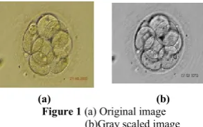
The cleaved embryo images used in this paper are acquired by Hoffman Modulation Contrast (HMC) microscopes and the hospital supported is KLE Dr. Prabhakar Kore Hospital and MRC, Belagavi. The boundary of cells represent ‘ridge’ features instead of edge features, because the pixels around the boundary are darker. There is no significant difference between intensities inside and outside of cells. So, to extract cell boundary, a ‘ridge’ detector would be more desirable than an ‘edge’ detector.
The Hessian-based FrangiVesselness filter [2] is adopted to detect ridges in the image. Afterwards, it is combined with non-maximal suppression and hysteresis thresholding to extract one-pixel thick ridges. Finally, the small segments in the boundary image are removed. In the base work, the FrangiVesselness filter is used for enhancement of blood vessels in Digital Subtraction Angiography images [2]. Here, FrangiVessel filtering is used as ridge detector for cell boundary detection. The measure of ridge-likeliness is based on the eigenvalues of hessian matrix of each pixel.
(a) (b)
Figure 1 (a) Original image
(b)Gray scaled image
Consider the Taylor expansion of image I(x), in the local neighbourhood of point x0:
I(x0+δ x0) = I(x0,σ)+δ x0T∆0,σ δ x0+δx0T H0,σ δ x0 (1)
H0,σ is the hessian matrix computed in x0 with scale σ . For 2D image, the hessian matrix has to eigenvectors λ1 and λ2 , | λ1 | ≤ | λ2 |.
The “blobness measure” is defined as
RB = λ1 / λ2 (2)
The ratio of eigenvalues measures the ratio of change rates along two vertical directions. If the blobness measure is large, the change rates between two directions are close. Consequently, the blobness is large and the neighborhood of the corresponding pixel is more “blob-like”. If the blobness measure is small, the difference between change rates would be large. Consequently, the neighborhood of the corresponding
pixel is more “line-like”. The “second order structureness” is defined as
S = ||H ||F = √ λ12 + λ22 (3)
Having large hessian eigenvalues represents there is large value changes around the given pixel. Consequently, larger second order structureness represents that the pixel contains more structure information.
The “vessel likeliness measure” at a certain scale is defined as
V0 (σ) = 0 if λ2>0, exp (-RB
2
/ 2α2)(1- exp (-S2/ 2β2)) (4)
Here, α and β are parameters which are used to control the scale of “blobness measure” RB and the “second order structureness” S .σ is the scale of used for detect ridges. Large scale represents wide ridge.
V0 (σ) increases with the decrease of RB and the increase of S , which means small blobness measure and large second order structureness give large vessel likeness measure.
The vessel likeliness function would be maximized at the scale which corresponds to the size of ridge. Therefore, it is calculated over different scales, and the largest value of V0 will be considered as the vessel likeliness at that point.
V0 = max σ min ≤ σ ≤ σ max V0(σ) (5)
Figure 2 vesselness filtered embryo
Post processing
After acquiring the vessel likeness function V0(x) over the image, nonmaxima suppression and hysteresis thresholding is performed in the same manner as canny edge detector [3]. Then, small segments are removed from the edge image.
Figure 3 left-Image after non-maxima suppression and right- the image after
canny
embryos, and provided a basis for identifying potentially effective algorithms for this study. Semi-automatic and manual methods were required, so it is likely that algorithms would needto be developed and tuned for the Day 2 as well as Day 3 images. Automatic image analysis may help embryo selection and, consequently, lead to an improvement of the IVF process. Many work concerning automatic image analysis of early stage (days 1 to 2 or 5 post-fertilization) human embryos [1],[2],[3], to the best of our knowledge but still the efficiency remains poor. This is challenging due to varied appearance and quality of images.
Grading of Human Embryos
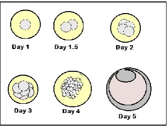
In vitro fertilization (IVF) is a process by which female’s egg cells are fertilized by sperm outside the body, in vitro. IVF belongs to assisted reproductive technology (ART), which is used in infertility treatments. In IVF procedure, retrieved oocytes are first fertilized and then are cultured for five days as depicted in Fig 2, later embryos will be transferred to the female’s uterus. In order to improve the chance of positive pregnancy, these embryos must be inspected and only the good quality embryos with the highest normal growth potential should be transferred. The selection process is done manually and requires experts (such as clinical embryologist) to grade the embryos according to their morphologies and development patterns at different growth stages and 5 pronuclear morphology .6
Fig 2 Embryo Development Stages
Unacceptably high rates of three or more embryo transfers in select countries resulting in multiple births and adverse perinatal outcomes7.
This makes the IVF embryo selection procedure subjective and becomes very laborious in a centre performing high volume (more number of cases). To support this time lapse imaging systems (Embryoscope, Primovision, Esco Miri L) have entered the market but are too expensive. This will be an added burden on the patient and hence cannot be afforded by all the units. Many efforts have been made to identify an efficient and fast method that recognizes quality of the blastomeres from a single embryo without the need of three dimensional image8, using blastomere size as biomarker for fragmentation9. Solutions that decrease IVF’s cost and/or systems that help in embryo scoring are very desirable. We are proposing a research towards an automated embryo grading system which analyzes various aspects of embryos and their growth, particularly at Fertilization (PN Score), 4cells stage, 8 cells stage and blastocyst stages.
Problem characteristics
Since implanting more than one embryo caused multiple pregnancies, it is better forboth the mother and the baby to try to minimize the number of embryos. An automated system that is able to achieve this would reduce the load onthe IVF screeners and provide a consistent and uniform selection of embryos forimplantation.Each clinic chooses a grading system according to many issues, such as the culturemedia available, the extra cost needed for longer culturing embryos and sometimes the ethicalrules of the country or even the religion. The clinic that agreed to support this work was KLE Dr Prabhakar Kore Hospital & MRC this clinic re-implants the embryos on Day 2 or Day 3 based on the subjective analysis, hence usesthe Cleaved embryo grading system in particular to choose the embryos. Hence theimages used in this study, were Day 2 or Day 3, and their general appearance was the 4 cell embryoor 8 cell seen in Fig 2.
Features to be extracted
Fig 3 Human Embryo Features
The Embryo features which can be extracted are Nucleoli, Pronuclei, cell number and size of the Blasomere. Map the characteristics and key features of the Day 2 or Day 3 embryo cells that wouldmake them suitable for implantation into features that can be detected by the system.
Pre-process the image to compensate for magnification and illuminationvariations in the microscope images. Develop and compare different image segmentation and
feature extractiontechniques appropriate to these images. Identify the most accurate image analysis techniques for
classifying theembryo as suitable for implantation. Investigate the performance of the approach using
images of embryos takenwith different microscope magnifications.
Edge detection techniques applied for three different edge-detectors, for pre-processing the image data for the Hough Transform. The Sobel edge-detector gave the poorest results with the lowest matching factors compared to the other two techniques. In the noisy images, the process found false cells with similar matching factors to those of the true cells, and for particularly noisy images, the technique failed to detect true cells and detected false cells with high matching factors. The lowest numbers of false cells that were found was using the 3x3 kernel, although it detected slightly fewer true cells than for the Sobel edge-detector.
Embryo detection using binary template matching
Contents of the template
Although the template could have contained a disk, whose size corresponds to the size of the blastomeres, it was expected that the matching process would be confused by the overlap of the cells, and so it was decided to design a template with a ring to match the edge of the blastomere, Fig 4.shows a sample of such a template. The thickness of the ring was created to be similar to the thickness of the cell.
Fig 4 Template
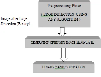
Each image is converted to its binary form after detecting its edges using different edge detection techniques as suggested in the previous report.
Fig 5 Binary image representation
As both the image and template are now in binary form, it is less complex and simpler to perform a simple AND operation between the two arrays and then count the number of matches (1s). In this case, the maximum value indicates the position of the best match. As with the previous techniques, all peaks are found for each image when processed with a particular ring diameter, and the redundant ones are eliminated.
The template can be compared with digital edged images got from edge detection techniques, then moving the template over the image will give us the matches with each image.
Fig 6 Flow Chart of Medical Preprocessing
The above process can be repeated for the set of images obtained from different edge detection algorithms.
Fig 7 Sobel Edge with different thresholds
As seen by the above result of edges the Sobel will give a low percentage of enhancements, but by increasing the threshold the clearance of edges can be enhanced. The Canny Edge and Prewitt have given a better edge enhancement comparatively. Each above edge detection technique suggested will manage to find cells that are almost a circle and matching can be obtained. There are also a cells which have to be detected in the background, The similarity percentage in this case may be higher, but unfortunately some of those can be false cells which are missing in the above algorithm.
Table 1 Number of count cell detected
Here we intend to implement several other cell detection algorithms and compare their performance in their ability to detect true cells. This stage will be considered as a pre-processing stage for a classification algorithm that can suggest success rate of implanting a given embryo. These classification algorithms will be soft computing algorithms like Artificial Neural Networks (ANNs). Currently, there are many types of ANNs available for such classification work. Suitability of specific type of ANN has to be studied.
DISCUSSIONS & FUTURE WORK
The primary objective of the study is to develop automated embryo grading system replacing a manual embryo grading by an embryologist. This can minimize interpersonal errors in grading and hence maintaining uniformity in the laboratory outcome. This ultimately lowers the overall cost associated with the time lapsed imaging systems. Automated morphological evaluation is likely to save the embryology staff a significant amount of time. Hence the efficiency of identifying the potential embryo for implantation may become easier which will ease the job of embryologists.
Acknowledgment
KLEDr MSSCET for allowing me to carry out my research work in this center.
References
1. Karlsson A. et al., “Automatic segmentation of zonapellucidainHMCimagesofhumanembryos.,”
inProcofthe 17th IntConf on Pattern Recognition, 2004, pp. 23–26.
2. Morales D.A. et al., “Automatic segmentation of zonapellucida in human embryo images applying an
active contourmodel,”
inProcofthe12thAnnualConfonMedical Image Underst. and Anal., 2008, pp. 209–213.
3. Perdersen U.D. et al., “A multiphase variational level set approach for modelling human embryos.,” in Proc of the 2ndIEEEWorkshoponvariational,GeometricandLevel Set Methods, 2003.
4. D. K. Gardner et al., “Single blastocyst transfer: a prospective randomized trial,” FertilSteril, vol. 81, pp. 551–555, 2004R. Nicole, “Title of paper with only first word capitalized”, J. Name Stand. Abbrev., in press. 5. Luengo-Oroz, M.A., Ledesma-Carbayo, M.J., Peyriéras,
N. and Santos, A., 2011. Image analysis for understanding embryo development: a bridge from microscopy to biological insights. Current opinion in genetics & development, 21(5), pp.630-637.
6. Improvement in the detection of egg fertility using digital image processing techniques. ISSN(p):2439-3968, volume 2, issue 4, Dr. N M Palanivelu M S, Ph .D
7. Selection of high potential embryos using time-lapse
imaging: the era of morphokinetics Javier
Herrero, Ph.D., Marcos Meseguer, Ph.D
8. Gonzalez, R. & Woods, R. 2002, Digital Image Processing (2nd ed.)., Prentice Hall.
9. Cladioanna, Loris Nanni, Alessandra Lmini and sabastinaPappalardo. 2013 Artificial intelligence techniques for embryo and oocyte classification. Reproductive Biomedicine online . 26 , 42-49
10. E Santos Filho, J. A. Noble and D Wells 2010 Toward a method for automatic grading of microscope human embryo images pp1289-1292. IEEE
11. Proceedings of an expert meeting: Alpha scientists in Reproductive Medicine and ESHRE Special Interest group of Embryology”.Human Reproduction Vol. 26, No.6 pp,1270-1283, 2011
12. R. Machtinger and C. Racowsky, “Morphological systems of human embryo assessment and clinical evidence,” ReproductiveBioMedicine Online, vol. 26, no. 3, pp. 210–221, 2013
*******
How to cite this article:
Sujata N Patil et al.2018, Performance Analysis And Classification of Human In Vitro Fertilized (Ivf) Embryos Using Vesselness Filters And Hough Transform Algorithm. Int J Recent Sci Res. 9(1), pp.23475-23479.