Microorganisms’ colonization and their antibiotic resistance pattern
in oro - tracheal tube
Alireza Abdollahi1*, Saeed Shoar2, Nasrin Shoar3
1Division of Pathology, Imam Khomeini Hospital, Tehran University of Medical Sciences Tehran, Iran. 2Department of Surgery, Shariati Hospital, Tehran University of Medical Sciences, Tehran, Iran.3School
of Medicine, Kashan University of Medical Sciences, Kashan, Iran.
Received: June 2011, Accepted: December 2012.
ABSTRACT
Background and Objectives: Recently, nosocomial infections have been discussed as a critical issue among intubated patients leading to significant morbidity and mortality. Hence, the pattern of microbiological colonization and antibiotic resistance are much valuable in this regard. We aimed to investigate the pattern of microorganism colonization and antibiotic resistance in patients with endotracheal tube or tracheostomy to propose a proper empirical antibiotic therapy in this setting.
Materials and Methods: This cross sectional study was conducted among 880 patients admitted in Imam Khomeini hospital between 2008 and 2011 who were subsequently intubated or underwent tracheostomy due to insufficient self ventilation. Samples for microbiological cultures were obtained after extubation and then sent to the central laboratory for further assessment. Antibiograms and microbiological cultures were obtained for each sample.
Results: Of 880 patients enrolled in this study, 531 (60.3%) were male and 349 (39.7%) were female.Nineteen different organisms were isolated including Acinetobacter (213, 24.2%), Pseudomonas aeruginosa (147, 16.7%), Staphylococcus aureus (106, 12%), Proteus mirabilis (90, 10.2%), and other organisms (324, 36.8%). Antibiotic resistance was mainly seen in Acinetobacter (ciprofloxacin, ceftazidim, cefepim, and penicillin), S. aureus (imipenem) and Klebsiella (pipracillin-tazobactam and ampicillin-sulbactam).
Conclusion: This study represents the most common microorganisms colonizing tracheal tube of hospitalized patients and their pattern of antibiotic resistance. Acinetobacter was the most common microorganism isolated from endotracheal tube. Hence, it may be possible to initiate the empiric antibiotic treatment before the results of culture are become available. Ciprofloxacin was also the most prevalent antibiotic revealing resistant pattern. Moreover, most of the microorganisms were sensitive to imipenem and pipracillin-tazobactam.
Keywords: Nosocomial Infection; Microorganism Colonization; Antibiotic Resistance; Tracheal Intubation.
INTRODUCTION
Nosocomial infection is a frustrating and budget consuming issue and due to the increased time of hospitalization it imposes a heavy burden on health
care resources (1). One of the most important types of this infection is pneumonia which commonly occurs in relation to the endotracheal intubation and mechanical ventilation named ventilation associated pneumonia (VAP) (2-5). Patients with mechanical ventilation have an increased risk for respiratory tract infection because the tube which has been inserted into the trachea reduces the clearance of bacteria and increases the leakage of secretion around the cuff of the tube and disable the cilliary tract by damaging to it (6). Because of decreased salivary secretion, colonization of oropharynx with Gram negative * Corresponding author: Alireza Abdollahi M.D.
Adress: Keshavarz Blvd., Imam Hospitals Complex, Tehran, Iran.
Tel: +98-9121220588 Fax: +98-21-88277321
bacteria is also probable (7-8).
In the literatures, the incidence of respiratory tract infection in relation to intubation and/ or mechanical ventilation has been reported to vary between 4% and 28% and this rate has been thought to be 21 times higher than in patients without endotracheal tube (9-13). Various studies have proposed different causative microorganism as the most common etio-logy for intubation related respiratory infections including Pseudomonas aeruginosa, A. baumannii, and methicillin resistant Staphylococcuus aureus (MRSA) or S. aureus in children (9, 14-19). The concerns related to the nosocomial infections are exacerbated by the presence of antibiotic resistant bacteria which increases morbidity rate and the associated costs (20). Extensive use of fluoroquinolones has lead to alterations in susceptibility patterns of microorganisms (10, 12-13, 17). Furthermore, inappropriate prescription of broad-spectrum antimicrobial agents has risen in the last decades with macrolides, fluoroquinolones, and third-generation cephalosporins at the top of the list (14-16, 19-20).
Our study hence aimed to determine the type of bacterial colonization and antibiotic sensitivity and resistance in patients with endotracheal intubation or tracheostomy in a tertiary care referral center to facilitate initiation of proper empirical antibiotic treatment in these patients.
MATeRIAlS AND MeThODS
Through a cross-sectional study in Imam Khomeini Hospital affiliated to Tehran University of Medical Sciences, 880 patients with positive culture of samples obtained from endotracheal tube or tracheostomy were enrolled in our investigation between 2008 and 2011. Patients had been intubated to maintain an airway, to be assisted by mechanical ventilation as a result of respiratory failure or just elective ventilation. Clinical impressions for necessitation of intubation or extubation were made by the responsible physicians based on clinical presentation of the patient and then endotracheal intubation (ET) was performed under a sterile technique; also extubation was done if the patients did assure clinically to be able to ventilate on his own without developing further complications. Patients with previous pneumonia or other airway infections were excluded from the study. Extubated ETs were cut by a sterile blade and the samples were
sent to the microbiological laboratory for organism isolation and microbiological culture (for 48 to 72 hours) and obtaining an antibiogram. The isolated colony was then confirmed for type of microorganism by PCR (21). Antibiotic susceptibility was also evaluated by disc diffusion method on Mueller Hinton agar according to the guidelines of Clinical and Laboratory Standards Institute (CLSI) (22). Positive and negative control was used for antibiogram.
The samples were obtained from patients admitted to 3 wards of our hospital including neonatal ICU (NICU), ICU and emergency room (ER). Patients or their relatives on behalf of them filled an informed consent to enroll the study. Research Ethics Committee of TUMS approved the study protocol and the study was then conducted in a period of 3 years.
Data were analyzed by Statistical Package for Social Sciences (SPSS, version 16, Chicago, Inc) and the values are reported as number (%).
ReSUlTS
Type of Organisms Male female <12 years (NICU) >12 years
Acinetobacter 123*(23.3%) 90*(25.8%) 1 (3%) 212*(25%)
Pseudomonas 94 (17.7%) 53 (15.2%) 4 (12.1%) 143 (16.9%)
S. aureus 57 (10.7%) 49 (14%) 4 (12.1%) 102 (12%)
Proteus mirabilis 65 (12.2%) 25 (7.2%) 1** (3%) 89 (10.5%)
Klebsiella 39 (7.3%) 28 (8%) 6*(18.2%) 61 (7.2%)
Entrobacter 36 (6.8%) 20 (5.7%) 1** (3%) 55 (6.5%)
E. coli 32 (0.6%) 22 (.3%) _ 54 (6.4%)
Citrobacter 18 (3.4%) 10 (2.9%) 1** (3%) 27 (3.2%)
Stenotrophomonas 12 (2.3%) 10 (2.9%) 3 (9.1%) 19 (2.2%)
Prodentia 13 (2.4%) 6 (1.7%) _ _
Streptococcus viridans 7 (1.3%) 9 (2.6%) _ _
S. epidermidis 7 (1.3%) 5 (1.4%) 3 (9.1%) 9 (1.1%)
Entrococcus 4 (0.8%) 7 (0.2%) 5 (15.2%) 6 (0.7%)
Seratia 8 (1.5%) 2 (0.6%) _ 10 (1.2%)
Candida albicans 6 (1.1%) 2 (0.6%) _ 8 (0.9%)
S. hemolyticus 2 (0.4%) 5 (1.4%) _ 7 (0.8%)
Proteus vulgaris 6 (1.1%) 1** (0.3%) _ 7 (0.8%)
Alcalginis 1** (0.2%) 5 (1.4%) 4 (12.1%) 2 (0.2%)
Salmonella 1** (0.2%) _ _ 1** (0.1%)
Total 531 (60.3%) 349 (39.7%) 33 (4%) 847 (96%) Table 1. Prevalence distribution of organisms isolated from different wards of the hospital.
* Most common ** Least common
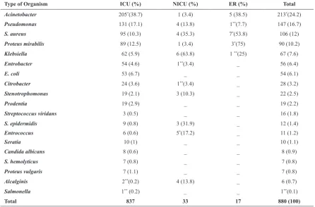
Type of Organism ICU (%) NICU (%) eR (%) Total
Acinetobacter 205*(38.7) 1 (3.4) 5 (38.5) 213*(24.2)
Pseudomonas 131 (17.1) 4 (13.8) 1**(7.7) 147 (16.7)
S. aureus 95 (10.3) 4 (35.3) 7*(53.8) 106 (12)
Proteus mirabilis 89 (12.5) 1 (3.4) 3*(75) 90 (10.2)
Klebsiella 62 (5.9) 6 (63.8) 1 **(25) 67 (7.6)
Entrobacter 54 (4.6) 1**(3.4) _ 56 (6.4)
E. coli 53 (6.7) _ _ 54 (6.1)
Citrobacter 24 (3.6) 1**(3.4) _ 28 (3.2)
Stenotrophomonas 19 (2.1) 3 (10.3) _ 22 (2.5)
Prodentia 19 (2.9) _ _ 19 (2.2)
Streptococcus viridans 3 (0.5) _ _ 16 (1.8)
S. epidermidis 9 (0.8) 3 (31.9) _ 12 (1.4)
Entrococcus 6 (0.6) 5*(17.2) _ 11 (1.2)
Seratia 10 (1) _ _ 10 (1.1)
Candida albicans 8 (0.6) _ _ 8 (0.9)
S. hemolyticus 7 (0.8) _ _ 7 (0.8)
Proteus vulgaris 7 (1.1) _ _ 7 (0.8)
Alcalginis 2**(0.2) 4 (13.8) _ 6 (0.7)
Salmonella 1** (0.2) _ _ 1**(0.1)
Total 837 33 17 880 (100)
Table 2. Differentiation of organisms based on age and sex.
to penicillin.
Antibiotic resistance and susceptibility pattern in general and categorized according to the type of isolates are presented in details in Table 4.
DISCUSSION
Infections are among the most important and the leading cause of mortality and morbidity in ICU. Endo-tracheal tubes are susceptible to infection and therefore it is important to be aware of the relevant factors and responsible organisms to take prompt action. The findings of this study would be helpful in selection of appropriate antibiotics. In this study, all the positive colonies obtained from the ET or tracheostomy cases were considered. The study by Simoni et al. (23) showed that 100% of samples from airway prosthesis are positive in culture; however, other studies have reported a positive culture rate between 0% and 33% in obtained samples from airway tubes. Cardinosa et al. have reported a positive culture result in 89% of their samples (24). The variation could be explained by the technique of intubation, clinical and individual characteristics of study population, colonization during intubation or lack of sufficient precautions for intubation due to the high work load in an emergency setting (25).
In our study, Gram negative bacteria were the most common isolated organisms including Acinetobacter and Pseudomonas aeruginosa which is in the same line with the study by Nardi et al. (36). However, in Cardinosa et al. study, during the first 24 hours of admission, Gram positive bacteria were the most common cause of infection and this could be as a result of the time of sampling which in Cardinosa study
was within the first 24 hours of admission but in our study the sample was done after the first 24 hours i.e. following extubation. In addition, McShane and Hone have showed coagulase-negative Staphylococci to be the most common source of infections among patients with ET (26). Amini et al. also conducted a descriptive study on distribution of isolated microorganisms from tracheal tube of ICU patients declaring that S. aureus (23.6%), Klebsiella spp. (23.3%), Acintobacter spp. (20.7%), P. aeroginosa (18.2%), E. coli (7.7%), and Enterobacter spp, were the most common isolates (27). Our study as well as the other ones in similar settings confirm that Acinetobacter, P. aeruginosa, and S. aureus are among the most 3 prevalent isolated organism from ET.
In contrast, Rello et al. in their study have demonstrated that P. aeruginosa is the most common causative organism for infection of ET and this could be the result of large number of patients with chronic obstructive pulmonary disease (COPD) and long time of intubation and even a previous history of antibiotic therapy (28). It should be noted that although there are minor variations in relative frequency of obtained organisms, different studies have reported varying sources of isolations as in our study we did report colonization of organisms into the ET tube while other studies may have considered ET-related infections in their study analysis.
In a study by Nazal –Matunog, most of the Gram negative bacteria were sensitive to ciprofloxacin compared with 3% resistant cases; there was amikacin resistance in 9.7% of the cases with the highest resistance to cefamandole (57%), cefotaxime (50%), and tobramycin (50%) (29). Khosravi et al. showed that Enterobacter spp. was the most prevalent organism colonized the endotracheal tubes (41.14%). It was followed by Pseudomonas aeruginosa (15.35%), E. coli (13.97.2%), coagulase negative staphylococci (14.76%), S. aureus (13.97%), and Proteus spp (0.79%) (30). Antimicrobial testing also Type of Antibiotic Ciprofloxacin Ceftazidime Imipenem Cefepime Pipracillin-tazobactam Ampi-sulbactam Penicillin
Acinetobacter 82% 97.4% 7.4% 100% 6.3% 27% 100%
S. Aureus 35.1% 80% 18.2 - - - 91.7%
Proteus mirabilis 55.1% 59.1% 8.1% 88.9% 5.6% 55.6%
-Klebsiella 33% 85.7% 1.8% 75% 14.3% 55%
-E. coli - - - 77.8% - -
-S. epidermidis - - - 88.9%
Table 3. Prevalence of resistance to antibiotics.
Table 4. Sensitivity to antibiotics in two organisms.
Microorganisms Vancomycin Cloxaciline
S. aureus 99% 33%
revealed that the most resistant Gram negative isolate is P. aeruginosa with highest resistance to cefixime (70.8%), and coagulase negative staphylococci as the most resistant Gram positive isolates with highest resistance to oxacillin (84.2%). The concern which is similarly subject to all the studies is the increasing prevalence of resistant P. aeruginosa in critically ill patients which may lead to significant mortality as well as huge widespread to other wards of the hospital. In a study by Semoni et al. all of the included samples consisted of more than 1 pathogen while in our study each culture has only one pathogen. However, in the same line with our results, all of the organisms in their study were aerobics including Streptococcus viridance, S. aureus and P. aeruginosa (23).
One of the limitations of this study was the fact that although antibiotic resistance pattern is important to intensive care physician, this is also heavily influenced by the antibiotic usage pattern in the study patients prior to obtaining the sample and also overall antibiotic usage pattern in the study institution. In this regard, the utility of the reported results to other centers is limited.
In conclusion, this study presents the most common microorganisms colonized from endo-tracheal tube of hospitalized patients and their pattern of antibiotic resistance. As our study showed, Acinetobacter is the most common microorganism isolated from endotracheal tube. Ciprofloxacin was also the most prevalent antibiotic revealing resistant pattern. Moreover, most of the microorganisms were sensitive to imipenem and pipracillin-tazobactam.
ACKNOleDGeMeNT
This paper is the result of a thesis and was financially supported by Research Council of Tehran University of Medical Sciences. The authors declare that there is no conflict of interests.
RefeReNCeS
Wenzel RP, Edmond MB. The impact of hospital-1.
acquired bloodstream infections. Emerg Infect Dis 2001; 7: 174-177.
Vincent JL. Nosocomial infections in adult intensive-2.
care units. Lancet 2003; 361: 2068-2077.
Hoffken G, Niederman MS. Nosocomial pneumonia: 3.
the importance of a de-escalating strategy for antibiotic treatment of pneumonia in the ICU. Chest 2002; 122: 2183-2196.
Appelgren P, Hellstrom I, Weitzberg E, Soderlund 4.
V, Bindslev L, Ransjo U. Risk factors for nosocomial intensive care infection: a long-term prospective analysis. Acta Anaesthesiol Scand 2001; 45: 710-719. Tejada Artigas A, Bello Dronda S, Chacon Valles E, 5.
Munoz Marco J, Villuendas Uson MC, Figueras P, et al. Risk factors for nosocomial pneumonia in critically ill trauma patients. Crit Care Med 2001; 29: 304-309. Craven DE, Kunches LM, Kilinsky V, Lichtenberg DA, 6.
Make BJ, McCabe WR. Risk factors for pneumonia and fatality in patients receiving continuous mechanical ventilation. Am Rev Respir Dis 1986; 133: 792-796. Adair CG, Gorman SP, Feron BM, Byers LM, Jones DS, 7.
Goldsmith CE, et al. Implications of endotracheal tube biofilm for ventilator-associated pneumonia. Intensive Care Med 1999; 25: 1072-1076.
Dennesen P, van der Ven A, Vlasveld M, Lokker L, 8.
Ramsay G, Kessels A, et al. Inadequate salivary flow and poor oral mucosal status in intubated intensive care unit patients. Crit Care Med 2003; 31: 781-786. Diaz E, Planas K, Rello J. [Infection associated with 9.
the use of assisted-ventilation devices]. Enferm Infecc Microbiol Clin 2008; 26: 465-470.
Chastre J, Fagon JY. Ventilator-associated pneumonia. 10.
Am J Respir Crit Care Med 2002; 165: 867-903. Fortaleza CM, Abati PA, Batista MR, Dias A. Risk 11.
factors for hospital-acquired pneumonia in nonventilated adults. Braz J Infect Dis 2009; 13: 284-288.
Chevret S, Hemmer M, Carlet J, Langer M. Incidence 12.
and risk factors of pneumonia acquired in intensive care units. Results from a multicenter prospective study on 996 patients. European Cooperative Group on Nosocomial Pneumonia. Intensive Care Med 1993; 19: 256-264.
Haley RW, Hooton TM, Culver DH, Stanley RC, Emori 13.
TG, Hardison CD, et al. Nosocomial infections in U.S. hospitals, 1975-1976: estimated frequency by selected characteristics of patients. Am J Med 1981; 70: 947-959.
Baker AM, Meredith JW, Haponik EF. Pneumonia in 14.
intubated trauma patients. Microbiology and outcomes. Am J Respir Crit Care Med 1996; 153: 343-349. Craig CP, Connelly S. Effect of intensive care unit 15.
nosocomial pneumonia on duration of stay and mortality. Am J Infect Control 1984; 12: 233-238.
Cunnion KM, Weber DJ, Broadhead WE, Hanson LC, 16.
Pieper CF, Rutala WA. Risk factors for nosocomial pneumonia: comparing adult critical-care populations. Am J Respir Crit Care Med 1996; 153: 158-162. Nseir S, Ader F, Lubret R, Marquette CH. 17.
Pathophysiology of airway colonization in critically ill COPD patient. Curr Drug Targets Apr; 12: 514-520. Papazian L, Bregeon F, Thirion X, Gregoire R, Saux P, 18.
Denis JP, et al. Effect of ventilator-associated pneumonia on mortality and morbidity. Am J Respir Crit Care Med 1996; 154: 91-97.
Bercault N, Boulain T. Mortality rate attributable to 19.
Crit Care Med 2001; 29: 2303-2309.
Diaz E, Lorente L, Valles J, Rello J. [Mechanical 20.
ventilation associated pneumonia]. Med Intensiva; 34: 318-324.
Xu B, Liu L, Li X, Wang X. A multiplex PCR assay for 21.
the rapid and sensitive detection of methicillin-resistant Staphylococcus aureus and simultaneous discrimination of Staphylococcus aureus from coagulase-negative staphylococci. J Food Sci 2012; 77: M638-642. Institute CaLS. Performance standards for antimicro-22.
bial susceptibility testing. Nineteenth informational supplement. Wayne, PA: CLSI; 2009.
Simoni P, Wiatrak BJ. Microbiology of stents in 23.
laryngotracheal reconstruction. Laryngoscope 2004; 114: 364-367.
Cardenosa Cendrero JA, Sole-Violan J, Bordes Benitez 24.
A, Noguera Catalan J, Arroyo Fernandez J, Saavedra Santana P, et al. Role of different routes of tracheal colonization in the development of pneumonia in patients receiving mechanical ventilation. Chest 1999; 116: 462-470.
Oncag O, Cokmez B, Aydemir S, Balcioglu T. 25.
Investigation of bacteremia following nasotracheal intubation. Paediatr Anaesth 2005; 15: 194-198. McShane AJ, Hone R. Prevention of bacterial 26.
endocarditis: does nasal intubation warrant prophylaxis? Br Med J (Clin Res Ed). 1986; 292: 26-27.
Amini M, Javanmard A, Davati A, Azimi G. Bacterial 27.
Colonization in Tracheal Tubes of ICU Patients. Iranian Journal of Pathology 2009; 4: 123-127.
Rello J, Ausina V, Ricart M, Puzo C, Quintana E, Net 28.
A, et al. Risk factors for infection by Pseudomonas aeruginosa in patients with ventilator-associated pneumonia. Intensive Care Med 1994; 20: 193-198. Nazal -Matunog CL, Rodriguez L, Mercy G, Alberto 29.
L, Adrian CP, Remedios C. Nosocomial Pneumonia in Mechanically Ventilated Patients. Phil J Microbiol Infect Dis 1993; 22: 11-16.
Khosravi AD, Parhizgari N, Montazeri EA, Mozaffari 30.