ORIGINAL
AR
TICLE
INTRODUCTION
Genetic diversity at the population level of a species plays an important role in the interactions of a species with the environment. These interactions will structure the ecosystem, so that spatial and temporal partitioning of genetic diversity will
*Corresponding author: Ali-Reza Ahmadi PhD. Address: Department of Biomedical Sciences, Alzahra University, Tehran, Iran.
Tel: (+98) 21 85692083
E-mail: [email protected]
Phytoplanktons and DNA barcoding:
Characterization and molecular analysis of phytoplanktons on the Persian Gulf
Esmat Alemzadeh1, Raheem Haddad1, Ali-Reza Ahmadi2*
1Department of Agricultural Biotechnology, Faculty of Engineering and Technology, Imam Khomeini
International University, Qazvin, Iran. 2Department of Biomedical Sciences, Alzahra University, Tehran, Iran.
Received: June 2013, Accepted: December 2013.
ABSTRACT
Background and Objectives: Phytoplanktons are organisms with a very high diversities and global distribution in different habitats. The high distribution of phytoplankton is due to ecological flexibility and their ability to tolerate different climatic conditions and environmental stress.Phytoplankton is the most sensitive biological indicators of water resources. The purpose of this study was to identify the phytoplankton species with emphasis on DNA bar-coding method. The study of phytoplankton variation and the identification of their species composition can provide useful information about the water quality.
Materials and Methods: In this research project, a clone library of the ribosomal small subunit RNA gene (18S rDNA) in the nuclear genome was constructed by PCR using A and SSU-inR1 primers, and then, after examining the clones, selected clones were sequenced.
Results: Eleven analyzed sequences were identified correctly and characterized by a similarity search of the GenBank database using BLAST (NCBI). In this study, we revealed a wide range of taxonomic groups in the Alveolata (Ciliphora and Dinophyceae), Stramenopiles (Bacillariophyta and Bicosoecida), Rhodophyta and Haptophyceae. Moreover, we found species of fungi and Metazoa (Arthropoda). Most of the sequences were previously unknown but could still be assigned to important marine phyla.
Conclusion: Clone library of 18S rDNA is an accurate method to identify marine specimens and it is recommended as an efficient method for phylogenic studies in marine environments. There seems to be a high diversity and abundance of small eukaryotes in the marine regions of Persian Gulf.
Keywords: Clone library, DNA Barcoding, Phytoplankton, 18S rDNA
occur. Such structuring has seldom been measured in the marine planktonic community and studies of
genetic diversity are virtually nonexistent in pelagic
ecosystems (1). Yet taxonomicidentification of aquatic
microorganisms has been historically a difficult
culture only a small part of the known biodiversity. The lack of knowledge of their breeding systems makes genetic or demographic studies difficult (1).
Traditionally, morphological characters have been used to identify species and monitor the species composition of environmental water samples (2). Morphological similarities and plasticity have frequently led to erroneous results in species
identification (2, 3). Therefore, much of the species information, including that regarding putative cryptic species, was lost, and the actual species composition of the ecosystem was not reflected in the species lists produced by field surveys.
In the last decade, the introduction of molecular techniques into microbial ecology has greatly increased our knowledge by identifying the smallest aquatic microorganisms and, more particularly, prokaryotes (4). DNA barcoding in general demands a molecular locus, being variable enough to discriminate on species level for the organisms under study and a molecular reference database for comparison. The similarity or divergence of the molecular sequence of an unknown organism to a vouchered reference sequence in the database is used as quality indicator for species identification. DNA barcoding of environmental samples requires DNA
extraction from an environmental pooled sample, PCR amplification of a target locus; cloning of the resulting PCR products, sequencing and the analysis (5). The small subunit (SSU) 18S rRNA gene is one of the most frequently used genes in phylogenetic studies and an important marker for random target PCR in environmental biodiversity screening (6). In general, rRNA gene sequences are easy to access due to highly conserved flanking regions allowing for the use of universal primers (5, 6). Clone libraries have been successfully applied to reveal enormous biodiversity of microbial communities in many habitats (7- 10). In this technique, the species composition can be revealed through cloning after PCR amplification
of environmental samples. Because many DNA sequences based on 18S rDNA have been deposited in GenBank, the 18S rDNA gene can provide a major advantage for the selection of a target DNA region. Accordingly, a high level of unexpected species diversity in aquatic ecosystems has been recovered from 18S rDNA clone libraries (11, 12).
In this study, we analyzed the biodiversity of eukaryotic plankton in environmental water samples by constructing a clone library of 18S rDNA from
waters of the Persian Gulf of Iran. Phylogenetic analyses of the 18S rDNA sequences were performed. The improved molecular monitoring method developed here will reduce the time, cost, and labor required to construct community analyses of aquatic systems. Moreover, this technique will enable analysis of more environmental samples without a great increase in effort, which will lead to more data being deposited in sequence databases and better overall results regarding the biodiversity of aquatic ecosystems.
MATERIALS AND METHODS
Study site and sampling. The study was conducted in the coastal water in marine areas of the Persian Gulf (Iran). Environmental water samples were collected from surface water using a net during April/May in 2010. The samples were concentrated by centrifugation at 2000 x g (Hettich D-78532, Tuttlingen, Germany) for 15 min and the supernatants were discarded. The pellets were conserved at -20°C for molecular analyses.
Nucleic acid extraction. Total DNA was extracted according to the method outlined by Doyle and Doyle (13). Samples were suspended in the CTAB
extraction buffer (3% CTAB, 0.1 mol L−1 Tris-HCl, pH 8.0, 0.01 mol L−1 EDTA,1.4 mol L−1 NaCl, 0.5% β-mercaptoethanol, 1% PVP). The mixture was incubated at 60°C for one hour with every 15 min shaking, and was cooled down to room temperature. Subsequently, 1 ml of chloroform:isoamyl alcohol (24:1 v/v) was added, mixed for 15 min at room temperature, and centrifuged for 10 min at 12000 rpm. The supernatants were transferred to new tubes and the previously described chloroform:isoamyl alcohol
extraction was repeated once. The supernatants were transferred to new tubes containing equal volumes of ice-cold isopropanol and incubated at –20°C for 30-60 min. After centrifugation at 12,000 rpm and 4°C for 10 min, the resulting pellets were washed with 70% (v/v) ethanol. Finally, the pellets were dissolved in TE buffer. The quality of DNA was checked by electrophoresis on the 1% agarose gel stained with ethidium bromide before PCR amplification.
primer SSU-inR1 (5΄-CACCAGACTTGCCCTCCA-3΄)
based on the conserved domain region of 18S rDNA (14). The PCR mixture (50 μl) contained about 20 ng of environmental DNA and primers using the PCR Master Mix (Fermentas, Burlington, USA). Reactions were carried out in an automated thermal cycler (Techne TC-212, England, UK) with the following cycle: initial denaturation at 94°C for 3 min followed by 35 cycles of denaturation at 94°C for 30 s, annealing at 55°C for 30s, and extension at 72°C for 1 min, with a final extension at 72°C for 7 min. Several PCR products (at least four 50 μl
samples) were pooled, separated on 0.8% agarose gel, then the amplified fragments with the expected
size were excised from the gel and purified using Gel
Purification Kit (Bioneer, Daejeon, Korea).
Cloning, unique clone selection and sequencing. These PCR products were used to construct one clone library according to manufacturer of the InsTAclone PCR Cloning Kit (Thermo Scientific-Fermentas, Waltham, USA). The purified fragments cloned into PTZ57/RT plasmid vector. The ligation samples were directly used to transform the competent Escherichia coli strain DH5α.
Around 100 putative positive recombinant clones from library were randomly picked. The presence of the 18S rRNA gene insert in positive colonies was checked by PCR amplification using the same primer set (A and SSU-inR1) that was used to evaluate the
environmental samples. The PCR products were then electrophoresed in 1.8% agarose gel to confirm the presence of the inserts. To confirm presence of the 18S rRNA gene insert, Plasmids were extracted from bacteria and examined by the restriction enzymes
EcoRI and PstI (Fermentas, Burlington, USA) for 12 h at 37°C and separated by electrophoresis in a 2.5% low-melting-point agarose gel (NuSieve GTG agarose). After screening, the recombinant plasmids were isolated and purified using a High Pure Plasmid Isolation Kit (Bioneer, Daejeon, Korea) and two representatives of each clone producing the same operational taxonomic unit (OTU) were selected and sequenced from plasmid products.
Phylogenetic analysis. To determine the first phylogenetic affiliation, each sequence was compared with sequences available in databases using BLAST from the National Center for Biotechnology Information and the Ribosomal Database Project. The nucleotide sequences with the highest max
scores that were identified by the BLAST searches were selected for use in the analysis. The 18S rDNA sequences were aligned using Clustal X ver. 1.8 (15) and then manually adjusted. Phylogenetic trees were constructed using MEGA 4.0 (16).
Nucleotide sequence accession numbers. Nucleotide sequences determined in this study have been deposited in GenBank under accession numbers
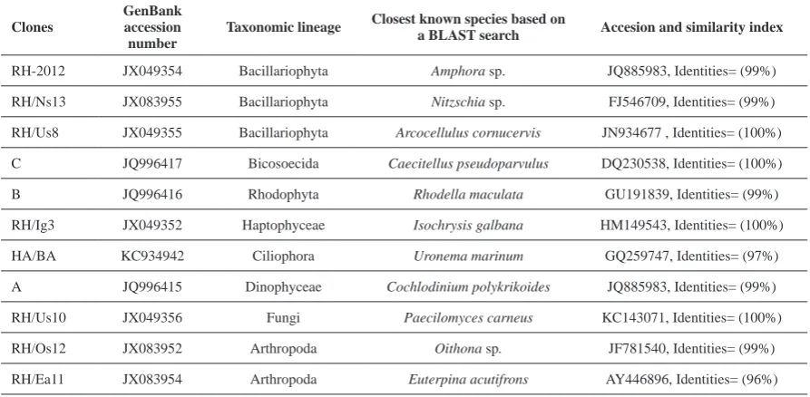
Table 1. Clones in the 18S rDNA clone library of the environmental water sample identified based on the results of a BLAST
search of the GenBank database.
Clones
GenBank accession number
Taxonomic lineage Closest known species based on a BLAST search Accesion and similarity index
RH-2012 JX049354 Bacillariophyta Amphora sp. JQ885983, Identities= (99%)
RH/Ns13 JX083955 Bacillariophyta Nitzschia sp. FJ546709, Identities= (99%) RH/Us8 JX049355 Bacillariophyta Arcocellulus cornucervis JN934677 , Identities= (100%) C JQ996417 Bicosoecida Caecitellus pseudoparvulus DQ230538, Identities= (100%)
B JQ996416 Rhodophyta Rhodella maculata GU191839, Identities= (99%)
RH/Ig3 JX049352 Haptophyceae Isochrysis galbana HM149543, Identities= (100%)
HA/BA KC934942 Ciliophora Uronema marinum GQ259747, Identities= (97%)
A JQ996415 Dinophyceae Cochlodinium polykrikoides JQ885983, Identities= (99%)
RH/Us10 JX049356 Fungi Paecilomyces carneus KC143071, Identities= (100%)
RH/Os12 JX083952 Arthropoda Oithona sp. JF781540, Identities= (99%)
listed in Table 1.
RESULTS
The objective of this work was to study the
taxonomic composition of the community of small eukaryotes. For this study, we used the SSU-inR1 and primers designed by Lee et al. (14) to avoid intron regions that produce PCR products with a long length.
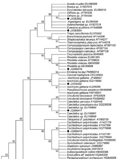
Several hundred white colonies were produced from cloning of the purified PCR products and screened by RFLP analysis to obtain the unique types. In total, 25 unique SSU rDNA sequences obtained from the library. After partial sequencing of the 25 region of the gene (560 bp on average), BLAST searches provided us with a further survey of the type of eukaryotic sequences present in our samples (Table 1). Based on the results of sequencing, those clones had the same 18S rDNA sequence. Therefore, we could easily isolate the unique clones, and this technique could greatly reduce the time and effort required for the clone isolation step. From the result of BLAST, all of the determined sequences corresponded to known species at the level of species and genus with a high sequence similarity (Table 1; >97% and 100% coverage). The phylogenetic tree constructed from environmental clone sequences and compared with those available in the GenBank database using NCBI/BLAST to search for related sequences (Fig. 1).
Phylotypes were affiliated with a wide variety of taxonomic groups including species from major eukaryotic lineages (Table 1) [Alveolata (2): Ciliphora (1) and Dinophyceae (1); Stramenopiles (4): Bacillariophyta (3) and Bicosoecida (1); Rhodophyta (1); Haptophyceae (1); Fungi (1) and Metazoa (2): Arthropoda (2)].
We isolated two phylotypes of Bacillariophyta
affiliated with Amphora sp. and Nitzschia sp. (99%). Environmental clones C, RH/Ig3, RH/Us8 and RH/Us10 were 100% homologous with Caecitellus
pseudoparvulus (Bicosoecida), Isochrysis
galbana (Haptophyceae), Arcocellulus cornucervis
(Bacillariophyta) and Paecilomyces carneus (Fungi) respectively. We also identified several clones with distinct taxonomic positions, including Dinophyceae (A), Fungi (RH/Us10), Bicosoecida (C), Rhodophyta (B), Haptophyceae (RH/Ig3) and Ciliophora (HA/ BA). In the case of metazoan species, we isolated
two clones that were closely related to Oithona sp.
(99%) and Euterpina acutifrons (96%).
DISCUSSION
For several years, the SSU rDNA has been used as the preferred marker to explore the diversity of microbial eukaryotic communities in a variety of environments, leading to the discovery of a huge hidden diversity. Consequently, the number of environmental sequences has increased exponentially and the necessity to place them correctly in phylogenetic trees to make
taxonomic inferences about the corresponding organisms has became crucial (17, 18).
Many of the central issues in antagonist debates about the ‘‘promise and perils’’ of DNA barcoding (5, 19-22) are essentially rooted in phylogenetics. It is the ambition of modern systematic to make classification
systems that reflect the patterns of descent of taxa. Alveolates contain both the autotrophic and the heterotrophic taxa. According to these results the ciliates (Ciliophora: Uronema marinum) are free-living heterotrophs. The dinoflagellates are marine (benthic or planktonic) photosynthetic autotrophs (Dinophyceae: Cochlodinium polykrikoides). Alveolata is one of the largest and most important assemblages of eukaryotic microorganisms recognized today (23). The marine alveolate group appeared to be the most abundant group, suggesting that these organisms are important components of marine picoplankton in the Persian Gulf waters.
Significant levels of alveolates in other open ocean and coastal environments were previously detected in libraries of 18S rDNA genes (11, 24). Similar sequences have also been found in other small eukaryotic genetic libraries from a surface sample and deep samples. It showed that they are undoubtedly abundant in the oceanic environment. The existence of many new alveolate lineages with poor branching order resolution might reflect the idea of an early radiation in evolutionary history within this phylum (25).
Fig. 1 Neighbor-joining tree obtained from the 18S
the novel stramenopile sequences within the different heterotrophic branches could not be unambiguously resolved, phylogenetic analyses (23) indicated that these organisms appeared before the chloroplast was acquired. Thus, the new sequences probably belong to heterotrophic organisms (28).
The Bacillariophyta group was previously described as a dominant member of mangrove microeukaryotic communities (29), and belongs to the Stramenopile or Heterokonta rank, which contain key oceanic algal classes (e.g., the ubiquitous diatoms) and heterotrophic groups such as the Bicosoecids (12). Molecular evaluation of microeukaryotic communities in environmental samples is becoming
Fig 1 --- Continued
an increasingly studied subject, indicating that the diversity of this group is higher than previously described. Indeed, very little is known about such diversity in many ecosystems (12, 30).
hand many evolutionary relationships among the eukaryotic taxa are not clear and the use of 18S rDNA clone libraries will be able to determine the phylogenetic positions of the environmental clones.
REFERENCES
1.
Medlin LK, Lange M, Nothig E. Genetic diversity in the marine phytoplankton: a review and a consideration of Antarctic phytoplankton. Antarct Sci 2000; 72: 325-333. 2. Medlin LK, Metfies K, Mehl H, Wiltshire, K, Valentin,K. Picoeukaryotic plankton diversity at the Helgoland time series site as assessed by three molecular methods. Microb Ecol 2006; 52: 53–71.
3. Evans KM, Wortley AH, Mann DG. An assessment of potential diatom “barcode” genes (cox1, rbcL, 18S and ITS rDNA) and their effectiveness in determining relationships in Sellaphora (Bacillariophyta). Protist 2007; 158: 349–364.
4. Lefranc M, The´not A, Lepe`re C, Debroas D. Genetic diversity of small eukaryotes in lakes differing by their trophic status. Appl Environ Microbiol 2005; 71: 5935–5942.
5. Wheeler QD. Losing the plot: DNA ‘‘barcodes’’ and taxonomy. Cladistics 2005; 21: 405–407.
6. Chenuil A. Choosing the right molecular genetic markers for studying biodiversity: from molecular evolution to practical aspects. Genetica 2006; 127: 101-120.
7. Bruemmer IH, Felske AD, Wagner-Doebler I. Diversity and seasonal changes of uncultured planctomycetales in river biofilms. Appl Environ Microbiol 2004; 70: 5094-5101.
8. Fuchs BM, Woebken DMVZ, Burkill P, Amann RI. Molecular identifica-tion of picoplankton populations on contrasting waters of the Arabian Sea. Aquat Microb Ecol 2005; 39: 145-157.
9. Mills HJ, Martinez RJ, Story S, Sobecky PA. characterization of microbial community structure in Gulf of Mexico gas hydrates: comparative analy-sis of DNA- and RNA- derived clone libraries. Appl Environ Microbiol 2005; 71: 3235-3247.
10. Sugita H, Nakamura H, Shimada T. Microbial communities associated with filter materials in recirculating aquaculture system of freshwater fish.
Aquaculture 2005; 243: 403-409.
11. Lo´pez-Garcı´a P, Rodrı´guez-Valera F, Pedro´ s-Alio´ C, Moreira D. Unexpected diversity of small eukaryotes in deep-sea Antarctic plankton. Nature
2001; 409: 603–607.
12. Moon-Van der Staay SY, De Wachter R, Vaulot D. Oceanic 18S rDNA sequences from picoplankton reveal unsuspected eukaryotic diversity. Nature. 2001; 409: 607–610.
13. Doyle JJ, Doyle JL. A rapid DNA isolation procedure for small quantities of fresh leaf tissue. Phytochem Bull 1987; 19: 11-15.
14. Lee s, Oak JH, Chung IK, Lee JA. Effective molecular examination of eukaryotic plankton species diversity
in environmental seawater using environmental PCR, PCR-RFLP, and sequencing. J Appl Phycol 2010; 22: 699-707.
15. Thompson JD, Gibson TJ, Plewniak F, Jeanmougin F, Higgins DG. The CLUSTAL_X windows interface: flexible strategies for multiple sequence alignment aided by quality analysis tools. Nucleic Acids Res
1997; 25: 4876–4882.
16. Tamura k, Dudley J, Nei M, Kumar S. MEGA 4: Molecular evolutionary genetics analysis (MEGA) software version 4.0. Mol Biol Evol 2007; 24: 1596-1599. 17. Berney C, Fahrni J, Pawlowski J. How many novel
eukaryotic ‘kingdoms’? Pitfalls and limitations of environmental DNA surveys. BMC Biol 2004; 2: 1–13. 18. Cavalier-Smith T. Only six kingdoms of life. Proc Biol
Sci 2004; 271: 1251–1262.
19. Hebert PDN, Gregory TR. The promise of DNA barcoding for taxonomy. Syst Biol 2005; 54: 852–859. 20. Lorenz JG, Jackson WE, Beck JC, Hanner R. The
problems and promise of DNA barcodes for species diagnosis of primate biomaterials. Philos Trans R Soc Lond B Biol Sci 2005; 360: 1869–1877.
21. Will KW, Rubinoff D. Myth of the molecule: DNA barcodes for species cannot replace morphology for identification and classification. Cladistics 2004; 20: 47–55.
22. Will KW, Mishler BD, Wheeler QD. The perils of DNA barcoding and the need for integrative taxonomy. Syst Biol 2005; 54: 844–851.
23. Leander BS, Keeling PJ. Morphostasis in alveolate evolution. Trends Ecol Evol 2003; 18: 395–402. 24. Dı´ez B, Pedro´ s-Alio´ C, Massana R. Study of genetic
diversity of eukaryotic picoplankton in different oceanic regions by small-subunit rRNA gene cloning and sequencing. Appl Environ Microbiol 2001; 67: 2932–2941.
25. Yuan J, Chen MY, Shao P, Zhou H, Chen YQ, Qu LH. Genetic diversity of small eukaryotes from the coastal waters of Nansha Islands in China. FEMS Microbiol Lett 2004; 240: 163–170.
26. Patterson DJ. Stramenopiles: chromophytes from a protistan perspective, p. 357–379. In J. C. Green, B. S. C. Leadbeater, and W. L. Diver (ed.), Chromophyte algae: problems and perspectives. 1989. Clarendon Press, Oxford, United Kingdom.
27. Leipe DD, Tong SM, Goggin CL, Slemenda SB, Pieniazek NJ, Sogin ML. 16S-like rDNA sequences from Developayella elegans, Labyrinthuloides haliotidis, and Proteromonas lacertae confirm that the stramenopiles are a primarily heterotrophic group.
Eur J Protistol 1996; 32: 449–458.
28. Fenchel T. The ecology of heterotrophic microflagellates. Adv Microb Ecol 1986; 9: 57–97. 29. Rajkumar M, Perumal P, Prabu VA, Perumal NV,
Rajasekar KT. Phytoplankton diversity in Pichavaram mangrove waters from south-east coast of India. J Environ Biol 2009; 30: 489–498.