ISSN 2319-7625 (Online) (An International Research Journal), www.chemistry-journal.org
Biosynthesis and Size Control of Ag Nanoparticles Using
Strawberry Fruit Extract
Shrikant R. Kulkarni
BRACT’s Vishwakarma Institute of Technology, Bibwewadi, Pune, M. S., INDIA. email : [email protected].
(Received on: July 15, 2015)
ABSTRACT
Bio-directed synthesis of nano particles is of interest to biologists, chemists and materials scientists alike, especially in the quest of greener methods for inorganic material synthesis. Biosynthesis of metal nano particles has been carried out by several groups of scientists using plants, fungi and bacteria, This is a report on a greener synthesis of Ag nano particles using strawberry fruit extract as a
reducing and capping agents. By adjusting the concentration of AgNO3 which is
used as a precursor and extract in aqueous solutions, colloids having a larger propensity of either anisotropic or spherical nano crystals could be obtained at room temperature.
The nano particles obtained were characterized by UV–visible spectroscopy, PSD, high-resolution TEM etc. The spherical particles obtained have a size of 40-50 nm as shown by TEM image. The Surface Plasmon Resonance absorption maximum (SPR) is in tandem with that for Ag nanoparticles formation. The red shift in SPR from 398 nm for Ag nanoparticles must be attributed to increase in particle size. The TEM image shows more or less uniform particle size, smooth surface morphology, and spherical size of the synthesized Ag nanoparticles. The particle size distribution histogram showcases similar size range and average particle size diameter in close harmony with SPR and TEM image.
In this research initiative a green pathway in the form of using Strawberry extract for synthesizing Ag nanoparticles with and without using a capping agent is taken followed by its characterization.
Keywords: PSD, TEM, SPR.
INTRODUCTION
378
physical properties which differ substantially from those of the bulk materials1,2. In particular, metal nanoparticles have caught considerable attention because of their unique magnetic, optical, electrical, and catalytic properties and their promising applications in nanoelectronics2,3,4,5. Some physical and solid state chemical methods have been developed for synthesizing metal nanostructures of varied morphology like nanowires, nanorods, nanobelts and nanodots3-7 in addition to wet-chemical methods8-15.
METHOD AND MATERIALS
The Chemicals used are 25 ml of AgNO3 (0.001M), known volume of Strawberry
fruit extract (Obtained by crushing weighed amount of fresh strawberry fruits on washing thoroughly with Milli-Q water) as a reducing and capping agent. AgNO3 used is AR grade
chemical. However, an effort has been made to see the effect of capping agent in the form of a green agent like Gum Arabic. Further, water used for preparation of solutions, extraction and dilution purpose is Millipore. Solutions have been prepared meticulously by weighing appropriate quantity of chemicals concerned and dissolution is brought about followed by dilution to the requisite strength in accordance with experimental requirement.
The experimental method followed consists of a semi batch reactor. A mixture of varied volume of Strawberry fruit extract as a reducing agent with and without addition of a given quantity of Gum Arabic as a capping agent is chilled for 30 minutes. The chilled mixture is then taken in the batch reactor and AgNO3 (0.001M) solution filled in burette is
allowed to run down into the reaction mixture at the flow rate of drop/sec. The reaction mixture is subjected to continuous stirring using magnetic stirrer during the progress of reaction from the time of beginning of addition of AgNO3 from burette. After a regular
interval of one minute the reaction mixture from the batch is collected for testing its absorbance. The graph was plotted using spectrophotometer (UV-1650PC, Shimadzu make) interfaced with the software (UV Probe). The absorbance was measured for all collected samples until the exhaustion of AgNO3 in the burette. UV-Vis Spectrophotometer was
calibrated before measurement are made and calibration curve was plotted by using Vitamin C (Ascorbic acid) containing Gum Arabic as a capping agent.
RESULTS AND DISCUSSION
UV-Visible spectroscopy is an important analytical technique to ascertain the formation and stability of metal nanoparticles in aqueous solution.
Fig.1 shows vials containing [A] Strawberry extract [B] Reddish dispersion onset of Ag NP’s synthesis [C] Concluding stage of Ag NP’s
Fig. 2. UV-Vis spectrum for Ag NP’s a) λmax at 415 nm with surfactant b) with surfactant
Fig.2 (A) shows the UV-Visible spectrum of silver nanoparticles from precursor AgNO3 (0.001M;25ml) using Strawberry fruit extract (30ml) as a reducing agent without
capping agent while Fig. 2(B) is on using Gum Arabic as a capping agent. The rapid colour change of the solution to golden yellow is indicative of the formation of silver nanoparticles. Surface Plasmon Resonance (SPR) band appears around 415 nm which is in harmony with the earlier researchers reported data16-19. The Surface Plasmon Resonance produces a peak near 415 nm with peak width at half maximum (FWHM) of the order of 50 to 80 nm. which indicates particle size of silver nanoparticles between 10 to 15 nm. This is further confirmed from particle size distribution graph which shows the mean size of nanoparticles range between 10 to 15 nm11-14 which is further confirmed by employing Scherrer formula.
Fig.3 (A) shows the UV-Visible spectrum of silver nanoparticles from precursor AgNO3 (0.001M;25ml) using Strawberry fruit extract (30ml) as a reducing agent without
capping agent Fig. 3 (B) shows the absorption spectrum for the kinetics of Ag nanoparticles synthetic route for 25 ml AgNO3 (0.001 M) and 40 ml Strawberry extract. Fig. 3 (C) Fig. 6
shows the absorption spectrum for the kinetics of Ag nanoparticles synthetic route for 50 ml AgNO3 (0.001 M) and 60 ml Strawberry extract while Fig. 3(D) shows the absorption
spectrum for the kinetics of Ag nanoparticles synthetic route for 25 ml AgNO3 (0.001 M) and
380
powdering, as received from the market. The sharpness in the SPR indicates the formation of spherical nanoparticles and more of isotropic and less of anisotropic nanoparticles. The FWHM of all colloidal Ag specimens are essentially identical (50 to 80 nm), which indicates that the samples were monodispersed. Further, the Surface Plasmon Resonance (SPR) without peak broadening but rather peak sharpness enhances indicating less increase in size of nanoparticles but morphology of the NP’s remain more of spherical in nature. Fig. 3(D) shows that on adding a green gum Arabic as a capping agent the FWHM is getting reduced to (30 to 50 nm) because of sharpness in UV absorption spectrum, which indicates that the particles are monodispersed. Further, the Surface Plasmon Resonance (SPR) remains constant without peak broadening with comparatively more peak sharpness than when we do not add a capping agent indicating reduction in size of nanoparticles and morphology of the NP’s still remain more of a spherical in nature which is confirmed by further characterization like PSD, FTIR, etc.
Fig. 3. UV spectrum for the kinetics followed in the Ag NP’s with [A] 30 ml [B] 40 ml [C] 60 ml [D] With Gum Arabic as a capping agent
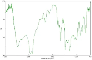
Fig. 4 shows the IR bands observed at 1317 and 1733 in strawberry fruit extract are characteristic of the C–O and C=O stretching modes of the carboxylic acid group. The amide I band appears as very strong band at 1619 cm_1 and amide II band as a medium broad shoulder at 1546 cm_1 . These amide I and II bands arise due to carboxyl stretch and N–H deformation vibrations in the amide linkages of the proteins9 present in it. The medium broad band at 1399cm_1 is the C–N stretching mode of aromatic amine group7. The C–O–C and C– OH vibrations9 of the protein appear as a very strong IR band at1022 cm_1. Further, the band due to C–O stretching at1314 cm_1 is intense in the spectrum of silver nanoparticles.
Fig.5. XRD image for Ag NP’s Fig. 6. Histogram showing particle size distribution for Ag NP’s
Fig. 6 shows the histogram showing the Ag NP’s particle size diameter ranges from 7.5 nm to 32.5 nm and average particle size diameter comes out be 13 nm which is in good agreement with that calculated from Scherrer formula and UV spectroscopy.
CONCLUSIONS
The method used here is hitherto unreported, inexpensive, nontoxic, eco-friendly, abundantly available green reagent for the consistent and rapid synthesis of Silver nanoparticles. The nanoparticles were characterized by a variety of standard analytical techniques namely, UV-Visible Spectroscopy and Particle size distribution (PSD).
A green approach for the synthesis of Ag nanoparticles using Strawberry fruit extract as a green reducing agent with and without a capping agent. Here Gum Arabic is tried as a green capping agent is successfully employed. Ag nanoparticles have been successfully synthesized and the characteristic wavelength of absorption (λmax = 415 nm) is in good
agreement with the reported wavelength in the earlier literature and research papers. The red shift in λmax (Bathochromic effect) to certain degree than the earlier reported wavelength of
the order of 398 nm may be attributed to somewhat higher particle size and distinct morphology. Overlay absorption spectrum too with time, the characteristic wavelength for maximum absorption shows no change and remains at about 415 nm.. The use of pure strawberry fruit extract is quite effective without using any capping or stabilizing agent as the strawberry fruit extract serves a dual purpose as a reducing and capping agent in particular when it is sufficiently concentrated and in adequate quantity. More is it concentrated and quantity more is it reducing capacity which leads to formation of monodispersed, almost
5 10 15 20 25 30 35
0 5 10 15 20 25 N u m b er ( % )
382
spherical and crystalline Ag Np’s. The process can be optimized for achieving a greater degree of efficiency, yield of Ag NP’s. The process is purely green in all respects and therefore eco-friendly in nature.
REFERENCES
1. M. P. Pileni, “Control of the size and shape of inorganic nanocrystals at various scales from nano to macrodomains,” Journal of Physical Chemistry C, Vol. 111, No. 26, pp, 9019-9038, (2007).
2. J. Chen, B. J. Wiley, and Y. Xia, “One-dimensional nanostructures of metals : large-scale synthesis and some potential applications,” Langmuir, Vol. 23, No. 8, pp, 4120-4129, (2007).
3. P. V. Kamat, “Photophysical, photochemical and photocatalytic aspects of metal nanoparticles,” Journal of Physical Chemistry B, Vol. 106, No. 32, pp.7729-7744, (2002). 4. Haes, A. J., Haynes, C. L., McFarland, A. D., Schatz, G. C., Van Duyne, R. P. and Zou,
S., Plasmonic materials for surfaceenhanced sensing and spectroscopy. Mater. Res. Bull., 30, 368–375 (2005).
5. Elechiguerra, J. L., Burt, J. L., Morones, J. R., Camacho-Bragado, A., Gao, X., Lara, H. H. and Yacaman, J. M., Interaction of silver nanoparticles with HIV-1. J.
Nanobiotechnol., 3, doi: 10.1186/1477-3155-3-6 (2005).
6. Morones, J. R., Elechiguerra, J.L., Camacho, A., Holt, K., Kouri, J. B., Ramirez, J. T. and Yacaman, M. J., The bactericidal effect of silver nanoparticles. Nanotechnology, 16, 2346–2353 (2005).
7. Markovich, G., Leff, D. V., Chung, S. W., Soyez, H. M., Dunn, B. and Heath, J. R., Parallel fabrication and single-electron charging of devices based on ordered, two-dimensional phases of organically functionalized metal nanocrystals. Phys. Lett., 70, 3107–3109 (1997).
8. Lewis, L. N., Chemical catalysis by colloids and clusters. Chem. Rev., 93, 2693–2730 (1993). 9. Cushing, B. L., Kolesnichenko, V. L. and O’Connor, C. J., Recent advances in the
liquid-phase syntheses of inorganic nanoparticles. Chem. Rev., 104, 3893 (2004).
10. Sharma, V. K., Yngard, R. A. and Lin, Y., Silver nanoparticles: green synthesis and their antimicrobial activities. Adv. Colloid Interface Sci., 145, 83–96 (2009).
11. Hagerman, A. E., Riedl, K. M., Jones, G. A., Sovik, K. N., Ritchard, N. T., Hartzfeld, P. W. and Riechel, T. L., High molecular weight plant polyphenolics (tannins) as biological antioxidants. J. Agric. Food Chem., 46, 1887–1892 (1998).
12. McDonald, M., Mila, I. and Scalbert, A., Precipitation of metal ions by plant polyphenols: optimal conditions and origin of precipitation. J. Agric. Food Chem., 44, 599–606 (1996).
14. Bors, W., Foo, L. Y., Hertkorn, N., Michel, C. and Stettmaier, K., Chemical studies of proanthocyanidins and hydrolyzable tannins. Antioxid. Redox Signal., 3, 995–1008 (2001).
15. Castanon, G. A., Nino-Martinez, N., Gutierrez, F., Martinez-Mendoza, J. R. and Ruiz, F., Synthesis and antibacterial activity of silver nanoparticles with different sizes. J. Nanopart. Res., 10, 1343–1348 (2008).
16. Liu, J., Qin, G., Raveendran, P. and Ikushima, Y., A facile and green synthesis, characterization, and catalytic function of b-Dglucose stabilized Au nanocrystals. Chem.
Eur. J., 12, 2131–2138 (2006).
17. Hassan, A. A., Sandre, O., Cabuil, V. and Tabeling, P., Synthesis of iron oxide nanoparticles in a microfluidic device: preliminary results in a co-axial flow millichannel. Chem. Commun., 1783–1785 (2008).