How protein coronas determine the fate of engineered
nanoparticles in biological environment
Ivona Capjak
1, Sandra Šupraha Goreta
2, Darija Domazet Jurašin
3, and Ivana Vinković Vrček
4Croatian Institute of Transfusion Medicine1, University of Zagreb, Faculty of Pharmacy and Biochemistry2, Ruđer
Bošković Institute3, Institute for Medical Research and Occupational Health4, Zagreb, Croatia
[Received in October 2017; Similarity Check in October 2017; Accepted in December 2017]
Nanomedicine is a booming medical field that utilises nanoparticles (NPs) for the development of medicines, medical devices, and diagnostic tools. The behaviour of NPs in vivo may be quite complex due to their interactions with biological
molecules. These interactions in biological fluids result in NPs being enveloped by dynamic protein coronas, which serve as an interface between NPs and their environment (blood, cell, tissue). How will the corona interact with this environment will depend on the biological, chemical, and physical properties of NPs, the properties of the proteins that make the corona, as well as the biological environment. This review summarises the main characteristics of protein corona and
describes its dynamic nature. It also presents the most common analytical methods to study the corona, including examples
of protein corona composition for the most common NPs used in biomedicine. This knowledge is necessary to design NPs that will create a corona with a desired efficiency and safety in clinical use.
KEY WORDS: hard corona; nano-bio interface; nanomedicine; soft corona
Nanomedicine is a growing medical field that utilises nanomaterials for new applications in medicine, including their clinical use in disease diagnosis and treatment (1-3). According to the European Commission (4), 'Nanomaterial' means a natural, incidental or manufactured material containing particles, in an unbound state or as an aggregate or as an agglomerate and where, for 50 % or more of the particles in the number size distribution, one or more external dimensions is in the size range 1-100 nm. However,
in medicine the term nanoparticle includes particles with
dimensions of up to 1000 nm.
Due to a large surface-area-to-volume ratio, nanoparticles (NPs) have exceptional functional and structural properties
that make them suitable to carry many diagnostic and
therapeutic agents (5). Recent advances in nanomedicine have resulted in the development of biodegradable nanodrug delivery systems, nanocrystals for magnetic resonance imaging (MRI), and luminescent NPs for multiplexed molecular diagnostics (1, 3, 5, 6).
Because of the small size, NPs can enter almost every part of the body, including tissues, organs, and organelles (mitochondria, lysosomes, and endosomes) by different routes (e.g., inhalation, ingestion, injection, or physical contact with cuts or wounds) (2, 3, 7-9).
There are many types of NPs, including polymeric NPs,
liposomes, carbon nanotubes, quantum dots, or metal-based
NPs (gold, silver iron oxide, silica, titanium dioxide, etc.). Owing to the exceptional properties, these NPs may be used
for targeted drug and contrast delivery, photothermal
therapy, optical sensing, biochromatography, bioanalytical electrochemistry, biocidal agents and coatings, or a variety
of bioassays (1-3, 5).
Their distribution, excretion, metabolism, and pharmacokinetics may be quite complex and pose a
challenge for developing safe and effective nano-based biomedical agents. One of the key issues to resolve is rapid NP uptake and clearance by the reticuloendothelial system (RES), active vs. passive targeting, and penetration into
tumour tissues (5).
Even though thousands of research papers have already been published on the interaction between NPs and
biological systems, little is still known about the mechanistic
details of these interactions (5). What are the biological interfaces that facilitate the interaction between NPs and cell components? This question should be addressed from the perspective of colloidal chemistry (10).
Blood as a biological medium contains more than a thousand biomolecules like proteins, lipids, and nucleic
acids (1, 10). As soon as NPs enter the medium, ions, small molecules, proteins, and cells compete to adsorb on the NP surface due to its high reactivity (11). Plasma proteins have a critical role in creating nano-bio interfaces, as they opsonise NPs and form coronas (5, 12-16).
What kind of a protein corona forms around an NP's surface will depend on the NP's properties (size, shape, composition, surface functional groups, and surface charges), biophysical properties of the biological medium (blood, interstitial fluid, or cell cytoplasm), and the time of
interaction. In other words, how will proteins adsorb on Correspondence to: Ivana Vinković Vrček, Institute for Medical Research
on protein-protein interactions.
Once formed, a protein corona will determine the physicochemical behaviour of an NP. Its properties are more
important in determining the biological response
(agglomeration, cellular uptake, circulation lifetime, signalling, kinetics, transport, accumulation, and toxicity) than NP's properties. In other words, to find out what will be the distribution, metabolism, and elimination of NPs in the body before it is applied in clinical practice, one needs to determine how protein corona affects them.
Knowing how to control the formation of the protein corona is crucial for most clinical uses of NPs (9, 10, 17, 18). This review summarises the current knowledge on the nano-bio interface between NPs and proteins.
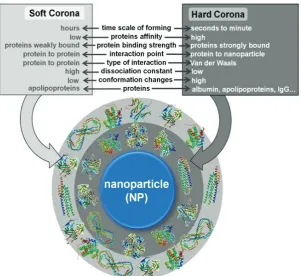
Dynamic nature of protein corona
When NPs come in contact with biological components, a nano-bio interface is formed. What makes it dynamic is a number of physicochemical interactions and thermodynamic exchanges between NP and biomolecular surfaces (7, 19-21). The dynamic nature of the nano-bio interface between NPs and proteins is best described by soft and hard coronas (Figure 1). Proteins with higher affinity for NP surface will exchange easily and quickly forming
the hard corona, while proteins with low affinity exchange slowly forming the soft corona (19). The hard corona
proteins interact directly with NP surface, while the soft corona proteins interact with the hard corona proteins via
weak protein-protein interactions. The time needed for corona formation differs between the hard and soft corona.
a minute, while the formation of the soft corona may take
hours or even days, as proteins with higher affinity replace those with lower affinity (21). This process depends on protein concentrations and the composition of the biological
environment.
Some suggest that even at low plasma concentrations,
corona proteins will completely envelope the surface of an NP and modify its nature and physicochemical properties (19). Soft corona proteins can also interact with the hard corona proteins, as they desorb from NP surface and free the slot for other biomolecules to interact. All these
exchanges are based on competitive adsorption and
desorption of proteins, which depends on interaction time, protein concentrations, and their adsorption affinity for the NP. These exchanges, known as the Vroman effect (22, 23)
have two stages. In the early stage, proteins adsorb rapidly with the highest association rates, and in the late stage proteins with short residence times are being replaced by proteins with slower association rates but longer residence
times (24).
This dynamic nano-bio system is determined by hydrodynamic, electrostatic, electrodynamic, solvent, and
steric interactions (Table 1) (25, 26).
This is why the nano-bio interface changes continuously in a biological environment (Figure 2), especially in the living cells, where different cell products are being secreted. When NPs move from one biological compartment to another, protein corona will change its profile. Some proteins form only transient complexes with NPs, while
others will be tightly bound, depending on the specific NP type and the biological fluids in which NPs are suspended.
Protein adsorption on and interaction with NPs may induce conformational changes or crowding of proteins on the NPs surface or the formation of reactive oxygen species (ROS) that will cause oxidative damage to the adsorbed proteins (26). If a hydrophobic or charged protein sequence interacts with a hydrophobic or charged part of NP surface, this will induce thermodynamically favourable changes. Conformational changes of proteins induced by their interaction with NPs are typically irreversible (21) and may affect the downstream protein-protein interaction, cellular
signalling, and DNA transcription, which directly affect enzyme activity (24). At the same time, protein binding that changes the shape, size, and surface charge of NPs will directly affect the agglomeration, cellular uptake, circulation lifetime, signalling, kinetics, transport, accumulation, and toxicity of NPs in a biological environment (10, 27).
A complex consisting of an NP and its protein corona
is a new entity that cells can see (10, 28). For easier understanding, hard and soft coronas are usually presented as layers (Figure 1). The outer layer (soft corona) does not allow the inner layer (hard corona) to interact with the cell
Force Range (nm) Origin and properties
Hydrodynamic
interactions 102-106
Long-range interactions; induced by particles moving in a viscous fluid, bulk transport, shear, lift, and Brownian diffusion; Increases the collision between NPs
and other surfaces in the system Electrostatic
interactions 1-100
Coulomb interactions; induced by attraction or collision between charged interfaces and counter- or repel co-ions; characterized by the formation of an
electrostatic double layer Electrodynamic
interactions 1-100
Van der Waals interactions that describe interactions between randomly oriented dipoles, between dipole and induced dipole, and fluctuating dipole and induced
dipole
Solvent interactions 1-10 Interactions between lyophobic or lyophilic materials and solvent molecules
Steric interactions 1-100 Repulsive interactions with other interfaces; induced by adsorbed polymer layers on NPs surface; increase stability of individual NPs, but can interfere in cellular uptake
on the thickness of the outer layer.
Figure 3 shows the types of interactions between NPs
and the biological medium. These interactions promote or
inhibit: (a) the adsorption of ions, detergents, and other molecules from the medium, (b) attachment/detachment of proteins, (c) competitive binding, (d) steric hindrance on the NPs surface, (e) formation of two or more layers on NP surface, (e) NP dissolution and/or degradation, (f) surface reconstruction, and (g) accumulation and/or agglomeration of NPs (12, 27, 30, 31).
The most important physicochemical properties of an NP that define protein corona formation and fate are chemical composition, shape, curvature, surface functionalisation and structure, porosity, crystallinity,
heterogeneity, roughness, and hydrophobicity/hydrophilicity
(2-4). Furthermore, effective surface charge, aggregation
state, stability, biodegradability, and dissolution properties
of the NP surface layer are also important parameters that need to be considered for the investigation of the nano-bio interface (4, 32). For example, surface curvature of an NP affects protein-binding affinities. Greater curvature makes
the corona thicker but decreases protein-protein interactions
and conformational changes of the adsorbed proteins. Higher surface charge increases corona thickness as well as conformational changes of proteins (13, 27, 30, 31, 33). It may also trigger protein denaturation (27). Higher
hydrophobicity increases corona thickness and
conformational changes of proteins, as well as the opsonisation rate (14, 34).
determine the long-range and short-range forces governing the nano-bio interface (Table 1) (10, 35). Long-range forces originate from attractive van der Waals and repulsive
electrostatic double-layer interactions, while short-range
forces arise from charge, steric interactions, depletion, and solvent interactions (Table 2) (10, 35).
Understanding how each physicochemical parameter
of an NP affects corona formation is a key to designing new, efficient, and secure nanomaterials. Then these properties can be optimised, NPs pre-coated, and their surfaces functionalised to obtain the nature of the protein corona that would render an NP biocompatible (23). One should also take into account environmental factors, such as temperature, pH, protein concentrations, and time of interaction. NPs may also change adsorption, accumulation,
degradation, agglomeration, dissolution, distribution, and
clearance patterns after the protein corona has been formed, while the proteins forming the corona may pass through conformational changes, free energy release, restructuration, or change their binding profile and kinetics (10, 36, 37).
Mechanistic investigation of protein corona
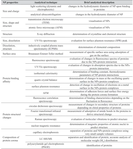
The properties of metallic NPs can be investigated with a range of spectroscopic, electrophoretic, and microscopic methods (Table 2). The same methods can also be used to study protein corona formation and composition. The most common methods for determining NP properties are the transmission electron microscopy (TEM), light scattering techniques, and UV-visible and fluorescence spectroscopy.
Newly synthesised metallic NPs are usually characterised in the medium used for their synthesis, such as water. Although quite demanding, NP evaluation in pure water is
much less complicated than in any biological matrix.
Complexes that form between NPs and proteins are most often analysed with mass spectrometry (MS)-based
proteomics. Spectroscopic methods like ultraviolet-visible
(UV-Vis) and fluorescence spectroscopy, and circular dichroism (CD) are used to investigate nano-bio interface
binding interactions due to their robustness and high sensitivity. UV-Vis spectroscopy can be used to measure
the rate of protein binding as a function of change in
plasmon λmax over time. Fluorescence spectroscopy acquires the intrinsic fluorescence of the protein and can therefore measure binding to NPs. CD spectroscopy uses changes in the chiral properties of a protein to predict changes in its secondary structure. Measured interactions between plasma proteins and NPs can be quantified using several kinetic models and equations (38-40). All the methods described in Table 2 are quite accessible and straightforward to evaluate nano-bio interface in pure water or a simple buffer
system. In complex media like blood plasma or cellular
matrix, however, analytical performance and interpretation of results may become very complex.
NP properties Analytical technique Brief analytical description
Size and charge
light scattering (dynamic and
electrophoretic) changes in the hydrodynamic diameter of NP upon binding to proteins analytical ultracentrifugation changes in the hydrodynamic diameter of NP
Size, shape and structure
transmission electron microscopy
(TEM) visualisation of NPs
atomic force microscopy (AFM) visualisation of NPs
Structure X-ray diffraction determination of crystalline and chemical structure
Size, dissolution UV-Vis spectroscopy evaluation for surface plasmon resonance (SPR) peak Dissolution,
structure inductively coupled plasma mass spectrometry (ICPMS) determination of elemental composition Surface area Braunauer-Emmet-Teller method measurement of specific surface area using adsorption of gas on the surface
Protein binding affinity
fluorescence spectroscopy evaluation of changes in fluorescence spectra of proteins due to the NPs-protein interaction
UV-Vis spectroscopy evaluation of changes in absorption spectra due to the NPs-protein interaction
isothermal calorimetry determination of binding constant, thermodynamic parameters of NP-protein interactions
quartz crystal balance determination of changes in mass at the oscillating quartz surface in the NPs-protein complexes
surface plasmon resonance detection of change in oscillation of electrons on a metal surface in the NPs-protein complexes
AFM determination of adhesion forces and surface free energy during the protein corona formation fluorescence correlation
spectroscopy determination of binding characteristics depending on the fluctuation in florescence
Protein structural changes
circular dichroism spectroscopy measurement of changes in secondary structure of proteins depending on chiral properties of proteins Fourier transformed infrared
spectroscopy measurement of adsorption of amide bonds in the proteins to derive structural change Raman spectroscopy evaluation of molecular vibrations to predict structure
nuclear magnetic resonance determination of magnetic properties of atomic nuclei to predict structure
Composition of protein corona
capillary electrophoresis separation of proteins and NPs-protein complexes using very small sample volumes
LC-MS/MS separation and identification of protein, accurate analysis of molecular weight (M
w) distribution
polyacrylamide gel electrophoresis
Table 3 Composition of protein corona by NP type
NP type Proteins detected in corona Reference
Polystyrene NPs coagulation factors, immunoglobulins, lipoproteins, acute phase proteins, complement proteins, plasminogen, anti-CD4, c4a, albumin 1, 23,42
Latex NPs albumin, apolipoproteins, immunoglobulins, hemoglobin, haptoglobins 1, 23
Copolymer NPs albumin, apolipoprotiens, fibrinogen, immunoglobulins, C4BP-α-chain 1 Supraparamagnetic
iron oxide NPs albumin, α-1-antitrypsin, fibrinogen chains, immunoglobulin chains, transferrin, transthyretin 1, 41, 42 Gold NPs albumin, fibrinogen chains, apolipoprotein A1, transport proteins, coagulation factors, tissue development proteins 1, 23, 42
Carbon nanotubes fibrinogen chains, immunoglobulin light chains, fibrin, albumin, ApoA1, complement, component proteins, fibronectin 1, 42
SiO2 NPs immunoglobulins, lipoproteins, complement proteins, coagulation proteins, acute phase proteins, cell proteins, serum proteins 1, 23, 19
TiO2NPs, ZnONPs,
SiO2 NPs
albumin, immunoglobulins, fibrinogen, transferrin, apolipoprotein A1,
complement proteins, immunoglobulin light chains, fibrin, albumin, fibronectin 1, 23 Magnetic NPs albumin, apolipoprotein A1, complement factors, vitronectin, hemoglobin 1
Citrate-coated AgNPs
albumin, α-1-antiproteinase, α-2-HS-glycoprotein, apolipoprotein A1, serotransferrin, macroglobulin, α-fetoprotein, apolipoprotein B100,
α-2-antiplasmin, complement C3, β-2-glycoprotein 1, fetuin-B, inter-α-trypsin inhibitor heavy chain H1, hemoglobin foetal subunit β, inter-α-trypsin inhibitor heavy chain H3, inter-α-trypsin inhibitor heavy chain H2, hemoglobin subunit α,
complement factor B, hemopexin, serpin A3-6
46
AgNPs coated with polyvinylpyrrolidone
albumin, α-2-HS-glycoprotein, α-1-antiproteinase, apolipoprotein A1, serotransferrin, α-2-macroglobulin, α-fetoprotein, apolipoprotein B100, complement C3, α-2-antiplasmin, inter-α-trypsin inhibitor heavy chain H1, fetuin-B, β-2-glycoprotein 1, hemoglobin foetal subunit beta, inter-α-trypsin
inhibitor heavy chain H3, inter-α-trypsin inhibitor heavy chain H2, vitamin D-binding protein, transthyretin, hemoglobin subunit α, complement factor B
46
The most extensively studied biological environment
for protein corona is human blood plasma. Protein layer(s) that adsorb on NPs in blood can affect their uptake and distribution in the cells. For example, fibrinogen, immunoglobulin G (IgG), or complement factors are believed to promote phagocytosis and removal of NPs from the bloodstream, while human serum albumin (HSA) and apolipoproteins prolong their circulation time in blood (19).
In the early stage of corona formation, albumin, IgG, fibrinogen, and apolipoproteins seem to adsorb rapidly on metallic NPs in plasma (13, 35, 41). These proteins are found in the hard coronas of all studied NPs and are replaced by apolipoproteins and coagulation factors in the slow phase of corona formation (35). Changes in biological environment also reflect on a corona composition. Walkey and Chan (21)
use the term adsorbome to denote a group of 125 most
common plasma proteins in the corona. Table 3 lists some
of them by the type of NP.
Only two to six of them strongly adsorb on metallic NPs. For most metallic NPs, the corona is dominated by albumin (42), which is at the same time the most abundant
protein in plasma. Although it has a negative net charge at
pH 7.4, albumin contains 60 positively charged lysine
residues, which enable its interaction with both positively
corona complexes with NPs regardless of their net charge (36). These coronas are similar in size and effective surface
charge, but their behaviour in contact with the cell will
differ. Fleischert and Payne (36) believe that cationic NPs alter the structure of albumin proteins in the corona, while anionic NPs do not. These structural changes affect the behaviour of the albumin-NP complex at the cellular level, so that cell receptors bind coronas formed around anionic NPs and redirect those formed around cationic NPs to scavenger receptors. Another difference is that albumin adsorbs much more on the surface of anionic than cationic NPs (23% vs. 8% of surface coverage, respectively) (36).
of nanoparticles
As the NP-corona complex is actually “what the cell sees” (28), it is more important to determine the biological response (i.e., immunogenicity) to the complex than the properties of an NP alone (43). For most biomedical
purposes, hard corona will likely improve the interaction
between NPs and proteins, membranes, phospholipids, endocytic vesicles, organelles, and DNA (44-46).
Corona is what controls which type of biomolecule will it bind and how, how will the NP-corona complex interact
with cells receptors, and what will its distribution and
elimination be (45-46). For a nano-drug delivery system it is important to define the affinity, stoichiometry, kinetics, and the concentrations of NPs for their interaction with specific proteins. At the moment, however, we still have a lot to learn. The biggest challenge for researches is to find
out how protein corona could contribute to nanodrug distribution in vivo.
Conflicts of interest
None to declare.
REFERENCES
1. Saptarshi SR, Duschl A, Lopata AL. Interaction of nanoparticles with proteins: relation to bio-reactivity of the nanoparticle. J Nanobiotechnol 2013;11:26. doi: 10.1186/1477-3155-11-26
2. Huang R, Carney RP, Stellacci F, Lau BL. Protein-nanoparticle interactions: the effects of surface compositional and structural heterogeneity are scale dependent. Nanoscale 2013;5:6928-35. doi: 10.1039/c3nr02117c
3. Jurašin DD, Ćurlin M, Capjak I, Crnković T, Lovrić M, Babič M, Horák D, Vinković Vrček I, Gajović S. Sufrace coating affects behavior of metallic nanoparticle in a bilogical environment. Beilstein J Nanotechnol 2016;7:246-62. doi: 10.3762/bjnano.7.23.
4. European Commission. Commission recommendation of 18 October 2011 on the definition of nanomaterial (Text with EEA relevance) (2011/696/EU) [displayed 3 November 2017]. Available at https://ec.europa.eu/research/industrial_ technologies/pdf/policy/commission-recommendation-on-the-definition-of-nanomater-18102011_en.pdf
5. Lane LA, Qian X, Smith AM, Nie S. Physical chemistry of nanomedicine: understanding the complex behaviours of nanoparticles in vivo. Annu Rev Phys Chem 2015;66:521-47. doi: 10.1146/annurev-physchem-040513-103718
6. Ding F, Radic S, Chen R, Chen P, Geitner NK, Brown JM, Ke PC. Direct observation of a single nanoparticle-ubiquitin corona formation. Nanoscale 2013;5:9162-9. doi: 10.1039/ c3nr02147e
7. Duran N, Silveira CP, Duran M, Martinez DST. Silver nanoparticle protein corona and toxicity: a mini-review. J Nanobiotechnology 2015;13:55. doi: 10.1186/s12951-015-0114-4
8. Docter D, Westmeier D, Markiewicz M, Stolte S, Knauer SK, Stauber RH. The nanoparticle biomolecule corona:
2015;44:6094-121. doi: 10.1039/C5CS00217F
9. Kononenko V, Narat M, Drobne D. Nanoparticle interaction with the immune system. Arh Hig Rada Toksikol 2015;66:97-108. doi:10.1515/aiht-2015-66-2582
10. Nel AE, Mädler L, Velegol D, Xia T, Hoek EMV, Somasundaran P, Klaessig F, Castranova V, Thompson M. Understanding biophysicochemical interactions at the nano-bio interface. Nat Mater 2009;8:543-57. doi: 10.1038/ nmat2442
11. Tavanti F, Pedone A, Menziani MC. Competitive binding of proteins to gold nanoparticles disclosed by molecular dynamics simulations. J Phys Chem C 2015;119:22172-80. doi: 10.1021/acs.jpcc.5b05796
12. Cedervall T, Lynch I, Foy M, Berggad T, Donnelly S, Cagney G, Linse S, Dawson K. Detailed identification of plasma proteins adsorbed on copolymer nanoparticles. Angew Chem Int Ed 2007;46:5754-6. doi: 10.1002/anie.200700465 13. Lundqvist M, Stigler J, Elia G, Lynch I, Cedervall T, Dawson
KA. Nanoparticle size and surface properties determine the protein corona with possible implications for biological impacts. Proc Natl Acad Sci 2008;105:14265-70. doi: 10.1073/pnas.0805135105
14. Cedervall T, Lynch I, Lindman S, Berggård T, Thulin E, Nilsson H, Dawson KA, Linse S. Understanding the nanoparticle-protein corona using methods to quantify exchange rates and affinities of proteins for nanoparticles. Proc Natl Acad Sci USA 2007;104:2050-5. doi: 10.1073/ pnas.0608582104
15. Lynch I, Dawson KA, Linse S. Detecting cryptic epitopes created by nanoparticles. Sci STKE 2006;2006(327):pe14. doi: 10.1126/stke.3272006pe14
16. Gref R, Lück M, Quellec P, Marchand M, Dellacherie E, Harnisch S, Blunk T, Müller RH. ‘Stealth’ corona-core nanoparticles surface modified by polyethylene glycol (PEG): influences of the corona (PEG chain length and surface density) and of the core composition on phagocytic uptake and plasma protein adsorption. Colloids Surf B Biointerfaces 2000;18:301-13. doi: 10.1016/S0927-7765(99)00156-3 17. Kittler S, Greulich C, Gebauer JS, Diendorf J, Treuel L, Ruiz
L, Gonzalez-Calbet JM, Vallet-Regi M, Zellner R, Köller M, Epple M. The influence of proteins on the dispersability and cell-biological activity of silver NP. J Mater Chem 2010;20:512-8. doi: 10.1039/B914875B
18. Koshkina O, Lang T, Thiermann R, Docter D, Stauber RH, Secker C, Schlaad H, Weidner S, Mohr B, Maskos M, Bertin A. Temperature-triggered protein adsorption on polymer-coated nanoparticles in serum. Langmuir 2015;31:8873-81. doi: 10.1021/acs.langmuir.5b00537
19. Monopoli MP, Walczyk D, Campbell A, Elia G, Lynch I, Bombelli FB, Dawson KA. Physical-chemical aspects of protein corona: relevance to in vitro and in vivo biological impacts of nanoparticles. J Am Chem Soc 2011;133:2525-34. doi: 10.1021/ja107583h
20. Miclăuş T, Bochenkov VE, Ogaki R, Howard KA, Sutherland DS. Spatial mapping and quantification of soft and hard protein coronas at silver nanocubes. Nano Lett 2014;14:2086-93. doi: 10.1021/nl500277c
of high molecular-weight kininogen, factor-XII, and fibrinogen in plasma at interfaces. Blood 1980;55:156-9. PMID: 7350935
23. Aggarwal P, Hall JB, McLeland CB, Dobrovolskaia MA, McNeil SE. Nanoparticle interaction with plasma proteins as it relates to particle biodistribution, biocompatibility and therapeutic efficacy. Adv Drug Deliv Rev 2009;61:428-37. doi: 10.1016/j.addr.2009.03.009
24. Göppert TM, Müller RH. Polysorbate-stabilized solid lipid nanoparticles as colloidal carriers for of drugs to the brain: comparison of plasma protein adsorption patterns. J Drug Target 2005;13:179-87. doi: 10.1080/10611860500071292 25. Gebauer JS, Malissek M, Simon S, Knauer SK, Maskos M,
Stauber RH, Peukert W, Treuel L. Impact of the nanoparticle-protein corona on colloidal stability and nanoparticle-protein structure. Langmuir 2012;28:9673-9. doi: 10.1021/la301104a 26. Treuel L, Nienhaus GU. Toward a molecular understanding
of nanoparticle-protein interactions. Biophys Rev 2012;4:137-47. doi 10.1007/s12551-012-0072-0
27. Lynch I, Dawson KA, Linse S. Detecting cryptic epitopes created by nanoparticles. Sci STKE 2006;2006(327):pe14. doi: 10.1126/stke.3272006pe14
28. Walczyk D, Bombelli FB, Monopoli MP, Lynch I, Dawson KA. What the cell “sees” in bionanoscience. J Am Chem Soc 2010;132:5761-8. doi: 10.1021/ja910675v
29. Gessner A, Waicz R, Lieske A, Paulke B-R, Mäder K, Müller RH. Nanoparticles with decreasing surface hydrophobicities: influence on plasma protein adsorption. Int J Pharm 2000;196:245-9. doi: 10.1016/S0378-5173(99)00432-9 30. Lundqvist M, Stigler J, Cedervall T, Berggard T, Flanagan
MB, Lynch I, Elia G, Dawson K. The evolution of the protein corona around nanoparticles: a test study. ACS Nano 2011;5:7503-9. doi: 10.1021/nn202458g
31. Mahmoudi M, Lynch I, Ejtehadi MR, Monopoli MP, Bombelli FB, Laurent S. Protein-nanoparticle interactions: opportunities and challenges. Chem Rev 2011;111:5610-37. doi: 10.1021/cr100440g
32. Klein J. Probing the interaction of proteins and nanoparticles. Proc Natl Acad Sci USA 2007;104:2029-30. doi: 10.1073/ pnas.0611610104
33. Gessner A, Lieske A, Paulke BR, Müller RH. Influence of surface charge density on protein adsorption on polymeric nanoparticles: analysis by two-dimensional electrophoresis. Eur J Pharm Biopharm 2002;54:165-70. doi: 10.1016/S0939-6411(02)00081-4
34. Lindman S, Lynch I, Thulin E, Nilsson H, Dawson KA, Linse S. Systematic investigation of the thermodynamics of HSA adsorption to N-iso-propylacrylamide/N-tert-butylacrylamide copolymer nanoparticles. Effects of particle size and hydrophobicity. Nano Lett 2007;7:914-20. doi: 10.1021/ nl062743+
M, Laurent S, Tawil N, Yahia L, Mahmoudi M, editors. Springer Series in Biophysics. Vol. 15. Protein-nanoparticle interactions. Chapter 2. Berlin Heidelberg: Springer-Verlag; 2013. p. 21-44.
36. Fleischert CC, Payne CK. Nanoparticle-cell interactions: molecular structure of the protein corona and cellular outcomes. Acc Chem Res 2014;47:2651-9. doi: 10.1021/ ar500190q
37. Laera S, Ceccone G, Rossi F, Gilliland D, Hussain R, Siligardi G, Calzolai L. Measuring protein structure and stability of protein-nanoparticle systems. Nano Lett 2011;11:4480-4. doi: 10.1021/nl202909s
38. Eskandari K, Kamali M, Ramezani M, Safiri Z, Keihan AH, Rashidiani J, Kooshki H, Zarei H. The effect of hydrophobicity and hydrophilicity of gold nanoparticle on proteins structure and function. Int J Bio-Inorg Hybrid Nanomat 2013;2:465-70. 39. Cui M, Liu R, Deng Z, Ge G, Liu Y, Xie L. Quantitative study
of protein coronas on gold nanoparticles with different surface modifications. Nano Res 2013;7:345. doi: 10.1007/ s12274-013-0400-0
40. Boulos SP, Davis TA, Yang JA, Lohse SE, Alkilany AM, Holland LA, Murphy CJ. Nanoparticle-protein interactions: a thermodynamic and kinetic study of the adsorption of bovine serum albumin to gold nanoparticle surfaces. Langmuir 2013;29:14984-96. doi: 10.1021/la402920f 41. Sakulkhu U, Mahmoudi M, Maurizi L, Salaklang J, Hofmann
H. Protein corona composition of superparamagnetic iron oxide nanoparticles with various physico-chemical properties and coatings. Scientific Rep 2014;4:5020. doi: 10.1038/ srep05020
42. Karmali PP, Simberg D. Interactions of nanoparticles with plasma proteins: implication on clearance and toxicity of drug delivery systems. Expert Opin Drug Deliv 2011;8:343-57. doi: 10.1517/17425247.2011.554818
43. Lee YK, Choi E-J, Webster TJ, Kim S-H, Khang D. Effect of the protein corona on nanoparticles for modulating cytotoxicity and immunotoxicity. Int J Nanomedicine 2015;10:97-113. doi: 10.2147/IJN.S72998
44. Yallapu MM, Ebeling MC, Jaggi M, Chauhan SC. Plasma proteins interaction with curcumin nanoparticles: implications in cancer therapeutics. Curr Drug Metab 2013;14:504-15. PMCID: PMC4030727
45. Zook JM, Halter MD, Cleveland D, Long SE. Disentangling the effects of polymer coatings on silver nanoparticle agglomeration, dissolution and toxicity to determine mechanisms of nanotoxicity. J Nanopart Res 2012;14:1165. doi: 10.1007/s11051-012-1165-1
Nanomedicina je iznimno napredno medicinsko područje u kojem se iskorištavaju nanočestice za razvoj inovativnih lijekova, medicinskih pomagala i dijagnostičkih postupaka. U in vivo uvjetima ponašanje nanočestica može biti vrlo kompleksno zbog bliskih interakcija s biološkim molekulama. Zbog međudjelovanja nanočestica i proteina u biološkim tekućinama nastaje dinamička proteinska korona koja obavija nanočestice i tvori novo sučelje između nanočestica i okoliša u kojem se one nalaze (krv, stanice, tkiva). Ta međudjelovanja ovise o biološkim, kemijskim i fizikalnim svojstvima samih nanočestica i proteina, ali i samog biološkog okoliša. U ovom preglednom radu dan je prikaz glavnih karakteristika koji određuju proteinsku koronu te opis njezine dinamičke prirode. Prikazane su najvažnije analitičke metode za istraživanje proteinske korone te primjeri sastava proteinske korone za najčešće korištene vrste nanočestica u biomedicini. Takvo je znanje nužno za dizajn i razvoj učinkovitih i sigurnih nanomedicinskih proizvoda.
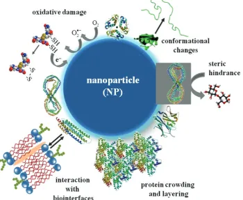
![Figure 3 Active interactions at the nano-bio interface [inspired by (10)]](https://thumb-us.123doks.com/thumbv2/123dok_us/10090743.1995752/4.595.121.473.470.756/figure-active-interactions-nano-bio-interface-inspired.webp)