S H O R T R E P O R T
Open Access
Oxidative stress mediates depot-specific
functional differences of human
adipose-derived stem cells
Sandhya Sriram
1, Chengxiang Yuan
1, Smarajit Chakraborty
1,6, Winson Tay
1,6, Min Park
2, Asim Shabbir
3,
Sue-Anne Toh
4, Weiping Han
5and Shigeki Sugii
1,2,6*Abstract
Background:Visceral (VS) fat depot is known to have defective adipogenic functions compared to subcutaneous (SC) fat, but its mechanism of origin is unclear.
Objective:We tested our hypothesis that the degree of oxidative stress in adipose-derived stem cells (ASCs) from these depots may account for this difference.
Methods:ASCs were isolated from VS (omental region) and SC (abdominal region) fat depots of human subjects undergoing bariatric surgery. ASCs from VS and SC fat were investigated for their cellular characteristics in reactive oxygen species (ROS), metabolism, gene expression, proliferation, senescence, migration, and adipocyte differentiation. ASCs were also treated with antioxidant ascorbic acid (vitamin C).
Results:We found that human VS-derived ASCs exhibit excessive oxidative stress characterized by high reactive oxygen species (ROS), compared to SC-derived ASCs. Gene expression analyses indicate that the VS-ASCs exhibit higher levels of genes involved in pro-oxidant and pro-inflammatory pathways and lower levels of genes in antioxidant and anti-inflammatory pathways. VS-ASCs have impaired cellular functions compared to SC-ASCs, such as slower proliferation, early senescence, less migratory activity, and poor adipogenic capability in vitro. Treatment with ascorbic acid decreased ROS levels drastically in VS-ASCs. Ascorbic acid treatment substantially improved proliferation, senescence, migration, and adipogenic capacities of compromised ASCs caused by high ROS.
Conclusions:This finding suggests the fat depot-specific differences of cellular defects originating from stem cell population. Considering clinical potentials of human ASCs for cell therapies, this also offers a possible strategy for improving their therapeutic qualities through antioxidants.
Keywords:Subcutaneous fat, Intra-abdominal fat, Mesenchymal stromal cells, Oxidative stress, Reactive oxygen species, Ascorbic acid
Introduction
Adipose-derived stem cells (ASCs) are the mesenchymal stem cell (MSC) type that exhibits cellular characteristics potentially useful for regenerative medicine, which in-cludes multipotent and migratory capacities [1,2]. Due to the relative ease in isolation, abundance, and therapeutic
potencies, a number of clinical trials involving ASCs are currently undertaken worldwide in diverse disease indica-tions [3]. Within white fat tissue, ASCs are prevalent and believed to be the origin of mature adipocytes [1,4]. There are at least two different types of white adipose tissue, sub-cutaneous (SC) fat depot residing under the skin and vis-ceral (VS) fat depot surrounding internal organs, which exhibit distinct adipogenic phenotypes. For example, in contrast to SC fat, VS fat is characterized by poor lipid storage capacity, higher inflammation, altered adipokine release, and insulin resistance [5, 6]. As a result, the ex-pansion of VS adiposity during obesity is associated with
© The Author(s). 2019Open AccessThis article is distributed under the terms of the Creative Commons Attribution 4.0 International License (http://creativecommons.org/licenses/by/4.0/), which permits unrestricted use, distribution, and reproduction in any medium, provided you give appropriate credit to the original author(s) and the source, provide a link to the Creative Commons license, and indicate if changes were made. The Creative Commons Public Domain Dedication waiver (http://creativecommons.org/publicdomain/zero/1.0/) applies to the data made available in this article, unless otherwise stated. * Correspondence:[email protected]
1
Fat Metabolism and Stem Cell Group, Singapore Bioimaging Consortium (SBIC), Agency for Science, Technology and Research (A*STAR), 11 Biopolis Way #02-02, Singapore 138667, Singapore
2Duke-NUS Medical School, 8 College Road, Singapore 169857, Singapore
various metabolic complications including diabetes, hypertension, hyperlipidemia, and cardiovascular diseases. It was previously demonstrated in rodents and human that progenitor and stem cell population show fat depot-specific differences in adipogenic capacities in vitro [7–10]. Stem cells from VS fat are poorly differentiated into mature adipocytes compared to SC-derived cells, but molecular mechanisms underlying this difference are completely understood. Oxidative stress resulting from in-creased reactive oxygen species (ROS) is known to cause cellular damages leading to genomic instability, senes-cence, and apoptosis. The involvement of oxidative stress in obesity and diabetes is relatively well studied; numerous studies have been performed on diet-induced obesity ani-mal models and human obese patients to determine the effects of oxidative stress/ROS levels in metabolic organs and systemic metabolism [11–15]. Functions of fat tissue and adipocytes are greatly affected by intracellular ROS levels, though mechanisms leading to the oxidative stress in fat are still not clear [16]. Little is known about the ROS profiles and its effect on cells derived from the ana-tomically distinct SC and VS fat depots. In this manu-script, we investigated possible roles of oxidative stress in mediating depot-specific differences in human ASCs.
Results and discussion
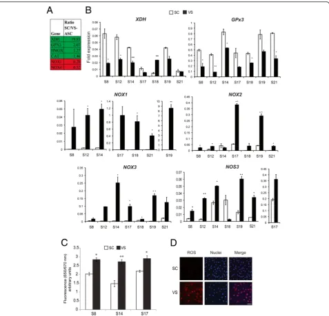
In our previous publication [10], we performed microarray and ontological analysis on human ASCs isolated from SC and VS depots, in order to identify genes underlying their molecular differences. We recognized a set of genes in-volved in either generating or detoxifying ROS to be dif-ferentially expressed in SC- and VS-ASCs, as shown in Fig.1a. We performed quantitative real-time PCR analysis and confirmed that a subset of those genes are consist-ently and differentially expressed across different subjects (Fig.1b). SC-ASCs show upregulation of genes implicated in antioxidant activities including xanthine dehydrogenase (XDH) and glutathione peroxidase 3 (GPx3) in the major-ity of subjects. On the other hand, VS-ASCs generally have upregulated expression of oxidative stress-inducing genes such as NADPH oxidases 1, 2, and 3 (NOX1,
NOX2, NOX3) and nitric oxide synthase 3 (NOS3). Among those found in microarray analysis, antioxidants catalase (CAT) and heme oxygenase-1 (HMOX1) were higher in five out of seven SC-ASCs (Additional file 1: Figure S1). Other oxidative stress-related genes, such as
NOX4, NOX5, GPx1, GPx2, GPx4, SOD1, SOD2, and
SOD3, exhibit variable expressions across subjects, indi-cating that different factors may influence these genes to varying degrees (Additional file1: Figure S1). Based on ex-pression of these genes, we anticipated higher oxidative stress in VS-ASCs. By using a specific fluorescent dye, we confirmed that reactive oxygen species (ROS) is present at significantly higher levels in VS-ASCs than in SC-ASCs
(Fig. 1c, d). The same finding was observed when low
passage VS-ASCs and SC-ASCs were compared
(Additional file1: Figure S2).
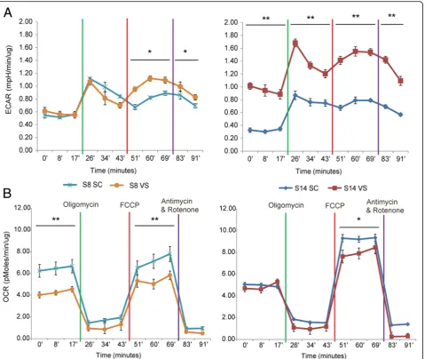
Seahorse XF Analyzer was employed to investigate glycolytic and oxidative metabolism of SC- and VS-ASCs. As shown in Fig. 2, the extracellular acidification rate (ECAR) tends to be increased in VS-ASCs compared to SC-ASCs, suggesting that VS-ASCs generally have higher glycolysis activity (Fig. 2a). In contrast, VS-ASCs show a lower oxygen consumption rate (OCR) than SC-ASCs, es-pecially under maximal respiratory states, indicating that VS-ASCs have less oxidative metabolism capacity (Fig.2b). Increased ROS leads to mitochondrial dysfunctions, which is observed in many pathological conditions [17]. It is speculated that excessive ROS production in VS-ASCs shifts metabolism away from oxidative phosphorylation and toward glycolytic activity.
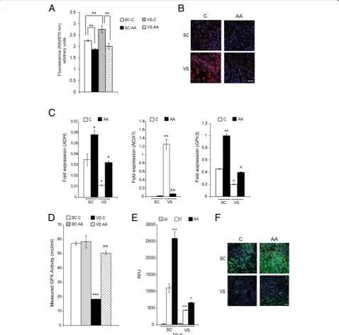
Next, we investigated the effects of ascorbic acid (vitamin C), a water-soluble natural antioxidant and powerful scav-enger of ROS [18,19]. Ascorbic acid was found to potently reduce ROS levels of both SC- and VS-ASCs, as revealed by ROS-specific signal changes (Fig. 3a, b). Expression levels of antioxidant genesXDHandGPx3are upregulated by ascorbic acid in both ASCs, while pro-oxidant NOX1 gene is significantly downregulated by ascorbic acid treat-ment in VS-ASCs (Fig.3c). The activity of glutathione per-oxidase (GPx), an antioxidant enzyme, is lower in VS-ASCs than in SC-ASCs, but significantly reversed by treatment with ascorbic acid (Fig. 3d). The abilities of both ASCs to differentiate into mature adipocytes are significantly im-proved when ascorbic acid is supplemented during adipo-genic stimulation (Fig. 3e, f). In order to validate if the effects on adipogenesis are specifically mediated by ROS, SC-ASCs were incubated with ROS-causing hydrogen per-oxide (H2O2) and treated with vitamin E, which is another antioxidant and specific ROS scavenger. As expected, H2O2 induced ROS and inhibited adipocyte differentiation of ASCs (Additional file1: Figure S3). Treatment with vitamin E, however, at least partially reversed the ROS level and sig-nificantly improved the adipogenic defect induced by H2O2 (Additional file1: Figure S3).
significantly delayed senescence of VS-ASCs (Fig. 4d, e). Rather unexpectedly, Seahorse analysis indicates that treatment of ascorbic acid generally decreases both ECAR (glycolysis) and OCR (oxidative metabolism) activities in VS-ASCs (Additional file1: Figure S4).
Collectively, our data indicate that antioxidants im-prove cellular dysfunctions of undifferentiated and dif-ferentiated ASCs caused by high oxidative stress. It is
antioxidants for improving general ASC properties. Since ASCs, along with other MSCs, have been actively investi-gated for cell therapies, it is worth considering that inclu-sion of stable derivatives of ascorbic acid or other antioxidants in culture media may help achieve greater therapeutic abilities of ASCs for clinical applications.
Conclusion
Our studies demonstrate that high ROS is observed in visceral fat-derived ASCs and associated with their al-tered oxidative metabolism and cellular dysfunctions, compared to subcutaneous fat-derived ASCs. Treatment with antioxidant ascorbic acid leads to improvement of adipocyte differentiation, proliferation, migration, and early senescence. These results point to depot-specific
differences in oxidative stress that may be caused by mo-lecular factors (e.g., pro-oxidant and antioxidant genes) and environmental cues (e.g., inflammation). In addition, our data suggest the potential use of antioxidants for im-proving ROS-mediated dysfunction of ASCs for future clinical applications.
Methods
Isolation and culture of ASCs
White adipose tissue (WAT) was isolated from the sub-cutaneous (abdominal region) depot and visceral depot (omental region) from human subjects undergoing bariat-ric surgery, which was approved by the Domain Specific Review Board at National Healthcare Group, Singapore. ASCs were isolated and enriched by serial passage culture Fig. 2ASCs exhibit depot-specific differences in mitochondrial respiration and glycolysis.aGraph showing ECAR (extracellular acidification rate) by Seahorse XF analyses each in triplicates for SC- and VS-ASCs from two representative subjects (S8 and S14).bGraph showing OCR (oxygen consumption rate) by Seahorse XF analyses each in triplicates for SC- and VS-ASCs from two subjects. Statistical significance was calculated by ANOVA. *p< 0.05, **p< 0.01
Fig. 4Ascorbic acid improves stem cell dysfunctions of VS-ASCs.aProliferation assay using a methylene blue staining method each in triplicates in S17 SC- and VS-ASCs with and without 50μM ascorbic acid (AA) treatment at various time points as indicated. Statistical significance was calculated by ANOVA. *p< 0.05 when compared to SC-C; ^p< 0.05 when compared to VS-C.bRepresentative images (× 10) showing migration assay by the scratch test for S17 SC-ASCs 16 h post AA treatment. Scale bar represents 100μm.cGraph showing average hours taken for migration from S8, S14, and S17 SC- and VS-ASCs with and without AA treatment in the scratch test (n= 3). *p< 0.05 when compared to VS-C. dRepresentative images (× 10) showing senescence test usingβ-galactosidase in S8 SC- and VS-ASCs with and without AA treatment.e Quantification of the average percentage of cells exhibiting positiveβ-galactosidase staining from S18 and S19 SC- and VS-ASCs with and without AA treatment (n= 2). **p< 0.01 when compared to VS-C
of stromal vascular fractions (SVF) using ASC media (DMEM, 15% FBS, 1× GlutaMAX, 1× NEAA, Pen/Strep, 5 ng/ml basic FGF), as described previously [8, 10, 20]. Subject information is found in Additional file1: Table S1. As previously described, we routinely checked the expres-sion of MSC markers (CD73, CD90, and CD105) and trili-neage differentiation in ASC culture. Only cells with similar passage numbers (up to p8) were used for any comparative studies. A stock solution of L-ascorbic acid (Sigma: A4403) was made freshly and added to media with a maximum of 48 h for ascorbic acid treatment.
Cellular analyses
Adipogenesis of ASCs was performed as previously described with minor modifications [8, 10]. Briefly, after 2 days of overconfluence, cells were subjected to differentiation cocktail (0.5 mM isobutylmethyl-xanthine, 1μM dexamethasone, 167 nM insulin, and 100μM indomethacin) for 6 days, followed by main-tenance media containing 167 nM insulin for 6 more days. Cells were stained with AdipoRed (Lonza). ROS was detected by a specific dye CellROX Deep Red Re-agent (Thermo Fisher Scientific) and counter-stained with nuclear staining with Hoechst 33342 (Thermo Fisher Scientific), according to the manufacturer’s in-structions. Stained cells were imaged by either
Ima-geXpress Micro (Molecular Devices) or TS100
microscope (Nikon). Proliferation rates were mea-sured by using the Methylene Blue and SpectraMax spectrophotometer (Molecular Devices) according to the previous publication [21]. Migration was mea-sured by the scratch assay, recording time taken to fill the gap scratched with p200 pipet tips, as previously reported [22]. Senescence of cells was assessed by staining with beta-galactosidase (Thermo Fisher Sci-entific) as in the manufacturer’s instruction. For cel-lular and mitochondrial respiration analysis, Seahorse XF24 Analyzer (Agilent) was used accordingly to the manufacturer’s protocol. Six thousand ASCs were seeded in 24-well microplates and adhered for 1 day with ASC media. For the ascorbic acid treatment study, the drug (50μM) was added at this stage. Then, cells were switched to the assay media (Seahorse XF Base Medium supplemented with 1% FBS, 1 mM pyruvate, 2 mM glutamine, and 10 mM glucose). OCR and ECAR were measured, with sequential addition of metabolic disruptors, oligomycin, carbonyl cyanide p-trifluoromethoxy-phenylhydrazone (FCCP), rote-none, and antimycin A. The data were normalized to the protein concentrations that had been estimated with the BCA protein assay, and analyzed using Prism version 5 software (GraphPad). For all the cellular assays,“ tripli-cates” refers to technical replicates where experiments were performed in each cell type from three independent
culture wells.n= 2 or 3 indicates that values were calcu-lated from cells of two or three subjects.
Quantitative real-time PCR
Real-time qPCR was performed as described previously [8, 10]. Briefly, total RNA was extracted using Trizol re-agent (Thermo Fisher Scientific) and treated with DNase I to remove genomic DNA. cDNA conversion was done by the RevertAid H minus first-strand cDNA synthesis kit (Fermentas). Relative mRNA levels were calculated and normalized to that of GAPDH. The sequence of primers is found in Additional file1: Table S2.
Additional file
Additional file 1: Figure S1.Gene expression studies of additional ROS-related genes that are not included in Figure1b.Figure S2.VS-ASCs have increased ROS when compared to SC-ASCs in the lower passage. Figure S3.Effects on adipogenesis are specifically mediated by ROS. Figure S4.Ascorbic acid treatment generally decreases both glycolytic and oxidative respirations.Table S1.List of subjects used for this study. Table S2.List of RT-qPCR primers-oligos 5′to 3′. (DOCX 3731 kb)
Acknowledgements
We thank Kosuke Takeda for the preceding microarray analysis and Subha Subramanian, Wee Kiat Ong, and other members of Fat Metabolism and Stem Cell Group, Laboratory of Metabolic Medicine, Microscopy Core, Mechanobiology Institute at National University of Singapore, and Singapore Bioimaging Consortium-Nikon Imaging Centre (SBIC-NIC) for the help with our studies.
Funding
This work was supported by intramural funding from Biomedical Research Council of Agency for Science, Technology and Research (A*STAR) to Sh. Su, Singapore-China Joint Grant Project #1412424003 to Sh. Su and M.P, Developmental Programme Grant (#1334k00083) from A*STAR Joint Council Office to Sa.Sr and Sh.Su, and funding from the Singapore National Medical Research Council (NMRC) to S-A.T.
Availability of data and materials
The datasets used and/or analyzed during the current study are available from the corresponding author upon request.
Authors’contributions
SaS contributed to the conception and design, collection and/or assembly of data, data analysis and interpretation, and manuscript writing. CY contributed to the collection and/or assembly of data, data analysis and interpretation, and manuscript writing. SC contributed to the collection and/or assembly of data, data analysis and interpretation, and manuscript writing. WT contributed to the collection and/or assembly of data and data analysis. MP contributed to the collection and/or assembly of data. AS contributed to the provision of study material or patients and administrative support. SAT contributed to the provision of study material or patients and administrative support. WH contributed to the financial support, administrative support, and manuscript writing. ShS contributed to the conception and design, financial support, administrative support, data analysis and interpretation, and manuscript writing. All authors made final approval of the manuscript.
Ethics approval and consent to participate
Use of human patient-derived cells was conducted with informed consent obtained for each subject, approved by the National Healthcare Group Domain Specific Review Board, Singapore, and performed in accordance with its relevant regulations.
Competing interests
ShS is a co-founder of Celligenics Pte Ltd., which has not had any financial or scientific influence on this study. The other authors declare that they have no competing interests.
Publisher’s Note
Springer Nature remains neutral with regard to jurisdictional claims in published maps and institutional affiliations.
Author details
1Fat Metabolism and Stem Cell Group, Singapore Bioimaging Consortium
(SBIC), Agency for Science, Technology and Research (A*STAR), 11 Biopolis Way #02-02, Singapore 138667, Singapore.2Duke-NUS Medical School, 8
College Road, Singapore 169857, Singapore.3Department of Surgery,
National University Hospital, 5 Lower Kent Ridge Road, Singapore 119074, Singapore.4Department of Medicine, Yong Loo Lin School of Medicine, National University of Singapore, 14 Medical Drive, Singapore 117599, Singapore.5Laboratory of Metabolic Medicine, Singapore Bioimaging
Consortium (SBIC), Agency for Science, Technology and Research (A*STAR), 11 Biopolis Way, Singapore 138667, Singapore.6Present address: Institute of Bioengineering and Nanotechnology (IBN), Agency for Science, Technology and Research (A*STAR), 31 Biopolis Way #07-01, Singapore 138669, Singapore.
Received: 5 December 2018 Revised: 28 March 2019 Accepted: 22 April 2019
References
1. Gimble JM, Katz AJ, Bunnell BA. Adipose-derived stem cells for regenerative medicine. Circ Res. 2007;100(9):1249–60.
2. Ong WK, Sugii S. Adipose-derived stem cells: fatty potentials for therapy. Int J Biochem Cell Biol. 2013;45(6):1083–6.
3. Lim MH, Ong WK, Sugii S. The current landscape of adipose-derived stem cells in clinical applications. Expert Rev Mol Med. 2014;16:e8.
4. Tang QQ, Lane MD. Adipogenesis: from stem cell to adipocyte. Annu Rev Biochem. 2012;81:715–36.
5. Tran TT, Yamamoto Y, Gesta S, Kahn CR. Beneficial effects of subcutaneous fat transplantation on metabolism. Cell Metab. 2008;7(5):410–20.
6. Després JP, Lemieux I. Abdominal obesity and metabolic syndrome. Nature. 2006;444(7121):881–7.
7. Macotela Y, Emanuelli B, Mori MA, Gesta S, Schulz TJ, Tseng YH, Kahn CR. Intrinsic differences in adipocyte precursor cells from different white fat depots. Diabetes. 2012;61(7):1691–9.
8. Ong WK, Tan CS, Chan KL, Goesantoso GG, Chan XH, Chan E, Yin J, Yeo CR, Khoo CM, So JB, Shabbir A, Toh SA, Han W, Sugii S. Identification of specific cell-surface markers of adipose-derived stem cells from subcutaneous and visceral fat depots. Stem Cell Reports. 2014;2(2):171–9.
9. Tran TT, Kahn CR. Transplantation of adipose tissue and stem cells: role in metabolism and disease. Nat Rev Endocrinol. 2010;6(4):195–213. 10. Takeda K, Sriram S, Chan XH, Ong WK, Yeo CR, Tan B, Lee SA, Kong KV,
Hoon S, Jiang H, Yuen JJ, Perumal J, Agrawal M, Vaz C, So J, Shabbir A, Blaner WS, Olivo M, Han W, Tanavde V, Toh SA, Sugii S. Retinoic acid mediates visceral-specific adipogenic defects of human adipose-derived stem cells. Diabetes. 2016;65(5):1164–78.
11. Bondia-Pons I, Ryan L, Martinez JA. Oxidative stress and inflammation interactions in human obesity. J Physiol Biochem. 2012;68(4):701–11. 12. Fernandez-Sanchez A, Madrigal-Santillan E, Bautista M, Esquivel-Soto J,
Morales-Gonzalez A, Esquivel-Chirino C, Durante-Montiel I, Sanchez-Rivera G, Valadez-Vega C, Morales-Gonzalez JA. Inflammation, oxidative stress, and obesity. Int J Mol Sci. 2011;12(5):3117–32.
13. Furukawa S, Fujita T, Shimabukuro M, Iwaki M, Yamada Y, Nakajima Y, Nakayama O, Makishima M, Matsuda M, Shimomura I. Increased oxidative stress in obesity and its impact on metabolic syndrome. J Clin Invest. 2004; 114(12):1752–61.
14. Gariballa S, Afandi B, Abuhaltem M, Yassin J, Habib H, Ibrahim W. Oxidative damage and inflammation in obese diabetic Emirati subjects supplemented with antioxidants and B-vitamins: a randomized placebo-controlled trail. Nutr Metab (Lond). 2013;10(1):21.
15. Savini I, Catani MV, Evangelista D, Gasperi V, Avigliano L. Obesity-associated oxidative stress: strategies finalized to improve redox state. Int J Mol Sci. 2013;14(5):10497–538.
16. Castro JP, Grune T, Speckmann B. The two faces of reactive oxygen species (ROS) in adipocyte function and dysfunction. Biol Chem. 2016;397(8):709–24. 17. Liemburg-Apers DC, Willems PH, Koopman WJ, Grefte S. Interactions
between mitochondrial reactive oxygen species and cellular glucose metabolism. Arch Toxicol. 2015;89(8):1209–26.
18. Niki E. Action of ascorbic acid as a scavenger of active and stable oxygen radicals. Am J Clin Nutr. 1991;54(6 Suppl):1119S–24S.
19. Padayatty SJ, Katz A, Wang Y, Eck P, Kwon O, Lee JH, Chen S, Corpe C, Dutta A, Dutta SK, Levine M. Vitamin C as an antioxidant: evaluation of its role in disease prevention. J Am Coll Nutr. 2003;22(1):18–35.
20. Sugii S, Kida Y, Berggren WT, Evans RM. Feeder-dependent and feeder-independent iPS cell derivation from human and mouse adipose stem cells. Nat Protoc. 2011;6(3):346–58.
21. Oliver MH, Harrison NK, Bishop JE, Cole PJ, Laurent GJ. A rapid and convenient assay for counting cells cultured in microwell plates: application for assessment of growth factors. J Cell Sci. 1989;92 ( Pt 3:513–8.