R E V I E W
Open Access
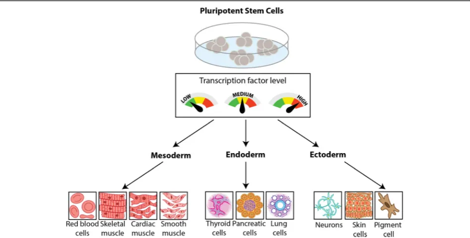
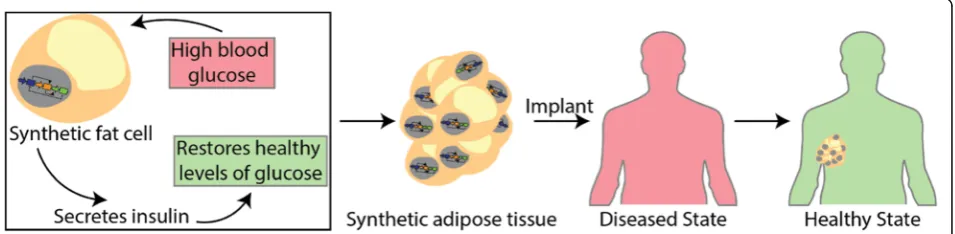
Genetic circuits to engineer tissues with
alternative functions
C. P. Healy and T. L. Deans
*Abstract
Persistent and complex problems arising with respect to human physiology and pathology have led to intense
investigation into therapies and tools that permit more targeted outcomes and biomimetic responses to
pathological conditions. A primary goal in mammalian synthetic biology is to build genetic circuits that exert fine
control over cell behavior for next-generation biomedical applications. In pursuit of this, synthetic biologists have
engineered cells endowed with genetic circuits with sensor that are capable of reacting to a variety of stimuli and
responding with targeted behavior. Here, we highlight how synthetic biology approaches are being used to
program cells with novel functions for therapeutic applications, and how they can be used in stem cells to improve
differentiation outcomes. These approaches open the possibilities for engineering synthetic tissues for employing
personalized medicine and to develop next-generation biomedical therapies.
Introduction
Synthetic biologists use bottom-up approaches to
assem-ble genetic parts into more complex gene circuits to
enable the programming of new functions into cells.
This approach combines individual gene expression
parts, or modules, that can be characterized
independ-ently and used to build novel genetic circuits by
combin-ing multiple modules that interact with each other to
perform a defined function in cells. The inception of
synthetic biology started with engineering prokaryotes
with novel functions [1
–
18], and efforts to engineer
mammalian cells soon followed. Mammalian synthetic
biology has traditionally focused on transcriptional and
post-transcriptional regulation to program cells with
new functions. These efforts include programming
feed-back [19
–
21], controlling gene expression levels [22
–
32],
implementing Boolean logic functions [33
–
39], and
tar-geting specific disease states [40
–
46]. More recent
approaches that target specific locations in the genome
using zinc finger (ZF) proteins, transcription activator-like
effectors (TALEs), and clustered regulatory interspaced
short palindromic repeats (CRISPR) coupled with a
modi-fied Cas9 protein with its nuclease activity removed
(dCas9), have been used to interrogate endogenous
transcription factors [47
–
56]. These studies have enabled
the interrogation of endogenous DNA sequences to better
understand the role of natural transcriptional networks
within cells to better understand how cells control these
networks [57,
58]. The details of these genetic tools have
been extensively reviewed elsewhere [59
–
63], therefore,
here we aim to provide a framework for using genetic
cir-cuits to design new therapies by engineering tissues with
alternative functions.
Pluripotent stem cells are cells that have the potential
to produce any cell or tissue in the body.
In the early development of complex organisms,
pluri-potent stem cells undergo specialized decision-making
in a remarkably ordered process to yield tissue patterns,
morphogenesis, and organogenesis [64
–
68]. The
under-lying mechanisms of this lineage specification process is
not fully understood. However, coordinated clusters of
transcription factors, or gene networks, have emerged as
key regulators of stem cell pluripotency and
differenti-ation. Additionally, studies have shown that
dysregula-tion of these natural gene networks contributes to the
onset of cancer and tissue degeneration, thereby
under-lying multiple types of human disease.
Stem cells can naturally direct their lineage commitment
by controlling the timing and level of expression of key
transcription factors resulting in desired differentiation
pathways [69]. Previous work to recapitulate transcription
factor expression in stem cells to drive differentiation into
© The Author(s). 2019Open AccessThis article is distributed under the terms of the Creative Commons Attribution 4.0 International License (http://creativecommons.org/licenses/by/4.0/), which permits unrestricted use, distribution, and reproduction in any medium, provided you give appropriate credit to the original author(s) and the source, provide a link to the Creative Commons license, and indicate if changes were made. The Creative Commons Public Domain Dedication waiver (http://creativecommons.org/publicdomain/zero/1.0/) applies to the data made available in this article, unless otherwise stated.
* Correspondence:[email protected]
desired lineages included the overexpression of key
tran-scription factors [70
–
73]. These studies demonstrated
improved desired differentiation outcomes, however, this
method often produces inefficient cell yields, relies on
subpopulation selection, and generates heterogeneous cell
types [74]. These challenges have recently led to efforts
from synthetic biologists to implement genetic circuits
capable of tight gene control that provide precise spatial
and temporal expression of key transcription factors in
stem cells.
In this review, we provide a framework for
implement-ing synthetic biology in stem cells to direct stem cell
differentiation into desired lineages. We detail studies
that have implemented genetic circuits in stem cells and
discuss the outcomes of these studies on the robustness
of driving stem cell fate decisions. We next consider
using synthetic biology to design artificial tissues that
are endowed with alternative functions to provide new
therapies for diseased states.
Stem cells and synthetic biology
Stem cells play an important role in the development
and regeneration of human tissues. A universal network
of endogenous transcription factors control cell fate and
continuously send and respond to physiological signals
that adjust their cell-type specific gene expression. For
example, the overexpression of the master transcription
factors Oct4, Sox2, Klf4, and c-Myc is capable of
over-riding previously made cell fate choices to convert
som-atic cell types into a pluripotent state [75
–
81]. However,
the differentiation of pluripotent precursor cells into
adult cell types requires tightly controlled spatial and
temporal gene expression dynamics of lineage-specific
master transcription factors. Stem cell differentiation
and the development of organs involves a complex
co-ordination of both intrinsic and extrinsic cues that
con-trol cell behavior. This coordination of cues is critical for
stem cells to make fate decisions and for robust tissue to
develop.
Significant efforts are currently underway to program
stem cells with genetic circuits to push their
differenti-ation
into
desired
lineages.
Implementing
genetic
circuits to dynamically control gene expression (e.g.
transcription factor expression) in stem cells is thought
to improve differentiation outcomes because these
circuits are able to replicate the dynamic gene
expres-sion patterns that are observed during development.
Recently, a new genetic circuit was constructed coupling
genetic parts from a mold,
Neorospora crassa,
and the
bacterial Lac repressor system to create an orthogonal
genetic switch to be used in mammalian cells [27]. After
confirming tunability in immortalized cell lines for proof
of concept performance, the tight gene control and
tun-ability of gene expression of this genetic switch were
demonstrated in pluripotent stem cells. These results
suggest that synthetic biologists can program stem cells
with artificial decision-making abilities that can be used
to direct stem cell fate into desired lineages. For
example, using genetic circuits that control the level and
timing of expression of multiple transcription factors, it
is possible to tune key cell fate regulators at various
differentiation checkpoints to drive the differentiation of
stem cells into one or many desired cell fates (Fig.
1).
To demonstrate the utility of using genetic circuits to
drive decision-making in cells, a two-way
communica-tion genetic circuit was engineered to mimic the natural
gene expression patterns during angiogenesis, the
forma-tion of blood vessels [82]. Cell-to-cell communicaforma-tion
was the framework for this synthetic network.
Specific-ally, sender cells were programmed with a genetic circuit
to constitutively express tryptophan synthase (TrpB
26),
an enzyme from
E. coli
, which converts indole (a
com-pound in the media) into L-tryptophan. The authors also
engineered receiver cells with a genetic circuit designed
to allow the cells to sense the secreted L-tryptophan
and, in response, turn on the expression of a reporter
gene, secreted alkaline phosphatase (SEAP). Next, the
authors used this engineered cell-cell communication
system to implement enhanced angiogenesis. During
natural angiogenesis, two transcription factors, vascular
endothelial growth factor (VEGF) and angiopoietin-1
(Ang1), function in a sequential and coordinated fashion
to produce mature blood vessels [83]. In the engineered
cell-cell communication system, the sender and receiver
cells each possessed genetic circuits that generated
output genes expressed at different times, to guide
cellu-lar differentiation and produce blood vessels. Due to the
relatively small diffusion length of nutrients into tissues,
vascularization strategies, such as angiogenesis in newly
formed tissue, will be critical to the success of
engi-neered tissues.
the differentiation of pancreatic progenitor stem cells into
mature insulin producing
β
cells [76,
84]. Recently, a
gen-etic circuit that functions as a band-pass filter was built to
dynamically control the expression of the three
master-regulator transcription factors [85]. This genetic circuit
enabled the timely coordination of the three transcription
factors, which produced a homogeneous population of cells
that demonstrated robust insulin production over cells
pro-duced using traditional growth factor and chemical based
techniques. This study emphasizes the need for the
temporal regulation of gene expression during cell fate
decisions.
In addition to controlling when key transcription factors
turn on during differentiation, a recent study has shown
that some cell fate pathways require pulsing the expression
of key transcription factors [72]. In this study, the pulsing
expression of Gata6 in human induced pluripotent stem
(iPS) cells initiated the formation of all three germ layers
giving rise to a complex three-dimensional (3D)
multicellu-lar tissue construct, or organoid, that exhibited a liver
bud-like phenotype. Without pulsing only one germ layer
formed. Genetic circuits enable fine-tuned control over the
expression of transcription factors, suggesting that the
pos-sible gene expression patterns that can be implemented
using genetic circuits are effectively limitless. Patterns
in-cluding pulsing, tuning, oscillations are all within the realm
of possibilities. Therefore, genetic circuits offer
extraordin-arily precise control over gene expression and cell fate that
will likely transform their applicability in basic science and
clinical research.
Replicating physiological functions in alternative cell
types
Precise control over the intensity, duration, and timing
of gene expression have advanced our abilities to direct
stem cell fate into desired lineages, in addition to
devel-oping organoids. Using the same genetic tools, synthetic
biologists have also created therapeutic cells that are
capable of sensing and responding to various signals in a
therapeutic fashion [43
–
45,
86
–
104]. For example, in
two separate studies, genetic circuits were used to
regu-late glucose levels in the bloodstream of diabetic mice.
In the first study, the expression and secretion of the
glucagon-like peptide 1 (GLP-1), a peptide that has the
ability to decrease blood sugar levels in the blood by
enhancing to secretion of insulin [105], was controlled
in human embryonic kidney (HEK) 293 cells using an
optogenetic-controlled genetic circuit [106]. This genetic
circuit allowed the implanted cells to detect blue light
and, in response, initiate the transcription of GLP-1,
causing blood glucose levels to fall in diabetic mice. In
the second study, Chinese hamster ovarian (CHO) cells
were engineered with a genetic circuit that produced
insulin in response to decreasing pH levels. This study
demonstrated controlled production of insulin when
environmental pH dropped below the physiological
Fig. 1Tools in synthetic biology to drive stem cell differentiation. Genetic tools built by synthetic biologists enable tight control of generange [41]. When these engineered cells were implanted
into diabetic mice, they were able to restore insulin and
glucose levels to the same level as that of healthy mice.
In addition to engineering therapeutic cells for
meta-bolic disorders, a recent study demonstrated the use of a
genetic circuit to endow HEK293 cells with the ability to
control an inflammatory response [107]. This circuit was
comprised of three basic modules to detect and respond
to inflammatory signals: a sensor to sense inflammation
signals; an amplifier with positive feedback to ensure
sustainability of the response; and an effector that
neu-tralized the inflammatory response. Therapeutic cells
that are endowed with the ability to keep the body
’
s
in-flammatory response in check are an exciting advance in
the field because these types of cells can be implanted
after surgery to prevent a prolonged inflammatory
re-sponse from hindering may proper healing and to allow
for the restoration of healthy levels of inflammatory
cytokines.
Future directions
Mammalian synthetic biologists have made great strides
in engineering novel genetic tools to tightly regulate
gene expression in various cell types. These genetic tools
have been used for directing stem cell differentiation to
produce desired cell lineages, to make organoids, and to
engineer therapeutic cells to sense and respond to
disease. With these accomplishments under our belts, it
stands to reason that synthetic biologists can engineer
implantable mini tissues, or organoids, that have been
engineered to sense and respond to disease.
Rather than trying to recreate a failing pancreas for
diabetic patients, can we engineer implantable adipose
tissue (fat) with the ability to regulate blood glucose
levels? Studies have shown that a patients
’
own fat can
be harvested and injected back into the individual
’
s
joints to help alleviate joint pain [108]. In fact, Lipogems
are an FDA approved system that are small bits of fat
removed individuals, washed, and reinjected into various
joints for those suffering from spinal conditions, joint
pain, arthritis, or rotor cut tears [109
–
112]. One can
start to image engineering personalized synthetic adipose
tissue by first making iPS cells from a patient
’
s skin cells,
and programming them with various genetic circuits:
one to drive the differentiation into adipose cells, and
the other with a program to regulate blood glucose
levels (Fig.
2). These small synthetic tissues can then be
injected under the armpit, or other unnoticeable
loca-tions, to regulate blood glucose levels over long periods
of time. Of course, once one accepts the idea of
inject-able synthetic fat tissue, it
’
s easy to imagine engineering
other synthetic organs (e.g. skin) that can function in
various ways to improve the health of an individual.
Organoids are another area where synthetic biologists
can have a significant impact. Unlike a purified tissue,
like adipose tissue, organoids are miniaturized versions
of an organ that can be isolated from organ progenitor
cells, or pluripotent stem cells, and differentiated to
form an organ-like structure. These organoids have
mul-tiple cell types that self-organize to create a structure
similar to an organ found in vivo [113]. Because multiple
cell types are in organoids, this offers opportunities to
engineer more complex interactions (e.g. synthetic
pattern formation, spatial and temporal communication
between cell types, etc.) that may be required to
recap-itulate failing organs whose function cannot be replaced
with a single tissue. For example, a synthetic organoid
can be engineered with genetic circuits that can mimic
healthy pathways to alter underlying disease states and
rewire them to restore the healthy state [114].
Conclusion
Engineered therapeutic cells that are endowed with
gen-etic circuits have the potential to transform basic science
and medicine. Using genetic circuits to tightly control
the expression of transcription factors has shown to
significantly improve differentiation outcomes. With the
improvements to controlling gene expression in cells
that continue to be built by synthetic biologists, we will
continue to push the envelope of cell engineering
possi-bilities. Altogether, these efforts will result in the rapid
and precise engineering of cells, tissues, and organoids
that will lead to transformative clinical applications.
Acknowledgements
We would like to thank the Deans Lab for lively discussions about this review.
Funding
We gratefully acknowledge the funding from the University of Utah startup funds, the National Science Foundation CAREER Program (CBET-1554017), the Office of Naval Research Young Investigator Program (N00014–16-1-3012), and the National Institute of Health Trailblazer Award (1R21EB025413–01).
Availability of data and materials
Data sharing is not applicable to this article because no datasets were generated or analyzed during the current study.
Authors’contributions
TLD conceived of and drafted the manuscript. CPH and TLD edited the manuscript and make the figures. Both authors read and approved of the final manuscript.
Ethics approval and consent to participate Not applicable.
Consent for publication Not applicable.
Competing interests
The authors declare that they have no competing interests.
Publisher
’
s Note
Springer Nature remains neutral with regard to jurisdictional claims in published maps and institutional affiliations.
Received: 28 February 2019 Accepted: 17 April 2019
References
1. Anderson JC, Clarke EJ, Arkin AP, Voigt CA. Environmentally controlled invasion of cancer cells by engineered bacteria. J Mol Biol. 2006;355:619–27. 2. Archer EJ, Robinson AB, Suel GM. Engineered E. coli that detect and
respond to gut inflammation through nitric oxide sensing. ACS Synth Biol. 2012;1:451–7.
3. Bonnet J, Yin P, Ortiz ME, Subsoontorn P, Endy D. Amplifying genetic logic gates. Science. 2013;340:599–603.
4. Brophy JA, Voigt CA. Principles of genetic circuit design. Nat Methods. 2014;11:508–20.
5. Callura JM, Cantor CR, Collins JJ. Genetic switchboard for synthetic biology applications. Proc Natl Acad Sci U S A. 2012;109:5850–5.
6. Cameron DE, Bashor CJ, Collins JJ. A brief history of synthetic biology. Nat Rev Microbiol. 2014;12:381–90.
7. Cheng AA, Lu TK. Synthetic biology: an emerging engineering discipline. Annu Rev Biomed Eng. 2012;14:155–78.
8. Daniel R, Rubens JR, Sarpeshkar R, Lu TK. Synthetic analog computation in living cells. Nature. 2013;497:619–23.
9. Elowitz MB, Leibler S. A synthetic oscillatory network of transcriptional regulators. Nature. 2000;403:335–8.
10. F. Farzadfard and T. K. Lu,“Synthetic biology. Genomically encoded analog memory with precise in vivo DNA writing in living cell populations,” Science,vol. 346, p. 1256272, Nov 14 2014.
11. Roquet N, Lu TK. Digital and analog gene circuits for biotechnology. Biotechnol J. 2014;9:597–608.
12. Siuti P, Yazbek J, Lu TK. Synthetic circuits integrating logic and memory in living cells. Nat Biotechnol. 2013;31:448–52.
13. Yang L, Nielsen AA, Fernandez-Rodriguez J, McClune CJ, Laub MT, Lu TK, et al. Permanent genetic memory with >1-byte capacity. Nat Methods. 2014;11:1261–6.
14. Gupta S, Bram EE, Weiss R. Genetically programmable pathogen sense and destroy. ACS Synth Biol. 2013;2:715–23.
15. Hwang IY, Tan MH, Koh E, Ho CL, Poh CL, Chang MW. Reprogramming microbes to be pathogen-seeking killers. ACS Synth Biol. 2014;3:228–37. 16. Saeidi N, Wong CK, Lo TM, Nguyen HX, Ling H, Leong SS, et al. Engineering
microbes to sense and eradicate Pseudomonas aeruginosa, a human pathogen. Mol Syst Biol. 2011;7:521.
17. Gardner TS, Cantor CR, Collins JJ. Construction of a genetic toggle switch in Escherichia coli. Nature. 2000;403:339–42.
18. Kohanski MA, Collins JJ. Rewiring bacteria, two components at a time. Cell. 2008;133:947–8.
19. Bloom RJ, Winkler SM, Smolke CD. A quantitative framework for the forward design of synthetic miRNA circuits. Nat Methods. 2014;11:1147–53. 20. Bloom RJ, Winkler SM, Smolke CD. Synthetic feedback control using an
RNAi-based gene-regulatory device. J Biol Eng. 2015;9:5. 21. Stapleton JA, Endo K, Fujita Y, Hayashi K, Takinoue M, Saito H, et al.
Feedback control of protein expression in mammalian cells by tunable synthetic translational inhibition. ACS Synth Biol. 2012;1:83–8.
22. Endo K, Hayashi K, Inoue T, Saito H. A versatile cis-acting inverter module for synthetic translational switches. Nat Commun. 2013;4:2393. 23. Weber W, Fussenegger M. Engineering of synthetic mammalian gene
networks. Chem Biol. 2009;16:287–97.
24. Tigges M, Marquez-Lago TT, Stelling J, Fussenegger M. A tunable synthetic mammalian oscillator. Nature. 2009;457:309–12.
25. Chen X, Wang X, Du Z, Ma Z, Yang Y. Spatiotemporal control of gene expression in mammalian cells and in mice using the LightOn system. Curr Protoc Chem Biol. 2013;5:111–29.
26. Basu S, Mehreja R, Thiberge S, Chen MT, Weiss R. Spatiotemporal control of gene expression with pulse-generating networks. Proc Natl Acad Sci U S A. 2004;101:6355–60.
27. Fitzgerald M, Gibbs C, Shimpi AA, Deans TL. Adoption of the Q transcriptional system for regulating gene expression in stem cells. ACS Synth Biol. 2017;6:2014–20.
28. Deans TL, Cantor CR, Collins JJ. A tunable genetic switch based on RNAi and repressor proteins for regulating gene expression in mammalian cells. Cell. 2007;130:363–72.
29. Andrianantoandro E, Basu S, Karig DK, Weiss R. Synthetic biology: new engineering rules for an emerging discipline. Mol Syst Biol. 2006;2:1–14. 30. Farzadfard F, Perli SD, Lu TK. Tunable and multifunctional eukaryotic
transcription factors based on CRISPR/Cas. ACS Synth Biol. 2013;2:604–13. 31. Auslander S, Fussenegger M. From gene switches to mammalian designer
cells: present and future prospects. Trends Biotechnol. 2013;31:155–68. 32. Bacchus W, Aubel D, Fussenegger M. Biomedically relevant circuit-design
strategies in mammalian synthetic biology. Mol Syst Biol. 2013;9:691. 33. Y. Liu, Y. Zeng, L. Liu, C. Zhuang, X. Fu, W. Huang, et al., "Synthesizing AND
gate genetic circuits based on CRISPR-Cas9 for identification of bladder cancer cells," Nat Commun,vol. 5, p. 5393, Nov 6 2014.
34. Miyamoto T, Razavi S, DeRose R, Inoue T. Synthesizing biomolecule-based Boolean logic gates. ACS Synth Biol. 2013;2:72–82.
35. Lohmueller JJ, Armel TZ, Silver PA. A tunable zinc finger-based framework for Boolean logic computation in mammalian cells. Nucleic Acids Res. 2012; 40:5180–7.
36. Rinaudo K, Bleris L, Maddamsetti R, Subramanian S, Weiss R, Benenson Y. A universal RNAi-based logic evaluator that operates in mammalian cells. Nat Biotechnol. 2007;25:795–801.
37. Auslander S, Auslander D, Muller M, Wieland M, Fussenegger M. Programmable single-cell mammalian biocomputers. Nature. 2012;487:123–7.
38. Purcell O, Lu TK. Synthetic analog and digital circuits for cellular computation and memory. Curr Opin Biotechnol. 2014;29C:146–55. 39. Ghosh P, Bibb LA, Hatfull GF. Two-step site selection for
serine-integrase-mediated excision: DNA-directed integrase conformation and central dinucleotide proofreading. Proc Natl Acad Sci U S A. 2008;105:3238–43.
41. Auslander D, Auslander S, Charpin-El Hamri G, Sedlmayer F, Muller M, Frey O, et al. A synthetic multifunctional mammalian pH sensor and CO2 transgene-control device. Mol Cell. 2014;55:397–408.
42. Auslander S, Auslander D, Fussenegger M. Synthetic biology-the synthesis of biology. Angew Chem Int Ed Engl. 2017;56:6396–419.
43. Auslander S, Fussenegger M. Engineering gene circuits for mammalian cell-based applications. Cold Spring Harb Perspect Biol. 2016;8:1–17.
44. Bugaj LJ, Schaffer DV. Bringing next-generation therapeutics to the clinic through synthetic biology. Curr Opin Chem Biol. 2012;16:355–61. 45. Dobrin A, Saxena P, Fussenegger M. Synthetic biology: applying biological
circuits beyond novel therapies. Integr Biol (Camb). 2015:409–30.
46. Folcher M, Fussenegger M. Synthetic biology advancing clinical applications. Curr Opin Chem Biol. 2012;16:345–54.
47. Yeo NC, Chavez A, Lance-Byrne A, Chan Y, Menn D, Milanova D, et al. An enhanced CRISPR repressor for targeted mammalian gene regulation. Nat Methods. 2018;15:611–6.
48. Gaj T, Gersbach CA, Barbas CF 3rd. ZFN, TALEN, and CRISPR/Cas-based methods for genome engineering. Trends Biotechnol.
2013;31:397–405.
49. Gilbert LA, Larson MH, Morsut L, Liu Z, Brar GA, Torres SE, et al. CRISPR-mediated modular RNA-guided regulation of transcription in eukaryotes. Cell. 2013;154:442–51.
50. Larson MH, Gilbert LA, Wang X, Lim WA, Weissman JS, Qi LS. CRISPR interference (CRISPRi) for sequence-specific control of gene expression. Nat Protoc. 2013;8:2180–96.
51. Gonzalez F, Zhu Z, Shi ZD, Lelli K, Verma N, Li QV, et al. An iCRISPR platform for rapid, multiplexable, and inducible genome editing in human pluripotent stem cells. Cell Stem Cell. 2014;15:215–26.
52. Kiani S, Beal J, Ebrahimkhani MR, Huh J, Hall RN, Xie Z, et al. CRISPR transcriptional repression devices and layered circuits in mammalian cells. Nat Methods. 2014;11:723–6.
53. Nissim L, Perli SD, Fridkin A, Perez-Pinera P, Lu TK. Multiplexed and programmable regulation of gene networks with an integrated RNA and CRISPR/Cas toolkit in human cells. Mol Cell. 2014;54:698–710.
54. Sander JD, Joung JK. CRISPR-Cas systems for editing, regulating and targeting genomes. Nat Biotechnol. 2014;32:347–55.
55. Konermann S, Brigham MD, Trevino AE, Joung J, Abudayyeh OO, Barcena C, et al. Genome-scale transcriptional activation by an engineered CRISPR-Cas9 complex. Nature. 2015;517:583–8.
56. Dai WJ, Zhu LY, Yan ZY, Xu Y, Wang QL, Lu XJ. CRISPR-Cas9 for in vivo gene therapy: promise and hurdles. Mol Ther Nucleic Acids. 2016;5:e349. 57. Khalil AS, Lu TK, Bashor CJ, Ramirez CL, Pyenson NC, Joung JK, et al. A
synthetic biology framework for programming eukaryotic transcription functions. Cell. 2012;150:647–58.
58. Rao CV. Expanding the synthetic biology toolbox: engineering orthogonal regulators of gene expression. Curr Opin Biotechnol. 2012;23:689–94. 59. T. L. Deans, "Parallel Networks: Synthetic Biology and Artificial Intelligence,"
ACM Journal on Emerging Technologies in computing systems (JETC),vol. 11, pp. 21:1–21:11, 2014.
60. Deans TL, Grainger DW, Fussenegger M. Synthetic biology: innovative approaches for pharmaceutics and drug delivery. Adv Drug Deliv Rev. 2016;105:1–2.
61. MacDonald IC, Deans TL. Tools and applications in synthetic biology. Adv Drug Deliv Rev. 2016;105:20–34.
62. Weisenberger MS, Deans TL. Bottom-up approaches in synthetic biology and biomaterials for tissue engineering applications. J Ind Microbiol Biotechnol. 2018;45:599–614.
63. Kitada T, DiAndreth B, Teague B, Weiss R. Programming gene and engineered-cell therapies with synthetic biology. Science. 2018;359. 64. Abdelalim EM, Emara MM. Advances and challenges in the
differentiation of pluripotent stem cells into pancreatic beta cells. World J Stem Cells. 2015;7:174–81.
65. Pettinato G, Wen X, Zhang N. Formation of well-defined embryoid bodies from dissociated human induced pluripotent stem cells using
microfabricated cell-repellent microwell arrays. Sci Rep. 2014;4:7402. 66. Singh VK, Kalsan M, Kumar N, Saini A, Chandra R. Induced pluripotent stem
cells: applications in regenerative medicine, disease modeling, and drug discovery. Front Cell Dev Biol. 2015;3:2.
67. Swelstad BB, Kerr CL. Current protocols in the generation of pluripotent stem cells: theoretical, methodological and clinical considerations. Stem Cells Cloning. 2009;3:13–27.
68. Deans TL, Elisseeff JH. Stem cells in musculoskeletal engineered tissue. Curr Opin Biotechnol. 2009;20:537–44.
69. Boyer LA, Lee TI, Cole MF, Johnstone SE, Levine SS, Zucker JP, et al. Core transcriptional regulatory circuitry in human embryonic stem cells. Cell. 2005;122:947–56.
70. Ferreira R, Ohneda K, Yamamoto M, Philipsen S. GATA1 function, a paradigm for transcription factors in hematopoiesis. Mol Cell Biol. 2005;25:1215–27.
71. Gutierrez L, Tsukamoto S, Suzuki M, Yamamoto-Mukai H, Yamamoto M, Philipsen S, et al. Ablation of Gata1 in adult mice results in aplastic crisis, revealing its essential role in steady-state and stress erythropoiesis. Blood. 2008;111:4375–85.
72. Guye P, Ebrahimkhani MR, Kipniss N, Velazquez JJ, Schoenfeld E, Kiani S, et al. Genetically engineering self-organization of human pluripotent stem cells into a liver bud-like tissue using Gata6. Nat Commun. 2016;7:10243. 73. Shivdasani RA, Fujiwara Y, McDevitt MA, Orkin SH. A lineage-selective knockout
establishes the critical role of transcription factor GATA-1 in megakaryocyte growth and platelet development. EMBO J. 1997;16:3965–73.
74. Wobus AM, Boheler KR. Embryonic stem cells: prospects for developmental biology and cell therapy. Physiol Rev. 2005;85:635–78.
75. Aoi T, Yae K, Nakagawa M, Ichisaka T, Okita K, Takahashi K, et al. Generation of pluripotent stem cells from adult mouse liver and stomach cells. Science. 2008;321:699–702.
76. Ariyachet C, Tovaglieri A, Xiang G, Lu J, Shah MS, Richmond CA, et al. Reprogrammed stomach tissue as a renewable source of functional beta cells for blood glucose regulation. Cell Stem Cell. 2016;18:410–21. 77. Takahashi K, Yamanaka S. Induction of pluripotent stem cells from mouse
embryonic and adult fibroblast cultures by defined factors. Cell. 2006;126:663–76.
78. Raab S, Klingenstein M, Liebau S, Linta L. A comparative view on human somatic cell sources for iPSC generation. Stem Cells Int. 2014;2014:768391. 79. Kim JS, Choi HW, Choi S, Do JT. Reprogrammed pluripotent stem cells from
somatic cells. Int J Stem Cells. 2011;4:1–8.
80. Yu J, Vodyanik MA, Smuga-Otto K, Antosiewicz-Bourget J, Frane JL, Tian S, et al. Induced pluripotent stem cell lines derived from human somatic cells. Science. 2007;318:1917–20.
81. Yamanaka S. Strategies and new developments in the generation of patient-specific pluripotent stem cells. Cell Stem Cell. 2007;1:39–49. 82. Bacchus W, Lang M, El-Baba MD, Weber W, Stelling J, Fussenegger M.
Synthetic two-way communication between mammalian cells. Nat Biotechnol. 2012;30:991–6.
83. Freeman M. Feedback control of intercellular signalling in development. Nature. 2000;408:313–9.
84. Zhou Q, Brown J, Kanarek A, Rajagopal J, Melton DA. In vivo reprogramming of adult pancreatic exocrine cells to beta-cells. Nature. 2008;455:627–32.
85. Saxena P, Heng BC, Bai P, Folcher M, Zulewski H, Fussenegger M. A programmable synthetic lineage-control network that differentiates human IPSCs into glucose-sensitive insulin-secreting beta-like cells. Nat Commun. 2016;7:11247.
86. Ye H, Charpin-El Hamri G, Zwicky K, Christen M, Folcher M, Fussenegger M. Pharmaceutically controlled designer circuit for the treatment of the metabolic syndrome. Proc Natl Acad Sci U S A. 2013;110:141–6.
87. Lienert F, Lohmueller JJ, Garg A, Silver PA. Synthetic biology in mammalian cells: next generation research tools and therapeutics. Nat Rev Mol Cell Biol. 2014;15:95–107.
88. Haellman V, Fussenegger M. Synthetic biology-toward therapeutic solutions. J Mol Biol. 2016;428(5 Pt B):945–962.
89. Higashikuni Y, Chen WC, Lu TK. Advancing therapeutic applications of synthetic gene circuits. Curr Opin Biotechnol. 2017;47:133–41. 90. Karlsson M, Weber W. Therapeutic synthetic gene networks. Curr Opin
Biotechnol. 2012;23:703–11.
91. Khalil AS, Collins JJ. Synthetic biology: applications come of age. Nat Rev Genet. 2010;11:367–79.
92. Kis Z, et al. Mammalian synthetic biology: emerging medical applications. J R Soc Interface. 2015;12(106).
93. Kojima R, Aubel D, Fussenegger M. Novel theranostic agents for next-generation personalized medicine: small molecules, nanoparticles, and engineered mammalian cells. Curr Opin Chem Biol. 2015;28:29–38. 94. Lu TK, Khalil AS, Collins JJ. Next-generation synthetic gene networks. Nat
95. Mathur M, Xiang JS, Smolke CD. Mammalian synthetic biology for studying the cell. J Cell Biol. 2017;216:73–82.
96. Schukur L, Fussenegger M. Engineering of synthetic gene circuits for (re-)balancing physiological processes in chronic diseases. Wiley Interdiscip Rev Syst Biol Med. 2016;8:402–22.
97. Slomovic S, Pardee K, Collins JJ. Synthetic biology devices for in vitro and in vivo diagnostics. Proc Natl Acad Sci U S A. 2015;112:14429–35.
98. Teixeira AP, Fussenegger M. Synthetic biology-inspired therapies for metabolic diseases. Curr Opin Biotechnol. 2017;47:59–66.
99. Xie M, Haellman V, Fussenegger M. Synthetic biology-application-oriented cell engineering. Curr Opin Biotechnol. 2016;40:139–48.
100. Ye H, Aubel D, Fussenegger M. Synthetic mammalian gene circuits for biomedical applications. Curr Opin Chem Biol. 2013;17:910–7. 101. Hennig S, Rodel G, Ostermann K. Artificial cell-cell communication as an
emerging tool in synthetic biology applications. J Biol Eng. 2015;9:13. 102. Xie M, Fussenegger M. Designing cell function: assembly of synthetic gene
circuits for cell biology applications. Nat Rev Mol Cell Biol. 2018;19:507–25. 103. Weber W, Fussenegger M. Molecular diversity--the toolbox for synthetic
gene switches and networks. Curr Opin Chem Biol. 2011;15:414–20. 104. Ye H, Fussenegger M. Synthetic therapeutic gene circuits in mammalian
cells. FEBS Lett. 2014;588:2537–44.
105. Deacon CF, Plamboeck A, Moller S, Holst JJ. GLP-1-(9-36) amide reduces blood glucose in anesthetized pigs by a mechanism that does not involve insulin secretion. Am J Physiol Endocrinol Metab. 2002;282:E873–9. 106. Ye H, Daoud-El Baba M, Peng RW, Fussenegger M. A synthetic optogenetic
transcription device enhances blood-glucose homeostasis in mice. Science. 2011;332:1565–8.
107. Smole A, Lainscek D, Bezeljak U, Horvat S, Jerala R. A synthetic mammalian therapeutic gene circuit for sensing and suppressing inflammation. Mol Ther. 2017;25:102–19.
108. Cattaneo G, De Caro A, Napoli F, Chiapale D, Trada P, Camera A. Micro-fragmented adipose tissue injection associated with arthroscopic procedures in patients with symptomatic knee osteoarthritis. BMC Musculoskelet Disord. 2018;19:176.
109. Panchal J, Malanga G, Sheinkop M. Safety and Efficacy of Percutaneous Injection of Lipogems Micro-Fractured Adipose Tissue for Osteoarthritic Knees. Am J Orthop (Belle Mead NJ). 2018;47.
110. Randelli P, Menon A, Ragone V, Creo P, Bergante S, Randelli F, et al. Lipogems product treatment increases the proliferation rate of human tendon stem cells without affecting their Stemness and differentiation capability. Stem Cells Int. 2016;2016:4373410.
111. Tremolada C, Colombo V, Ventura C. Adipose tissue and mesenchymal stem cells: state of the art and Lipogems(R) technology development. Curr Stem Cell Rep. 2016;2:304–12.
112. Tremolada C, Ricordi C, Caplan AI, Ventura C. Mesenchymal stem cells in Lipogems, a reverse story: from clinical practice to basic science. Methods Mol Biol. 2016;1416:109–22.
113. Lancaster MA, Knoblich JA. Organogenesis in a dish: modeling development and disease using organoid technologies. Science. 2014;345:1247125.