R E V I E W
Open Access
Transcranial direct current stimulation
(tDCS) for improving capacity in activities
and arm function after stroke: a network
meta-analysis of randomised controlled
trials
Bernhard Elsner
1,2*, Gert Kwakkel
3,4,5,6, Joachim Kugler
1and Jan Mehrholz
1,2,7Abstract
Background:Transcranial Direct Current Stimulation (tDCS) is an emerging approach for improving capacity in activities of daily living (ADL) and upper limb function after stroke. However, it remains unclear what type of tDCS stimulation is most effective. Our aim was to give an overview of the evidence network regarding the efficacy and safety of tDCS and to estimate the effectiveness of the different stimulation types.
Methods:We performed a systematic review of randomised trials using network meta-analysis (NMA), searching the following databases until 5 July 2016: Cochrane Central Register of Controlled Trials (CENTRAL), MEDLINE, EMBASE, CINAHL, AMED, Web of Science, and four other databases. We included studies with adult people with stroke. We compared any kind of active tDCS (anodal, cathodal, or dual, that is applying anodal and cathodal tDCS concurrently) regarding improvement of our primary outcome of ADL capacity, versus control, after stroke. PROSPERO ID: CRD42016042055.
Results:We included 26 studies with 754 participants. Our NMA showed evidence of an effect of cathodal tDCS in improving our primary outcome, that of ADL capacity (standardized mean difference, SMD = 0.42; 95% CI 0.14 to 0. 70). tDCS did not improve our secondary outcome, that of arm function, measured by the Fugl-Meyer upper extremity assessment (FM-UE). There was no difference in safety between tDCS and its control interventions, measured by the number of dropouts and adverse events.
Conclusion:Comparing different forms of tDCS shows that cathodal tDCS is the most promising treatment option to improve ADL capacity in people with stroke.
Keywords:Stroke, Recovery of function, Transcranial direct current stimulation, Review, Meta-analysis
Background
An emerging approach for enhancing neural plasticity and hence rehabilitation outcomes after stroke is non-invasive brain stimulation (NIBS). Several stimulation procedures are available, such as repetitive transcranial magnetic stimulation (rTMS) [1], transcranial direct
current stimulation (tDCS) [2–4], transcranial alternat-ing current stimulation (tACS) [5], and transcranial pulsed ultrasound (TPU) [6]. In recent years a consider-able evidence base for NIBS has emerged, especially for rTMS and tDCS.
tDCS is relatively inexpensive, easy to administer and portable, hence constituting an ideal adjuvant therapy during stroke rehabilitation. It works by applying a weak and constant direct current to the brain and has the ability to either enhance or suppress cortical excitability, with effect lasting up to several hours after the
* Correspondence:[email protected] 1
Department of Public Health, Dresden Medical School, Technical University Dresden, Fetscherstr. 74, 01307 Dresden, Germany
2Physiotherapy, SRH University of Applied Health Sciences Gera, Gera, Germany
Full list of author information is available at the end of the article
stimulation [7–9]. Hypothetically, this technique makes tDCS a potentially useful tool to modulate neuronal in-hibitory and excitatory networks of the affected and the non-affected hemisphere post stroke to enhance, for ex-ample, upper limb motor recovery [10, 11]. Three differ-ent stimulation types can be distinguished.
In anodal stimulation, the anodal electrode (+) usually is placed over the lesioned brain area and the reference electrode over the contralateral orbit [12]. This leads to subthreshold depolarization, hence promoting neural excitation [3].
In cathodal stimulation, the cathode (−) usually is placed over the non-lesioned brain area and the ref-erence electrode over the contralateral orbit [12], leading to subthreshold polarization and hence inhi-biting neural activity [3].
Dual tDCS means the simultaneous application of anodal and cathodal stimulation [13].
However, the literature does not provide clear guide-lines, not only regarding the tDCS type, but also regard-ing the electrode configuration [14], the amount of current applied and the duration of tDCS, or the ques-tion if tDCS should be applied as a standalone therapy or in combination with other treatments, like robot-assisted therapy [15].
Rationale
There is so far conflicting evidence from systematic re-views of randomised controlled trials on the effective-ness of different tDCS approaches after stroke. For example, over the past two decades more than 30 rando-mised clinical trials have investigated the effects of dif-ferent tDCS stimulation techniques for stroke, and there are 55 ongoing trials [16]. However, the resulting net-work of evidence from randomised controlled trials (RCTs) investigating different types of tDCS (i.e., anodal, cathodal or dual) as well as their comparators like sham tDCS, physical rehabilitation or pharmacological agents has not yet been analyzed in a systematic review so far.
A network meta-analysis (NMA), also known as mul-tiple treatment comparison meta-analysis or mixed treatment comparison analysis, allows for a quantitative synthesis of the evidence network. This is made possible by combining direct evidence from head-to-head com-parisons of three or more interventions within rando-mised trials with indirect evidence across randorando-mised
trials on the basis of a common comparator [17–20].
Network meta-analysis has many advantages over trad-itional pairwise meta-analysis, such as visualizing and fa-cilitating the interpretation of the wider picture of the evidence and improving understanding of the relative merits of these different types of neuromodulation when
compared to sham tDCS and/or another comparator such as exercise therapy and/or pharmacological agents [21, 22]. By borrowing strength from indirect evidence to gain certainty about all treatment comparisons, net-work meta-analysis allows comparative effects that have not been investigated directly in randomised clinical tri-als to be estimated and ranked [22, 23].
Objective
The aim of our systematic review with NMA was to give an overview of the evidence network of randomised con-trolled trials of tDCS (anodal, cathodal, or dual) for im-proving capacity in activities of daily living (ADL) and upper limb function after stroke, as well as its safety, and to estimate and rank the relative effectiveness of the different stimulation types, while taking into account po-tentially important treatment effect modifiers.
Methods
Protocol and registration
We published a study protocol, which has been regis-tered in the PROSPERO database under the ID CRD42016042055. Our protocol adheres to the PRISMA extension statement for NMA [24].
Role of the funding source
There was no funding source for this study.
Eligibility criteria
Information sources
We searched the following databases until 5 July 2016: Cochrane Central Register of Controlled Trials (CEN-TRAL; the Cochrane Library; 2016, Issue 7), MEDLINE (from 1948), EMBASE (from 1980), CINAHL (from 1982), AMED (from 1985), Web of Science (from 1899), Physiotherapy Evidence Database, Rehabdata, COM-PENDEX (from 1969) and INSPEC (from 1969). There were no language restrictions. In order to identify fur-ther published and unpublished trials, we searched trial registers and reference lists, hand-searched conference proceedings and contacted authors and equipment manufacturers.
Search
The search strategy for MEDLINE can be found in Add-itional file 1. This search strategy was adapted for the other databases.
Study selection
One review author (BE) excluded obviously irrelevant studies by reading titles and abstracts. We retrieved the full text of the remaining studies, which were then ranked by two review authors (JK, BE) as relevant, pos-sibly relevant, or irrelevant according to our inclusion criteria. Two review authors (JK, JM) decided whether the possibly relevant publications fitted the PICOS strat-egy (Patient, Intervention, Comparison, Outcome, Study type) of our research question. We excluded all trials ranked as irrelevant and tested all trials ranked as rele-vant or possibly relerele-vant for inclusion. Disagreements were resolved by discussions with all review authors.
Data collection process
Two review authors (BE, JM) independently extracted trial and summary outcome data from the trials.
Data items
We used checklists to independently assess the following items: (1) methods of random sequence generation, (2) methods of allocation concealment, (3) blinding of out-come assessors, participants and personnel, (4) use of an intention-to-treat analysis, (5) adverse effects and drop-outs, (6) important differences in prognostic factors, (7) participants (number, age, time from stroke onset to study entry), (8) comparison (details of interventions in treatment and control groups, duration of treatment and details of co-interventions in the groups) and (9) out-comes at the end of the study.
Geometry of the network
The geometry of the network characterizes the relation and precision of direct comparisons. At the level of type of intervention we analyzed what type of tDCS (anodal,
cathodal, or dual) was compared with a particular con-trol intervention. The geometry of the network was assessed by generating network graphs [26]. Each type of intervention represents a node in the network. Rando-mised comparisons between interventions are shown as links between the nodes. Multi-arm studies are indicated by colored polygons [27].
Risk of bias within individual studies
We assessed risk of bias of included studies by the Cochrane risk of bias tool at study and at outcome level [28]. The results were incorporated into our sensitivity analysis, where only studies with low risk of bias were included. We presented the results for each outcome in a separate figure.
Summary measures
Considering studies that used the same outcome meas-ure for an outcome, we calculated Mean Differences (MD) and their corresponding 95% Confidence Intervals (CI). Including studies that did not use the same out-come measure, but did measure the same underlying construct, we calculated Standardized Mean Differences (SMD) and their corresponding 95% CIs. We expected the number of dropouts and adverse events to be rare and therefore calculated the Risk Difference (RD) and corresponding 95% CIs as the summary measure. For all comparisons we generated contrast-based forest plots. We performed a relative ranking of the competing inter-ventions according to their P-scores [27]. The P-score of an intervention, which may range from 0 to 1 and, can be interpreted as the mean certainty of its superiority and thus is comparable to its surface under the cumula-tive ranking curve (SUCRA) [26, 29]. All statistical ana-lyses have been performed with the statistical software R version 3.2.2 [30], package“netmeta”version 0.8–0 [27].
Planned method of analysis
This network meta-analysis was based on a frequentist weighted least squares approach [31, 32], which is roughly equivalent to maximum likelihood estimation [29]. This approach follows the graph-theoretical meth-odology and allows incorporation of multi-arm trials [32]. The analysis is based on two assumptions: (a) inde-pendence of studies and (b) consistency of the
under-lying effects (transitivity assumption) [29]. We
considered the treatment effects to differ between the proof-of-concept trials, and therefore applied a random-effects model to estimate summary treatment random-effects, based on treatment contrasts.
Assessment of inconsistency
multivariate meta-analysis to test the homogeneity and inconsistency assumptions [33, 34]. This Q statistic con-tains one within-design Q statistic and one between-design Q statistic, the latter being calculated on the basis of a full design-by-treatment interaction random-effects model, thus embracing the concept of design inconsist-ency [34]. We assessed inconsistinconsist-ency locally (that is be-tween pairwise comparisons) using the net heat plot [34]. The net heat plot is based on a global chi-squared test for inconsistency based on the comparison of an in-consistency model and a in-consistency model [34]. It iden-tifies inconsistency between specific direct evidence in the network and provides possible drivers [34].
Risk of bias across studies
We assessed the risk of bias regarding selective reporting by comparing the published protocol of a study with the corresponding full text obtained by our electronic search.
Additional analyses
We considered allocation concealment, blinding of out-come assessor and intention-to-treat analysis to be po-tentially important effect modifiers and incorporated them in our sensitivity analysis. Although we conducted random-effects analyses regardless of the level of hetero-geneity, we defined the choice of analysis method (fixed-effect versus random-(fixed-effects model) a priori as another potentially important effect modifier, and therefore assessed the effect of heterogeneity on estimated treat-ment comparisons by visually inspecting Bland-Altman plots [29].
We also conducted a post-hoc meta-regression ana-lysis of all sham-controlled studies in order to identify low- and high-inference moderator variables (i.e., cod-ings) that may have biased outcomes of ADL capacity or arm function, as measured by UE-FM [35]. For the high-interference codings, we investigated the impact of the publication year to investigate novelty effects. For low-interference codings based on information of the trial it-self, we investigated factors such as time since stroke, electric current [mA], duration of stimulation session [min], number of stimulation sessions, electrode size [cm2], current density [mA/cm2], electric charge per
ses-sion [C], and charge density per sesses-sion [C/cm2]. We
used the simultaneous entry approach to fit the meta-regression model [36].
Results
Study selection
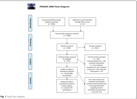
We screened 5709 unique records and assessed 176 full-text articles for eligibility. We included 30 trials with 868 participants in a qualitative analysis and 26 studies with
754 participants in a quantitative synthesis (meta-ana-lysis). Figure 1 shows the flow of studies.
Study characteristics
Twenty-four of the 30 studies (80%) were RCTs, and the remaining six studies (20%) were randomised crossover trials. The sample sizes of the included studies ranged from four [37] to 96 [38]. The mean (SD) sample size was 26 (22) with a median sample size of 20. The mean age in the experimental groups ranged from 43 to 67 years and that in the control groups from 45 to 75 years. The mean time since stroke ranged from 3 days to 8 years. The current applied ranged from 1 mA to 2 mA, while the number of stimulation sessions ranged from five to 30.
A comprehensive summary of the characteristics of the included trials examining tDCS for improving ADL capacity and upper limb function, and its safety, can be found in Additional file 2.
Presentation of network structure
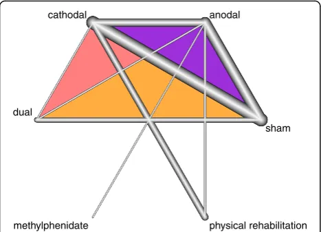
Figure 2 shows a network graph comparing anodal, cath-odal, and dual tDCS with their control interventions for improving ADL capacity after stroke. Figure 3 shows a network graph comparing anodal, cathodal, and dual tDCS with their control interventions for improving arm function (measured by the upper extremity UE-FM) after stroke. Figure 4 shows a network graph comparing tDCS with their control interventions regarding safety (measured by the number of dropouts and adverse events).
Summary of network geometry
A total of 284 patients received active tDCS to improve their ADL capacity (number of studies = 12, number of study arms = 27) [38–49]. The intervention types stud-ied were mostly cathodal tDCS (seven study arms with 167 participants) [38, 43, 44, 46, 49–51], anodal tDCS (six study arms with 88 participants) [38, 39, 43, 46, 48, 52–56], or dual tDCS (three study arms with 29 partici-pants) [40, 41, 47]. A total of 163 participants received sham tDCS as a comparator intervention (number of studies = 10) [37–41, 44–46]. Two study arms with 45 participants used physical rehabilitation comparators like virtual reality and physical therapy [44, 45].
A total of 302 patients received active tDCS to im-prove their arm function, as measured by UE-FM (num-ber of studies = 16, num(num-ber of study arms = 35) [38, 39,
43, 44, 46–57]. The intervention types studied were
Fig. 1Study flow diagram
anodal
cathodal
dual
physical rehabilitation sham
Fig. 2Network graph of tDCS for improving ADL capacity after stroke. The thicker the edge, the lower the standard error of this comparison. Colored polygons indicate multi-arm studies
anodal
cathodal
dual
physical rehabilitation sham
comparator intervention (number of study arms = 14) [38, 39, 43, 46–51, 53–57]. Two study arms with 30 par-ticipants used physical rehabilitation comparators like virtual reality and physical therapy [44, 52].
We identified 26 trials (number of study arms = 57) with 754 participants which investigated tDCS for improving ADL capacity or arm function and extracted data regard-ing the safety of tDCS (number of dropouts and adverse effects) [37–47, 49–56, 58–62]. The intervention types studied were mostly anodal tDCS (16 study arms with 174 participants) [37–39, 42, 43, 46, 48, 52–56, 58, 60–62], cathodal tDCS (11 study arms with 170 participants) [37, 38, 42–46, 49, 51, 58, 59], and dual tDCS (seven study arms with 65 participants) [40, 41, 47, 57, 58, 61, 63].
Additional file 3 shows the risk of bias within studies of tDCS for improving ADL capacity, arm function, and safety.
Additional file 4 shows a possible approach to presenting data from studies examining the effects of tDCS on ADL capacity and arm function, as well as the safety of the inter-ventions (anodal, cathodal, dual or sham tDCS, physical re-habilitation interventions and methylphenidate).
Synthesis of results
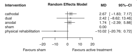
Table 1 provides a comparison of effect estimates ob-tained from the network meta-analysis with effect esti-mates obtained from direct comparisons by pairwise meta-analysis of tDCS for improving ADL capacity. Table 2 shows the ranking of the treatments by P-score, and Fig. 5 shows the forest plot of tDCS for improving ADL capacity after stroke.
Table 3 provides a comparison of effect estimates ob-tained from the network meta-analysis with effect esti-mates obtained from direct comparisons by pairwise meta-analysis for improving arm function after stroke.
Table 4 shows the ranking of treatments by P-score, and Fig. 6 shows the forest plot of tDCS for improving arm function after stroke.
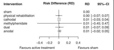
Table 5 provides a comparison of effect estimates ob-tained from the network meta-analysis with effect esti-mates obtained from direct comparisons by pairwise meta-analysis. Table 6 shows the ranking of treatments by P-score, and Fig. 7 shows the forest plot of the safety of tDCS for improving ADL capacity and arm function after stroke.
Exploration for inconsistency
Significant inconsistency, which means disagreement be-tween direct and indirect comparisons, was not ob-served. Visual inspection of the net heat plots of the three comparisons did not yield excess inconsistency, and formal testing did not detect statistically significant design inconsistency (Q = 1.29; df = 2;p= 0.52 for ADL capacity; Q = 7.3; df = 3;p= 0.06 for arm function, and Q = 0.88; df = 2;p= 0.66 for safety). The accompanying net heat plots can be found in Additional file 5.
Risk of bias across studies
We assessed the risk of bias qualitatively for each outcome by visual inspection of its risk-of-bias graph. We assessed the risk of bias regarding tDCS for improving ADL cap-acity and arm function and its safety as low to unclear. The risk-of-bias graphs can be found in Additional file 3.
anodal cathodal
dual
methylphenidate physical rehabilitation sham
Fig. 4Network graph of the safety of tDCS (measured by number of dropouts and adverse events) after stroke. The thicker the edge, the lower the standard error of this comparison. Colored polygons indicate multi-arm studies
Table 1League table for comparing network estimates with direct estimates of tDCS for improving ADL capacity
Cathodal 0.15 (−0.2; 0.5) – 0.43 (0.0; 0.8) 0.1 (−0.3; 0.4)
0.14 (−0.2; 0.5) Physical rehabilitation – – –
0.19 (−0.4; 0.8) 0.04 (−0.7; 0.8) Dual 0.23 (−0.3; 0.8) –
0.39 (0.1; 0.7) 0.25 (−0.3; 0.8) 0.25 (−0.4; 0.9) Anodal 0.13 (−0.2; 0.5)
0.42 (0.2; 0.7) 0.28 (−0.2; 0.8) 0.23 (−0.8; 0.3) 0.03 (−0.3; 0.3) Sham
Results of additional analyses
Our prespecified sensitivity analysis on the effects of methodological quality regarding our primary outcome measure (ADL capacity) included four studies with 247 participants with proper allocation concealment, blind-ing of outcome assessor and intention-to-treat analysis. Cathodal tDCS remained the most effective stimulation type (Standardized Mean Difference, SMD = 0.4; 95% CI: 0.1 to 0.7). The full results of our subgroup analysis can be found in Additional file 6.
As prespecified in our protocol, we used random-effects models for each comparison, and additionally compared them with fixed-effects models in order to as-sess the effect of heterogeneity on estimated treatment outcomes. Visual inspection of Bland-Altman plots did not yield any systematic differences between the results of fixed- and random-effects models for any of the out-comes. The results of our sensitivity analyses can be found in Additional file 6.
We could not identify any statistically significant effect moderators of tDCS for improving ADL capacity or arm function. The detailed results of our meta-regression analyses can be found in Additional file 7.
Discussion
This systematic review with a network meta-analysis in-cluded 12 randomized controlled trials with 284 partici-pants examining the effect of tDCS on our primary outcome, that of ADL capacity. We found evidence of a
significant moderate effect in favor of cathodal tDCS, whereas no significant effects were found for the other active tDCS (i.e., dual tDCS, anodal tDCS, and sham tDCS) or control interventions. Sixteen studies with 302 participants examined our secondary outcome, that of upper limb motor function as measured with Upper Ex-tremity Fugl-Meyer Motor scores (UE-FM). We found no evidence of an effect of cathodal tDCS, dual tDCS, anodal tDCS, sham tDCS, or physical rehabilitation in-terventions. Finally, our analysis of 26 trials with 754 participants found no statistically significant differences between sham tDCS, physical rehabilitation interven-tions, cathodal tDCS, methylphenidate, dual tDCS, and anodal tDCS, regarding our other secondary outcome, that of the safety of tDCS or its control interventions as revealed by the number of dropouts and adverse events.
The results of this network meta-analysis in terms of our primary outcome, ADL capacity, are in line with a recent Cochrane review examining the effects of tDCS in improving activities, arm and lower extremity func-tion, muscle strength, and cognition [16]. Due to the methodological limitations inherent in traditional pair-wise meta-analyses the authors of that review could only draw pairwise comparisons, not taking into account the existing evidence network. Furthermore, in order to avoid multiple testing, the authors had to combine treat-ment groups with different types of tDCS into a single tDCS group, thus maybe masking possible differences between different tDCS types. In a pre-specified formal subgroup analysis for their primary outcome of activities, the authors tried to estimate the treatment effects of the different tDCS types (anodal, cathodal, and dual tDCS). The analysis suggested a favorable effect of cathodal tDCS for improving ADL after stroke (SMD 0.33, 95% CI 0.10 to 0.57; six studies with 301 participants), whereas there was no effect for anodal (SMD -0.04, 95% CI -0.35 to 0.27; five studies with 164 participants) or dual tDCS (SMD 0.30, 95% CI -0.39 to 0.99; two studies with 33 participants), which is in accordance with our findings.
The relative superiority of cathodal tDCS might be due to a downregulation of the overactive non-affected brain hemisphere as a result of insufficient interhemi-spheric inhibition and with that, restoring the balance of excitatory and inhibitory interactions between both hemispheres [64–67]. From this point of view, cathodal tDCS should rather be regarded as supporting the down-regulation of overactivity of the non-lesioned
hemi-sphere. This might contrast with the model of
‘vicariation of function’[68, 69] which suggests that un-affected brain areas ‘take over’functions of the affected hemisphere [64, 69]. Recently, doubts have been raised about whether this model may be oversimplified or even incorrect and new models have been proposed, such as
Table 2treatment rankings by P-score of tDCS for improving ADL capacity
Treatment P-Score
Cathodal 0.87
Physical rehabilitation 0.62
Dual 0.57
Anodal 0.25
Sham 0.18
Treatments are listed in order of relative ranking. The P-Score, ranging from 0 to 1, describes the mean degree of certainty about a particular treatment be-ing better than another treatment
Intervention
cathodal
physical rehabilitation dual
anodal sham
−0.6 −0.2 0 0.2 0.4 0.6
Random Effects Model
Favours sham Favours active treatment
SMD
0.42 0.28 0.23 0.03 0.00
95%−CI
[ 0.15; 0.69] [−0.20; 0.75] [−0.29; 0.76] [−0.25; 0.32]
Fig. 5Forest plot of tDCS for improving ADL capacity after stroke (12 studies with 284 participants). Treatments are listed in order of relative ranking. SMD = standardized mean difference,
the bimodal balance-recovery model, which links
inter-hemispheric balancing to the brain’s remaining
struc-tural reserve [64].
The optimal stimulation paradigm, in terms of the se-lection of participants likely to benefit from tDCS, the electrode size and location, the amount of direct current applied and the duration of administration remains to be established [14, 64, 70]. Besides the above-mentioned neurophysiological explanation for the finding of super-iority of cathodal tDCS, there might also be methodo-logical reasons. For example, the majority of participants in randomised studies of tDCS for improving ADL cap-acity were treated with cathodal tDCS (167 out of 284 participants, 59%). Hence, this intervention might have the greatest statistical power in showing evidence, whereas dual tDCS was the least powered intervention.
Regarding our secondary outcome (i.e., function of the upper paretic limb), our results are in line with two sys-tematic reviews with pairwise meta-analysis. Tedesco Triccas and colleagues (2015) included genuine RCTs with multiple sessions of tDCS for improving the func-tion of the upper paretic limb after stroke [71]. They in-cluded nine studies with 371 participants. Their analysis did not reveal any statistically significant effect of active tDCS at the end of the intervention period (SMD 0.11, 95% CI -0.17 to 0.38). The other systematic review of the effects of anodal tDCS on upper extremity function and cortical excitability in people with stroke also yielded no evidence of effect (SMD 0.39, 95% CI -0.17 to 0.94) [72]. There have also been systematic reviews with
contradicting results: Butler and colleagues (2013) exam-ined the effect of anodal tDCS on upper limb motor re-covery in people with stroke and included randomised controlled trials, non-randomised trials and pre–post tri-als. Their analyses revealed a statistically significant beneficial effect of tDCS on upper limb function (SMD 0.49, 95% CI 0.18 to 0.817; seven studies with 168 partic-ipants) [73]. One reason for the discrepancy between their results and ours might be that the authors also in-cluded non-randomised studies, and that their meta-analyses suffered from multiplicity.
We found evidence of an effect of tDCS for improving ADL capacity, but not for improving arm function. Since there is only a weak association between paresis of one upper limb after stroke and ADL scores, one could argue that the improvement in ADL capacity may be not based on an improvement of the paretic arm itself, but rather on a generalized treatment effect, or on chance.
A recent systematic review with pairwise meta-regression explored several stimulation variables, like electrode size, electric current, current density, tDCS duration, number of sessions, electric charge, total elec-tric charge, and total elecelec-tric charge density [74]. The authors included ten comparisons of eight RCTs with 213 participants which measured arm function after stroke. They identified pad size, charge density, and current density as potentially relevant effect modifiers in studies measuring arm function by UE-FM, by entering each of the variables in an inverse variance-weighted
Intervention
cathodal dual anodal sham
physical rehabilitation
−20 −10 0 10 20
Random Effects Model
Favours sham Favours active treatment
MD
2.67 2.42 1.74 0.00 −10.02
95%−CI
[ −1.83; 7.17] [ −8.62; 13.46] [ −2.39; 5.88]
[−20.76; 0.72]
Fig. 6Forest plot of tDCS for improving arm function after stroke (16 studies with 302 participants). Treatments are listed in order of relative ranking. MD = mean difference [UE-FM points], CI = confidence interval. Sham is the reference category
Table 4Treatment rankings by P-score of tDCS for improving arm function
Treatment P-Score
Cathodal 0.76
Dual 0.66
Anodal 0.65
Sham 0.41
Physical rehabilitation 0.03
Treatments are listed in order of relative ranking. The P-Score, ranging from 0 to 1, describes the mean degree of certainty about a particular treatment being better than another treatment
Table 3League table for comparing network estimates with direct estimates of tDCS for improving arm function
Cathodal – −0.63 (−5.4; 4.2) 4.35 (0.6; 8.1) 3.76 (−7.9; 15.4)
0.25 (−11.7;12.2) Dual – 2.47 (−6.0; 11.0) –
0.93 (−4.0; 5.9) 0.67 (−11.1; 12.5) Anodal 1.45 (−3.5; 6.4) 19.00 (9.4; 28.6)
2.67 (−2.7; 9.0) 2.4 (−8.6; 13.5) 0.67 (−12.5; 11.1) Sham –
13.48 (−1.8; 7.2) 12.44 (−3.0; 27.8) 11.8 (1.38; 22.1) 10.02 (0.72; 20.8) Physical rehabilitation
linear meta-regression. We incorporated 19 comparisons of 16 studies with 302 participants measuring arm func-tion by UE-FM, and could not find any statistically
sig-nificant potential effect modifier. This might be
explained by our different sample as well as our different
approach to data extraction and meta-regression
analysis.
To our knowledge, our review, including 26 genuine RCTs with a total of 754 participants, is the most com-prehensive review so far of the effects and safety of tDCS regarding ADL capacity and arm function. However, our study has several limitations. These concern the level of individual studies and outcomes in the review as well as that of the review itself. At the level of individual studies, there is the concern about overestimating treatment ef-fects and safety due to unclear or sometimes even high risk of bias, and the fact that the reporting of adverse events was often unsatisfactory. However, our sensitivity analysis regarding methodological quality was in accord-ance with the results of our main analysis. Another as-pect is that there was methodological and clinical heterogeneity among the included studies regarding study type (the majority of included studies were phase II studies with rather small sample sizes, hence prone to the risk of baseline imbalance), age of the participants, time since stroke, dosage of stimulation, electrode loca-tion, base therapy (i.e., concurrent treatment) and level
of initial severity. This may be due to the fact that the optimal stimulation paradigm still has to be established, since theoretical assumptions about the interaction be-tween motor learning and tDCS-enhanced brain plasti-city are still weak. This includes the optimal electrode placement. In popular electrode settings most of the current is redirected by the skin covering the skull, hence being unable to ‘trigger’ neurons effectively [14]. Although tDCS easily could be coupled with novel tech-nologies like, for example robot-assisted training, its added value to rehabilitation outcomes has been limited so far [15]. The bimodal balance recovery model might represent a further step towards a patient-tailored ap-proach to tDCS. But if an interaction effect is assumed between motor learning (base therapy) and brain plasti-city (tDCS), tDCS should start earlier. This, however, was not supported by our data.
All clinical trials did employ a simplistic dose strategy of tDCS, assuming increased or decreased excitability of stimulated brain areas under the anodal and cathodal electrode, respectively (a detailed qualitative description of interventions can be found in Additional file 2). How-ever, recent dose-response studies suggest that anodal or cathodal tDCS follows a complex, non-linear intensity-dependent effect on neuronal networks [10, 75]. The electric fields induced by tDCS applied in current doses in humans are found not sufficient in themselves to
Table 6Treatment rankings by P-score of the safety of tDCS (measured by drop-outs and adverse events during the intervention phase)
Treatment P-Score
Sham 0.60
Physical rehabilitation 0.57
Cathodal 0.50
Methylphenidate 0.49
Dual 0.46
Anodal 0.38
Treatments are listed in order of relative ranking. The P-Score, ranging from 0 to 1, describes the mean degree of certainty about a particular treatment being better than another treatment
Intervention
sham
physical rehabilitation cathodal
methylphenidate dual
anodal
−0.4 −0.2 0 0.2 0.4
Risk Difference (RD)
Favours active treatment Favours sham
RD
0.00 0.00 0.01 0.01 0.01 0.01
95%−CI
[−0.07; 0.07] [−0.03; 0.04] [−0.45; 0.47] [−0.07; 0.09] [−0.02; 0.05]
Fig. 7Forest plot of the safety of tDCS for improving ADL capacity or arm function after stroke (26 studies with 754 participants). Treatments are listed in order of relative ranking. RD = Risk Difference, CI = confidence interval. Sham is the reference category
Table 5League table for comparing network estimates with direct estimates of tDCS for safety of tDCS
Sham – 0.00 (−0.9; 0.1) – 0.01 (−0.1; 0.1) 0.01 (−0.0; 0.1)
0.00 (−0.1; 0.1) Physical rehabilitation 0.01 (−0.1; 0.0) – – 0.00 (−0.2; 0.2)
0.00 (−0.0; 0.0) 0.00 (−0.1; 0.1) Cathodal – 0.00 (−0.7; 0.7) 0.00 (−0.1; 0.1)
0.01 (−0.5; 0.5) 0.01 (−0.5; 0.5) 0.01 (−0.5; 0.5) Methylphenidate 0.00 (−0.5; 0.5)
0.01 (−0.1; 0.1) 0.01 (−0.1; 0.1) 0.01 (−0.1; 0.1) 0.00 (−0.5; 0.5) Dual 0.00 (−0.2; 0.2)
0.01 (−0.1;0.0) 0.01 (−0.1; 0.1) 0.01 (−0.0; 0.0) 0.00 (−0.5; 0.5) 0.00 (−0.1; 0.1) Anodal
trigger spikes but rather to activate neurons at sub-threshold level [10, 64]. Current animal studies rather suggest that cathodal and anodal tDCS may respectively, introduce dendritic hyper- or depolarization of neural membranes [10, 64]. The tDCS induced polarization of membranes of the apical dendrite will differ from that of soma and basal dendrite and dependent on the direction (i.e., inward (anodal) or outward (cathodal) current [10].
In other words, the localization of hyper-and
depolarization in the cortex will differ in the same neuron dependent on its cellular composition and its position in relation with the cortex surface. As a conse-quence, there is now strong evidence that tDCS may in-duce long-term potentiation (LTP) and long-term depression (LTD) of stimulated neuronal pools [10, 64], which are fundamental for Hebbian and non-Hebbian forms of neuronal plasticity [76]. Furthermore, anodal tDCS induced LTP may enhance the secretion of brain derived nerve growth factors (BDNF) such as GAP43 [77, 78], change interneuronal activity and metabolism of glia cells [10]. The complexity of neuromodulation by tDCS suggests that a more sophisticated approach of tDCS is required to target neural networks effectively in a functional way [10].
Regarding the review level, there is the concern about violating the transitivity assumption, which means that in-cluded studies lack comparability. Violating the assump-tion of transitivity is more likely in larger treatment networks or in systematically different study conditions, like a wide variation in dates of study performance [24]. Neither of these was the case in our analyses. Although our formal analyses regarding inconsistency in the treat-ment networks were negative, this does not automatically mean that no inconsistency occurred [21]. Another point is that network meta-analyses require reasonably homoge-neous studies, which is why we restricted our analysis to the post-intervention effects of tDCS. Since stroke is often a chronic disease, future network meta-analyses could also focus on the sustainability of effects of cathodal tDCS, ac-knowledging that the number of published trials that in-cluded long-term outcomes is rather small.
Conclusions
Our network meta-analysis of randomised controlled trials suggests that cathodal tDCS is the most promising treat-ment option when tDCS is used to improve ADL capacity and arm function in people with stroke. There is evidence of an effect of cathodal tDCS in terms of improving ADL capacity. There is no evidence of an effect of either cathodal or any other tDCS stimulation type in terms of improving the function of the upper paretic limb after stroke, as mea-sured by UE-FM. No difference regarding safety (in terms of dropouts and adverse events) was seen between different types of tDCS and their control interventions.
Next to improve the methodological quality of thee proof-of-concept trials, future trials in humans need to im-prove reporting the exact dose of tDCS including the elec-trode montage (elecelec-trode size and position) allowing to replicate findings [9]. In particular, the present meta-analysis shows that tDCS trials should improve the meth-odological quality of research. In particular there is room for improvement with respect to allocation concealment, report of drop outs and accompanying intention-to-treat analyses, as well as report of adverse events and long-term outcomes post intervention.Finally, the current network meta-analysis also suggests that the scientific rigor of differ-ent types of tDCS hamper and require a better understand-ing of its underlyunderstand-ing workunderstand-ing mechanism [64].
This finding suggests that not only methodological ca-veats but also technical limitations and insufficient fun-damental knowledge about the dose-dependent working mechanism of tDCS may have influenced the current scientific rigor of this promising therapy.
Additional files
Additional file 1:Search strategy for MEDLINE. (PDF 48 kb) Additional file 2:Characteristics of included studies. (PDF 92 kb) Additional file 3:Risk of bias of included studies. (PDF 101 kb) Additional file 4:Presentation of outcomes of included studies. (PDF 90 kb) Additional file 5:Inconsistency tables and net heat plots. (PDF 70 kb) Additional file 6:Results of sensitivity analyses. (PDF 123 kb) Additional file 7:Results of post hoc meta-regression. (PDF 86 kb)
Abbreviations
95% CI:95% confidence interval; ADL: Activities of daily living; BDNF: Brain derived nerve growth factors; LTD: Long-term depression; LTP: Long-term potentiation; M1: Primary motor cortex; MD: Mean difference; NIBS: Non-invasive brain stimulation; NMA: Network meta-analysis; RD: Risk difference; rTMS: Repetitive transcranial magnetic stimulation; SMD: Standardised mean difference; SUCRA: Surface under the cumulative ranking curve;
tACS: Transcranial alternating current stimulation; tDCS: Transcranial direct current stimulation; TPU: Transcranial pulsed ultrasound; UE-FM: Upper extremity Fugl-Meyer assessment
Acknowledgements Not applicable.
Funding
There was no funding.
Availability of data and materials
The datasets supporting the conclusions of this article are included within the article and its additional files.
Authors’contributions
Ethics approval and consent to participate Not applicable.
Consent for publication Not applicable.
Competing interests
The authors declare that they have no competing interests.
Publisher’s Note
Springer Nature remains neutral with regard to jurisdictional claims in published maps and institutional affiliations.
Author details
1Department of Public Health, Dresden Medical School, Technical University Dresden, Fetscherstr. 74, 01307 Dresden, Germany.2Physiotherapy, SRH University of Applied Health Sciences Gera, Gera, Germany.3Department of Rehabilitation Medicine, VU University Medical Center, MOVE Research Institute Amsterdam, Amsterdam, The Netherlands.4Neurorehabilitation, Amsterdam Rehabilitation Research Center Reade, Amsterdam, The Netherlands.5Neuroscience Campus Amsterdam, VU University Amsterdam, Amsterdam, The Netherlands.6Department of Physical Therapy and Human Movement Sciences, Northwestern University, Evanston, IL, USA.
7
Wissenschaftliches Institut, Private Europäische Medizinische Akademie der Klinik Bavaria in Kreischa GmbH, Kreischa, Germany.
Received: 13 February 2017 Accepted: 30 August 2017
References
1. Barker AT, Jalinous R, Freeston IL. Non-invasive magnetic stimulation of human motor cortex. Lancet. 1985;1:1106–7.
2. Bindman LJ, Lippold OC, Redfearn JW. The action of brief polarizing currents on the cerebral cortex of the rat (1) during current flow and (2) in the production of long-lasting after-effects. J Physiol. 1964;172:369–82. 3. Nitsche MA, Paulus W. Excitability changes induced in the human motor cortex
by weak transcranial direct current stimulation. J Physiol. 2000;527(Pt 3):633–9. 4. Priori A, Berardelli A, Rona S, Accornero N, Manfredi M. Polarization of the
human motor cortex through the scalp. Neuroreport. 1998;9:2257–60. 5. Antal A, Boros K, Poreisz C, Chaieb L, Terney D, Paulus W. Comparatively
weak after-effects of transcranial alternating current stimulation (tACS) on cortical excitability in humans. Brain Stimul. 2008;1:97–105.
6. Tufail Y, Matyushov A, Baldwin N, Tauchmann ML, Georges J, Yoshihiro A, Tillery SI, Tyler WJ. Transcranial pulsed ultrasound stimulates intact brain circuits. Neuron. 2010;66:681–94.
7. Nitsche MA, Paulus W. Sustained excitability elevations induced by transcranial DC motor cortex stimulation in humans. Neurology. 2001;57:1899–901. 8. Nitsche MA, Nitsche MS, Klein CC, Tergau F, Rothwell JC, Paulus W. Level of
action of cathodal DC polarisation induced inhibition of the human motor cortex. Clin Neurophysiol. 2003;114:600–4.
9. Woods AJ, Antal A, Bikson M, Boggio PS, Brunoni AR, Celnik P, Cohen LG, Fregni F, Herrmann CS, Kappenman ES, et al. A technical guide to tDCS, and related non-invasive brain stimulation tools. Clin Neurophysiol. 2016;127:1031–48. 10. Jackson MP, Rahman A, Lafon B, Kronberg G, Ling D, Parra LC, Bikson M.
Animal models of transcranial direct current stimulation: methods and mechanisms. Clin Neurophysiol. 2016;127:3425–54.
11. Zimerman M, Heise KF, Hoppe J, Cohen LG, Gerloff C, Hummel FC. Modulation of training by single-session transcranial direct current stimulation to the intact motor cortex enhances motor skill acquisition of the paretic hand. Stroke. 2012;43:2185–91.
12. List J, Lesemann A, Kubke JC, Kulzow N, Schreiber SJ, Floel A. Impact of tDCS on cerebral autoregulation in aging and in patients with cerebrovascular diseases. Neurology. 2015;84:626–8.
13. Vines BW, Cerruti C, Schlaug G. Dual-hemisphere tDCS facilitates greater improvements for healthy subjects’non-dominant hand compared to uni-hemisphere stimulation. BMC Neurosci. 2008;9:103.
14. Rampersad SM, Janssen AM, Lucka F, Aydin U, Lanfer B, Lew S, Wolters CH, Stegeman DF, Oostendorp TF. Simulating transcranial direct current stimulation with a detailed anisotropic human head model. IEEE Trans Neural Syst Rehabil Eng. 2014;22:441–52.
15. Simonetti D, Zollo L, Milighetti S, Miccinilli S, Bravi M, Ranieri F, Magrone G, Guglielmelli E, Di Lazzaro V, Sterzi S. Literature review on the effects of tDCS coupled with robotic therapy in post stroke upper limb rehabilitation. Front Hum Neurosci. 2017;11:268.
16. Elsner B, Kugler J, Pohl M, Mehrholz J. Transcranial direct current stimulation (tDCS) for improving activities of daily living, and physical and cognitive functioning, in people after stroke. Cochrane Database Syst Rev. 2016;3: CD009645.
17. Ioannidis JP, Karassa FB. The need to consider the wider agenda in systematic reviews and meta-analyses: breadth, timing, and depth of the evidence. BMJ. 2010;341:c4875.
18. Bafeta A, Trinquart L, Seror R, Ravaud P. Reporting of results from network meta-analyses: methodological systematic review. BMJ. 2014;348:g1741. 19. Mills EJ, Bansback N, Ghement I, Thorlund K, Kelly S, Puhan MA. Multiple
treatment comparison meta-analyses: a step forward into complexity. Clin Epidemiol. 2011;3:193-202.
20. Li T, Puhan MA, Vedula SS, Singh S, Dickersin K. Network meta-analysis-highly attractive but more methodological research is needed. BMC Med. 2011;9:1–5. 21. Mills EJ, Thorlund K, Ioannidis JP. Demystifying trial networks and network
meta-analysis. BMJ. 2013;346:f2914.
22. Lu G, Ades AE. Combination of direct and indirect evidence in mixed treatment comparisons. Stat Med. 2004;23:3105–24.
23. Caldwell D, Ades A, Higgins J. Simultaneous comparison of multiple treatments: combining direct and indirect evidence. BMJ. 2005;331:897–900. 24. Hutton B, Salanti G, Caldwell DM, Chaimani A, Schmid CH, Cameron C,
Ioannidis JP, Straus S, Thorlund K, Jansen JP, et al. The PRISMA extension statement for reporting of systematic reviews incorporating network meta-analyses of health care interventions: checklist and explanations. Ann Intern Med. 2015;162:777–84.
25. Gandiga PC, Hummel FC, Cohen LG. Transcranial DC stimulation (tDCS): a tool for double-blind sham-controlled clinical studies in brain stimulation. Clin Neurophysiol. 2006;117:845–50.
26. Salanti G, Ades AE, Ioannidis JP. Graphical methods and numerical summaries for presenting results from multiple-treatment meta-analysis: an overview and tutorial. J Clin Epidemiol. 2011;64:163–71.
27. Rücker G, Schwarzer G, Krahn U, König J. netmeta: network meta-analysis with R. 2014:23. https://cran.r-project.org/web/packages/netmeta/netmeta. pdf. Accessed 5 Feb 2016.
28. Higgins JPT, Altman DG, JAC S. Chapter 8: assessing risk of bias in included studies. In: Higgins JPT, green S (editors). Cochrane handbook for systematic reviews of interventions version 5.1.0 [Updated march 2011]: The Cochrane Collaboration; 2011. http://handbook-5-1.cochrane.org/.
29. Schwarzer G, Carpenter JR, Rücker G. Meta-analysis with R. Heidelber: Springer; 2015.
30. R Core Team: R: a language and environment for statistical computing. R Foundation for Statistical Computing, Vienna, Austria. 2015. https://www.R-project.org/.
31. Rücker G. Network meta-analysis, electrical networks and graph theory. Res Syn Meth. 2012;3:312-24.
32. Rucker G, Schwarzer G. Reduce dimension or reduce weights? Comparing two approaches to multi-arm studies in network meta-analysis. Stat Med. 2014;33:4353–69.
33. Higgins JPT, Jackson D, Barrett JK, Lu G, Ades AE, White IR. Consistency and inconsistency in network meta-analysis: concepts and models for multi-arm studies. Res Syn Meth. 2012;3
34. Krahn U, Binder H, König J. A graphical tool for locating inconsistency in network meta-analyses. BMC Med Res Methodol. 2013;13:1–18. 35. Cooper H. Hypotheses and problems in research synthesis. New York:
Russell Sage Foundation; 2009.
36. Mundry R, Nunn C. Stepwise model fitting and statistical inference: turning noise into signal pollution. Amer Nat. 2009;173:119–23.
37. Boggio PS, Nunes A, Rigonatti SP, Nitsche MA, Pascual-Leone A, Fregni F. Repeated sessions of noninvasive brain DC stimulation is associated with motor function improvement in stroke patients. Restor Neurol Neurosci. 2007;25:123–9.
38. Hesse S, Waidner A, Mehrholz J, Tomelleri C, Pohi M, Werner C. Combined transcranial direct current stimulation and robot-assisted arm training in subacute stroke patients: an exploratory, randomized multicentertrial. Neurorehabil Neural Repair. 2011;25:838–46.
effects of tDCS combined with constraint-induced movement therapy in poststroke patients. Neurorehabil Neural Repair. 2011;9:819–29. 40. Di Lazzaro V, Dileone M, Capone F, Pellegrino G, Ranieri F, Musumeci G,
Florio L, Di Pino G, Fregni F. Immediate and late modulation of Interhemipheric imbalance with bilateral Transcranial direct current stimulation in acute stroke. Brain Stimul. 2014a;7:841–8.
41. Di Lazzaro V, Dileone M, Capone F, Pellegrino G, Ranieri F, Musumeci G, Florio L, Di Pino G, Fregni F. Immediate and late modulation of Interhemipheric imbalance with bilateral Transcranial direct current stimulation in acute stroke. Brain Stimul. 2014b;7:841–8.
42. Khedr E, Shawky O, El-Hammady D, Rothwell J, Darwish E, Mostafa O, Tohamy A. Effect of anodal versus cathodal transcranial direct current stimulation on, stroke rehabilitation: a pilot randomized controlled trial. Neurorehabil Neural Repair. 2013;27:592–601.
43. Kim D-Y, Lim J-Y, Kang EK, You DS, Oh M-K, Oh B-M, Paik N-J. Effect of transcranial direct current stimulation on motor recovery in patients with subacute stroke. Am J Phys Med Rehabil. 2010;89:879–86.
44. Lee SJ, Chun MH. Combination Transcranial direct current stimulation and virtual reality therapy for upper extremity training in patients with subacute stroke. Arch Phys Med Rehabil. 2014;95:431–8.
45. Qu YP, Wu DY, Tu XQ, Qian L, Yang YB, Geng H. Effect of transcranial direct current stimulation on relieving upper-limb spasticity after stroke. [Chinese]. Chin J Cerebrovasc Dis. 2009;6(11):586–9.
46. Rocha S, Silva E, Foerster A, Wiesiolek C, Chagas AP, Machado G, Baltar A, Monte-Silva K. The impact of transcranial direct current stimulation (tDCS) combined with modified constraint-induced movement therapy (mCIMT) on upper limb function in chronic stroke: a double-blind randomized controlled trial. Disabil Rehabil. 2016;38:653–60.
47. Straudi S, Fregni F, Martinuzzi C, Pavarelli C, Salvioli S, Basaglia N. tDCS and robotics on upper limb stroke rehabilitation: effect modification by stroke duration and type of stroke. Biomed Res Int. 2016;2016 (no pagination) 48. Tedesco Triccas L, Burridge J, Hughes A, Verheyden G, Desikan M, Rothwell
J. A double-blinded randomised controlled trial exploring the effect of anodal transcranial direct current stimulation and uni-lateral robot therapy for the impaired upper limb in sub-acute and chronic stroke. Neuro Rehabilitation. 2015;37:181–91.
49. Wu D, Qian L, Zorowitz RD, Zhang L, Qu Y, Yuan Y. Effects on decreasing upper-limb poststroke muscle tone using transcranial direct current stimulation: a randomized sham-controlled study. Arch Phys Med Rehabil. 2013;94:1–8. 50. Fusco A, Assenza F, Iosa M, Izzo S, Altavilla R, Paolucci S, Vernieri F. The
ineffective role of cathodal tDCS in enhancing the functional motor outcomes in early phase of stroke rehabilitation: an experimental trial. Biomed Res Int. 2014;2014
51. Nair DG, Renga V, Lindenberg R, Zhu L, Schlaug G. Optimizing recovery potential through simultaneous occupational therapy and non-invasive brain-stimulation using tDCS. Restor Neurol Neurosci. 2011;6:411–20. 52. Cha HK, Ji SG, Kim MK, Chang JS. Effect of Transcranial direct current
stimulation of function in patients with stroke. J Phys Ther Sci. 2014;26:363–5. 53. Rossi C, Sallustio F, Di Legge S, Stanzione P, Koch G. Transcranial direct
current stimulation of the affected hemisphere does not accelerate recovery of acute stroke patients. Eur J Neurol. 2013;20:202–4.
54. Viana RT, Laurentino GEC, Souza RJP, Fonseca JB, Silva Filho EM, Dias SN, Teixeira-Salmela LF, Monte-Silva KK. Effects of the addition of transcranial direct current stimulation to virtual reality therapy after stroke: a pilot randomized controlled trial. Neuro Rehabilitation. 2014;34:437–46. 55. Allman C, Amadi U, Winkler AM, Wilkins L, Filippini N, Kischka U, Stagg CJ,
Johansen-Berg H. Ipsilesional anodal tDCS enhances the functional benefits of rehabilitation in patients after stroke. Sci Transl Med. 2016;8:pp.330re1. 56. Sattler V, Acket B, Raposo N, Albucher J-F, Thalamas C, Loubinoux I, Chollet
F, Simonetta-Moreau M. Anodal tDCS combined with radial nerve stimulation promotes hand motor recovery in the acute phase after ischemic stroke. Neurorehabil Neural Repair. 2015;29:743–54. 57. Lindenberg R, Renga V, Zhu LL, Nair D, Schlaug G. Bihemispheric brain
stimulation facilitates motor recovery in chronic stroke patients. Neurology. 2010:2176–84.
58. Fusco A, De Angelis D, Morone G, Maglione L, Paolucci T, Bragoni M, Venturiero V. The ABC of tDCS: Effects of Anodal, Bilateral and Cathodal Montages of Transcranial Direct Current Stimulation in Patients with Stroke-A Pilot Study. Stroke Res Treat. 2013;2013:837595.
59. Fusco A, Iosa M, Venturiero V, De Angelis D, Morone G, Maglione L, Bragoni M, Coiro P, Pratesi L, Paolucci S. After vs. priming effects of anodal
transcranial direct current stimulation on upper extremity motor recovery in patients with subacute stroke. Restor Neurol Neurosci. 2014;32:301–12. 60. Mortensen J, Figlewski K, Andersen H. Combined transcranial direct current
stimulation and home-based occupational therapy for upper limb motor impairment following intracerebral hemorrhage: a double-blind randomized controlled trial. Disabil Rehabil. 2016;38:637–43.
61. Sik BY, Dursun N, Dursun E, Sade I, SahIn E. Transcranial direct current stimulation: the effects on plegic upper extremity motor function of patients with stroke. J Neurol Sci Turk. 2015;2:320–34.
62. Wang QM, Cui H, Han SJ, Black-Schaffer R, Volz MS, Lee YT, Herman S, Latif LA, Zafonte R, Fregni F. Combination of transcranial direct current stimulation and methylphenidate in subacute stroke. Neurosci Lett. 2014;569:6–11.
63. Ang KK, Guan C, Phua KS, Wang C, Teh I, Chen CW, Chew E. Transcranial direct current stimulation and EEG-based motor imagery BCI for upper limb stroke rehabilitation. Conf Proc IEEE Eng Med Biol Soc. 2012;2012:4128–31. 64. Di Pino G, Pellegrino G, Assenza G, Capone F, Ferreri F, Formica D, Ranieri F,
Tombini M, Ziemann U, Rothwell JC, Di Lazzaro V. Modulation of brain plasticity in stroke: a novel model for neurorehabilitation. Nat Rev Neurol. 2014;10:597–608.
65. Nowak DA, Grefkes C, Ameli M, Fink GR. Interhemispheric competition after stroke: brain stimulation to enhance recovery of function of the affected hand. Neurorehabil Neural Repair. 2009;23:641–56.
66. Duque J, Hummel F, Celnik P, Murase N, Mazzocchio R, Cohen LG. Transcallosal inhibition in chronic subcortical stroke. NeuroImage. 2005;28:940–6. 67. Murase N, Duque J, Mazzocchio R, Cohen LG. Influence of interhemispheric
interactions on motor function in chronic stroke. Ann Neurol. 2004;55:400–9. 68. Jaillard A, Martin CD, Garambois K, Lebas JF, Hommel M. Vicarious function
within the human primary motor cortex? A longitudinal fMRI stroke study. Brain. 2005;128:1122–38.
69. Buma F, Kwakkel G, Ramsey N. Understanding upper limb recovery after stroke. Restor Neurol Neurosci. 2013;31:707–22.
70. Floel A. tDCS-enhanced motor and cognitive function in neurological diseases. NeuroImage. 2014;85(Pt 3):934–47.
71. Tedesco Triccas L, Burridge JH, Hughes AM, Pickering RM, Desikan M, Rothwell JC, Verheyden G. Multiple sessions of transcranial direct current stimulation and upper extremity rehabilitation in stroke: a review and meta-analysis. Clin Neurophysiol. 2015a:946–55.
72. Bastani A, Jaberzadeh S. Does anodal transcranial direct current stimulation enhance excitability of the motor cortex and motor function in healthy individuals and subjects with stroke: a systematic review and meta-analysis. Clin Neurophysiol. 2012;123:644–57.
73. Butler AJ, Shuster M, O'Hara E, Hurley K, Middlebrooks D, Guilkey K. A meta-analysis of the efficacy of anodal transcranial direct current stimulation for upper limb motor recovery in stroke survivors. J Hand Ther. 2013;26:162–71. 74. Chhatbar PY, Ramakrishnan V, Kautz S, George MS, Adams RJ, Feng W.
Transcranial direct current stimulation post-stroke upper extremity motor recovery studies exhibit a dose-response relationship. Brain Stimul. 2016;9:16–26. 75. Jamil A, Batsikadze G, Kuo HI, Labruna L, Hasan A, Paulus W, Nitsche MA.
Systematic evaluation of the impact of stimulation intensity on neuroplastic after-effects induced by transcranial direct current stimulation. J Physiol. 2016;4: 1273-88.
76. Cooke SF, Bliss TV. Plasticity in the human central nervous system. Brain. 2006;129:1659–73.
77. Fritsch B, Reis J, Martinowich K, Schambra HM, Ji Y, Cohen LG, Lu B. Direct current stimulation promotes BDNF-dependent synaptic plasticity: potential implications for motor learning. Neuron. 2010;66:198–204.