R E V I E W
Open Access
Copper signalling: causes and
consequences
Julianna Kardos
1*, László Héja
1, Ágnes Simon
1, István Jablonkai
1, Richard Kovács
2and Katalin Jemnitz
1Abstract
Copper-containing enzymes perform fundamental functions by activating dioxygen (O
2) and therefore allowing chemical
energy-transfer for aerobic metabolism. The copper-dependence of O
2transport, metabolism and production
of signalling molecules are supported by molecular systems that regulate and preserve tightly-bound static
and weakly-bound dynamic cellular copper pools. Disruption of the reducing intracellular environment, characterized
by glutathione shortage and ambient Cu(II) abundance drives oxidative stress and interferes with the bidirectional,
copper-dependent communication between neurons and astrocytes, eventually leading to various brain disease forms.
A deeper understanding of of the regulatory effects of copper on neuro-glia coupling via polyamine metabolism may
reveal novel copper signalling functions and new directions for therapeutic intervention in brain disorders associated
with aberrant copper metabolism.
Keywords:
Redox disproportionation and speciation of copper, Dynamic copper pool, Copper-rich aggregates, GSH/
GSSG ratio, Copper chelate therapy, Neuro-glia coupling
Background
Copper is a generally utilized heavy metal [
1
] with a
toxic limit beyond 10
μM [
2
,
3
]. At low
concentra-tions, copper ion is an essential micronutrient that
plays a variety of functions in biological systems.
Cop-per containing enzymes and transcription factors are
essential for cellular integrity, energy production,
sig-nalling, proliferation, oxidation and radiation defence.
Research concerning acute or chronic toxicity of
cop-per due to its deficiency or excess is growing rapidly
and interest in the subject is pervasive [
4
–
12
].
Never-theless, the pertinent redox status-dependent
chela-tion [
13
–
19
] and regulatory mechanisms [
20
–
32
] are
still being elucidated.
Recently, copper-related mechanisms have been
sug-gested as therapeutic targets for important indications
such as cancer [
33
], microbial defence [
34
–
37
], chronic
lung inflammation [
38
], influenza A [
39
],
neurodegen-erative diseases including Alzheimer’s disease (AD),
Parkinson’s disease (PD) and prion disease along with
disorders linked to copper homeostasis such as Menkes
disease (MD) or Wilson’s disease (WD) [
40
–
43
].
Ele-vated copper levels in the serum and tissue of cancer
patients also suggest the involvement of copper in
tumour growth [
44
,
45
].
Our review will focus on biologically-relevant and
emer-ging features of copper-dependent processes such as
redox disproportionation, the properties of the chemical
species generated (acid-base character, ligands, geometry
etc. [
46
,
47
]), the interaction between copper and sulfur
redoxomes, the underlying redox signalling, along with
the
“dark side”
where copper metabolism has been linked
to compromised or fatal conditions [
48
–
50
].
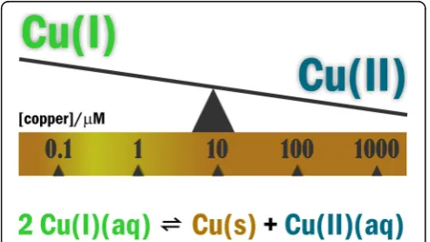
The redox capability of copper
Evidence for the incorporation of oxygen atoms from
dioxygen (O2) into oxidation products of
cuproenzyme-catalyzed reactions in nature was first published in 1955
[
51
]. Since the pioneering work of Osamu Hayaishi, and
independently Howard S. Mason a consensus has been
achieved as to the involvement of Cu(I)
disproportion-ation (redox) equilibria 2Cu(I)(aq)
Cu(0)(s) + Cu(II)(aq)
(
Eq. 1.
) in the aqueous reduction of O2
to water (
see
[
52
–
54
] and citations included). The value of + 0.37
V
relating K=Cu(II)]/[Cu(I)]
2= 10
6M
−1indicates that
aer-obic organisms can effectively utilize O2
when excess
* Correspondence:[email protected]
1Functional Pharmacology Research Group, Institute of Organic Chemistry,
Research Centre for Natural Sciences, Hungarian Academy of Sciences, Magyar Tudósok körútja 2, Budapest 1117, Hungary
Full list of author information is available at the end of the article
Cu(I) is sufficient [
46
]. This condition can be achieved
within a copper concentration range of 10
−7M
to 10
− 6M (Fig.
1.
) Within the 10
−4-10
−3M
range, however,
the reduced Cu(I) form is minimally present, which
would impair oxidative energy-transfer. The pertinent
copper-containing enzymes such as cytochrome c
oxi-dase (COX) [
55
] or copper, zinc superoxide dismutase
(Cu, Zn-SOD1) [
56
–
58
] are involved in the
mitochon-drial electron transport chain [
59
] or in the dismutation
of superoxide radical anion (O2
•-) to hydrogen peroxide
(H2O2), respectively. It is worth noting that the higher
oxidation state of copper, Cu(III) may also shape the redox
activation of the cytosolic copper pool and contributes to
hydroxylation of phenolate substrates [
60
–
62
].
Cuprous Cu(I) ion possesses both electron
“donor”
and
“acceptor”
attributes, and redox capability via the
one-electron transfer charge-disproportionation
be-tween the
“donor”
and
“acceptor”
Cu(I) yielding Cu(II)
and Cu(0). This ability of Cu(I) to disproportionate is
fundamental, not only to vital functions related to O2
transport, regulation of respiration, neuronal
differen-tiation and signal transmission [
63
,
64
], but also to the
instability of the copper ionome [
26
]. We know that
uncontrolled redox reactions of copper that can be
deleterious to life [
12
,
65
–
75
], however, here we focus
on and re-consider the controlled, redox
capability-related signalling of copper that may be important for
neurobiology.
Copper homeostasis
An evaluation of the effects of copper under normal
and pathological conditions depends on an accurate
knowledge of copper concentrations present in vivo.
In spite of this, a bewildering feature of efforts to
examine the role of copper in biological processes is
the limited data available on the relative distribution
of copper between organs, tissues, cell types and
sub-cellular compartments in mammals [
2
,
44
,
76
–
83
].
From a practical viewpoint, the lack of the information
makes it unrealistic to determine the recommended
concentration of copper in drinking water. In addition
to its biological variance, the significant differences in
copper levels that exist in habitats and diets may also
explain difficulties in determining the impact of
cop-per on biological systems [
2
]. Moreover, multiple
com-parisons of existing data are compromised by the use
of varying techniques, characteristically atomic
ab-sorption spectroscopy (AAS), flameless atomic
absorp-tion
spectroscopic
technique
(FAAS),
inductively
coupled plasma-atomic emission spectrometry
(ICP-AES) (Tables
1
and
2
) and radiotracer detection or
di-verse sample preparing protocols. Data obtained by
FAAS on brain tissue samples taken from 38 brain
re-gions of 7 males within 2
–
4 h after death showing no
macroscopic signs of disease [
77
,
78
] disclosed
signifi-cant copper concentration differences between brain
areas, grey versus white matter cells, and between
in-dividuals. Brain copper concentrations were inversely
correlated with age. It is worth noting that
measure-ments of total copper levels may not necessarily reflect
the biologically active metal pools [
84
].
Transition metals in biological tissues have been
evalu-ated by atom absorption spectroscopy or radiotracer
de-tection techniques, and more recently by the laser
ablation inductively-coupled plasma mass spectrometry
(LA-ICP-MS), secondary ion mass spectrometry, X-ray
fluorescence microscopy (XFM), X-ray absorbance
spec-troscopy (XAS), micro particle-induced X-ray emission,
and electron microscopy. Innovative imaging technologies
of transitional metals were reviewed recently [
85
–
90
]. The
recent development of recognition-based copper sensors
and reaction-based copper indicators has allowed
fluores-cence imaging of labile copper pools [
91
–
95
]. Recent
ad-vances in non-destructive analytical methods will likely
enable the assessment of copper dynamics over short,
medium or long time scales that are relevant to signalling,
metabolism and nutrition or aging.
These technologies have made possible a deeper
un-derstanding of copper dynamics and distribution.
Signifi-cant relationships regarding the levels of Ctr1, Atox1,
ATP7A/ATP7B and copper concentrations in the human
brain have been identified by the combined application
of ICP-MS spectrometry, Western blot and
immunohis-tochemistry. Copper and ATP7A levels in the
substantia
nigra
and in the
cerebellum
, respectively, have been
found to be significantly greater compared to other brain
regions [
96
]. New insights into the relative distribution
of copper among elements including P, S, Cl, K, Ca, Fe,
Zn within the
choroid plexus
(CP), ventricle system, and
surrounding brain tissue have been provided by XFI
techniques. In agreement with the known abundance of
specific metal transporters, the elemental maps indicate
that Zn, Fe and Cu are present within the CP, where the
blood-cerebrospinal fluid barrier is primarily located
[
96
–
99
]. Investigating the relationships between age,
copper levels, and regulatory genes in the neurogenesis
active sub-ventricular zone (SVZ) and CP has revealed i)
age-related increases in Cu levels in both areas; ii) an
age-related increase in MTs in SVZ, and iii) an
age-related decrease and increase in Ctr1 in SVZ and
CP, respectively [
98
]. These and past [
100
] findings
sug-gest a specific role for copper in the development of
brain tissue. The development of new imaging methods
should provide a basis for further examination of the
genuine labile copper pools, and related redox signalling
within the brain.
From atomic structure to Speciations shaping dynamic
copper Pool and Signalling
Among transition metal elements in brain, copper ranks
third only to iron and zinc in pervasiveness. Yet, its
dis-proportionation chemistry is unique due to its electronic
structure (3
s
23
p
63
d
104
s
1) characterized by small energy
differences between 3
d
and 4
s
orbitals that allows for
strong hybridization effects and electron tunneling [
101
,
102
]. The easily convertible redox states Cu(I) and
Cu(II) generate distinguishable bioligand variations
(spe-ciation). Indeed, axial symmetry distortion of Cu(II)
aquo-complexes leads to extremely fast exchange of
water (near to 10
10s
−1) [
103
,
104
]. This copper electron
transfer-coupled structural alteration of coordination at
copper sites in proteins [
105
,
106
] can be envisaged as a
molecular machine [
107
–
109
] switched on and driven
by the redox disproportionation of copper. These
mo-lecular motions permit straight energy transfer from O2
to intrinsic cellular processes, potentially supporting fast
neuronal signalling and remodelling of neuro-glia
coup-ling [
110
] within the brain.
The extremely diverse copper speciation may be
rep-resented by a collection of copper bioligands including
small ions and molecules such as sulfide ion, amino
acids like His, Cys, Met, Asp, Tyr, Thr, Gly;
neuro-transmitters such as ATP, norepinephrine [
111
];
γ-aminobutyric acid (GABA) [
112
], and constituents of
dense core vesicle cargo neurotrophins ([
113
] and
ref-erences cited) inositol phosphates (IPs) [
114
],
low-density lipoproteins (LDL) [
115
]. Redox propensity of
chelates between copper and pertinent peptides
(tri-peptide glutathione (γ-L-glutamyl-L-cysteinylglycine:
GSH) [
116
,
117
]; peptide fragments of matricellular
calcium-binding glycoprotein (secreted protein, acidic
and rich in cysteine: SPARC) Gly-His-Lys (GHK) (for
a recent review see [
118
] and proteins
(metallothio-nein, ceruloplasmin, albumin, macroglobulin,
transcu-prein [
3
,
19
,
119
–
122
]), prion protein PrP
C[
65
],
amylin [
123
]) may present specific feature of transport
and storage of copper. Likewise, many cuproproteins
with redox, or redox-with-transport functions (mono-,
di-, tetranuclear cupredoxins nitrite reductase, laccase,
Cu, Zn-SOD1, amine oxidase CuAO, galactose
oxi-dase, hemocyanin, tyrosinase, catechol oxioxi-dase, COX,
N2O reductase, menaquinol NO reductase et cetera)
[
47
],
copper-transporting
ATPases
(Cu-ATPases,
ATP7A and ATP7B) [
124
–
126
], divalent metal
trans-porter DMT1 [
127
], copper transporters and
chap-erons Ctr1, Ctr2, Atox1 and CCS [
128
,
129
], diverse
group of bacterial periplasmic copper binding proteins
(CopC) [
130
] are known. It is to note, that major
mo-lecular players of growth or metabolism DNA [
131
] or
biogenic polyamines (pAs) [
132
] also bind copper. It is
to note, that the four metal binding sites of albumin
are partially selective, transporting not only Cu(II) but
Table 1
Average concentration of copper in human organs
Sumino et al. [82] Margalioth et al. [44] Hamilton et al. [364] Yoo et al. [83] Lech & Sadlik [365] Haswell [79] Bárány et al. [76]
FAAS AAS AAS ICP-AES FAAS AAS ICP-MS
μg/g wet tissue
brain 5.1 3.10 3.32
liver 9.9 7.8 5.60 3.47
kidney 2.6 1.80 2.1 1.80 2.15
stomach 1.44 1.10
intestines 2.1 1.54
lung 1.3 0.97 1.91
spleen 1.2 0.88 1.23
heart 3.3 2.40 3.26
bile 3.60
blooda 1.2 0.97 0.85 0.99 0.95
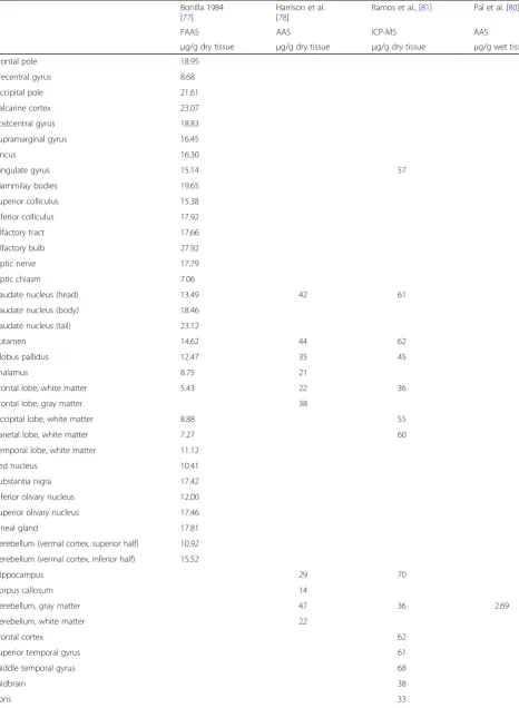
Table 2
Average concentration of copper in different brain areas
Bonilla 1984 [77]
Harrison et al. [78]
Ramos et al., [81] Pal et al. [80]
FAAS AAS ICP-MS AAS
μg/g dry tissue μg/g dry tissue μg/g dry tissue μg/g wet tissue
Frontal pole 18.95
Precentral gyrus 8.68
Occipital pole 21.61
Calcarine cortex 23.07
Postcentral gyrus 18.83
Supramarginal gyrus 16.45
Uncus 16.30
Cingulate gyrus 15.14 57
Mammilay bodies 19.65
Superior colliculus 15.38
Inferior colliculus 17.92
Olfactory tract 17.66
Olfactory bulb 27.92
Optic nerve 17.79
Optic chiasm 7.06
Caudate nucleus (head) 13.49 42 61
Caudate nucleus (body) 18.46
Caudate nucleus (tail) 23.12
Putamen 14.62 44 62
Globus pallidus 12.47 35 45
Thalamus 8.75 21
Frontal lobe, white matter 5.43 22 36
Frontal lobe, gray matter 38
Occipital lobe, white matter 8.88 55
Parietal lobe, white matter 7.27 60
Temporal lobe, white matter 11.12
Red nucleus 10.41
Substantia nigra 17.42
Inferior olivary nucleus 12.00
Superior olivary nucleus 17.46
Pineal gland 17.81
Cerebellum (vermal cortex, superior half) 10.92
Cerebellum (vermal cortex, inferior half) 15.52
Hippocampus 29 70
Corpus callosum 14
Cerebellum, gray matter 47 36 2.69
Cerebellum, white matter 22
Frontal cortex 62
Superior temporal gyrus 61
Middle temporal gyrus 68
Midbrain 38
also Zn(II), Ni(II), Cd(II), Pt(II), V(IV)O and Au(I)
[
133
]. Besides, the rather unique redox stability of
Cu(II) bound to the the
N
-terminal albumin sequence
could also be explained by the presence of the axially
coordinated water [
133
], presenting less-distorted
pyr-amidal symmetry [
103
].
Through the application of multiple complementary
approaches, two subsets of total copper can be
distin-guished: the static, tightly bound and the dynamic,
rela-tively weakly bound (labile or exchangeable) pools [
134
].
Most of the copper uptake in cells takes place through
the Ctr1, whereas ATP7A and ATP7B prevent excess
copper accumulation within cells [
125
,
126
]. The
mem-brane protein Ctr1 is considered as the major entry
pathway for copper into eukaryotic cells. Although it is
currently the sole identified transporter for copper
up-take, the existence of Ctr1-independent copper entry by
as yet unknown transporters has been suggested [
135
,
136
]. Copper entrance requires its prior reduction by
cell surface metalloreductases, as Ctr1 mediates
trans-port of Cu(I) only, whereas ceruloplasmin, which carries
half of the copper in blood plasma, delivers it as Cu(II)
to the cell membrane [
137
]. Copper uptake is regulated
mainly by Ctr1 translocation between the membrane
surface and intracellular vesicles on demand, however,
the Ctr1 protein has been shown to be degraded more
rapidly under conditions of high copper excess [
136
].
Binding events in the His- and Met-rich extracellular
amino terminal domain of vertebrate Ctr1 may support
both reduction and transfer of copper from the carriers
to the transporter [
138
]. Questions arise how
Ctr1-bound copper moves outside-in down the peptide chain
and dissociates? The human transporter is a symmetric
Ctr1 trimer shaping a cone-like pore, which becomes
wider in the outside-in direction from approximately
8
Å
to 22
Å
[
129
]. Cu(I) may traverse from the
extracel-lular binding site through the cone to the HisCysHis
motif near the intracellular carboxyl terminus of the
protein by exchanging neighbouring Met of the
con-served Met-XXX-Met Cu-binding motifs positioned
along the pore interface. Higher stability of Cu(I)-Cys
versus Cu(I)-Met could be the driving force for Cu(I)
passage [
136
]. As far as the intracellular Cu(I) discharge
pathways are concerned, Cys containing small peptides,
such as GSH, or the
“
antioxidant peptide
”
Atox1 may
contribute to Cu(I) release from the carrier. The
typic-ally high intracellular concentration of GSH (
cca
.
10 mM [
139
]) may produce shifting of the binding
equi-librium towards GSH bound Cu(I) suggesting that GSH
can efficiently collect copper [
140
,
141
] bound to the
intracellular HisCysHis binding crevice of Ctr1.
Alterna-tively, Atox1 can also pick up HisCysHis bound Cu(I)
and shuttles it to cytoplasmic metal-binding domains in
ATP7A and ATP7B (also called MD and WD proteins,
respectively) [
16
,
63
,
142
–
144
]. As suggested previously,
the fast exchange of amino acid residues surrounding
Cu(I) can readily explain entropy-compensation
phe-nomena in course of dynamic interconversion of Cu-Cys
coordinations during chaperon-target hopping [
144
].
The astonishing fact that free copper is undetectable
within cells is due to the existence of copper
chaper-ones, such as CCS which binds and transfers copper
directly to its final target Cu, Zn-SOD1 [
145
,
146
].
One can assume a novel type of protein-protein
inter-action delivering copper to its protein target
destina-tions intracellularly [
147
]. It has been suggested that
the exchange of copper between a variety of
target-specific cytosolic chaperones and their targets in
dis-tinct compartments is driven by an increase in the
copper binding affinity [
148
]. The speciation of copper
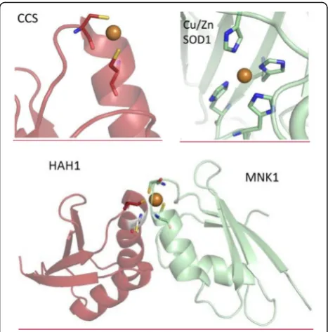
sites in the CCS chaperone for target Cu, Zn-SOD1
and in the HAH1 chaperone for the soluble cytosolic
domains (Menkes protein, MNK1) of the target
ATP7A (Fig.
2.
) [
148
–
151
] indicates, that the first
do-main of CCS, the sixth dodo-main of the MNK1 and the
HAH1 binds Cu(I) through two cysteines in a
Cys-XX-Cys motif. The Cu, Zn-SOD1 protein binds
copper via four His residues. Reportedly, the values of
the apparent dissociation constants of Cu(I) towards
chaperones and their intracellular targets may vary
mostly in the range of 0.01
pM
to 0.1
fM
[
148
]. These
estimates, however, turned to be erroneous as
demon-strated by Shoshan and co-workers [
152
]. By taking
into consideration oligomeric species of
Cu(I)-dithio-threitol, the modified calculations conclude affinity
values several orders of magnitude higher, an
observa-tion that deserves further comments. The affinities of
copper sensors and indicators associated with novel
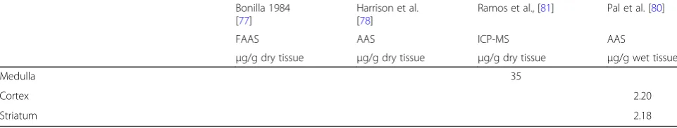
Table 2
Average concentration of copper in different brain areas
(Continued)
Bonilla 1984 [77]
Harrison et al. [78]
Ramos et al., [81] Pal et al. [80]
FAAS AAS ICP-MS AAS
μg/g dry tissue μg/g dry tissue μg/g dry tissue μg/g wet tissue
Medulla 35
Cortex 2.20
imaging technologies may not allow fluorescence imaging
of strongly bound copper in chaperones or targets, but
possibly will permit detection of chaperone-Cu(I)-target
complex formation (Fig.
2.
lower panel), as characterized
by several orders of magnitude lower affinity for the
com-plex formation equilibria MNK1 +
HAH1MNK1-Cu(I)--HAH1 (
Eq. 2.
) [
151
]. In this case, variations of AAS (total
pool) and fluorescence imaging (labile pool) data could
give rise to proper assess of the strongly bound copper
pool.
Intracellular cu(I), GSH and the concept of coupled
“
Redoxomes
”
As outlined above, intracellular copper exists in an
im-mense variety of static forms that involve a multiple
oxidation states with favoured ligand speciations (
see
below) or mixed-valent copper complexes. However, it
also can change by reacting with
“self”
(
see
Eq. 1.
)
within sub-nanometer distances of multiple copper
sites of vital peptides, proteins and enzymes [
101
,
102
,
105
,
106
,
153
–
156
]. A somewhat similar distinction can
be made for a sulfur
“redoxome”, a redox
reaction-coupled proteomic network comprising numerous
sul-fur oxidation states and species and reactions with
sulfur-containing peptides, proteins and enzymes, as
well as the reaction of GSH with
“self”
yielding
glutathi-one disulfide (GSSG) (2GSH
→
GSSG) [
157
–
159
].
Im-portantly, these
“self”-reacting copper and sulfur
“redoxomes”
also interact with each other through the
prominent chelation of Cu(I) by thiols of either the
antioxidant copper chaperone protein Atox1 or GSH
[
63
,
142
,
160
–
162
]. Supporting this concept, the GSH/
GSSG ratio was found to be the most sensitive
indica-tor of copper intoxication (and subsequent oxidative
stress) [
11
]. Moreover, sulfur-doped copper clusters are
relatively stable and abundant [
163
]. In the Cu-S-Cu
unit found within active sites of copper-sulfur proteins
like COX, the S
−bridging a Cu2
+component displays a
short Cu-Cu distance and a small Cu-S-Cu bond angle,
which are essential for the electron transport performed
by COX [
163
–
165
]. With this in mind, toxicity of
cop-per excess in mammalian cells is explained by
obstruct-ing the control of the
“interactome”
of copper-sulfur
containing
“redoxomes”
[
166
–
169
].
Because of its charge density and polarizability, the
oxi-dized cupric Cu(II) ion would tend to be found in
com-plex with
“hard”
bases such as H2O, OH
−, RNH2
etc.
(N-or O-ligands), while the
“soft”
acid Cu(I) does favour
“soft”
bases such as RS
−and CN
−ligands [
46
]. These trends in
the stability of coordination complexes [
170
,
171
] predict
that the reduced cuprous Cu(I) ion would prefer the
for-mation of complexes with
“S-ligands”
such as GSH [
172
].
Importantly, GSH can also represent
“N- or O-ligands”
for Cu(II), assisting disproportionation and O2
•−dismut-ase activity of Cu(I). Indeed, the complex equilibrium
sys-tem GSH-Cu(I) can switch to the oxidized GSSG-Cu(II)
one [
173
]. Taken together, these observations have been
used to classify the speciation of Cu(I) with GSH as a key
feature accompanying redox homeostasis [
11
].
Although the dissociation constant for the
GSH-Cu(I) equilibrium has been predicted to be about 9
pM
[
148
] (GSH-Cu(I)), this value has been called into
question [
174
,
175
]. Specifically, in an experimental
model of GSH-Cu(I), the formation of the tetranuclear
[Cu4(GS)6] cluster was observed as the major species
within the range of pH from 5.5 to 7.5 [
175
]. The
clus-ter formation equilibrium predicts that [Cu4(GS)6]
limits free Cu(I)(aq) to the sub-femtomolar
concentra-tion range in eukaryotes. These findings suggest that
the affinity of GSH towards Cu(I) may be orders of
magnitude higher than previously thought [
148
]. If
valid in vivo, not only the high intracellular GSH
con-centration but the high-affinity formation of the
[Cu4(GS)6] cluster would also force the
membrane-cytosol transfer of Cu(I) from Ctr1 to GSH. It is
note-worthy, that bacteria capture copper surplus through
the cytosolic protein Csp3s, which forms tetranuclear
Cu(I) thiolate clusters [
176
] [Cu4(S-Cys)5]
−,
[Cu4(S-Cys)6]
2−, and [Cu4(S-Cys)5(O-Asn)]
−. In order to avoid
toxicity of cytosolic copper overload, eukaryotes gain
control over excess by MTs [
177
], including the
brain-specific MT3 (growth inhibitory factor) binding.
In fact, using XRF microscopy with sub-micron
reso-lution, Sullivan et al. [
178
,
180
] demonstrated the
presence of Cu-rich aggregates in astrocytes of the
dentate gyrus and rostral migratory stream in the rat
brain. These aggregates contain CuxSy
clusters with a
sulfur/Cu(I) ratio consistent with that of the Cu-MT
complex. Apparently, both age-dependent [
98
] and
overload-evoked changes [
177
–
180
] can be related to
the copper-binding capacity of MTs.
Direct and indirect effects leading to sudden and
catastrophic hemolytic anemia due to the direct toxic
effects of copper on red blood cells has been described
in the past [
181
–
190
]. Nevertheless, the observation
that during chronic copper poisoning in sheep there is
decreased antioxidant capacity directly correlating with
the level of serum copper [
191
] putting GSH at the
centre of anti-ROS protection [
192
]. Underscoring the
importance of this role, the level of GSH in
erythro-cytes is an inheritable trait [
193
]. Unfortunately, it is
hard to obtain valid GSH and GSH/GSSG data from
biological samples [
194
].
Central regulation and storage of copper: Copper
deficiency and toxicity disorders
ATP7A and ATP7B are highly abundant in the liver, yet
disruptions in their transport functions affect the central
nervous system (CNS). This is reflected in the sex-linked
recessive CNS disorder observed in males with symptoms
of copper deficiency (MD) arising from a mutant ATP7A
pump. In contrast, a mutant ATP7B pump leads to copper
toxicity in the autosomal recessive WD. These
“brain”
dis-eases suggest that the homeostasis of copper in the liver is
essential for normal brain function [
195
–
197
]. It has been
known for a long time, that WD is characterized by the
accumulation of copper in tissues, particularly in the liver
and brain ([
198
–
201
] and references cited). The
biosyn-thesis, folding, localization, turnover and protein
inter-action network, of the most frequent copper transporter
ATP7B mutant causing toxic accumulation of copper in
WD has recently been described [
202
]. By targeting this
network
with
specific
siRNAs,
correction
of
the
localization of ATP7B-mutant restored copper levels to an
acceptable range. Decreased stability associated with
increased structural dynamics has been ascribed to
disease-causing point-mutations in the metal-binding
do-mains of WD protein [
203
]. Another fatal liver injury, the
Indian childhood cirrhosis (ICC), was also found to be
as-sociated with heavy deposits of copper, though in all other
respects it was different from WD [
204
].
Besides their significance in the overall copper efflux
and balance, ATP7A and ATP7B play a critical role in
copper transport between intracellular compartments.
In hepatocytes, ATP7A and ATP7B are located mainly
in the trans-Golgi network and supply copper for
in-corporation into copper-dependent enzymes such as
tyrosinase, peptidylglycine amidating monooxygenase,
dopamine monooxygenase, lysyl oxidase, and
cerulo-plasmin [
205
]. At high intracellular copper
concentra-tion, the carriers are translocated reversibly to the
plasma membrane (ATP7A typically to the basolateral,
ATP7B to the apical surface) where they efflux excess
copper from the cell [
206
].
In food and water, the average daily intake of copper
in the US is about 1 mg [
207
], which is relatively low.
Most humans and animals are able to control excess
amounts of copper by either decreased absorption or
in-creased excretion. Ingestion of toxic amount of copper
(> 10
mg/day
) or acute or chronic environmental
expos-ure, such as occupational hazard, accidents, release from
copper pipes, initially affects the liver, the first organ of
copper deposit. Many factors that alter copper
metabol-ism influence the progress of chronic copper poisoning.
The toxicity remains subclinical until the copper that is
stored in the liver is released in massive amounts. The
lethal dose of copper is about 10-20
g
[
207
]. Initial
symptoms of acute overdoses may be metallic taste,
gastrointestinal distress that can progress to
cardiovas-cular collapse, coma and death within hours. Hepatic
symptoms arise after 24
h
to 72
h
of exposure, and are
characterized by marked elevations in serum
amino-transferase levels, hepatic failure, elevation in
pro-thrombin time and jaundice. Erosion of epithelial lining
of the gastrointestinal tract, acute tubular necrosis in
the kidney was also reported. Blood copper
concentra-tions can increase suddenly, causing lipid peroxidation
and intravascular hemolysis [
207
,
208
].
supported by the large number of clinical syndromes
linked to either copper excess or shortage [
197
,
210
,
211
]. Several reviews have summarized results of genetic,
biochemical and structural approaches concerning
cellu-lar copper homeostasis and related disorders [
116
,
212
,
213
], yet the entire network of events that regulate
cop-per transport and intracellular disposition has not been
fully explored.
As Ctr1 cannot transport bivalent copper, some
ingested Cu(II) avoids the liver and passes rapidly into
the systemic circulation where can target albumin
[
135
]. Following entry into hepatocytes, Cu(I) binds the
initial acceptor GSH, which delivers it to the different
copper chaperones, such as Atox1, CCS and COX17
that partition copper into distinct intracellular
com-partments [
116
]. Nevertheless, the landscape of Cu(I)
trafficking to chaperons via GSH may change. Recent
data on femtomolar [
175
] versus picomolar [
148
]
affin-ities of GSH towards Cu(I) raise the role for [Cu4(GS)6]
preserving Cu(I). In fact, the Cu(I) availability is highly
associated with GSH level of the cell. Ogra et al. [
214
]
observed that depletion of GSH led to decreased copper
in the bile and blood but increased copper in the liver.
The decreased GSH level resulted in an oxidative
envir-onment in the liver that made Cu(I) less bioavailable. In
addition, the redox state of the cells influences the
ac-tivity of copper pumps. GSH deficiency inhibits ATP7A
and ATP7B resulting in the intracellular accumulation
of copper [
136
].
Synaptic release of copper
The concentration of copper in the cerebrospinal fluid
(~ 70-80
μM) is rather high in comparison to serum
(12-24
μ
M
) [
215
,
216
], raising the possibility of specific
copper signalling in the brain. As outlined in previous
sections, most cellular copper is strongly bound to
pro-teins, yet the disposition of loosely bound copper can be
detected by novel imaging technologies. This labile
cop-per pool is believed to be associated with redox
signal-ling. Labile copper has been found in the soma of
cerebellar granule and cortical pyramidal neurons, in
addition to the neuropil in the cerebellar and cerebral
cortices, hippocampus and spinal cord [
217
].
The observation on the release of zinc from brain
tis-sue during activity published in Nature in 1984 [
218
]
provided initial evidence that transition metals could be
directly involved in signalling [
112
,
134
,
219
–
229
]. Initial
evidence suggested the potential of copper to modulate
brain activity by affecting central inhibition. These
in-clude findings such as the pro-convulsant effects of a
hitherto unidentified endogenous substance containing
copper [
112
,
230
], or depolarization-induced co-release
of endogenous copper with the major inhibitory
neuro-transmitter
γ-aminobutyric acid (GABA) in different
experimental models of nerve terminals in vitro
(synap-tosomal fraction) and ex vivo (
median eminence
) [
225
].
Conclusions from
67Cu uptake and release
measure-ments performed in hypothalamic slices by the presence
of action potential blocker tetrodotoxin [
231
,
232
] or
de-termination of depolarization-induced copper release
from nerve endings by AAS [
225
] raised the concept of
copper signalling in the brain. Findings, such as the
N-methyl-
D
-aspartate
(NMDA)
receptor
activation-induced ATP7A trafficking to the plasmamembrane in
the hippocampus [
233
,
234
] have have provided new
support for a role for copper efflux in mechanisms of
excitotoxicity.
During the past 30 years, there has been a renaissance
of interest and an expanded view of the contributions of
copper to brain function and pathophysiology, as
reflected in follow-up statistics, and throughout the
lit-erature [
18
,
42
,
63
,
69
,
235
–
248
] (Fig.
3.
). One may
speculate about copper speciation and/or mechanisms of
copper uptake and release. Apart from trafficking in
complex with various neurotransmitters, carrier peptides
and proteins, or as part of the protein cargo of
extracel-lular vesicles [
39
] there is also the potential for copper
uptake as a result of autophagy [
29
,
249
].
also conlude that a copper concentration of 100
μ
M
can
exist transiently within the synaptic cleft.
Copper can diffuse out of the synapse driven by the
lower extrasynaptic concentration (1
μ
M
) [
216
].
More-over, the extrasynaptic copper concentration has been
estimated to be in the nanomolar range based on the
cellular and network excitability produced by
bath-applied copper in the CA1 area of the rat hippocampal
slice [
244
]. (This effect was primarily explained by the
ability of copper to interfere with Hodgkin–Huxley
con-ductances rather than the synaptic effects of copper
[
255
].) Using a second-generation fluorescent copper
sensor in combination with XFM, Dodani et al. [
256
]
have observed that neural activity triggers copper
traf-ficking from the cell body toward dendrites and revealed
that these copper fluxes are calcium-dependent. This
work
provided
direct
imaging
evidence
that
complemented prior studies on bulk copper release
[
225
,
232
]. Applying fluorescent copper indicators with
improved hydrophilicity Dodani et al. [
256
] identified
la-bile copper sources in the developing retina, and
dem-onstrated that they modulate spontaneous activity of
neural circuits via the copper transporter Ctr1, referred
to as a
‘
copper ion channel
’
(
see
also section
“
The
source-target-physiology
scheme
for
therapeutic
intervention
”
).
Copper Dyshomeostasis and brain disorders
Chronic copper intoxication causes region-specific
cop-per accumulation in the CNS of male Wistar rats,
fol-lowing intra-peritoneal injections of copper lactate
(0.15
mg
Cu/100
g
body weight) daily for 90 days. In
these animals, copper content, but not that of zinc or
iron was found to be significantly elevated in the cortex,
cerebellum and striatum as determined by atomic
ab-sorption spectrophotometry [
257
]. Remarkably, metal
dis-homeostasis has been widely accepted as a hallmark
of several neurodegenerative diseases, such as prion, AD,
PD, amyotrophic lateral sclerosis (ALS), Huntington’s
chorea (HC) ([
257
,
258
] and references cited). The
anti-oxidant responses to copper overloads (0–30
mg/kg
) in
rat brains showed markedly decreased brain GSH and
GSH/GSSG ratio after chronic copper exposure. Copper
overloads are characterized by a t1/2
of 9-10
h
for the
de-crease in GSH and of 4
h
for decreases in the GSH/
GSSG ratio, the latter being the most sensitive indicator
of copper excess [
11
].
Prion diseases
The mainly
α-helical folded prion protein PrP
Cis
expressed in the enteric nervous system, e.g. in enteric
nerve fibers/terminals and glia within the myenteric
sub-mucosal plexuses (inguinea pigs, mice), suggesting a role
in the regulation of ileal contractility [
259
]. Additional
beneficial roles for PrP
Cmay arise from the discovery, that
prion is an agonist at the G-protein coupled Adgrg6
re-ceptor, known to regulate demyelinization-linked
neur-opathy [
260
,
261
]. Copper has long been associated with
the formation of protease-resistant,
β-sheet enriched
“scrapie”
conformation of prion protein PrP
Sc, which has
been considered the critical step in the neurodegenerative
prion diseases known as transmissible spongiform
en-cephalopathies [
43
,
262
]. Recently, Giachin et al. [
263
]
proposed that there is a non-octarepeat copper binding
region [
264
] of PrP
Cwhich switch to the infectious PrP
Scunder acidic conditions. The only known prion disease
observed in wildlife is the chronic wasting disease (CWD).
Dietary magnesium and copper have been linked to
in-flammatory events in CWD pathogenesis [
265
].
Import-antly, geographical regions where CWD is absent have
significantly higher concentration of magnesium, and
re-gion where CDW is endemic show a higher magnesium/
copper ratio in the water. Prion diseases share
characteris-tics of
“prion-like”
neurodegenerative diseases in terms of
the involvement of proteins (α-synuclein, amyloid
β, and
tau) forming amyloid deposits [
266
].
Alzheimer
’
s disease
The metal theory of AD [
43
,
267
–
273
] (but
see
the
ad-vice of Schrag et al. [
274
]) predicts that the disregulation
of copper/zinc levels by proteins known to be involved
in AD-related neurodegeneration may lead to the
accu-mulation of amyloid fibers and oxidative stress. Indeed,
by using XFM high areal concentration of copper has
been detected in amyloid beta (Aβ) plaques of the
hippo-campal
gyrus dentatus
sub-region in a mouse model of
AD [
275
]. These data corroborate previous findings on
the high-affinity interaction between Cu(II) and the
histidine binding motif of Aβ
[
276
], along with the role
for Aβ
as a synaptic Cu(II) scavenger [
277
]. In addition,
the experimental
‘halo’
effect in copper maps may
indi-cate co-localization of copper with a
‘ring’
rich in lipids,
observed around the Aβ
plaque in AD models [
278
] and
human AD sections [
279
]. This suggests a potential
as-sociation between Cu-catalyzed oxidative stress and
plaque formation [
280
]. However, the question remains
as to whether changes in metal distribution are the cause
or the consequence of the plaque formation and
pro-gression of AD [
275
] or other progressive
neurodegener-ative diseases. For example, the neuropathology seen in
AD may also characterize individuals with Down
syn-drome [
281
,
282
], ALS or HC. By supporting a common
pathway for familial and sporadic ALS, the pathological
inclusions
containing
SOD1
fibrils
may
hold
amyloid-like properties [
283
]. Abnormal copper
accu-mulation in the striatum of HC patients has been linked
to the copper binding facilitated formation of
amyloid-copper transporter Ctr1, rlike bodies of the huntingtin
(Htt) protein [
284
,
285
]. Differential effects of ATP7A
and ATP7B regulating copper metabolism MURR1
do-main protein 1 (COMMD1) on the formation of mutant
Cu, Zn-SOD1 fibrils (increase) or parkin inclusions
(de-crease) as well as the Htt aggregates (unaltered),
how-ever, suggest mechanistic diversity [
286
].
Parkinson
’
s disease
There is evidence that alterations in copper homeostasis
play a role in PD with excess copper leading to neuronal
cell death and
α-synuclein aggregation [
121
,
287
]. It is
noteworthy in this context, that the depletion of GSH [
70
]
is a very early symptom in the course of PD [
288
].
Amyl-oid fibre formation in type-2 diabetes [
289
] may also
facili-tate PD, due to the acceleration of
α-synuclein amyloid
formation by islet amyloid polypeptide amylin [
290
].
Dis-ruption of retromer, a conserved heterotrimeric protein
complex consisting of VPS35, VPS29 and VPS26, has been
observed in a number of diseases including PD [
291
],
resulting in disregulation of the retrieval and recycling of
vital proteins [
292
]. Furthermore, the mutation of VPS35
increases copper toxicity in yeast, a likely outcome of the
copper transporter miss-trafficking [
293
]. In fact, the
endosomal retrieval and recycling of the copper
trans-porter ATP7A is retromer-dependent in human cells
[
294
]. Protecting the cargo of regulatory membrane
pro-teins such as copper transporters and pumps via the
retro-mer shipment may be critical in age-related health. It will
be important to consider further the link between
retro-mer complexes and copper homeostasis.
Multiple sclerosis
identified in the sera of patients suffering from the
chronic inflammatory CNS disorder multiple sclerosis
(MS). Feeding the copper chelator
bis-cyclohexanone-oxalyldihydrazone (cuprizone, CTZ) reduces the
mye-lin sheath and activates microglial and astroglial cells
in the CNS, providing a reproducible and reversible
model of pathologic processes underlying white and
gray matter demyelination [
296
–
301
]. Expression of
Kir4.1 autoantigen has been studied in the brain of
CTZ-fed mice and revealed the induction of Kir4.1
protein in microvessels of the cerebral cortex [
302
].
The antioxidant functions of MTs [
303
] may have a
role in MS, as suggested by the elevated level of MTs
induced by CTZ in astroglia, while the oligodendroglia
express low levels of MTs, which may contribute to
their oxidative stress vulnerability [
304
,
305
]. Apart
from MS modelling, several lines of evidence suggest
that CTZ intoxication is an excellent paradigm to
study pathology and/or therapy of epilepsy or
schizo-phrenia as well. However, mechanistic clues claiming
either copper deficiency or copper build-up
(associ-ated with hydrazide formation-dependent enzyme
in-hibition) remain contradictory [
306
].
Chelate therapy
The restoration of copper homeostasis is mostly relevant
to WD [
119
,
307
], although neurodegenerative ([
308
–
312
], but see [
313
] versus [
314
]) or inflammatory [
38
,
208
] diseases can also be related. Before the disease
pro-gresses to liver and brain (WD) or lung (inflammation),
the excess copper can be limited by Cu(II) reduction,
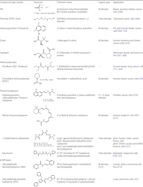
Zn(II) addition and administration of Cu(II) chelating
li-gands such as tetrathiomolybdate (TM), triethylene
tet-ramine (Trientine, TETA, Trien) or D-penicillamine
(D-pen) [
119
,
207
] for limiting excess copper.
Due to its high level in proliferating tissues, copper
can also promote angiogenesis and cancer development
[
315
]. Hence, the Cu(II) lowering therapy has also
po-tential in treating cancer (breast, colorectal, leukemia,
lung, prostate) by copper chelating compounds (Table
3
).
A range of targets and/or mechanisms of action have
been suggested for the antiproliferative activity of the
Cu(II) chelate forming compounds. These include
prote-asome inhibitors and apoptosis inducers [
316
], DNA and
protein interactions [
317
,
318
], ROS generation [
319
],
oxidative stress [
320
], integrin
β4 up-regulation [
321
],
Schiff base copper complex formation [
318
,
322
]. (For a
comprehensive knowledge of copper ion complexes as
anticancer agents we refer to reviews [
323
,
324
]).
Considering the redox activity of potential anticancer
Cu(II) chelates (Table
3
and references cited) [
323
–
327
]
one possible consequence is that the high level of copper
in proliferating tissue could also reduce oxidative redox
potential which may in turn increase cancer cell
proliferation [
45
,
50
,
328
,
329
]. The redox imbalance
could be targeted by chelate formation coupled Cu(II)
reduction in the proliferating tissue. Indeed, the
revers-ible one-electron reduction of Cu(II) does occur, as
ex-emplified by the thiosemicarbazone complex of Cu(II) in
the elesclomol [
330
,
331
] (Table
3
).
It is interesting to note the effect of metformin [
50
,
332
–
334
], which is a first line diabetes II drug and has
been shown to increase healthy life span irrespective of
its anti-diabetes effect. Its copper chelating ability [
335
]
may suggest an anti-aging role for copper.
The source-target-physiology scheme for therapeutic
intervention
The advent of imaging techniques that gained insight
into the dynamics of labile copper pool made it possible
to look beyond the molecular-level interactions in
cop-per homeostasis and examine network-level dynamic
in-terplays shaping copper signalling. The source-target-n
physiology (STP) scheme suggested by Chang [
336
]
includes labile, neuronal copper pools in the Golgi
com-partment as a source, signal propagation via postsynaptic
membrane receptor/ion channel target (the Cu(I)
trans-porter Ctr1), and copper-dependent spontaneous activity
of the neural network (physiology). Vesicular storage of
canonical neurotransmitters with copper suggesting
their co-release has also been described. Furthering the
interactions between compartments within neurons, we
conceive that cellular-level copper signalling between
neurons and astrocytes, an emerging cell type of the
brain, also exists and may play a fundamental part in
the brain’s information processing.
Table 3
Copper chelating compounds with anticancer activities
Compound type (name) Structure Chemical name Ligand type Application
TM Ammonium tetra-thiomolybdate
Bis-Choline tetrathio molybdate
Bi-dentate Breast, prostate, kidney cancer cells [208]
Trientine (TETA, Trien) N,N′-Bis(2-aminoethyl) ethane-1,2-diamine
Tetra-dentate Colorectal cancer cells [366]
Hydroxyquinoline (Clioquinol) 5-Chloro-7-iodo-8-hydroxy quinoline Bi-dentate AD and human breast cancer cells [309,316]
D-pen 3-Mercapto-D-valine Bi-dentate Human leukemia and breast
cancer cells [319]
Captopril
D-3-Mercapto-2-methyl-propionyl-L-proline
– Mammary ductal carcinoma cell line [367,368]
Dithiocarbamates
Disulfiram (DSF, Antabuse) 1-(Diethylthio-carbamoyl-disulfanyl)-N,N-diethyl-methane-thioamide
– Human breast, lung cancer cells [315,369]
Pyrrrolidine dithiocarbamate (PDTC)
Pyrrolidine-1-carbodithioic acid Bi-dentate Human breast cancer cells [344]
Thiosemicarbazone
Hydroxyquinoline-carboxaldehyde– Thiosemi-carbazone
8-Hydroxy-quinoline-2-carbox-aldehyde– thio-semicarbazone
R = H tetra-dentate
Prostate cancer cells [370]
Retinal thiosemicarbazone 9-cis-Retinal thiosemi-carbazone Bi-dentate Human leukemic cell U937 [317]
1,2-Bis(thiosemi-carbazones) H2gts: glyoxal-bis(thiosemi-carbazone)
atsm: diacetyl-bis(4-methylthio-semi-carbazone)
ptsm: pyruvaldehyde-bis(4-methylthio-semicarbazone)
Tetra-dentate atsm: human colon cancer tumor cells
ptsm: Ehrlich ascites and EMT6 tumour cells [371]
Elesclomol N’1,N’3-Dimethyl-N’1,N’3 -bis(phenyl-carbonothioyl)propanedihydrazide
Tetra-dentate Metastatic melanoma cells [320,331]
Schiff-bases
Salicylaldehyde-benzoylhydrazone (SBH)
N′-[(2-Hydroxyphenyl) methylidene] benzohydrazide
Bi-dentate [372]
Human adeno-carcinoma cell line [373]
Salicylaldehyde-pyrazole-hydrazone (SPH)
(E)- N′ -(2-Hydroxy-benzylidene)-1-benzyl-3-phenyl-1H-pyrazole-5-carbohydrazide
233
,
234
,
236
–
243
,
245
,
254
,
255
,
336
,
339
,
340
,
342
–
362
]. GABA can be synthesized from the pA
putres-cine
by
copper-containing
CuAO
in
astrocytes.
CuAOs perform the oxidation of primary amines such
as spermine, spermidine and putrescine to aldehydes
and ammonia, producing H2O2
as a by-product.
Putrescine-derived GABA is released by the inside-out
(reverse) action of glial GABA transporter subtypes.
The increased GABA release and the generated tonic
inhibition thereby modulate the power of gamma
range oscillation in the CA1 region in vivo. The
con-centration of cytosolic and extracellular putrescine has
been determined to be 22
nmol/g
and 12
nmol/g
,
re-spectively [
339
]. In contrast, copper may decrease
tonic inhibition via acting on delta subunit-containing
extrasynaptic GABAA
receptors [
235
,
237
,
246
], thus
adding a new layer to disinhibitory mechanisms in
copper-rich brain areas.
Conclusions
Despite multifaceted roles for copper observed in
vari-ous brain diseases and tumours, the copper signalling
theme is still in its initial stages. However, our
increas-ing understandincreas-ing of dynamic copper pools supports
the idea of neuronal activity-dependent Cu(I)
trans-mission affecting astroglia network signaling and
astroglia-neuron metabolic cooperation. Rather than
simply reflecting copper excess, copper-rich
aggre-gates - likely in astrocytes and not in neurons - are a
sign of a disturbed network. Brain diseases linked to
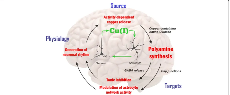
Fig. 4Copper signaling via neuro-glia coupling. Astroglia, a previously neglected cell type of the brain [340], operate a variety of copper-dependent metabolic functions [6,80,240,341,342]. For this reason, in addition to synaptic and extrasynaptic copper signalling by way of excitatory/ inhibitory receptors and ionic channels [22,234,235,237–244,246,255,336,345–355], we place copper-dependent production of pAs in astrocytes [338] and correlated gap-junction modulation in the centre of this option. The proposed scheme conjectures activity-dependent changes of copper pools [179,180] and polyamines (pAs), produced by CuAOs in astrocytes. First, an enhanced gap junction communication can be achieved by pAs [356–358], possibly promoting activity-dependent synchronization [339,359]. Second, major inhibitory neurotransmitter gamma-aminobutyric acid (GABA) formed from pAs is released by astrocyte-specific GABA transporter [360]. Acting on its extrasynaptic receptor, GABA elevates tonic inhibition and enhances the fast (gamma band) neural oscillations [360]. These ways, the steady-state pA level in astrocytes determined by copper-dependent forming and consuming can be associated with neural circuit activity [244,255,362]
Table 3
Copper chelating compounds with anticancer activities
(Continued)
Pyridine-carboxaldehyde-phenylpyrimidyl-hydrazone (Pyimpy)
1-Phenyl-1-(pyridin-2-yl)-2-(pyridin-2-ylmethylene)hydrazine
Tri-dentate Rat breast tumor cells [322]
Hydroxy naphthaldehyde imine (HL)
1-(((2-((2-Hydroxy-propyl)amino) ethyl)imino) methyl) naphthalene-2-ol)
oxidative stress [
363
] change the GSH/GSSG ratio and
thereby automatically affect the copper homeostasis, as
GSH is the immediate partner, along with various
chaper-ones, that takes Cu(I) from the transporter. Therefore,
Cu(I) distribution is disturbed and might in turn enhance
oxidative stress at copper-containing deposits or limit Cu,
Zn-SOD1 activity in regions with decreased copper level.
Closer understanding of copper signalling and its
vulner-ability opens up new perspectives improving chelate
ther-apy approaches against brain diseases and tumour.
Abbreviations
AAS:Atomic absorption spectroscopy; AD: Alzheimer’s disease; ALS: Amyotrophic lateral sclerosis; Atox1, CCS, HAH1, COX17: Copper chaperones; ATP7A, ATP7B, Cu-ATPases: Copper-transporting P-type ATPases; Aβ: Amyloid beta; CNS: Central nervous system; CopC: Bacterial peripasmic copper binding proteins; COX, mitochondrial complex IV: Cytochrome c oxidase (EC 1.9.3.1); CP:Choroid plexus; Ctr1, Ctr2: Copper(I) transporters; CTZ: cuprizone (bis-cyclohexanoneoxalyl-dihydrazone); Cu, Zn-SOD1: Copper, zinc superoxide dismutase; CuAO: Copper aminooxidase; CWD: Chronic wasting disease; DMT1: Divalent metal transporter; D-pen:D-penicillamine; EM: Electron microscopy; FAAS: Flameless atomic absorption spectroscopy; GABA: Gamma-aminobutyric acid; GHK: SPARC fragment Gly-His-Lys; GSH: Gluthation (gamma-L-glutamyl-L-cysteinylglycine); GSSG:: Gluthation disulfide; HC: Huntington’s chorea; Htt: Huntingtin protein; ICC: Indian childhood cirrhosis; ICP-AES: Inductively coupled plasma - atomic emission spectrometry; IPs: Inositol phosphates; Kir4.1: Inwardly rectifying K+ion channel 4.1;; LA-ICP-MS: Laser ablation - inductively coupled plasma - mass spectrometry; LDL: Low-density lipoproteins; MD: Menkes disease; MNK1: Menkes protein (soluble cytosolic ATP7A domain); MT: Metallothionein;μ -PIXE: Micro - particle induced X ray emission; MS: Multiple sclerosis; COMMD1: MURR1 domain protein 1; NMDA: N-methyl-D-aspartate; PD: Parkinson’s disease; pAs: Polyamines; PrPC: Prion protein,αhelical (Adgrg6 receptor agonist); PrPSc: Prion protein,βsheet enriched (“scrapie”); SPARC: Secreted protein, acidic and rich in cysteine; STP: Source-target-physiology; SVZ: Sub-ventricular zone; TSPP: Tetrakis-(4-sulfophenyl)-porphine; TM: Tetrathiomolybdate; Trientine: TETA, Trien (Triethylene tetramine); WD: Wilson’s disease; XFM: X-ray fluorescence microscopy
Acknowledgements
We do thank Susan Amara for her valuable comments and criticism.
Funding
This work was supported by grants KMR_12-1-2012-0112 TRANSRAT, VEKOP-2.1.1-15-2016-00156 and OTKA K124558.
Author’s contribution
LH participated in the design, helped to draft the manuscript, and carried out documentation materials. AS participated assessed relevant bioinformatics. IJ and KJ evaluated organic chemistry and liver toxicity studies, respectively. RK participated in the design and helped to draft the manuscript. JK participated in the design, coordinated the study and helped to draft the manuscript. All authors read and approved the final manuscript.
Ethics approval and consent to participate Not relevant.
Consent for publication Not relevant.
Competing interests
The authors declare that they have no competing interests.
Publisher
’
s Note
Springer Nature remains neutral with regard to jurisdictional claims in published maps and institutional affiliations.
Author details
1Functional Pharmacology Research Group, Institute of Organic Chemistry,
Research Centre for Natural Sciences, Hungarian Academy of Sciences, Magyar Tudósok körútja 2, Budapest 1117, Hungary.2Institute of Neurophysiology, Charité-Universitätsmedizin, Berlin, Germany.
Received: 8 August 2018 Accepted: 24 September 2018
References
1. Crichton RR, Pierre J-L. Old iron, young copper: from Mars to Venus. Biometals. 2001;14:99–112.
2. Georgopoulos PG, Roy A, Yonone-Lioy MJ, Opiekun RE, Lioy PJ. Environmental copper: its dynamics and human exposure issues. J Toxicol Environ Health B Crit Rev. 2001;4:341–94.
3. Tapia L, González-Agüero M, Cisternas MF, Suazo M, Cambiazo V, Uauy R, González M. Metallothionein is crucial for safe intracellular copper storage and cell survival at normal and supra-physiological exposure levels. Biochem J. 2004;378:617–24.
4. Alghobashy AA, Alkholy UM, Talat MA, Abdalmonem N, Zaki A, Ahmed IA, Mohamed RH. Trace elements and oxidative stress in children with type 1 diabetes mellitus. Diab Metab Synd Obesity Targets Ther. 2018;11:85–92. 5. Angelé-Martínez C, Nguyen KV, Ameer FS, Anker JN, Brumaghim JL.
Reactive oxygen species generation by copper(II) oxide nanoparticles determined by DNA damage assays and EPR spectroscopy. Nanotoxicology. 2017;11:278–88.
6. Bulcke F, Dringen R, Scheiber IF. Neurotoxicity of copper. Adv Neurobiol. 2017;18:313–43.
7. Fu Y, Chang F-MJ, Giedroc DP. Copper transport and trafficking at the host −bacterial pathogen interface. Acc Chem Res. 2014;47:3605–13. 8. Gaetke LM, Chow-Johnson HS, Chow CK. Copper: toxicological relevance
and mechanisms. Arch Toxicol. 2014;88:1929–38.
9. Ladomersky E, Petris MJ. Copper tolerance and virulence in bacteria. Metallomics. 2015;7:957.
10. Sadiq S, Ghazala Z, Chowdhury A, Büsselberg D. Metal toxicity at the synapse: presynaptic, postsynaptic, and long-term effects. J Toxicol. 2012:132671. 11. Semprine J, Ferrarotti N, Musacco-Sebio R, Saporito-Magriñá C, Fuda J, Torti H,
Castro-Parodi M, Damiano A, Boveris A, Repetto MG. Brain antioxidant responses to acute iron and copper intoxications in rats. Metallomics. 2014;6:2083–9. 12. Shimberg GD, Ok K, Neu HM, Splan KE, Michel SLJ. Cu(I) disrupts the
structure and function of the nonclassical zinc finger protein tristetraprolin (TTP). Inorg Chem. 2017;56:6838–48.
13. Bagchi P, Morgana MT, Bacsa J, Fahrni CJ. Robust affinity standards for cu(I) biochemistry. J Am Chem Soc. 2013;135:18549–59.
14. Ceko MJ, Aitken JB, Harris HH. Speciation of copper in a range of food types by X-ray absorption spectroscopy. Food Chem. 2014;164:50–4.
15. Guo M, Dong P, Feng Y, Xi X, Shao R, Tian X, Zhang B, Zhu M, Meng X. A two-photon fluorescent probe for biological cu (II) and PPi detection in aqueous solution and in vivo. Biosens Bioelectron. 2017;90:276–82. 16. Hatori Y, Yan Y, Schmidt K, Furukawa E, Hasan NM, Yang N, Liu C-N,
Sockanathan S, Lutsenko S. Neuronal differentiation is associated with a redox-regulated increase of copper flow to the secretory pathway. Nat Commun. 2016;7:10640.
17. Jiang X, Chen J, BajićA, Zhang C, Song X, Carroll SL, Cai Z-L, Tang M, Xue M, Cheng N, Schaaf CP, Li F, MacKenzie KR, Ferreon ACM, Xia F, Wang MC, Maletić-SavatićM, Wang J. Quantitative real-time imaging of glutathione. Nat Commun. 2017;8:16087.
18. Krishnamoorthy L, Cotruvo JA Jr, Chan J, Kaluarachchi H, Muchenditsi A, Pendyala VS, Jia S, Aron AT, Ackerman CM, Wal MNV, Guan T, Smaga L, Farhi SL, New EJ, Lutsenko S, Chang CJ. Copper regulates cyclic AMP-dependent lipolysis. Nature Chem Biol. 2016;12:586–92.
19. Sendzik M, Pushie MJ, Stefaniak E, Haas KL. Structure and affinity of cu(I) bound to human serum albumin. Inorg Chem. 2017;56:15057–65. 20. Argüello JM, Raimunda D, González-Guerrero M. Metal transport across
biomembranes: emerging models for a distinct chemistry. J Biol Chem. 2012;287:13510–7.
21. Brown DR, Schmidt B, Kretzschmar HA. Effects of copper on survival of prion protein knockout neurons and glia. J Neurochem. 1998;70:1686–93. 22. Gaier ED, Eipper BA, Mains RE. Copper signaling in the mammalian nervous
23. Jiang WD, Liu Y, Hu K, Jiang J, Li SH, Feng L, Zhou XQ. Copper exposure induces oxidative injury, disturbs the antioxidant system and changes the Nrf2/ARE (CuZnSOD) signaling in the fish brain: protective effects of myo-inositol. Aquat Toxicol. 2014;155:301–13.
24. Jack H, Kaplan JH, Maryon EB. How mammalian cells acquire copper: An essential but potentially toxic metal. Biophys J. 2016;110:7–13. 25. Kumar D, Mains RE, Eipper BA. 60 years of POMC: from POMC and
αMSH to PAM, molecular oxygen, copper and vitamin C. J Mol Endocrinol. 2016;56:T63–76.
26. Malinouski M, Hasan NM, Zhang Y, Seravalli J, Lin J, Avanesov A, Lutsenko S, Gladyshev VN. Genome-wide RNAi ionomics screen reveals new genes and regulation of human trace element metabolism. Nat Commun. 2014;5:3301.
27. Merker K, Hapke D, Reckzeh K, Schmidt H, Lochs H, Grune T. Copper related toxic effects on cellular protein metabolism in human astrocytes. Biofactors. 2005;24:255–61.
28. Ogra Y, Tejima A, Hatakeyama N, Shiraiwa M, Wu S, Ishikawa T, Yawata A, Anan Y, Suzuki N. Changes in intracellular copper concentration and copper-regulating gene expression after PC12 differentiation into neurons. Sci Rep. 2016;6:33007.
29. Seo Y, Cho YS, Huh YD, Park H. Copper ion from Cu2O crystal induces AMPK-mediated autophagy via superoxide in endothelial cells. Mol Cells. 2016;39:195–203.
30. Wittung-Stafshede P. Unresolved questions in human copper pump mechanisms. Q Rev Biophys. 2015;48:471–8.
31. Xiao Z, Wedd AG. The challenges of determining metal-protein affinities. Natural Prod Rep. 2010;27:768–89.
32. Young TR, Wedd AG, Xiao Z. Evaluation of cu (I) binding to the E2 domain of the amyloid precursor protein–a lesson in quantification of metal binding to proteins via ligand competition. Metallomics. 2018;10:108–19.
33. Blockhuys S, Wittung-Stafshede P. Roles of copper-binding proteins in breast cancer. Int J Mol Sci. 2017;18:E871.
34. Neumann W, Gulati A, Nolan EM. Metal homeostasis in infectious disease: recent advances in bacterial metallophores and the human metal-withholding response. Curr Op Chem Biol. 2017;37:10–8.
35. Srivastava S, Panda S, Li Z, Fuhs SR, Hunter T, Thiele D, Hubbard SR, Skolnik EY. Histidine phosphorylation relieves copper inhibition in the mammalian potassium channel KCa3.1. eLife. 2016;5:e16093.
36. Sun TS, Ju X, Gao HL, Wang T, Thiele DJ, Li JY, Wang ZY, Ding C. Reciprocal functions of Cryptococcus neoformans copper homeostasis machinery during pulmonary infection and meningoencephalitis. Nat Commun. 2014;5:5550.
37. Wiemann P, Perevitsky A, Lim FY, Shadkchan Y, Knox BP, Landero Figueora JA, Choera T, Niu M, Steinberger AJ, Wüthrich M, Idol RA, Klein BS, Dinauer MC, Huttenlocher A, Osherov N, Keller NP. Aspergillus fumigatus copper export machinery and reactive oxygen intermediate defense counter host copper-mediated oxidative antimicrobial offense. Cell Rep. 2017;19:1008–21. 38. Liu L, Geng X, McDermott J, Shen J, Corbin C, Xuan S, Kim J, Zuo L, Liu Z. Copper
deficiency in the lungs of TNF-a transgenic mice. Front Physiol. 2016;7:234. 39. Cypryk W, Lorey M, Puustinen A, Tuula A, Nyman TA, Matikainen S. Proteomic
and bioinformatic characterization of extracellular vesicles released from human macrophages upon influenza a virus infection. J Proteome Res. 2017; 16:217–27 (“The Immune System and the Proteome 2016.”).
40. Bandmann O, Weiss KH, Kaler SG. Wilson’s disease and other neurological copper disorders. Lancet Neurol. 2015;14:103–13.
41. Bánsági B, Lewis-Smith D, Pal E, Duff J, Griffin H, Pyle A, Müller JS, Rudas G, Aranyi Z, Lochmüller H, Chinnery P, Horvath R. Phenotypic convergence of Menkes and Wilson disease. Neurol Genet. 2016;2:e119.
42. Manto M. Abnormal copper homeostasis: mechanisms and roles in neurodegeneration. Toxics. 2014;2:327–45.
43. Viles JH. Metal ions and amyloid fiber formation in neurodegenerative diseases. Copper, zinc and iron in Alzheimer’s, Parkinson’s and prion diseases. Coord Chem Rev. 2012;256:2271–84.
44. Margalioth EJ, Schenker JG, Chevion M. Copper and zinc levels in normal and malignant tissues. Cancer. 1983;52:868–72.
45. Gupte A, Mumper RJ. Elevated copper and oxidative stress in cancer cells as a target for cancer treatment. Cancer Treat Rev. 2009;35:32–46.
46. Delhaize E, Loneragan JF, Webb J. Copper in plants. Its relation to soils and availability to animals. Chapter 1. In: McC Howell J, Gawthorne JM, editors. Copper in Animals and Man, vol. volume I and II. Boca Raton, Florida: CRC Press, Inc.; 1987. p. 1–19.
47. Lu Y. Cupredoxins. In: McCleverty J, Meyer TJ, editors. Comprehensive Coordination Chemistry II: From Biology to Nanotechnology, vol. 8 (Biocoordination Chemistry, Lawrence Que, Jr. and William B. Tolman, Eds.); 2003. p. 91–122.
48. Brewer GJ. Copper-2 ingestion, plus increased meat eating leading to increased copper absorption, are major factors behind the current epidemic of Alzheimer’s disease. Nutrients. 2015;7:10053–64.
49. Bulcke F, Dringen R. Handling of copper and copper oxide nanoparticles by astrocytes. Neurochem Res. 2016;41:33–43.
50. Watson JD. Type 2 diabetes as a redox disease. Lancet. 2014;383:841–3. 51. Que L. 60 years of dioxygen activation. J Biol Inorg Chem. 2017;22:171–3. 52. Koppenol WH, Stanbury DM, Bounds PL. Electrode potentials of partially
reduced oxygen species, from dioxygen to water. Free Radic Biol Med. 2010;49:317–22.
53. Armstrong DA, Huie RE, Koppenol WH, Lymar SV, Merenyi G, Neta P, Ruscic B, Stanbury DM, Steenken S, Wardman P. Standard electrode potentials involving radicals in aqueous solution: inorganic radicals. Pure Appl Chem. 2015;87:1139–50.
54. Shepard EM, Dooley DM. Inhibition and oxygen activation in copper amine oxidases. Acc Chem Res. 2015;48:1218–26.
55. Lyons JA, Aragão D, Slattery O, Pisliakov AV, Soulimane T, Caffrey M. Structural insights into electron transfer in caa3-type cytochrome oxidase. Nature. 2012;487:514–8.
56. Furukawa Y, O’Halloran TV. Posttranslational modifications in cu, Zn-superoxide dismutase and mutations associated with amyotrophic lateral sclerosis. Antioxid Redox Signal. 2006;8:847–67.
57. Lamb AL, Torres AS, O’Halloran TV, Rosenzweig AC. Heterodimeric structure of superoxide dismutase in complex with its metallochaperone, nature Struct. Biol. 2001;8:751–5.
58. Zou Y, Sun Y, Zhu Y, Ma B, Nussinov R, Zhang Q. Critical nucleus structure and aggregation mechanism of the C-terminal fragment of copper−zinc superoxide dismutase protein. ACS Chem Neurosci. 2016;7:286–96. 59. Shimada A, Kubo M, Baba S, Yamashita K, Hirata K, Ueno G, Nomura T,
Kimura T, Shinzawa-Itoh K, Baba J, Hatano YE, Miyamoto A, Murakami H, Kumasaka T, Owada S, Tono K, Yabashi M, Yamaguchi Y, Yanagisawa S, Sakaguchi M, Ogura T, Komiya R, Yan J, Yamashita E, Yamamoto M, Ago H, Yoshikawa S, Tsukihara T. A nanosecond time-resolved XFEL analysis of structural changes associated with CO release from cytochrome c oxidase. Sci Adv. 2017;3:e1603042.
60. Itoh A. Developing mononuclear copper−active-oxygen complexes relevant to reactive intermediates of biological oxidation reactions. Acc Chem Res. 2015;48:2066–74.
61. Keown W, Gary JB, Stack TDP. High-valent copper in biomimetic and biological oxidations. J Biol Inorg Chem. 2017;22:289–305.
62. Pham AN, Xing G, Miller CJ, Waite TD. Fenton-like copper redox chemistry revisited: hydrogen peroxide and superoxide mediation of copper-catalyzed oxidant production. J Catal. 2013;301:54–67.
63. Hatori Y, Lutsenko S. The role of copper chaperone Atox1 in coupling redox homeostasis to intracellular copper distribution. Antioxidants. 2016;5:25. 64. Quist DA, Diaz DE, Liu JJ, Karlin KD. Activation of dioxygen by copper metalloproteins and insights from model complexes. J Biol Inorg Chem. 2017;22:253�