IS O L A T IO N , CH ARA CTERIZA TION AND EXPR ESSIO N OF
cDNAs ENC OD IN G H U M A N AND MARMOSET
CYTOCHROM ES P450'>
by
M ano] S hantilal N a n ]I
A thesis submitted in partial fulfilment of the requirements for the degree of Doctor of Philosophy in the University of London
Department of Biochemistry University College London
ProQuest Number: 10016727
All rights reserved
INFORMATION TO ALL USERS
The quality of this reproduction is dependent upon the quality of the copy submitted.
In the unlikely event that the author did not send a complete manuscript and there are missing pages, these will be noted. Also, if material had to be removed,
a note will indicate the deletion.
uest.
ProQuest 10016727
Published by ProQuest LLC(2016). Copyright of the Dissertation is held by the Author.
All rights reserved.
This work is protected against unauthorized copying under Title 17, United States Code. Microform Edition © ProQuest LLC.
ProQuest LLC
789 East Eisenhower Parkway P.O. Box 1346
A b s t r a c t
The expression of individual cytochromes P450 (P450s) has become a
valuable tool for studying the structure- function relationship of these proteins
and their metabolic capacities. A full-length cDNA encoding human CYP2A6
was expressed both in the baculovirus/Sf9 insect cell system and as a fusion protein with the maltose binding protein (MSP) in Escherichia coli (E. coli)
cells. The expressed proteins were detected by SDS-PAGE and western blotting. MBP-CYP2A6 fusion protein was located in the cell membrane
fraction of E. coli cells. In Sf9 insect cells transfected with a recombinant baculovirus, CYP2A6 was located in the microsomal membranes.
M BP-CYP2A6 fusion protein expressed in E. coli was functionally inactive towards the CYP2A6 substrate, coumarin. Although the degradation
of MBP-CYP2A6 was not detected by western blot analysis, spectral analysis showed a strong P420 component suggesting misfolding of the polypeptide
due to the interaction with the MBP domain. Attempts to purify MBP-CYP2A6
from E. coli were not successful. Baculovirus expressed CYP2A6 was found to
be enzymatically active towards the metabolism of coumarin but not testosterone. Endogenous NADPH-cytochrome P450 reductase in Sf9 cells
did interact with the expressed CYP2A6. However, the amounts of this protein were not sufficient for the amount of CYP2A6 expressed and catalytic studies
required the addition of exogenous NADPH-cytochrome P450 reductase to obtain maximum CYP2A6 activity. CYP2A6 was purified from Sf9 cells by
affinity chromatography.
A cDNA encoding a CYP2A was isolated from marmoset liver total RNA
by reverse transcription and PCR. When compared to the sequence of
CYP2A6, marmoset CYP2A cDNA contained a deletion of a nucleotide C after
the initiation codon which changed the reading frame. Although not useful for
heterologous studies, the cDNA was used as a probe for northern blot
analysis of marmoset liver total RNA isolated from the livers of untreated or phénobarbital treated animals. CYP2A mRNA was induced 20-fold on
D E D IC A T IO N
T o
a c k n o w l e d g m e n t s
I would like to thank Dr. E. A. Shephard for all her help and inspiration
throughout the course of my studies. Her patience and caring nature are
admirable. I would also like to thank Dr. I. R. Phillips for encouragement and
suggestions, Dr. P. Clair for his training and guidance on expression of P450s
in the baculovirus/Sf9 insect cell system, Dr. 0 . Bonfils and Dr. P. Maurel of
INSERM, Paris, for CYP2A7 antibodies, Susannah Lindey for her help in
sequencing human and marmoset cDNA clones, and all my friends and
colleagues in lab 101 for providing fun and support, especially through difficult
times. Special thanks go to Andrew Elia, May Akrawi and Richard McCombie
for listening to my problems and helping me in proof reading and printing of
this thesis. Finally I would like to acknowledge the Medical Research Council
and Cancer Research Campaign for the provision of financial support to
LIST of A
b b r e v i a t i o n sA260 absorbance at 260 nm
A adenine
ATP adenine triphosphate
bp base pair
BSA bovine serum albumin
C cytosine
cDNA complentary deoxyribonucleic acid
Ci Curie
cpm counts per minute
dATP deoxy adenosine triphosphate
dCTP deoxycytidine triphosphate
ddATP dideoxyadenosine triphosphate
ddCTP dideoxycytidine triphosphate
ddGTP dideoxyguanosine triphosphate
ddTTP dideoxythymidine triphosphate
DEAE diethylaminoethyl
DMSO dimethyl sulfoxide
DNA deoxyribonucleic acid
DNase deoxyribonuclease
dNTP deoxynucleoside triphosphate
D IT dithiothreitol
EDTA ethylenediaminetetraacetic acid
ig immunoglobulin
IPTG isopropyl-1-thio-B-D-galactoside
Kb kilobase
MBP maltose-binding protein
mRNA messenger ribonucleic acid
NAD nicotinamide adenine dinucleotide
OD260 optical density at 260
oligo oligonucleotide
ORF open reading frame
ori origin of replication
PAGE polyacrylamide gel electrophoresis
PCR polymerase chain reaction
PEG polyethylene glycol
PMSF phenylmethylsulfonyl fluoride
RNA ribonucleic acid
RNase ribonuclease
S D S sodium dodecyl sulfate
T thymine
TBE Tris/borate buffer
TBS Tris-buffered saline
TE Tris/EDTA buffer
TEMED N,N,N' ,N'-tetramethyl-ethylenediamine
TLC thin-layer chromatography
Tris tris (hydroxymethyl) aminomethane
UV ultraviolet
Ta b l e of Co n t e n t s
Chapter 1: Introduction ... 15
1.1 General characteristics of cytochromes P450 ... 16
1.2 Multiplicity of P450s and reactions catalysed by these proteins ... 18
1.2.1 Mechanism of P450 catalysed reactions ... 19
1.2.2 Nomenclature ... 23
1.2.3 Evolution of P450s ... 26
1.3 Regulation, tissue specific expression and catalytic activities of cytochromes P450 ... 29
1.3.1 P450S involved in the metabolism of xenobiotics ... 31
1.3.2 Steroidogenic and cholesterol metabolising P450S ... 43
1.4 Tools for predicting P450-mediated metabolism of xenobiotics ... 45
1.4.1 Cloning of genes and heterologous expression ... 46
1.4.2 Expression systems ... 47
1.4.2.1 E. CO// ... 49
1.4.2.2 Yeast ... 50
1.4.2.3 Mammalian cells ... 51
C hapter 2: M aterials and M ethods ... 56
2.1 Bacterial growth media ... 57
2.1.1 Liquid media ... 57
2.1.2 Media containing agar ... 58
2.1.3 Antibiotics ... 58
2.2 Bacterial strains ... 58
2.3 Isolation of plasmid DNA ... 59
2.3.1 Growth and harvesting of bacteria on a large scale ... 59
2.3.2 Isolation of plasmid DNA on a large scale ... 59
2.3.3 Small scale plasmid preparation... ... 61
2.4 Restriction endonuclease digestion ... 61
2.5 Electrophoresis of DNA ... 62
2.5.1 Stock solutions ... 62
2.6 Amplification of CYP2A sequences ... 63
2.6.1 Reverse transcription ... 63
2.6.2 Polymerase chain reaction (PCR) ... 64
2.7 Preparation of full-length cDNA insert for subcloning into plasmid vector pUC19 ... 65
2.7.1 Restriction endonuclease digest and gel electrophoresis .. 65
2.7.2 Isolation of DNA fragments from agarose gels ... 65
2.7.2.1 Elution of DNA from DEAE-cellulose membranes .. 65
2.7.2.2 Recovery of DNA from agarose gels using liquid nitrogen ... 66
2.7.3 Quantitation of DNA fragment isolated ... 67
2.8 Subcloning of cDNAs into the plasmid vector pUC19 ... 67
2.8.1 Ligation of cDNAs into the plasmid vector pUC19 ... 67
2.9 Standard transformation of E c o //JM 109 ... 68
2.9.1 Solutions ... 68
2.9.2 Plating out of pUCI 9 transformants ... 70
2.9.3 Preparation of recombinant plasmid DNA ... 70
2.10 DNA sequence determination ... 71
2.10.1 Isolation of plasmid DNA ... 71
2.10.2 Dénaturation of the template DNA ... 72
2.10.3 Annealing of primer to template DNA ... 72
2.10.4 Sequencing reactions ... 72
2.10.5 Labelling reaction ... 73
2 .1 0 .6 Termination reactions ... 73
2.10.7 Gel electrophoresis of sequencing reactions ... 74
2.11 [a-32P] Radioactive labelling of DNA ... 75
2.11.1 Random primer labelling ... 75
2.12 Northern blotting ... 76
2.12.1 Solutions ... 76
2.12.2 Gel preparation ... 77
2.12.3 Sample preparation and gel electrophoresis ... 77
2.12.4 RNA transfer preparation ... 78
2.12.5.1 Pre-hybridization of the filter ... 79
2.12.5.2 Hybridization of the filter ... 79
2.12.5.3 Filter washes ... 80
2.15 Sodium dodecyl sulphate polyacrylamide gel electrophoresis (SDS-PAGE) ... 80
2.16 Western blotting ... 82
2.17 Expression of human CYP2A6 in E. coli ... 85
2.17.1 Construction of the fusion plasmid ... 85
2.17.2 Bacterial expression and cellular fractionation ... 86
2.17.3 Spectral analysis and coumarin 7-hydroxylase (COM) activity of MBP-CYP2A6 fusion protein ... 87
2.17.4 Purification of MBP-CYP2A6 fusion protein ... 87
2.17.4.1 Analysis of Affinity of MBP-CYP2A6 fusion protein for amylose resin) ... 89
2.17.4.2 Analysis of the effect of detergent on affinity of MBP-CYP2A6 fusion protein to amylose resin ... 89
2.18 Baculovirus mediated expression of human CYP2A6 ... 89
2.18.1 Construction of recombinant transfer plasmid ... 89
2.18.2 Maintenance and culture of insect cells ... 90
2.18.2.1 Passage of cells ... 90
2.18.2.2 Long term cell storage ... 91
2.18.3 Homologous recombination ... 91
2.18.4 Identification and purification of the recombinant virus ... 92
2.18.5 Amplification of virus stocks ... 94
2.18.6 Preparation of Sf9 cellular extracts ... 94
2.18.7 Enzymatic activity of baculovirus expressed CYP2A6 ... 96
2.18.7.1 Coumarin 7-hydroxylase (COH) activity of the expressed CYP2A6 ... 96
2.18.7.2 Testosterone hydroxylase activity ... 99
Chapter 3: Results and Discussion ... 102
3.1 Amplification of a Human CYP2A6 sequence ... 104
3.2 Characterization of human CYP2A6 cDNA ... 104
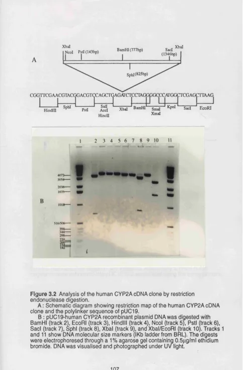
3.2.1 Restriction endonuclease analysis of the human CYP2A cDNA clone ... 106
3.2.2 Validation of the cloned CYP2A cDNA by sequencing ... 106
3.3 isolation and characterization of the marmoset CYP2A sequence . . . 110
3.4 Expression of human CYP2A6 in E.Coli ... 115
3.4.1 Construction of pMAL-c-CYP2A6 fusion plasmid ... 119
3.4.2 Construction of pMAL-p-CYP2A6 fusion plasmid ... 119
3.4.3 Expression of fusion protein using pMAL-p-CYP2A6 fusion plasmid ... 121
3.4.4 Cellular localization of MBP-CYP2A6 fusion protein ... 123
3.4.5 Spectral analysis of MBP-CYP2A6 fusion protein ... 127
3.5 Purification of MBP-CYP2A6 fusion protein ... 129
3.5.1 Affinity of MBP-CYP2A6 fusion protein to amylose resin . . . 131
3.5.2 Purification of MBP-CYP2A6 fusion protein by Chromatography on p-chloramphetamine-coupled Sepharose 131 3.6 Baculovirus mediated expression of human CYP2A6... 135
3.6.1 Replacement of the polyhedrin gene through homologous recombination ... 138
3.6.2 isolation of recombinant viruses and expression of CYP2A6 ... 141
3.6.3 Sub-cellular localization of expressed CYP2A6 ... 141
3.7 Enzymatic activity of baculovirus-expressed CYP2A6 ... 146
3.7.1 Coumarin 7-hydroxylase activity of the expressed CYP2A6 148 3.7.2 Analysis of metabolism of testosterone using microsomes from Sf9 cells infected with recombinant baculovirus expressing CYP2A6 ... 152
L IS T of F
i g u r e sFigure P ag e
Figure 1.1 Examples of reactions catalyzed by cytochromes ... 20
P450 Figure 1.2 Mechanism of P450 catalyzed [reactions... 22
Figure 1.3 Phylogenetic tree of P450 gene superfamily ... 27
Figure 1.4 Evolution of P450 gene superfamily ... 30
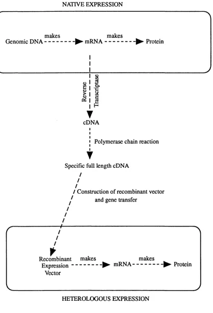
Figure 1.5 Cloning of specific DNA sequences and their subsequent expression in heterologous cell... 48
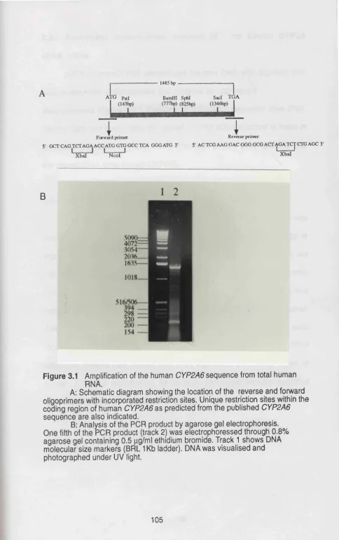
Figure 3.1 Amplification of the human CYP2A6 sequence from ... 105
total human RNA. Figure 3.2 Analysis of the human CYP2A cDNA clone by ... 107
restriction endonuclease digestion. Figure 3.3A Sequencing strategy for human CYP2A cDNA clone ... 108
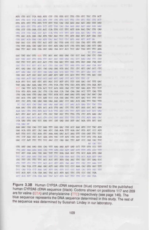
Figure 3.3B Human CYP2A cDNA sequence ... 109
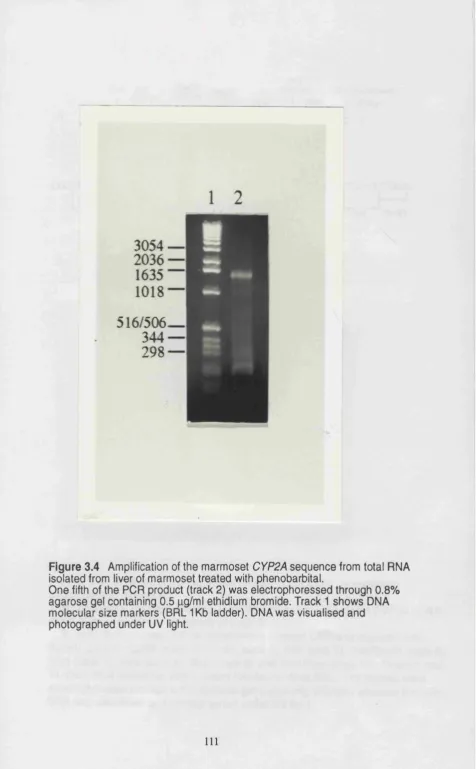
Figure 3.4 Amplification of the marmoset CYP2A sequence from total RNA isolated from liver of marmoset treated with phénobarbital ... I l l Figure 3.5 Analysis of the marmoset CYP2A cDNA clone by restriction endonuclease digestion ... 112
Figure 3.6 Marmoset CYP2A sequence ... 113
Figure 3.7 Northern blot analysis of marmoset liver RNA ... 114
Figure 3.8 pMal Vectors ... 117
Figure 3.9 Schematic representation of expression and purification using the MBP vectors ... 118
Figure 3.10 Restriction endonuclease digestion of plasmid DNA from putative positive transformants following transformation of E.co//JM109 cells with pMAL-c+CYP2A6 ligation mixture ... 120
Figure 3.12 Western immunoblot analysis of E. coli expressed
MBP-CYP2A6 fusion protein ... 124
Figure 3.13 Cellular localization of MBP-CYP2A6 fusion protein
(SDS/PAGE analysis) ... 125
Figure 3.14 Cellular localization of MBP-CYP2A6 fusion protein
(immunoblot an alysis)... 126
Figure 3.15 CO-Reduced difference spectrum of E. coli expressed MBP-CYP2A6 fusion protein ... 128
Figure 3.16 Affinity of MBP-CYP2A6 fusion protein to amylose resin . . . 130
Figure 3.17 Analysis of the effect of detergent concentration on the
affinity of MBP-CYP2A6 fusion protein for amylose resin .. 132 Figure 3.18 Purification of MBP-CYP2A6 fusion protein by
chromatography on p-chloramphetamine
coupled-Sepharose (analysis by S D S /P A G E )... 133
Figure 3.19 Purification of MBP-CYP2A6 fusion protein by chromatography on p-chloramphetamine-coupled
Sepharose (analysis by western blotting)... 134 Figure 3.20 Replacement of polyhedrin gene through
homologous recombination ... 140 Figure 3.21 Baculovirus transfer vector pAcC5 ... 142
Figure 3.22 Immunodetection of CYP2A6 ... 143 Figure 3.23 Analysis by SDS/polyacrylamide gel electrophoresis
of lysates and subcellular fractions of Sf9 cells ... 144 Figure 3.24 Western blot analysis of lysates and subcellular
fractions of Sf9 cells ... 145 Figure 3.25 Formation of 7-hydroxycoumarin from coumarin in a culture
medium of Sf9 cells infected with recombinant baculovirus
expressing CYP2A6 or wild type baculovirus (AcMNPV) . . . 149
Figure 3.26 Coumarin 7-hydroxlase activity in the microsomes from Sf9 cells infected with recombinant baculovirus or wild type
baculovirus(AcMNPV)... 151
Figure 3.27 Effect of adding exogenous NADPH-cytochrome P450
reductase on coumarin hydroxylase activity in microsomes
Figure 3.28 Effect of substracting COH activity with no added NADPH-cytochrome P450 reductase from the COH
activity obtained in the presence of added human
NADPH-cytochrome P450 reductase ... 154
Figure 3.29 Analysis of testosterone hydroxylase activity of baculovirus
expressed CYP2A6... 156
Figure 3.30 Purification of baculovirus expressed CYP2A6 by
chromatography on p-chloramphetamine- coupled Sepharose
(analysis by SDS/PAGE) ... 158 Figure 3.31 Purification of baculovirus expressed CYP2A6 by
chromatography on p-chloramphetamine- coupled
Sepharose(analysis by western blotting ) ... 159
Figure 3.32 Western blot analysis of baculovirus expressed CYP2A6 purified by chromatography on
p-chloramphetamine coupled Sepharose and concentrated using the Amicon centriprep-30 concentrator ... 160
T a b le s P ag e
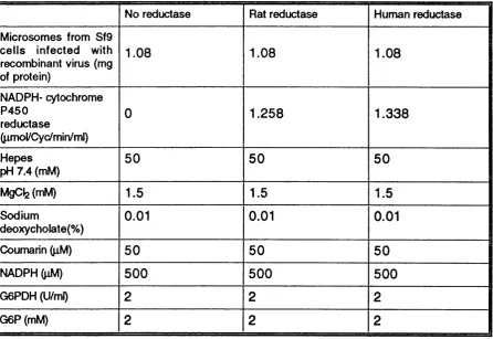
Table 1 Composition of reaction mixtures for the experiment on
effect of adding exogenous NADPH-cytochrome P450
reductase on COH activity of baculovirus expressed
c k a p t e r
1
1.1 General characteristics of cytochromes P450
Cytochrome P450 (P450) was identified in mammalian liver
microsomes as a reduced pigment that had an absorption band with a X^ax at
450nm after binding to carbon monoxide (Klingenberg 1958; Garfinkel 1958).
This pigment was further characterized, by Omura and Sato in 1964, as a
P450 haemoprotein. P450s are now known to be a family of proteins whose
members are present in biological sources as diverse as microorganisms,
plants and animals (Porter and Coon, 1991). In mammals, P450s are present
in almost all tissues (reviewed in Omura et a i, 1993).
P450S share the following characteristics: i) they contain a
noncovalently bound haem; ii) they are intrinsic membrane proteins firmly
bound to intracellular membranes and iii) they use reducing equivalents from
NADPH (and sometimes NADH) and an atom of oxygen derived from
molecular oxygen to oxygenate substrates.The reducing equivalents are
transferred to P450 via a second enzyme. P450s can be divided into two
major classes based on their intracellular location and the enzyme from which
they receive electrons.
The mitochondrial P450s, first found in mitochondria isolated from the
adrenal cortex (Harding at a i, 1964) are now known to be widely distributed
amongst animal organs. All steroidogenic organs and some non
steroidogenic organs including liver and kidney contain P450s in their
mitochondria. These enzymes are synthesized on m em brane-free
polyribosomes (Nabi at a i, 1983) as a large precursor and are then
transported into the mitochondria concomitant with the cleavage and the
P450S are bound to the inner mitochondrial membrane and receive electrons
from NADPH via two soluble redox proteins in the matrix,
NADPH-adrenodoxin reductase and NADPH-adrenodoxin (Baron et al., 1972). Several
mitochondrial P450s have been isolated from various animal sources, and
their enzymatic and molecular properties have been elucidated. These
proteins include the cholesterol side chain cleavage enzyme, steroid 11
p-hydroxylase, aldosterone synthase, and sterol 26 hydroxylase. Mitochondrial
P450S are involved in the metabolism of steroids and related physiological
substrates for example vitamin D3,
The majority of P450s are of the second class, found primarily in the
endoplasmic reticulum membranes. These proteins are synthesized on
membrane-bound polyribosomes and inserted directly into the lipid bilayer via
the signal sequence recognition system (Bar-Nun eta!., 1980; Sabatini et a!.,
1982). Microsomal P450s receive electrons from NADPH via the flavoprotein,
NADPH-cytochrome P450 reductase (Lu et a!., 1969) and in some cases, one
electron is derived from NADH via cytochrome bs (Pompon and Coon, 1984).
Microsomal P450s play a central role in the metabolism of endogenous
substances such as steroids and fatty acids (Guengerich eta!., 1986), and in
the detoxification of foreign substances (xenobiotics) including plant toxins,
drugs and environmental pollutants (Guengerich e ta !., 1986) and in the
activation of procarcinogens (Guengerich, 1988). Most P450s, particularly
those found in the liver, function to convert hydrophobic substances to more
hydrophilic derivatives that can be easily eliminated from the body directly or
after conjugation with water soluble agents such as glucuronic acid and
is then easily passed from the body via urine or bile. This is usually a
detoxification process, but in some instances, foreign substances are
converted to products with much greater cytotoxicity, mutagenicity and
carcinogenicity {Guengerich, 1988) This property of P450s emphasizes the
importance of understanding their multiplicity, substrate specificities, and
regulation.
1.2 Multiplicity of P450s and reactions cataiysed by these proteins
The vast array of foreign substances to which organisms are exposed
makes it impractical to have one enzyme for each compound, or even each
class of compounds. Thus, while most cellular functions tend to be very
specific, metabolism of foreign substances requires enzymes with diverse
substrate specificity. Much of this role is assumed by P450s.
Partial purification of a form of P450 from phénobarbital treated rabbit
liver microsomes was first reported by Lu and Coon (1968). Over the next 20
years it became evident through protein purification experiments that multiple
forms of P450 exist in mammals and other species. Studies using systems of
purified P450s reconstituted with lipid and NADPH-cytochrome P450
reductase (for example, Haugen and Coon, 1976; Coon eta!., 1975) revealed
that individual forms of P450 can exhibit either highly specific or less specific
overlapping substrate positional or jstereospecificities: (reviewed by
Guengerich etaL, 1986).
Most of the P450 mediated reactions begin with transfer of electrons
from NAD(P)H to either NADPH-cytochrome P450 reductase in the
microsomal system or a ferredoxin reductase and a nonhaem iron protein in
reductive activation of molecular oxygen followed by the insertion of one
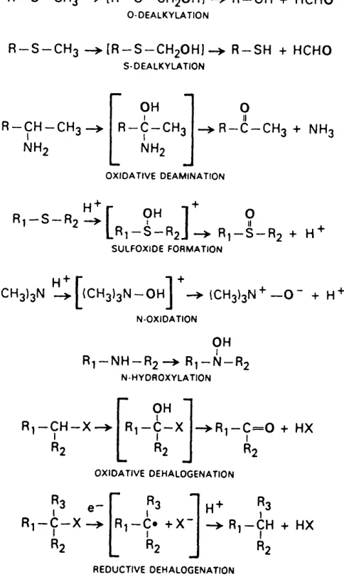
oxygen atom into the substrate. Reactions that have been demonstrated to be
catalysed by P450s include hydroxylation, epoxidation, peroxygenation,
deamination, désulfuration, dehalogenation and reduction (Nebert and
Gonzalez, 1987). Examples of P450 mediated reactions are shown in figure
1
.
1.
1.2.1 Mechanism of P450 catalysed reactions
The active site of P450 contains iron protoporphyrin IX bound in part by
hydrophobic forces. The fifth ligand is a thiolate anion provided by a cysteine
residue, a feature that contributes to the unusual spectral and catalytic
properties of P450s, and the sixth coordinate position may be occupied by an
exchangeable water molecule. Upon reduction of iron, molecular oxygen can
be bound in the sixth position (Poulos etaL, 1987). Stoichiometric studies on
monooxygenase reactions catalysed by P450 indicated the consumption of
one molecule each of NADPH and oxygen to introduce one oxygen atom to
the substrate molecule, indicating the supply of two electrons from NADPH to
one P450 molecule during one cycle of reaction (Cooper et a i, 1977).
Stoichiometry of the P450 catalyzed hydroxylation reaction:
P 4 5 0
R H + O 2 + N A D P H + H+---► N A D P + + H2O + R O H (11
R — CHg —► R — CH2OH
A L IP H A T IC O X ID A T IO N
T V
A R O M A T IC H Y D R O X Y L A T IO N
R — N H — C H3 —> (R — N H — C H2O H I —> R — N H2 + H C H O N -D E A L X Y L A T IO N
R — 0 — C H3 —> [R — 0 — C H2O H I —> R — OH + H C H O O -D E A L K Y L A TIO N
R — S — C H3 —► (R — S — C H2O H ] —> R — S H + H C H O s -D E A L K Y L A T IO N
R-CH-CHo I NH; OH R-C-CH3 I NH;
R — c — C H3 + N H3
O X ID A T IV E D E A M IN A T IO N
R i - S - R ; ^ r 9^ ] 9
L ^ i ^ R2J —^ R] — S — R; + H
S U L FO X ID E FO R M A TIO N
H+ r 1 +
(CH3)3N —^ — OH I —^ (CH3)3N —O + H
N -O X ID A T IO N
OH R'j — NH — R; —> R^ — N — R;
N H Y D R O X Y L A T IO N
R ,-C H -X
OH
Ri —C — X R ,-C = 0 + HX
O X ID A T IV E DE H A LO G EN A TIO N
Ri — C — X Ri — C* + X R'J — CH + HX R2
R E D U C TIV E DEH A LO G EN A TIO N
A cyclic mechanism for P450 catalysed monooxygenation was
postulated in accordance with the known stoichiometry of the hydroxylation
reaction (equation 1 ) and on the basis of observations of substrate induced
spectral changes and spectral detection of the oxygenated form of P450
(Estabrook ef a/., 1971). Further insight into the mechanism has also been
gained from studies of the crystallized form of P450camfrorn Pseudomonas
putida (Poulos etal., 1992). A schematic diagram of the overall P450 reaction
cycle is shown in figure 1.2.
The first step in the reaction cycle is the binding of substrate to the
active site of P450 resulting in the conversion of low- spin ferric haem in the
enzyme to a high spin-ferric state. This facilitates the uptake of one reducing
equivalent from the electron transfer system (in microsomes, this electron is
transferred by NADPH-cytochrome P450 reductase from NADPH) to form a
substrate- bound ferrous form (step 2).
Substrates that undergo reduction rather than oxygenation such as
epoxides, N-oxides, nitro and azo compounds and lipid hydroperoxides,
accept two electrons in stepwise manner as shown to give RH (H )2. To initiate
the oxidative reactions the ferrous enzyme reacts with molecular oxygen to
form a ternary complex of oxygen , substrate and ferrous P450 commonly
referred to as Oxy-P450. Introduction of a second reducing equivalent into the
Oxy-P450 (step 4) is mandatory for the oxygenation reaction to occur. In
microsomes, the second reducing equivalent is provided by NADPH
cytochrome P450 reductase or in some cases, cytochrome bs (as an
additional electron donor) (Pompon and Coon, 1984). The next step (5) is not
well understood but involves splitting of the 0-0 bond with the uptake of two
ROH
RH
(ROH)Fe®*
7
1
(RH)Fe**
/e
/
(R -)(F e -0 H )3 *^ ^ _ _
(RH)Fe^*
4
\
t
' e-,2H '
I
XOH XOOH
3
H
2O - 4 - ^
RH(Fe-O)®*
4 (RH)Fe3+(0ÿ)
"
1
' ^
i
Figure 1.2 Mechanism of P450 catalyzed reactions
Fe represents the heme iron atom at the active site, RH the substrate, RH(H)^ a reduction product, ROH a monooxygenation product and XOOH a peroxy compound that can serve as an alternative oxygen donor
Oxygen insertion into the substrate (step 6) is believed to involve hydrogen
abstraction from the substrate and the recombination of the resulting transient
hydroxyl and carbon radicals to give the product (Groves et al., 1978).
Dissociation of the product, ROH, then restores the P450 to the ferric state
(step 7).
Also indicated, in figure 1.2, is the way in which a peroxy compound
may substitute for molecular oxygen and reducing equivalents in what is
termed as the peroxide shunt. Hemolytic cleavage is envisioned with the
formation of an iron- bound hydroxyl radical capable of hydrogen abstraction
from the substrate (White and Coon, 1980).
Much remains to be learned about the factors controlling regio and
stereo specificity in P450 catalysed reactions. Heterologous cDNA based
expression of large amounts of individual P450s will greatly aid such studies.
Some of the systems that have been used for the heterologous expression of
P45ÜS are described in section 3 of this thesis.
1.2.2 Nomenclature
The confusing nature of P450 nomenclature has been one of the
hallmarks of P450 research. Most laboratories involved in the purification of
P450S developed their own system of nomenclature, often based on enzyme
characteristics, which included typical P450 inducing agents or substrates for
the purified enzyme or the sequential order in which a particular research
group had purified a particular series of P450s. For example, CYP1A1 has
been designated P-450-C, P450pNF-B, P-4482, P-450MC-1, P-450 isozyme 6
as a result of the ever increasing number of P450s to be isolated and the
broad and overlapping substrate specificity of these enzymes.
Characterization based on full length amino acid sequences was impeded by
technical difficulties and the required labour intensive procedures inherent in
this biochemical approach.
A major breakthrough in the structural analysis of P450s came with the
application of recombinant DNA technology (Fujii-Kuriyama et a/., 1982)
which subsequently resulted in the isolation and sequence determination of
many individual P450 forms. Sequence analysis has provided the basis for a
unified nomenclature system (Nebert ef a/., 1991).
In the P450 gene superfamily, families and subfamilies have been
defined on the basis of amino acid sequence similarity. A P450 protein
sequence from one gene family is defined as usually having less than or
equal to 40% amino acid identity to a P450 protein from any other family.
Within a single family, the P450 protein sequences are > than 40% identical.
Sequences of P450 proteins within the same subfamily are > 55% identical
within the same species.
Recommendations for naming a P450 gene or cDNA include the root
symbol C Y P (cyp for the mouse) denoting P450, an Arabic number
designating the P450 family, a letter indicating the subfamily and an Arabic
number representing the individual gene. With the mouse genes or cDNAs,
the final number is generally preceded by a hyphen. P (p in mouse) after a
gene number is used to denote a pseudogene. The same nomenclature is
recommended for the corresponding gene product (enzyme). For example,
CYP1A1 (cyp1a-1 , in mouse) for the gene and cDNA, and CYP1A1 for the
One of the problems with the P450 nomenclature system Is In
distinguishing allelic variants of one protein from closely related P450 forms.
For example, rodent P450 genes can differ by only a few bases. Allelic
variants can be distinguished by genetic crosses In rodents when the
enzymes can be resolved by two dimensional electrophoresis (Rampersaud
and Walz, 1987). Allelic variants can also be distinguished by direct analysis
of genomic DNA using the polymerase chain reaction (PCR) or Southern
blotting following genetic crosses of rodents and by linkage studies using
large pedigrees In humans.
Another problem faced by the P450 nomenclature system Is a
reasonable method to determine orthologous P450s. Genes or proteins In two
or more species are said to be orthologous If both are believed to have
evolved from a single ancestral gene present at the time of divergence of the
species. This Is problematic when multiple forms of P450s exist In certain
subfamilies. For example rats, mice, rabbits and humans contain many P450s
In the CYP2C subfamily, however, no two P450s between these different
P450S display the same catalytic activities. In addition, a single CYP2C P450
In rat does not have significantly higher sequence similarity with a single P450
In the same subfamily In humans than with the other P450s In the human
CYP2C subfamily. The same applies when comparisons are made between
other species. Thus P450 forms In the CYP2C family are given Individual form
designations. In contrast. In the CYP2E subfamily, most species except rabbits
contain a single P450 form designated CYP2E1 that has qualitatively similar
catalytic activities across species (Gonzalez, 1 9 # ) . A recent update of the
P450 nomenclature, and genes Isolated, has been published by Nelson etal.,
1.2.3 Evolution of P 450s
Based on the existence of two distinct classes of P450s, the possible
pathways of evolution giving rise to mammalian P450s can be hypothesized.
The mitochondrial P450s may have arisen from bacterial P450s such as
P450cam (CYP101) from Pseudomonas putida and other prokaryotes that use
an iron sulphur protein and flavoprotein as electron donors. Microsomal
P450s might have evolved from the mitochondrial forms by transfer of genetic
material from the mitochondrial genome to the nucleus. Alternatively, P450
and NADPH cytochrome P450 reductase may have arisen from a fusion
protein such as that found in Bacillus megaterium followed by the separation
of the two domains. In fact, the mammalian CYP3 gene family and
NADPH-cytochrome P450 reductase are both found on human chromosome 7
(Yamano e fa /., 1989).
Much of the insight into the evolution of P450s has come from a
correlation of catalytic activities of the P450s with the information obtained
from the examination of phylogenetic trees of the P450s. Construction of the
phylogenetic tree is based on amino acid sequence relatedness of different
P450 forms. The percentage differences calculated after pairwise sequence
comparisons are converted to evolutionary distance in units defined as
accepted point mutations (Nelson and Strobel, 1987). Evolutionary distance
(d) Is a mathematical approximation of the number of mutations per 100 amino
acids that have occurred since a divergence event. The tree (figure 1.3) is
constructed using the unweighted-pair-group method of analysis (UPGMA).
Proper interpretation of data in a phylogenetic tree requires calibration of d
values with real time. This is generally achieved by using divergence times
C Y P G E N E S U P E R FA M ILY
Q>
---0
4.0 3.0 2.0 1.0
EVOLUTIONARY DISTANCE
Figure 1.3 Phylogenetic tree of P450 gene superfamily
ago) and more recent divergence times (for example, mammalian radiation at
about 80 million years ago and rat-mouse divergence at 17 million years ago).
From an analysis of the P450 phylogenetic tree (figure 1.3.), it appears
that the oldest mammalian P450s closely related to the bacteria and yeast
P450s, are the cholesterol 7a-hydroxylase (CYP7) and the P450 aromatase
(CYP19) involved in bile acid and oestrogen synthesis respectively.
Two other “old” P450s are the cholesterol 27-hydroxylase (CYP27) and
the cholesterol side chain cleavage enzyme (CYP11A1). These are related to
P450S that metabolize fatty acids such as the lauric acid hydroxylases of the
CYP4 family. Cholesterol and fatty acid metabolizing P450s are thought to
have been involved in the maintenance of membrane integrity of early
eukaryotes.(Gonzalez, 1989). A later evolutionary event, about 900 million
years ago, was the formation of the drug metabolising P450s. These diverged
into major drug and carcinogen metabolising enzymes of the CYP1, CYP2
and CYP3 families. More recently, between 400 and 600 million years ago, a
tremendous expansion in the CYP2 gene family has occurred. The reason for
the increase in the number of P450 genes during the past several million
years may be related to environmental factors including dietary components
and toxins.
An interesting feature of P450 gene evolution is the occurrence of
multiple genes, within a particular subfamily, that in many cases are present in
some species but not in others. These are thought to have arisen through the
process of gene duplication and the fixation of duplicated genes through
natural selection. For example, rabbits, rats and humans are known to have
humans contains a single gene, whereas two highly similar genes (CYP2E1
and CYP2E2) are present in rabbits (Gonzalez, 1989). On the basis of amino
acid sequence similarity of the two rabbit proteins, it is predicted that a gene
duplication occurred in this species about 1 0 million years ago, long after the
rabbit-rat-human spéciation.
The net result of recent gene duplications and gene conversion events
is that individual species contain their own unique P450 genes. A scheme for
the formation of P450 gene families, subfamilies and gene conversion is
shown in figure 1.4 Gene duplication, conversion or gene loss probably
account for some of the major interspecies differences in drug and carcinogen
metabolism that have been observed over the years. Although humans have
more recently been exposed to a vast number of man made chemicals, it is
apparent, given an average unit evolutionary period for P450s of 4, that
human P450s cannot evolve rapidly enough to meet this challenge. This may
be an important factor underlying the increase in chemically induced diseases
such as cancer.
1.3 Regulation, tissue specific expression and catalytic activities
of cytochromes P450.
221 P450 genes and 12 putative pseudogenes have been
characterized (Nelson et a/., 1993). These genes have been described in
eleven prokaryotes and thirty one eukaryotes (including eleven mammalian
and three plant species). Of the thirty six gene families described, twelve
families exist in all mammals examined.
The mammalian P450 families can be functionally subdivided into two
ANCESTRAL P450 GENE
Species A
Species B
Gene duplication
Gene duplication
Gene duplication
and divergence
Mutation
Gene conversion
Gene loss
Figure 1.4 Evolution of P450 gene superfamily
A hypothetical scheme is shown in which two species, A and B evolve P450 genes. Gene duplications occur independently in both species. A second gene duplication has occured in species A while a mutation (X) has eliminated a gene in species B. Within the species A family, a gene has diverged from the other two due to changes in sequence and natural selection. This gene represented as a stripped rectangle, was then involved in a gene conversion with the neighbouring gene. After a period of time, species A and B have evolved their own distinct set of P450 genes.
those that primarily metabolize xenobiotics.
1.3.1 P450S involved in the metabolism of xenobiotics
CYP1 fam ily
The CYP1 family comprises of two genes (CYP1A1 and CYP1A2) in
every mammalian species so far examined. In humans C YP1A genes are
located on chromosome 15 (Jaiswal etal., 1987). CYP1A1 is associated with
high aromatic hydrocarbon hydroxylase activity and its expression is
dependent on the presence of inducers such as benzo (a) pyrene and can be
demonstrated in almost all tissues of an animal. The induction of the CYP1A1
gene is mediated through a cytosolic receptor called the Arylhydrocarbon
hydroxylase (Ah) receptor, that dimerises with the protein Arnt (Ah receptor
nuclear translocator), to initiate transcription of this gene by binding to
xenobiotic responsive elements (XREs) located in the 5’ flanking sequences
of this gene (Gonzalez etal., 1993).
Expression of the CYP1A1 gene has been correlated with development
of polycyclic aromatic hydrocarbon- associated cancers in rodents (Nebert,
1989). High and low-inducibility phenotypes have been found in humans
(Petersen et a!., 1991). A restriction fragment length polymorphism near the
CYP1A1 gene was found to be associated with increased lung cancer risk in
Japanese smokers (Nakachi et a!., 1993), however, the same association was
not found in a cohort study of Norwegian lung cancer patients (Tefre e ta l.,
1991). The biochemical basis of lung cancer association with CYP1A1 is not
known.
Human CYP1A1 cDNA has been isolated from a human liver cDNA
enzyme capable of N-hydroxylating acetylaminofluorine, hydroxylating
benzo(a)pyrene, and activating the food derived promutagen 2-amino-1
-methyl-6-phenyiimidazo [4,5-b] (PhIP) (McManus etal., 1990).
CYP1A2 is constitutively expressed in the liver and is responsible for
the activation of numerous promutagens and procarcinogens including
aflatoxin B1, heterocyclic arylamine promutagens and the drugs phenacetin
and caffeine (Guengerich and Shimada 1991). Gene regulation studies
demonstrated that a heterologous C Y P 1 A 2 promoter is activated by
3-methylcholanthrene in human hepatoma HepG2 cells and a region of DNA
that supports Ah receptor binding and promoter specific induction has been
identified (Quattrochi etal., 1994).
A human CYP1A2 cDNA has been cloned (Jaiswal et a!., 1987) and its
expression in B-lymphoblastoid cells (Crespi et al., 1990) produced an
enzyme capable of metabolically activating aflatoxin B1. The activity was
higher than other human P450s known to metabolize this compound. CYP1A2
therefore may be the principal enzyme functional in vivo under typical human
exposure concentrations of aflatoxin B1.
CYP2 family
CYP2 is the largest of the 12 mammalian P450 families and is divided
into 1 0 subfamilies.
CYP2A subfamily
The CYP2A subfamily has been extensively studied in rodents. Rats
and mice possess three CYP2A (CYP2A1, CYP2A2 and CYP2A3) and two
a/., 1993). CYP2A1 is highly specific for testosterone 7 a - hydroxylase activity.
In contrast, CYP2A2 exhibits a high testosterone 15a- hydroxylase activity.
CYP2A1 and CYP2A2 are liver specific, but they are regulated differently.
CYP2A1 production is increased in young male and female rats but the
expression of this gene is suppressed in males at the onset of puberty. In
contrast, CYP2A2 is never expressed in females and the gene is activated
only when males reach puberty (Matsunaga et a/., 1988). CYP2A3 is
constitutively expressed in the rat lung and absent from the liver. The mRNAs
encoding this protein are induced 3 fold by treatment of rats with
3-methylcholanthrene (Kimura etal., 1989 a).
The two P450S in the mouse CYP2A subfamily bear a high sequence
similarity with the rat CYP2A3 have been characterized (Lindberg and
Negishi, 1989). CYP2A4 carries out testosterone 15a-hydroxylation whereas
C Y P2A5-Cohh and CYP2A5- Coh'are allelic variants having high and low
coumarin 7-hydroxylase activities respectively and differing at only a single
nucleotide associated with the codon 117, Alai 17 in CYP2A5- Coh> and
V ail 17 in CYP2A5-Cohh (Lindberg and Negishi; 1989). The expression of the
Cyp2a-5 gene is induced by phénobarbital (Aida and Negishi; 1991) whereas
CYP2A3 gene expression is induced by 3-methylcholanthrene (Kimura etal.,
1989 a) suggesting that the regulatory elements controlling the CYP2A genes
have evolved differently in rats and mice. In humans, two distinct cDNAs have
been isolated, one of which (C Y P 2 A 6 ) encodes a P450 capable of
hydroxylating coumarin (Yamano etal., 1990). A second cDNA (CYP2A7) also
isolated from a human liver library (Yamano e ta l., 1990) encoded a protein
sequence to CYP2A6. Interestingly, CYP2A7 did not incorporate haem when
the cDNA was expressed using a vaccinia virus based system (Yamano etal.,
1990). It is possible that CYP2A7 contains an amino acid substitution that
renders it unstable. The most conspicuous feature of CYP2A6 is the extremely
large inter-individual variations observed at the protein, mRNA and enzyme
activity levels (Yamano etal., 1990, Palmer e tal., 1990, Rounio etal., 1988).
Large interspecies variation in COM activity has also been observed
(Pelkonen etal., 1985).
Depending on the human livers analysed, 40 to 100 fold differences in
COM activity were detected. Palmer et al 1990 found that the expression of
CYP2A6 mRNA varied 1000-fold between the individual samples they
analysed. In 10% to 20% of livers with high COM activity, CYP2A6 represents
only about 1% of total P450 indicating that CYP2A6 is a low abundance P450
(Yun et al., 1991). In a number of human livers, only trace amounts of CYP2A6
protein and associated enzyme activities were observed suggesting that the
protein may be inducible by some dietary components or drugs. Several
compounds have been reported to increase COM activity in mouse liver after
adminstration in vivo (Hahnemann et al., 1992). Examples include
phénobarbital, pyrazole and cobalt. As yet, inducibility of CYP2A6 by similar
compounds has not been demonstrated in humans. It is possible that the
C Y P 2A 6 gene is polymorphically expressed in humans and that the mutant
alleles in the population cause the marked variation in expression similar to
the findings with the C Y P 2 D 6 allele and the debrisoquine/sparteine
polymorphism (Kimura etal., 1989 b). This idea is supported by the presence
of CYP2A6V, a variant cDNA that was isolated from a human liver cDNA
(Yamano et al., 1990). As yet, it is not established whether this variant is
common within the human population and whether it accounts for the lack of
CYP2A6 protein activity observed in some human liver samples.
Coumarin has been used as an in vivo metabolic probe to test for the
presence of CYP2A6 polymorphism in humans (Cholerton at ai., 1992). These
studies have presented evidence for a wide inter-individual variation in
7-hydroxylation of coumarin. In the light of the possibility that a CYP2A6
polymorphism exists in humans, it will be interesting to determine whether the
expression of CYP2A6 is associated with an increased risk for chemically
induced cancer since CYP2A6 is capable of metabolically activating aflatoxin
B1, N-nitrosodiethylamine (Crespi at ai., 1991 a), and 1,3-butadiene
(Duescher and Elfarra; 1994). It would be interesting to determine whether
CYP2A6 is expressed in human lung, the site of tobacco smoke and air
pollution associated with carcinogen exposure. As yet, human CYP2A genes
have not been isolated, but a cluster is known to exist on human chromosome
19 (Phillips at ai., 1985).
CYP2B subfamily
The cDNAs for CYP2B1 and CYP2B2 in rats were the first P450 cDNAs
to be isolated and completely sequenced (Fujii-Kuriyama at ai., 1982).
CYP2B1 and CYP2B2 proteins exhibit 97% amino acid sequence similarity.
These enzymes have similar broad and overlapping substrate specificities but
CYP2B1 has two to ten fold higher activity (depending on the substrate) than
does CYP2B2 when these enzymes are purified and analysed for activity in a
reconstituted system (reviewed by Gonzalez, 198^). A third cDNA in the rat
have 77% amino acid sequence identity to CYP2B1 and CYP2B2. However,
the genes that encode CYP2B1, 2B2, and 2B3 are differentially regulated.
CYP2B1 is absent or expressed in a very low amount in the liver but is highly
inducible by phénobarbital treatment. CYP2B2 is constitutively expressed in
the liver and induced by phénobarbital. CYP2B3 is constitutively expressed
and not inducible by phénobarbital. Furthermore CYP2B1 is constitutively
expressed and not inducible by phénobarbital in lung and testis while
CYP2B2 is absent from these tissues regardless of treatment (reviewed by
Gonzalez, 198f). In the small intestine, CYP2B1 is constitutively expressed
and inducible by various agents including phénobarbital (Traber et al., 1988).
These observations suggest that the genes encoding CYP2B subfamily
members contain tissue specific enhancer and inducer control elements. To
date, no receptor for phénobarbital has been identified. Recently, two
cis-acting elements located between -199 and -183 and -72 and -31 of a CYP2B2
gene have been identified that bind nuclear proteins that are either more
abundant or activated in response to phénobarbital treatment (Shephard et
a/., 1994). It is known that induction of C YP2B 1/2 genes by phénobarbital
occurs through transcriptional activation (Waxman and Azaroff 1992).
Progress has been made in defining the molecular mechanism of
phénobarbital induction of P450s in bacteria. A region of DNA upstream of the
bacterial P450 gene CYP102 was found to confer transcriptional activation in
the presence of phénobarbital. This activation appears to be due to the
release of a repressor protein designated Bm3R1 that binds to a palindromic
operator sequence located immediately upstream of the CYP102 gene (Shaw
and Fulco 1992). It is not clear whether phénobarbital acts directly or indirectly
Two CYP2B cDNAs, CYP2B6 andi CYP2B7 have been Isolated from a
human liver cDNA library. CYP2B6 cDNA was expressed using the vaccinia
virus based system and the protein was able to catalyze the O- dééthylation of
7-ethoxycoumarin. The C Y P 2 B 7 cDNA deduced amino acid sequence
displayed 93% identity to that of C Y P 2 B 6 and contained a premature
termination codon due to a C to T transition (Yamano et al., 1989 b). A large
degree of inter individual variation in the expression of CYP2B protein and
mRNA has been observed in human liver specimens (Miles et a!., 1989,
Yamano et al ;1989 b). However, no evidence was presented to suggest the
presence of a known P450 inducing drug in patients whose liver specimens
contained high amounts of CYP2B mRNA or protein.
CYP2C subfam ily
The CYP2C subfamily is generally thought to represent a class of
constitutively expressed genes. A total of 9 rabbit and 8 rat C Y P 2C genes
have been identified to date (Nelson et al., 1993). In rats these enzymes are
noted for their sex specific and developmentally regulated expression
(reviewed by Gonzalez, 1989). In rabbits, none of the CYP2C enzymes are
uniquely expressed in males or females. In humans six CYP2C forms have
been identified through cDNA cloning (Nelson etal., 1993). CYP2C8 (Ged et
al., 1988) and CYP2C9 (Srivastava et al., 1991) have been purified. Another
P450 designated CYP2C10 (Ged et al., 1988) has identical cDNA deduced
amino acid sequence to CYP2C9 apart from an abrupt sequence change in
the 3' non-coding region of the cDNA. Could CYP2C10 be a cloning artefact?
The confirmation of the existence of C Y P 2 C 1 0 can be achieved by PCR
A member of the CYP2C subfamily is believed to encode a P450
responsible for the human S-mephenytoin polymorphism, which effects 2% to
5% of Caucasians and up to 27% of Japanese who lack S-mephenytoin
4-hydroxylase activity (Wilkinson et al., 1989). A C Y P 2C 9 cDNA isolated from
the Japanese population has been expressed in yeast (Yasumori etal., 1989)
and the expressed protein shows stereo selective metabolism of mephenytoin
but at low turnover rates. Proteins expressed from CYP2C9 cDNAs other than
that isolated from a Japanese liver cDNA library did not show S-mephenytoin
4-hydroxylase activity, but readily metabolized tolbutamide (Relling et al.,
1990).
Additionally, three cDNAs CYP2C17, C Y P 2 C 1 8 a n 6 CYP2C19 have
been isolated from a human liver cDNA library and their expression in COS
cells (Romkes et al., 1991) produced enzymes capable of metabolizing
racemic mephenytoin with CYP2C18 displaying high activities as compared to
CYP2C9 and CYP2C19. These observations suggest that CYP2C18 might be
a candidate for the P450 responsible for the mephenytoin polymorphism.
C YP2C 9 is also the major human hepatic P450 involved in the
metabolism of the therapeutically active S-isomer of the anticoagulant drug,
warfarin (Rettie et al., 1992). cDNA expressed C YP2C 9 catalyses
7-hydroxylation of S-warfarin and the 7-hydroxy metabolite is the major
metabolite found in humans after adminstration of the drug. Thus the cause of
large patient variability in warfarin effectiveness may be due to interindividual
variation in CYP2C9 expression. The locus of C Y P 2 C genes has been
CYP2D subfam ily
Mice and rats contain five CYP2D genes that are expressed in the liver
and kidney. Three CYP2D genes are present in humans and are expressed in
the liver, kidney and the intestine (Nelson etal., 1993). The rat and human
enzymes carry out the oxidation of the drugs debrisoquine,and bufuralol. In
humans, CYP2D6 is responsible for the debrisoquine/sparteine polymorphism
(Gonzalez and Meyer, 1991). About 7.5% of European and north American
Caucasians possess two copies of the mutant C Y P2D 6 alleles and are thus
unable to metabolize debrisoquine and over 20 other drugs. The three major
mutant C YP2D 6 alleles have been sequenced including one locus in which
the CYP2D6 gene is deleted (Hanioka et a!., 1990, Gaediak etal., 1991). The
majority of deficient metabolisers can be identified through PCR genotyping
(Broly etal., 1991, Daly etal., 1991,).
None of the mouse CYP2D genes encode a P450 able to metabolize
debrisoquine and other C Y P 2 D 6 substrates demonstrating a species
difference in the CYP2D subfamily. Heterologous cDNA expression studies
have demonstrated that CYP2D6 is capable of metabolically activating
4-(m ethylnitrosam ino)-1-(3-pyridyl)-1-butone (NNK), a tobacco smoke
nitrosamine carcinogen (Crespi et al., 1991 a). It has been observed that the
tobacco smoke associated lung cancer risk is less in smokers who lack a
functional C Y P 2 D 6 gene (Caporaso et al., 1991). A deficient metabolizer
genotype has also been shown to be predominant in patients with Parkinsons
disease suggesting that CYP2D6 expression offers some protection against
possible neurotoxins (Smith e ta l., 1992). The locus of human CYP2D genes
has been mapped to chromosome 2 2 and consists of CYP2D6, CYP2D7P,
four genes and includes a duplication of CYP2D7P (Heim and Meyer, 1992).
CYP2E subfamily
CYP2E1 is among the best conserved P450 forms in the CYP2 family.
A single CYP2E gene exists in humans, rats and mice and there are two very
similar genes in rabbits. The catalytic activities of CYP2E1 across species are
quite similar, suggesting that this enzyme might play a role in the metabolism
of some physiologically important compounds. In this respect, CYP2E1 was
shown to play a role in the metabolism of ketone bodies in the propandiol
pathway of glyconeogenesis (Koop and Casazza, 1985). CYP2E1 is a major
xenobiotic metabolising enzyme capable of metabolically activating numerous
low molecular weight solvents and carcinogens (Guengerich etal., 1991 a). At
least 15 promutagens and procarcinogens have been identified as being
mainly activated by CYP2E1 (Guengerich and Shimada, 1991). This P450 is
constitutively expressed in human liver and is probably influenced by inducers
many of which are CYP2E1 substrates, such as ethanol (Koop and Tierney,
1990). CYP2E1 is probably expressed in extrahepatic epithelial tissues,
especially after induction, for example CYP2E1 expression has been detected
in lymphocytes from uncontrolled diabetics (Song et a!., 1990).
Human CYP2E1 has been purified and was found to carry out the
metabolic activation of N-nitrosodimethylamine, similar to the rodent enzyme
(Wrighton et a/., 1987). These studies were confirmed by using
B-lymphoblastoid cell lines transfected with a human CYP2E1 cDNA (Crespi et
a/., 1990 b). These cell lines are very useful in determining the mutagenicity of
nitrosamines which are relatively inert in the commonly used Aimes test. The