RESEARCH
Comparative and molecular analysis
of MRSA isolates from infection sites and carrier
colonization sites
Khaled R. Alkharsah
1,2*, Suriya Rehman
1, Fatimah Alkhamis
1, Amani Alnimr
2, Asim Diab
2and Amein K. Al‑Ali
3Abstract
Background: Methicillin resistant Staphylococcus aureus (MRSA) constitutes a major global health concern causing hospital and community acquired infections. A wide diversity of MRSA genotypes are circulating in geographically related regions. Therefore understanding the molecular epidemiology of MRSA is fundamental to design control and clearance measures.
Methods: A total of 106 MRSA isolates from infection (51) and carrier colonization sites (55) are characterized geneti‑ cally based on SCCmec and MLST genotyping methods in addition to detection of PVL, TSST‑1 and enterotoxins.
Results: Sccmec‑IV was the most frequently detected genotype (77.3%) followed by genotype V (13.2%) and III (9.4%). SCCmec‑IVa was more prevalent among the carrier group (p value 0.002). CC80 was the most commonly identified clonal complex (CC). CC6 and CC22 were significantly more prevalent among the carrier group (p value 0.02 and 0.01, respectively). PVL was highly prevalent among the isolates (58.5%). PVL was detected in 70.6% of isolates from infection sites and 47.3% of isolates from carriers. All strains were sensitive to vancomycin, however, MRSA strains isolated from infection sites had significantly higher MICs compared to strains isolated from carrier colonization sites (p value 0.021). Five new sequence types mainly from the carrier group were identified and described in the study.
Conclusions: MRSA population is genetically very diverse among carriers and infected individuals. With SCCmec type IV being most prevalent, this suggests a community origin of most MRSA strains. Therefore very well designed surveillance and clearance strategies should be prepared to prevent emergence and control spread of MRSA in the community.
Keywords: MRSA, SCCmec, MLST, Carrier, Infection, Saudi Arabia, CA‑MRSA, HA‑MRSA
© The Author(s) 2018. This article is distributed under the terms of the Creative Commons Attribution 4.0 International License (http://creativecommons.org/licenses/by/4.0/), which permits unrestricted use, distribution, and reproduction in any medium, provided you give appropriate credit to the original author(s) and the source, provide a link to the Creative Commons license, and indicate if changes were made. The Creative Commons Public Domain Dedication waiver (http://creativecommons.org/ publicdomain/zero/1.0/) applies to the data made available in this article, unless otherwise stated.
Background
Methicillin-resistant Staphylococcus aureus (MRSA) causes a wide variety of infections including life threaten-ing infections. Consequently, MRSA is a global concern and it is one of the biggest threats in a health care facil-ity [1]. The emergence of S. aureus strains resistant to methicillin and other categories of β-lactam antimicrobi-als was reported as early as 1960s and was later attributed to the expression of a protein that binds penicillin with low affinity (PBP2a) [2]. PBP2a is encoded by mecA gene
(a small 2007 bp), a mobile extrinsic genetic element that is carried on a genomic island called staphylococcal cas-sette chromosome mec (SCCmec) [3]. According to the International Working Group on the Classification of Staphylococcal Cassette Chromosome Elements (IWG-SCC), eleven genotypes of MRSA have been identified based on the type of SCCmec [4]. SCCmec types I-III are generally detected in hospital acquired MRSA (HA-MRSA), which are the strains reported to cause hospital associated infections and outbreaks [5]. Other SCCmec types (IV, V or VII) are more frequently detected in com-munity acquired MRSA (CA-MRSA), which cause infec-tions in otherwise healthy individuals who had not been previously hospitalized [5]. However, CA-MRSA strains
Open Access
*Correspondence: [email protected]
2 Department of Microbiology, College of Medicine, Imam Abdulrahman
are increasingly reported in hospitals causing health-care associated infections [6]. Data from clinical and epi-demiologic studies provide convincing indication that attribute the increased virulence of CA-MRSA over the HA-MRSA to the lukF-PV and lukS-PV (PVL) genes, which make the Panton–Valentine leukocidin (PVL) [7]. PVL causes cell lysis and is considered as a genetic sym-bol for CA-MRSA strains [8], however, PVL positive HA-MRSA has been reported in different studies [7].
MRSA strains could be also grouped into biologically meaningful groups called clonal complexes (CC) based on sequencing fragments from seven housekeeping genes in a method called multi locus sequence typing (MLST) [9]. CC showed geographically distinct distribution. For example, sequence types (ST) such as ST5 and ST8, belonging to CC5 and CC8 respectively, are commonly found in the United States and Japan, whereas strains belonging to the clonal complexes CC22 are more com-monly found in Europe and Australia [10]. The ST80 (CC80) strain has been detected widely in the Middle East [11].
Hospitalization appears to present a patient with a major risk of infection with S. aureus. However, stud-ies have shown that about one-fifth to one-third of the population carry S. aureus in their nose and about 2–3% of people carry MRSA [12]. In health care workers, this percentage increases to approximately 5% [13]. S. aureus
carriers are another major risk factor and source of infec-tion. The transmission of MRSA in hospitals and com-munity needs to be better understood to determine the prevalence and peculiarity of circulating MRSA. Stud-ies conducted in European hospitals on MRSA carriers have shown large variations in genetic diversity and in the prevalence of MRSA strains [14, 15]. Screening and identification of hospitalized MRSA carriers will result in a more effective control of MRSA.
In the current study, we have characterized MRSA strains isolated from carriers or infected individuals from the Eastern Province of Saudi Arabia and compared their genotypes and antibiotic susceptibility patterns. A better understanding of the circulating MRSA strains will result in a greater control and management of the disease.
Methods
Study design and bacterial isolates
A cross-sectional study was conducted from January 2010 to September 2011 at King Fahd hospital of the University (KFHU) in Al-Khobar in the Eastern Prov-ince of Saudi Arabia. A total of 106 MRSA isolates were collected from the microbiology laboratory of the hos-pital during the period of the study. All of these isolates were identified using standard microbiology procedures were confirmed using Vitek II (biomerieux, France).
The isolates were confirmed for their resistance to oxa-cillin and cefoxitin using the disc diffusion method as described below. Clinical and demographic data were collected from patients’ medical records.
Ethical approval for the study was obtained from the Institutional Review Board at the University of Dammam (IRB-2013-08-023).
Antimicrobial susceptibility testing
Twenty-four antibiotics were used in disc diffusion method on Mueller–Hinton agar (Oxoid, Hampshire, England) according to the Clinical Laboratory Standard Institute [16]. All antibiotic discs were purchased from Oxoid (Oxoid, Hampshire, England). Vancomycin sus-ceptibility testing was performed by the E test strips (AB Biodisk, Sweden) [16].
Molecular genotyping
Extraction of DNA
Bacterial DNA was extracted using Qiagen DNA extrac-tion kit (Qiagen, Hilden, Germany) according to the manufacturer’s instructions. Extracted DNA was stored at − 80 °C till the time of analysis.
Detection of mecA and pvl genes
A multiplex PCR previously developed by Al-Talib et al. [17] was used to identify methicillin-resistant Staphy-lococcus by detecting the mecA gene. Additionally this multiplex PCR detects the presence of lukS gene of the Panton–Valentine leukocidin. To identify the genus
Staphylococcus and to discriminate S. aureus from coag-ulase negative staphylococci, the primer-cocktail com-prised primers targeting the 16S rRNA gene and the
femA gene, respectively [17]. The internal control prim-ers, originally included in Al-Talib protocol, were not included in our master mix. A laboratory isolate previ-ously known to be positive for mecA and PVL genes was used as positive control. A water negative control was used with each run.
Detection of other staphylococcal virulence toxins
Two sets of primers were used in two multiplex PCR reactions (A and B) for detection of the staphylococcal enterotoxins genes A to E, toxic shock syndrome toxin 1 (tsst-1), and exfoliative toxins A and B according to Mehrotra et al. [18].
SCCmec typing
methicillin resistance mecA gene [19]. The PCR program was followed as described by Ghaznavi-Rad et al. [19]. The PCR product was run on 1.8% metaphor agarose (Lonza, Rockland, USA) at 25 V overnight in 0.5 × TBE buffer for good separation and size-based discrimination of the DNA bands.
MLST genotyping and sequence analysis
The multi locus sequence typing (MLST) protocol for S. aureus was followed according to the instructions on the MLST.net website (http://saureus.beta.mlst.net/). The allelic profile of each isolate was obtained and compared to the database to obtain the sequence type (ST). eBurst algorithm (http://saureus.beta.mlst.net/#eBURST) was used to categorize MLST into groups and clonal com-plexes (CC).
Data analysis
Statistical analysis was performed using SPSS version 23 and OpenEpi software. Phylogenetic tree of the STs was performed by eBurst.
Results
The 106 MRSA isolates included in this study were obtained from 39 females (36.8%) and 67 males (63.2%) with an average age of 27.1 years (Table 1). Fifty-one strains (48.1%) were isolated from infection sites, includ-ing wound (16%), abscess (14.2%), skin and tissue infec-tions (5.7%), and lower respiratory tract infecinfec-tions (4.7%). MRSA strains isolated from ear, eye and throat infections
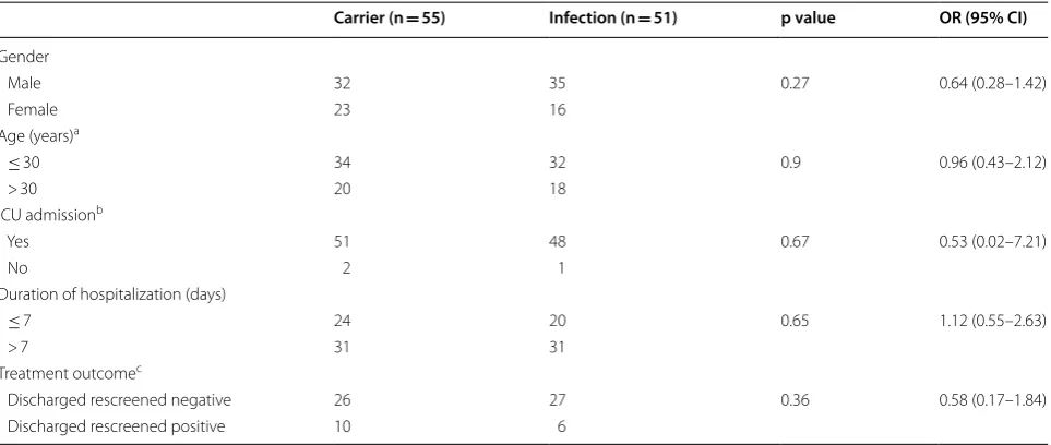
were less frequent (1.9% each). One strain was isolated from cerebrospinal fluid (0.94%) and one from a patient with a urinary tract infection (0.94%). Fifty-five strains (51.9%) were isolated from colonization sites such as nose (40.6%), throat (6.6%), axilla (2.8%) and groin (1.9%). MRSA strains isolated from colonization sites were iso-lated from Patients hospitalized for diseases not reiso-lated to MRSA infection. The detection of MRSA in these patients was performed as a part of infection control pol-icy which placed patients suspected or confirmed to be MRSA positive under contact isolation until decoloniza-tion is proven after successful prophylactic nasal mupi-rocin ointment and chlorhexidine body wash therapy. There was no statistically significant correlation between MRSA isolation from infection or colonization sites on the basis of age, duration of hospitalization, ICU admis-sion, or outcome of treatment (Table 1).
All MRSA isolates were positive for mecA, femA, and
sa442 genes. The most frequently detected SCCmec gen-otype was SCCmec-IV (77.3%) followed by SCCmec-V (13.2%), and III (9.4%). Further subtyping of SCCmec-IV revealed that subtypes SCCmec-IVa and SCCmec-IVc are the only sub-types prevalent among the isolates (41.5 and 58.5%, respectively) (Table 2). SCCmec-IVa genotype was more frequently found in MRSA isolated from the carriers compared to infected individuals (p value 0.002, OR 3.84) (Table 2).
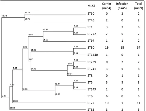
MLST genotyping of the MRSA isolates showed that ST80 is the most frequently encountered genotype in our study (34.9%) followed by ST22 (10.4%) (Table 2 and
Table 1 Frequency of MRSA isolates from carrier colonization sites and infection sites with demographic and clinical data
a One sample from each group has no data b Two samples from each group have no data c 37 patients discharged without further screen
Carrier (n = 55) Infection (n = 51) p value OR (95% CI)
Gender
Male 32 35 0.27 0.64 (0.28–1.42)
Female 23 16
Age (years)a
≤ 30 34 32 0.9 0.96 (0.43–2.12)
> 30 20 18
ICU admissionb
Yes 51 48 0.67 0.53 (0.02–7.21)
No 2 1
Duration of hospitalization (days)
≤ 7 24 20 0.65 1.12 (0.55–2.63)
> 7 31 31
Treatment outcomec
Discharged rescreened negative 26 27 0.36 0.58 (0.17–1.84)
Fig. 1). ST6 and ST22 were more frequently detected in the carrier group than the infection group (p value = 0.02 and 0.01, respectively) (Table 2). Other MLST genotypes, such as ST30, ST1930, ST239 and ST241 were more fre-quently detected among the infected patients but were statistically not significant (Table 2).
Toxic shock syndrome toxin 1 (TSST-1) was present in 14.2% of the isolates. Staphylococcal enterotoxin A (SEA) was the most frequently detected enterotoxin (35.8%)
(Table 2). SEB was significantly more prevalent among the carrier group compared to the infection group (p value 0.04) (Table 2).
Panton-Valentine leucocidin (PVL) was positive in 58.5% (62/106) of the total MRSA isolates. It was more frequently detected in the infection group of MRSA isolates (70.6%) compared to the carrier group isolates (47.3%) (p value 0.02, Table 2). However, when analyzing the prevalence of PVL among the MRSA isolates from Table 2 Molecular characterization of MRSA isolates from infection sites and carrier colonization sites
sea, seb, sec, sed, and see staphylococcal enterotoxin A, B, C, D, and E
eta and etb exfoliative toxin A and B
tst toxic shock toxin
a One of the isolates in infection group was a singleton in eBurst and could not be allocated to any CC b Includes also the 4 new STs from 6 samples according to eBurst
$ p value (two-tailed) from Hartley’s f test for equality of variance
Genotype Carrier (55) Infection (51) p value OR (95% CI)
SCCmec III 3 7 0.17 0.37 (0.07–1.48)
IVa 25 9 0.002 3.84 (1.59–9.81)
IVc 21 27 0.13 0.55 (0.25–1.20)
V 6 8 0.49 0.66 (0.20–2.1)
MLSTa CC1 (ST1, ST772) 5 8 0.30 0.53 (0.15–1.76)
CC5 (ST5, ST149) 4 5 0.64 0.71 (0.16–2.96)
CC6 (ST6) 6 0 0.02 Undefined
CC8 (ST8,ST239, ST241) 3 8 0.091 0.31 (0.06–1.19)
CC22 (ST22) 10 1 0.01 10.7 (1.70–242)
CC30 (ST30) 0 2 0.23 Undefined
CC45 (ST46) 2 0 0.27 Undefined
CC80 (ST80,ST1440)b 21 23 0.43 0.73 (0.33–1.59)
CC88 (ST88) 3 2 0.76 1.38 (0.20–12)
CC97 (ST97) 1 1 0.95 0.91 (0.02–36.08)
PVL Pos 26 36 0.02 0.38 (0.17–0.84)
Neg 29 15
sea Pos 16 22 0.14 0.54 (0.24–1.22)
Neg 39 29
seb Pos 7 1 0.04 7.18 (1.06–168.6)
Neg 48 50
sec Pos 0 0 NA NA
Neg 55 51
sed Pos 3 0 NA NA
Neg 52 51
see Pos 11 6 0.26 1.86 (0.63–5.88)
Neg 44 45
eta Pos 0 0 NA NA
Neg 55 51
etb Pos 0 0 NA NA
Neg 55 53
tst Pos 8 7 0.91 1.07 (0.35–3.34)
Neg 47 44
infection sites only based on whether these isolates are CA or HA-MRSA, PVL was more frequently detected in the CA-MRSA isolates but this difference was not statis-tically significant (Table 3).
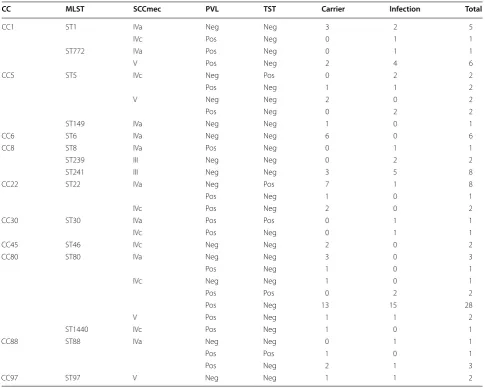
Clonal complex 80 (CC80) was the most frequent clonal complex detected in the study (41.51%) (Table 4)
followed by CC1 (12.26%), CC8 and CC22 (10.38% each), CC5 (8.49%), CC6 (5.66%), CC88 (4.71%). CC30, CC45, and CC97 were the least frequent isolates in the study (1.89% each) (Table 4). One new isolate was not possible to classify into any of the clonal complexes (0.94%) and seem to constitute singleton. Most of the clonal com-plexes harbored SCCmec type IV (IVa or IVc). SCCmec in ST239 and ST241 (CC8) was of type III only (Table 4).
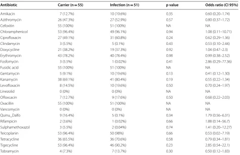
All MRSA isolates were sensitive to vancomycin, how-ever, MRSA strains isolated from infection sites had sig-nificantly higher minimum inhibitory concentrations (MIC) compared to strains isolated from carrier coloni-zation sites (p value 0.021) (Table 2). There was no other significant difference in the antibiotic susceptibility pat-tern between MRSA isolates from infection sites and car-rier colonization sites (Table 5). PVL was not associated with any antibiotic susceptibility pattern.
Fig. 1 Phylogenetic analysis of the MRSA MLST‑genotypes and their frequency among carrier and infection groups. 7 new types are not included in the analysis
Table 3 Prevalence of PVL among the MRSA isolates from infection sites
a According to our hospital infection control policy, the CDC classification of
MRSA was used to classify MRSA as CA or HA
CA-MRSAa
(n = 32) HA-MRSA
a
(n = 19) p value OR (95% CI)
PVL
Pos 24 12 0.4 1.7 (0.49–6.1)
Five new MLST alleles from seven samples were iden-tified in this study (Table 6). The isolates were obtained from scalp abscess, endotracheal aspirate, breast abscess, skin swab (cellulitis), urine, wound swab and one sam-ple from nasal swab (Table 6). These alleles produced new sequence types (STs) that have not been previously described in the MLST database. All except one were PVL positive and harbored the SCCmec-IV (Table 6).
Discussion
Extensive knowledge of the circulating MRSA strains is an important prerequisite for control and surveillance measures. Little is known about the genetic diversity of MRSA in the Middle East and the Gulf area. The Ara-bian Gulf countries attract a wide range of working man-power from different countries, which in turn enriches the microbial diversity in these countries. In the cur-rent study we characterized the MRSA isolates from
the Eastern Province in Saudi Arabia and compared the molecular types of these isolates from infection sites and from carrier colonization sites.
We did not find difference between the two groups regarding duration of hospitalization. Other studies have reported that MRSA infection prolongs the duration of hospitalization [20]. However, the type of infections in our study comprises mainly skin infections (abscess and wound) which do not necessarily require hospitalization. Interestingly, we found some molecular types confined to either the carrier or infection sites.
The most frequently detected SCCmec was type IV (IVa and IVc), which is in line with a previous study from the same region [21]. Type IV SCCmec is typically detected in MRSA isolates from samples outside hospitals [4, 5]. In our study SCCmec type IV strains were equally detected in carriers and from infection sites (Table 2). SCCmec subtype IVa was significantly associated with strains Table 4 Affiliation of MRSA isolates MLST, SCCmec, PVL, and TST to clonal complexes
CC MLST SCCmec PVL TST Carrier Infection Total
CC1 ST1 IVa Neg Neg 3 2 5
IVc Pos Neg 0 1 1
ST772 IVa Pos Neg 0 1 1
V Pos Neg 2 4 6
CC5 ST5 IVc Neg Pos 0 2 2
Pos Neg 1 1 2
V Neg Neg 2 0 2
Pos Neg 0 2 2
ST149 IVa Neg Neg 1 0 1
CC6 ST6 IVa Neg Neg 6 0 6
CC8 ST8 IVa Pos Neg 0 1 1
ST239 III Neg Neg 0 2 2
ST241 III Neg Neg 3 5 8
CC22 ST22 IVa Neg Pos 7 1 8
Pos Neg 1 0 1
IVc Pos Neg 2 0 2
CC30 ST30 IVa Pos Pos 0 1 1
IVc Pos Neg 0 1 1
CC45 ST46 IVc Neg Neg 2 0 2
CC80 ST80 IVa Neg Neg 3 0 3
Pos Neg 1 0 1
IVc Neg Neg 1 0 1
Pos Pos 0 2 2
Pos Neg 13 15 28
V Pos Neg 1 1 2
ST1440 IVc Pos Neg 1 0 1
CC88 ST88 IVa Neg Neg 0 1 1
Pos Pos 1 0 1
Pos Neg 2 1 3
isolated from carrier colonization sites. Sccmec type V, which is commonly associated with CA-MRSA infections [5], was equally detected in isolates from carriers and infection sites in our study. Similarly the sequence types ST6 and ST22 (except for one case) were only detected in MRSA isolates from carriers in our study.
PVL was highly prevalent among our isolates (56.5%), an observation which has been reported elsewhere [21, 22]. However, other studies reported a lower prevalence
(8–12%) [23, 24]. Moreover, PVL was significantly asso-ciated with MRSA strains isolated from infection sites compared to carriers. Among MRSA isolates from infec-tion sites, PVL was more frequently found in CA-MRSA, which is consistent with the literature [25]. Nonetheless, our study found that a large number of HA-MRSA iso-lates from infection sites harbor PVL (63.1%). This indi-cates that PVL-positive strains are invading hospitals and causing infections, which is in line with previous reports Table 5 Frequency of antibiotic resistance among the isolated strains
Antibiotic Carrier (n = 55) Infection (n = 51) p value Odds ratio (CI 95%)
Amikacin 7 (12.7%) 10 (19.6%) 0.35 0.60 (0.20–1.74)
Azithromycin 26 (47.3%) 27 (52.9%) 0.57 0.80 (0.37–1.72)
Cefoxitin 55 (100%) 51 (100%) NA NA
Chloramphenicol 53 (96.4%) 49 (96.1%) 0.94 1.08 (0.11–10.71)
Ciprofloxacin 27 (49.1%) 31 (60.8%) 0.24 0.62 (0.29–1.36)
Clindamycin 3 (5.5%) 5 (0.1%) 0.43 0.53 (0.10–2.44)
Doxycycline 21 (38.2%) 19 (37.3%) 0.92 1.04 (0.47–2.3)
Erythromycin 43 (78.2%) 40 (78.4%) 0.98 0.99 (0.38–2.52)
Fosfomycin 3 (5.5%) 1 (0.02%) 0.41 2.86 (0.29–77.36)
Fusidic acid 55 (100%) 51 (100%) NA NA
Gentamycin 5 (9.1%) 10 (19.6%) 0.13 0.41 (0.12–1.30)
Kanamycin 38 (69.1%) 41 (80.4%) 0.19 0.55 (0.22–1.34)
Levofloxacin 8 (14.5%) 10 (19.6%) 0.50 0.70 (0.24–1.97)
Linezolid 0 (0%) 0 (0%) NA NA
Ofloxcacin 7 (12.7%) 9 (17.6%) 0.50 0.68 (0.22–2.03)
Oxacillin 55 (100%) 51 (100%) NA NA
Vancomycin 0 (0%) 0 (0%) NA NA
Quinu_Dalfo 9 (16.4%) 5 (0.1%) 0.34 1.79 (0.56–6.31)
Rifampicin 2 (3.6%) 1 (0.02%) 0.66 1.88 (0.14–56.7)
Sulphamethoxazol 3 (5.5%) 2 (0.04%) 0.74 1.41 (0.20–12.27)
Teicoplanin 53 (96.4%) 50 (98%) 0.66 0.53 (0.02–7.19)
Tetracycline 36 (65.5%) 36 (70.6%) 0.58 0.79 (0.34–1.81)
Tigecycline 53 (96.4%) 46 (90.2%) 0.23 2.85 (0.54–22.1)
Tobramycin 4 (7.3%) 7 (13.7%) 0.30 0.50 (0.12–1.83)
Table 6 Characteristics of the new ST alleles found in the study
ND not determined, Pos positive, Neg negative
MLST genes CC SSCmec PVL Type of sample
arcC aroE glpF gmk pta tpi yqil
1 3 1 14 4 51 10 80 IVc Pos Scalp abscess
1 3 1 14 New 51 10 80 IVc Pos Endotracheal aspirate
1 New 1 14 11 51 10 80 IVc Pos Nasal swab
1 New 1 14 11 51 10 80 IVc Pos Breast abscess
1 New 1 14 11 51 10 80 IVc Pos Skin swab (cellulitis)
3 New 1 1 4 4 3 ND IVa Neg Urine
[26]. Despite the controversial role of PVL as virulence factor in MRSA pathogenesis [27, 28], it seems to play an important role in the successful evolutionary fitness of these strains [15]. Therefore, special control and clear-ance measure should be directed toward PVL-positive MRSA. Four MRSA strains were found to be simulta-neously positive for both PVL and TSST-1 genes in our study, which is otherwise uncommon [29]. One of the four isolates was obtained from a patient with severe necrotizing pneumonia that required ventilation and developed sepsis and one isolate was obtained from deep cutaneous abscess. The other two isolates came from surgical wound infection and colonization sites. A simi-lar finding has also been reported in the Middle East and other countries [29–32].
The most commonly identified clonal complex in our study was the CC80-MRSA-IV. This has been previously reported in several countries in the Middle East and North Africa and has also been reported from Riyadh in the central region of Saudi Arabia [11, 21, 22, 33–37]. This could indicate that it is the most common MRSA strain circulating in Saudi Arabia. This clone represents the European CA-MRSA clone. The spread of this clone in the Middle East and North Africa and the presence of sporadic cases in Europe suggest that this clone was introduced to Europe through migration from both the Middle East and North Africa. CC80-MRSA-IV isolates are usually PVL positive. However, PVL negative clones have been sporadically reported [11, 21, 35, 38, 39]. Four of the CC80-MRSA-IV isolates in our study were PVL negative. Since it has been suggested that PVL provides fitness to this isolate, in addition to the presence of this isolate in small numbers, it could be assumed that they are sporadic deletion variants. ST1440-IV was detected in only one sample in the present study. This isolate is very rare and has only been reported in Tunisia [40]. ST80-MRSA-V was identified in two cases in our study. This genotype has not been reported previously and seems to be a new isolate as all reported ST80 strains harbor the SCCmec genotype IV.
Two sequence types (ST1 and ST772) belonging to CC1 were identified in our study. Most ST1 strains were PVL negative and harbored SCCmec-IV, which may resemble the West Australian strain WA MRSA-1. The rest were PVL-positive strain, which may resemble the USA400 strain. ST772-MRSA-V is usually more distinct from the other CC1 isolates and is known as the Bengal Bay clone. This strain is more prevalent in India [41] and was also detected in many European countries, where it was linked to transmission associated with migration to these countries [42]. Similarly, the detection of this strain in Saudi Arabia could be explained by the presence of the large number of Indian workers in the country. The
detection of ST772-MRSA-IV has not been previously reported and it was isolated from one case in our study.
The UK-MRSA-15 strain, which is also called Barnim Epidemic strain (C22-MRSA-IV PVL-negative), was detected in 7.5% of our isolates and was found more fre-quently among carriers. Similar to other reports, it differs from the UK-MRSA-15 strain in that it is positive for the
tst1 gene and has the SSC-IVa [22]. The other PVL-posi-tive strain was detected in three cases and was confined to the carrier group. This strain has been reported in many other countries, including the neighboring United Arab Emirates [15].
Contrary to other studies from Riyadh in Saudi Ara-bia [21, 22], ST241-MRSA-III PVL-negative strain was the most frequently detected isolate belonging to CC8, which indicates interregional variation in the distribu-tion of these strains. This isolate has been reported in Kuwait, Tunisia, Taiwan and Poland [43–46]. The other CC8 strains were the ST239-MRSA-III and the USA300 (ST8-MRSA-IV PVL-positive) were previously reported from Riyadh region [21, 22].
Similar to previous reports from Saudi Arabia, PVL-negative and positive CC5 strains harboring the SCC-mec IV or V were identified in our study [21, 22]. These strains were isolated also in Qatar, Tunisia and Egypt [24, 44, 47, 48]. One sample in our study has genotypes simi-lar to the Maltese strain (CC 5/ST149) [49].
All the CC6 isolates were detected in the carrier group and they were also reported previously in Riyadh and the United Arab Emirate [15, 22], which indicates that it is an established strain in the region.
Other clonal complexes such as CC30, CC45 and CC97 were isolated from 1.9% of the cases each. ST30-MRSA-IV PVL-positive resembles the US1100 (southwest pacific clone) and is widespread in Europe and the Gulf region [15, 50]. Interestingly, CC45-MSSA strains were previously detected in Saudi Arabia from nasal coloniza-tion carriers [51]. Therefore, it is possible that this strain has acquired the mecA gene and transformed to MRSA. CC45-MRSA-IV was also reported previously in one case from Riyadh in Saudi Arabia [21] and one case from Hong Kong. Similarly, the CC97-MRSA-V was sporadi-cally reported from many countries, including the Middle Eastern region [15].
used to measure the MIC, E test, may have contributed to the increased MICs values [54]. However, a recent meta-analysis by van Hal et al. has shown that despite the methodology applied, these MICs close to the break point of 2.0, such as the isolates representing infected sites in this study, carry poor prognosis irrespective of the infection site [55]. Appropriate alternatives in such circumstances include daptomycin in cases of bacteremia and Linezolid in pneumonias.
Five new sequence types from seven samples are reported in this study (Table 6). All except one were iso-lated from infection sites. They belong to CC80 according to the eBust website. Due to the diversity of nationalities working and living in the Gulf region, further studies should be conducted on MRSA to reveal and monitor the emergence of new strains.
Conclusions
A highly diverse population of MRSA isolates is reported from both carriers and patients’ infection sites in our study from the Eastern Province in Saudi Arabia. Some of the isolate types were more prevalent among the car-riers more than the infection sites. Therefore, screening and control measures should consider the polyclonal nature of the problem. A good understanding of the genetic spectrum of the MRSA isolates and its preva-lence in the population calls for continuous surveillance and clearance measures. With SCCmec type IV being most prevalent; this suggests a community origin of most MRSA strains which is also supported by low resistance rates to clindamycin and sulfonamides. Therefore very well designed surveillance and intervention strategies, in particular restricting antimicrobial use, should be implemented to prevent emergence and control spread of MRSA in the community.
Abbreviations
MRSA: methicillin resistant Staphylococcus aureus; S. aureus: Staphylococcus aureus; PBP2a: penicillin binding protein 2a; mecA gene: methicillin resistance gene; SCCmec: staphylococcal cassette chromosome mec; HA‑MRSA: hospital acquired MRSA; CA‑MRSA: community acquired MRSA; lukF‑PV: leukocidin gene subunit F; lukS‑PV: leukocidin gene subunit S; PVL: Panton–Valentine leukocidin; CC: clonal complexes; MLST: multi locus sequence typing; ST: sequence types; femA: factor essential for mecA; PCR: polymerase chain reac‑ tion; ICU: intensive care unit; MIC: minimum inhibitory concentration.
Authors’ contributions
KRA: overall study design, raising fund, experimental design, and manuscript writing and editing. SR: experimental work and manuscript writing. FA: analysis of MLST sequences. AA: collection of bacterial isolates, collection of demo‑ graphic data, and manuscript reading and editing. AD: collection of bacterial isolates. AKA: manuscript writing and editing. All authors read and approved the final manuscript.
Author details
1 Department of Epidemic Diseases Research, Institute for Research and Medi‑
cal Consultations (IRMC), Imam Abdulrahman Bin Faisal University (IAU), P.O. Box 1982, Dammam 31441, Saudi Arabia. 2 Department of Microbiology,
College of Medicine, Imam Abdulrahman Bin Faisal University (IAU), P.O. Box 1982, Dammam 31441, Saudi Arabia. 3 Department of Biochemistry, Col‑
lege of Medicine, Imam Abdulrahman Bin Faisal University (IAU), P.O. Box 1982, Dammam 31441, Saudi Arabia.
Acknowledgements
The authors are grateful to Mr. Nestor Recella, Mrs. Janaika Yu Logan and the technical staff in the diagnostic microbiology laboratory (KFHU) for their technical assistance. The authors acknowledge the use of the S. aureus MLST database which is located at Imperial College London. This work is funded by the Deanship for Scientific Research at the University of Dammam (Project Number: 2013229).
Competing interests
The authors declare that they have no competing interests.
Availability of data
Please contact author for data requests.
Consent for publication Not applicable.
Ethics approval and consent to participate
Ethical approval for the study was obtained from the Institutional Review Board at the University of Dammam (IRB‑2013‑08‑023).
Funding
This work was funded by the Deanship for Scientific Research at the University of Dammam for Khaled R. Alkharsah (Project Number 2013229). The role of the funding body was financial support for the study and had no role in design of the study and collection, analysis, and interpretation of data and in writing the manuscript.
Informed consent Not applicable.
Publisher’s Note
Springer Nature remains neutral with regard to jurisdictional claims in pub‑ lished maps and institutional affiliations.
Received: 13 June 2017 Accepted: 8 March 2018
References
1. Conner‑Kerr TA, Sullivan PK, Gaillard J, Franklin ME, Jones RM. The effects of ultraviolet radiation on antibiotic‑resistant bacteria in vitro. Ostomy Wound Manag. 1998;44(10):50–6.
2. Chambers HF, Deleo FR. Waves of resistance: Staphylococcus aureus in the antibiotic era. Nat Rev Microbiol. 2009;7(9):629–41.
3. Wong H, Louie L, Lo RY, Simor AE. Characterization of Staphylococcus aureus isolates with a partial or complete absence of staphylococcal cas‑ sette chromosome elements. J Clin Microbiol. 2010;48(10):3525–31. 4. Liu J, Chen D, Peters BM, Li L, Li B, Xu Z, Shirliff ME. Staphylococcal chro‑
mosomal cassettes mec (SCCmec): a mobile genetic element in methicil‑ lin‑resistant Staphylococcus aureus. Microb Pathog. 2016;101:56–67. 5. David MZ, Daum RS. Community‑associated methicillin‑resistant
Staphy-lococcus aureus: epidemiology and clinical consequences of an emerging epidemic. Clin Microbiol Rev. 2010;23(3):616–87.
6. Seybold U, Kourbatova EV, Johnson JG, Halvosa SJ, Wang YF, King MD, Ray SM, Blumberg HM. Emergence of community‑associated methicillin‑ resistant Staphylococcus aureus USA300 genotype as a major cause of health care—associated blood stream infections. Clin Infect Dis. 2006;42(5):647–56.
8. Deresinski S. Methicillin‑resistant Staphylococcus aureus: an evo‑ lutionary, epidemiologic, and therapeutic odyssey. Clin Infect Dis. 2005;40(4):562–73.
9. Enright MC, Day NP, Davies CE, Peacock SJ, Spratt BG. Multilocus sequence typing for characterization of methicillin‑resistant and methicillin‑susceptible clones of Staphylococcus aureus. J Clin Microbiol. 2000;38(3):1008–15.
10. Otto M. MRSA virulence and spread. Cell Microbiol. 2012;14(10):1513–21. 11. Tokajian S. New epidemiology of Staphylococcus aureus infections in the
Middle East. Clin Microbiol Infect. 2014;20(7):624–8.
12. Chen CJ, Wang SC, Chang HY, Huang YC. Longitudinal analysis of methicillin‑resistant and methicillin‑susceptible Staphylococcus aureus carriage in healthy adolescents. J Clin Microbiol. 2013;51(8):2508–14. 13. Dulon M, Peters C, Schablon A, Nienhaus A. MRSA carriage among
healthcare workers in non‑outbreak settings in Europe and the United States: a systematic review. BMC Infect Dis. 2014;14:363.
14. Dulon M, Haamann F, Peters C, Schablon A, Nienhaus A. MRSA prevalence in European healthcare settings: a review. BMC Infect Dis. 2011;11:138. 15. Monecke S, Coombs G, Shore AC, Coleman DC, Akpaka P, Borg M, Chow
H, Ip M, Jatzwauk L, Jonas D, et al. A field guide to pandemic, epidemic and sporadic clones of methicillin‑resistant Staphylococcus aureus. PLoS ONE. 2011;6(4):e17936.
16. CLSI. Performance standards for antimicrobial susceptibility testing; eighteenth informational supplement. In CLSI document M100‑S26. Wayne: CLSI; 2016.
17. Al‑Talib H, Yean CY, Al‑Khateeb A, Hassan H, Singh KK, Al‑Jashamy K, Ravi‑ chandran M. A pentaplex PCR assay for the rapid detection of methicillin‑ resistant Staphylococcus aureus and Panton–Valentine Leucocidin. BMC Microbiol. 2009;9:113.
18. Mehrotra M, Wang G, Johnson WM. Multiplex PCR for detection of genes for Staphylococcus aureus enterotoxins, exfoliative toxins, toxic shock syndrome toxin 1, and methicillin resistance. J Clin Microbiol. 2000;38(3):1032–5.
19. Ghaznavi‑Rad E, Nor Shamsudin M, Sekawi Z, van Belkum A, Neela V. A simplified multiplex PCR assay for fast and easy discrimination of globally distributed staphylococcal cassette chromosome mec types in meticillin‑ resistant Staphylococcus aureus. J Med Microbiol. 2010;59(Pt 10):1135–9. 20. Rasmussen RV, Fowler VG Jr, Skov R, Bruun NE. Future challenges and
treatment of Staphylococcus aureus bacteremia with emphasis on MRSA. Future Microbiol. 2011;6(1):43–56.
21. Monecke S, Skakni L, Hasan R, Ruppelt A, Ghazal SS, Hakawi A, Slickers P, Ehricht R. Characterisation of MRSA strains isolated from patients in a hospital in Riyadh, Kingdom of Saudi Arabia. BMC Microbiol. 2012;12:146. 22. Senok A, Ehricht R, Monecke S, Al‑Saedan R, Somily A. Molecular char‑
acterization of methicillin‑resistant Staphylococcus aureus in nosocomial infections in a tertiary‑care facility: emergence of new clonal complexes in Saudi Arabia. New Microbes New Infect. 2016;14:13–8.
23. Moussa I, Shibl AM. Molecular characterization of methicillin‑resistant Staphylococcus aureus recovered from outpatient clinics in Riyadh, Saudi Arabia. Saudi Med J. 2009;30(5):611–7.
24. Abou Shady HM, Bakr AE, Hashad ME, Alzohairy MA. Staphylococcus aureus nasal carriage among outpatients attending primary health care centers: a comparative study of two cities in Saudi Arabia and Egypt. Braz J Infect Dis. 2015;19(1):68–76.
25. Vandenesch F, Naimi T, Enright MC, Lina G, Nimmo GR, Heffernan H, Liassine N, Bes M, Greenland T, Reverdy ME, et al. Community‑acquired methicillin‑resistant Staphylococcus aureus carrying Panton–Valentine leu‑ kocidin genes: worldwide emergence. Emerg Infect Dis. 2003;9(8):978–84. 26. Bukharie HA. Increasing threat of community‑acquired methicillin‑resist‑
ant Staphylococcus aureus. Am J Med Sci. 2010;340(5):378–81. 27. Bae IG, Tonthat GT, Stryjewski ME, Rude TH, Reilly LF, Barriere SL, Genter
FC, Corey GR, Fowler VG Jr. Presence of genes encoding the Panton–Val‑ entine leukocidin exotoxin is not the primary determinant of outcome in patients with complicated skin and skin structure infections due to methicillin‑resistant Staphylococcus aureus: results of a multinational trial. J Clin Microbiol. 2009;47(12):3952–7.
28. Otto M. A MRSA‑terious enemy among us: end of the PVL controversy? Nat Med. 2011;17(2):169–70.
29. Li Z, Stevens DL, Hamilton SM, Parimon T, Ma Y, Kearns AM, Ellis RW, Bryant AE. Fatal S. aureus hemorrhagic pneumonia: genetic analysis
of a unique clinical isolate producing both PVL and TSST‑1. PLoS ONE. 2011;6(11):e27246.
30. Al‑Bakri AG, Al‑Hadithi H, Kasabri V, Othman G, Kriegeskorte A, Becker K. The epidemiology and molecular characterization of methicillin‑resistant staphylococci sampled from a healthy Jordanian population. Epidemiol Infect. 2013;141(11):2384–91.
31. Harastani HH, Tokajian ST. Community‑associated methicillin‑resistant Staphylococcus aureus clonal complex 80 type IV (CC80‑MRSA‑IV) isolated from the Middle East: a heterogeneous expanding clonal lineage. PLoS ONE. 2014;9(7):e103715.
32. Holmes A, Ganner M, McGuane S, Pitt TL, Cookson BD, Kearns AM. Staphy-lococcus aureus isolates carrying Panton–Valentine leucocidin genes in England and Wales: frequency, characterization, and association with clinical disease. J Clin Microbiol. 2005;43(5):2384–90.
33. Tokajian ST, Khalil PA, Jabbour D, Rizk M, Farah MJ, Hashwa FA, Araj GF. Molecular characterization of Staphylococcus aureus in Lebanon. Epide‑ miol Infect. 2010;138(5):707–12.
34. Enany S, Yaoita E, Yoshida Y, Enany M, Yamamoto T. Molecular charac‑ terization of Panton–Valentine leukocidin‑positive community‑acquired methicillin‑resistant Staphylococcus aureus isolates in Egypt. Microbiol Res. 2010;165(2):152–62.
35. Udo EE, Sarkhoo E. The dissemination of ST80‑SCCmec‑IV community‑ associated methicillin resistant Staphylococcus aureus clone in Kuwait hospitals. Ann Clin Microbiol Antimicrob. 2010;9:31.
36. Bekkhoucha SN, Cady A, Gautier P, Itim F, Donnio PY. A portrait of Staphy-lococcus aureus from the other side of the Mediterranean Sea: molecular characteristics of isolates from Western Algeria. Eur J Clin Microbiol Infect Dis. 2009;28(5):553–5.
37. Ben Nejma M, Mastouri M, Jrad BB, Nour M. Characterization of ST80 Panton–Valentine leukocidin‑positive community‑acquired methicillin‑ resistant Staphylococcus aureus clone in Tunisia. Diagn Microbiol Infect Dis. 2013;77(1):20–4.
38. Djoudi F, Bonura C, Benallaoua S, Touati A, Touati D, Aleo A, Cala C, Fasciana T, Mammina C. Panton–Valentine leukocidin positive sequence type 80 methicillin‑resistant Staphylococcus aureus carrying a staphylo‑ coccal cassette chromosome mec type IVc is dominant in neonates and children in an Algiers hospital. New Microbiol. 2013;36(1):49–55. 39. Tristan A, Bes M, Meugnier H, Lina G, Bozdogan B, Courvalin P, Reverdy
ME, Enright MC, Vandenesch F, Etienne J. Global distribution of Panton– Valentine leukocidin—positive methicillin‑resistant Staphylococcus aureus, 2006. Emerg Infect Dis. 2007;13(4):594–600.
40. Mariem BJ, Ito T, Zhang M, Jin J, Li S, Ilhem BB, Adnan H, Han X, Hiramatsu K. Molecular characterization of methicillin‑resistant Panton–Valentine leukocidin positive staphylococcus aureus clones disseminating in Tuni‑ sian hospitals and in the community. BMC Microbiol. 2013;13:2. 41. D’Souza N, Rodrigues C, Mehta A. Molecular characterization of methicil‑
lin‑resistant Staphylococcus aureus with emergence of epidemic clones of sequence type (ST) 22 and ST 772 in Mumbai, India. J Clin Microbiol. 2010;48(5):1806–11.
42. Huh K, Chung DR. Changing epidemiology of community‑associated methicillin‑resistant Staphylococcus aureus in the Asia‑Pacific region. Expert Rev Anti Infect Ther. 2016;14(11):1007–22.
43. Boswihi SS, Udo EE, Al‑Sweih N. Shifts in the clonal distribution of methi‑ cillin‑resistant Staphylococcus aureus in Kuwait Hospitals: 1992–2010. PLoS ONE. 2016;11(9):e0162744.
44. Elhani D, Gharsa H, Kalai D, Lozano C, Gomez P, Boutheina J, Aouni M, Barguellil F, Torres C, Ben Slama K. Clonal lineages detected amongst tetracycline‑resistant meticillin‑resistant Staphylococcus aureus isolates of a Tunisian hospital, with detection of lineage ST398. J Med Microbiol. 2015;64(6):623–9.
45. Wang WY, Chiueh TS, Sun JR, Tsao SM, Lu JJ. Molecular typing and phenotype characterization of methicillin‑resistant Staphylococcus aureus isolates from blood in Taiwan. PLoS ONE. 2012;7(1):e30394.
46. Mlynarczyk A, Szymanek‑Majchrzak K, Grzybowska W, Durlik M, Deborska‑Materkowska D, Paczek L, Chmura A, Swoboda‑Kopec E, Tyski S, Mlynarczyk G. Molecular and phenotypic characteristics of methicillin‑ resistant Staphylococcus aureus strains isolated from hospitalized patients in transplantation wards. Transplant Proc. 2014;46(8):2579–82.
• We accept pre-submission inquiries
• Our selector tool helps you to find the most relevant journal • We provide round the clock customer support
• Convenient online submission • Thorough peer review
• Inclusion in PubMed and all major indexing services • Maximum visibility for your research
Submit your manuscript at www.biomedcentral.com/submit
Submit your next manuscript to BioMed Central
and we will help you at every step:
in a Qatari hospital from multinational patients. Clin Microbiol Infect. 2014;20(2):169–73.
48. Kechrid A, Perez‑Vazquez M, Smaoui H, Hariga D, Rodriguez‑Banos M, Vindel A, Baquero F, Canton R, Del Campo R. Molecular analysis of community‑acquired methicillin‑susceptible and resistant Staphylococcus aureus isolates recovered from bacteraemic and osteomyelitis infections in children from Tunisia. Clin Microbiol Infect. 2011;17(7):1020–6. 49. Scicluna EA, Shore AC, Thurmer A, Ehricht R, Slickers P, Borg MA, Coleman
DC, Monecke S. Characterisation of MRSA from Malta and the descrip‑ tion of a Maltese epidemic MRSA strain. Eur J Clin Microbiol Infect Dis. 2010;29(2):163–70.
50. Monecke S, Gavier‑Widen D, Hotzel H, Peters M, Guenther S, Lazaris A, Loncaric I, Muller E, Reissig A, Ruppelt‑Lorz A, et al. Diversity of Staphylococcus aureus isolates in European wildlife. PLoS ONE. 2016;11(12):e0168433.
51. Sarkar A, Raji A, Garaween G, Soge O, Rey‑Ladino J, Al‑Kattan W, Shibl A, Senok A. Antimicrobial resistance and virulence markers in methicillin
sensitive Staphylococcus aureus isolates associated with nasal coloniza‑ tion. Microb Pathog. 2016;93:8–12.
52. Wang G, Hindler JF, Ward KW, Bruckner DA. Increased vancomycin MICs for Staphylococcus aureus clinical isolates from a university hospital dur‑ ing a 5‑year period. J Clin Microbiol. 2006;44(11):3883–6.
53. Hidayat LK, Hsu DI, Quist R, Shriner KA, Wong‑Beringer A. High‑dose vancomycin therapy for methicillin‑resistant Staphylococcus aureus infec‑ tions: efficacy and toxicity. Arch Intern Med. 2006;166(19):2138–44. 54. Prakash V, Lewis JS 2nd, Jorgensen JH. Vancomycin MICs for methicillin‑
resistant Staphylococcus aureus isolates differ based upon the susceptibil‑ ity test method used. Antimicrob Agents Chemother. 2008;52(12):4528. 55. van Hal SJ, Lodise TP, Paterson DL. The clinical significance of vanco‑