R E S E A R C H A R T I C L E
Open Access
Functional divergence of the brain-size regulating
gene
MCPH1
during primate evolution and the
origin of humans
Lei Shi
1, Ming Li
1,2, Qiang Lin
1,2, Xuebin Qi
1and Bing Su
1*Abstract
Background:One of the key genes that regulate human brain size,MCPH1has evolved under strong Darwinian positive selection during the evolution of primates. During this evolution, the divergence ofMCPH1protein sequences among primates may have caused functional changes that contribute to brain enlargement. Results:To test this hypothesis, we used co-immunoprecipitation and reporter gene assays to examine the activating and repressing effects ofMCPH1on a set of its down-stream genes and then compared the functional outcomes of a series of mutantMCPH1proteins that carry mutations at the human- and great-ape-specific sites. The results demonstrate that the regulatory effects of humanMCPH1and rhesus macaqueMCPH1are different in three of eight down-stream genes tested (p73,cyclinE1andp14ARF), suggesting a functional divergence ofMCPH1 between human and non-human primates. Further analyses of the mutantMCPH1proteins indicated that most of the human-specific mutations could change the regulatory effects on the down-stream genes. A similar result was also observed for one of the four great-ape-specific mutations.
Conclusions:Collectively, we propose that during primate evolution in general and human evolution in particular, the divergence ofMCPH1protein sequences under Darwinian positive selection led to functional modifications, providing a possible molecular mechanism of howMCPH1contributed to brain enlargement during primate evolution and human origin.
Keywords:MCPH1,E2F1, Brain size, Primates, Evolution, Functional divergence
Background
A dramatic increase in brain size is one of the hallmarks of human evolution, but despite the significance of this trait, the causal molecular mechanism underlying this expansion is unclear [1]. Until recently, addressing this question with genetic tools has been difficult because the dramatically enlarged brain is a human-specific trait. Genetic studies of a rare brain developmental disorder, human autosomal primary microcephaly syndrome (MCPH, OMIM251200), have uncovered a set of genes that regulate brain development. To date, seven genes have been identified as being responsible for this syndrome:
MCPH1, also known as BRIT1 (BRCT-repeat inhibitor
of hTERT expression) [2,3], WDR62 (WD repeat domain
62;MCPH2) [4-6],CDK5RAP2 (cyclin-dependnet
kin-ase 5 regulatory associated protein 2; MCPH3) [7],
CEP152 (centrosomal protein 152 kDa; MCPH4) [8],
ASPM (abnormal spindle like microcephaly associated protein; MCPH5) [9], and CENPJ (centromeric protein J; MCPH6) [7] andSTIL(SCL/TAL1 interrupting locus; MCPH7) [10].
Previous evolutionary analyses of these microcephaly genes showed that four of them, ASPM, CDK5RAP2,
CENPJ and MCPH1, evolved rapidly under Darwinian
positive selection during the evolution of human and non-human primates [11-16]. ASPM also experienced positive selection across anthropoids [14-16], while
CDK5RAP2 and CENPJ showed accelerated rates of
non-synonymous substitutions over the course of pri-mate evolution [11,16]. The signal of positive selection
on MCPH1 was observed in the common ancestor of
* Correspondence:[email protected]
1State Key Laboratory of Genetic Resources and Evolution, Kunming Institute
of Zoology, Chinese Academy of Sciences, 32 East Jiao-Chang Road, Kunming, Yunnan 650223, PR China
Full list of author information is available at the end of the article
great apes and humans as well as in the human lineage [12], although another study onMCPH1only detected positive selection in the anthropoids as a whole and no particular acceleration in the human lineage [16]. This rapid evolution suggests these genes may have had a key role in the evolutionary enlargement of the brain, although the link ofCENPJandMCPH1to the evolution of gross brain size was not confirmed in the association analysis of absolute neonatal brain size among primates [16]. Among the four rapidly evolving microcephaly genes, onlyASPMhas been experimentally studied to detect the evolutionary consequence of protein sequence changes; mice carrying a truncated ASPM protein were shown to have reductions of both brain and testis size, while the transgenic mouse carrying human ASPM could rescue this phenotype, but did not cause any additional enlargement of the brain [17].
MCPH1was the first gene identified as being responsible for autosomal recessive primary microcephaly, charac-terized by significantly reduced brain volume, mental retardation and premature chromosome condensation (PCC) syndrome [2,18]. The MCPH1 gene encodes a 2,508-bp-long coding sequence (CDS) with 14 exons, spanning about 240 kb at 8p23. The MCPH1 protein contains three BRCA1-Carboxyl Terminal (BRCT) do-mains, including one N terminal BRCT domain and a tandem pair at the C terminus. Numerous studies have implied that the BRCT domains ofMCPH1function as the key component for protein-protein interaction; this seems likely as the interaction of theMCPH1 tandem BRCT domains with proteins likeE2F1and r-H2AX is required for the activation of cell cycle checkpoint, DNA repair and apoptosis [19-23]. Several studies have likewise suggested thatMCPH1may also function as a tumor suppressor [3,23].
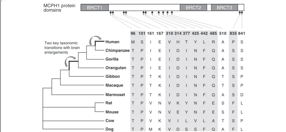
Evolutionary studies of MCPH1 have demonstrated a rapid change in protein sequence associated with the brain enlargement during primate evolution and human origin. Interestingly, during two key taxonomic transi-tions in primates, that is, between lesser apes and great apes, and between great apes and humans, absolute brain volume was greatly enlarged, and MCPH1 might be involved in this process [12]. Additionally, MCPH1is also highly polymorphic in human populations and still carries the molecular signature of on-going positive selection [12]. Human population studies have reported a sex-specific association between a MCPH1 sequence variant and brain volume [24,25]. These results suggest that the protein sequence changes, especially the human-specific changes ofMCPH1may have caused the functional changes that explain the genetic basis for the evolution of brain size in primates.
Previously, the MCPH1 protein has been shown to play an essential role during cell cycle and cell apoptosis
and it can physically interact with E2F1 to form a complex and bind the promoters of the target genes for regulating their transcriptional activities [19]. Beyond this,MCPH1alone can also function as a transcriptional regulator, and we previously demonstrated MCPH1could function as a transcriptional repressor [26]. Together, these regulatory mechanisms allow the experimental testing of the functional changes ofMCPH1during pri-mate evolution.
To detect if the protein sequence divergence ofMCPH1
among primates may confer any functional alterations, we selected eight known down-stream genes regulated by
E2F1andMCPH1:p73[19],p107[19],p18[27],p27[28],
p14ARF [19],Caspase7 [19], CyclinE1 [19] and hTERT
[26]. These genes are involved in cell proliferation and apoptosis, critical processes regulating brain development (see Additional file 1: Figure S1). We tested the activating effects (together with E2F1) and the repressing effects
(MCPH1 alone) of MCPH1 on these genes’ promoter
when introducing mutations at the sites containing human- and great-ape-specific amino acid changes. Our results demonstrated that most of the human-specific amino acid substitutions could influence the regulatory effects ofMCPH1on the down-stream genes, and a similar effect was also seen for one of the four great-ape-specific changes, suggesting that the species and lineage specific mutations ofMCPH1are indeed functional and potentially contributed to brain enlargement over the course of primate evolution.
Results
Identification of lineage-specificMCPH1amino acid substitutions
In order to identify lineage-specific amino acid substitutions,
MCPH1 orthologs of representative primate and other
mammalian species were obtained from the NCBI, EMBL and UniProt databases. We used a total of 11 species includ-ing 7 primates (human, great apes, lesser ape, Old World monkey and New World monkey) and 4 other representa-tive mammalian species (mouse, rat, dog and cow) (Figure 1). Using MUSCLE and Clustal W, we aligned the MCPH1
amino acid substitutions are shown in Additional file 3: Table S1 and the schematic map of theMCPH1protein domains labeled with the lineage-specific substitutions are shown in Figure 1. All these lineage-specific sites were selected to generate mutantMCPH1proteins for the reporter gene assays in order to test their functional effects.
Test of protein-protein interaction betweenMCPH1 andE2F1
Previous studies suggested human MCPH1 (hMCPH1) could interact with E2F1 in vitro and in vivo [19]. To test if the non-human primateMCPH1can also interact with E2F1, we cloned the rhesus macaque MCPH1
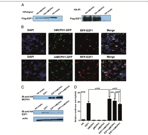
(mMCPH1). The results of the co-immunoprecipitation
assay showed that mMPCH1 can also directly interact withE2F1, and no difference was observed for the intensity of the interaction with E2F1 between hMCPH1 and
mMCPH1(Figure 2A). Given the established MCPH1
-E2F1interaction in human cell lines [19], this protein-protein interaction mechanism seems likely to have been conserved during primate evolution. We also conducted a cellular co-localization assay for both
mMCPH1 and hMCPH1, and the results indicated that
both were co-localized with E2F1(Figure 2B), consistent with the results of the co-immunoprecipitation assay.
Divergent effects ofhMCPH1andmMCPH1on transcriptional regulation
MCPH1interacts withE2F1to enhance its transactivation activity by forming a complex, which binds to the promoters of the E2F1 target genes, including p73,
Caspase and RAD51, among others [19]. We, therefore,
tested the enhancing effect of MCPH1 using the E2F4B reporter vector commonly used as the positive control in testing the transactivation activity ofE2F1[29]. The expression of the HA-tagged MCPH1 and E2F1 were verified by Western blot using anti-HA monoclonal antibodies (Figure 2C). Our results showed thathMCPH1
could significantly enhance the transactivation activity of
E2F1, but no significant effect was observed formMCPH1
and gMCPH1 (the gibbon copy ofMCPH1) (Figure 2D).
In addition, the difference betweenE2F1+hMCPH1and
E2F1 + mMCPH1 is significant (P <0.01, ANOVA test).
The same trend was also seen when comparing E2F1 +
hMCPH1andE2F1+gMCPH1, though not statistically sig-nificant (Figure 2D). Interestingly, the difference between
E2F1 + gMCPH1 and E2F1 + mMCPH1 is significant
(P<0.05, ANOVA test), suggesting continuum of func-tional divergence ofMCPH1during primate evolution with a major shift in function between Old World monkeys and apes and a probable further shift during human evolution (Figure 2D).
To check the conservation of the E2F1 binding sites (CGCGC), we aligned the promoter sequences of the target genes among representative primate species. We found that most of theE2F1binding sites are highly conserved from humans to marmosets (see Additional file 4: Figure S3), ruling out the possibility of binding bias due to promoter sequence divergence. TheE2F1protein sequences are also highly conserved among primate species (Additional file 5: Figure S4), ruling out the possibility that the different effect
of MCPH1 is caused by mutations in E2F1. Collectively,
BRCT1 BRCT2 BRCT3
Two key taxonomic transitions with brain enlargements
MCPH1 protein domains
Human
Chimpanzee
Gorilla
Orangutan
Gibbon
Macaque
Marmoset
Rat
Mouse
Cow
Dog
96 101 161 167 310 314 377 425 442 485 510 835 841
M S I E V H T Y L R A P S
T P I E I D I N F Q A S S
T P I E I D I N F Q A S S
T P I E I D I N F Q A S S
T P T K I D I N F Q T S P
T P T K I D I N F Q T S P
T P T K I D I N F Q A S D
T P V N V K Y N F E S F L
T P V N V E Y N F E S F L
T P V K V I L V L A T S P
T P M K V D S S F Q A F L
these results suggest a functional divergence ofMCPH1
between humans and nonhuman primates.
We further tested a set ofE2F1target genes (p73,p107,
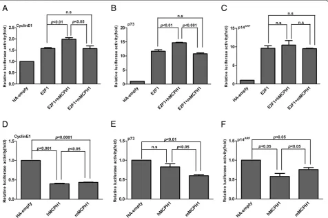
p18,p27, p14ARF,Caspase7and CyclinE1). The regulatory network of these genes is shown in Additional file 1: Figure S1. We found that the enhancing effect of
hMCPH1was significantly stronger thanmMCPH1for
CyclinE1 and p73 (Figure 3A, B) and a similar trend
was also observed forp14ARF, though not significantly (Figure 3C). No significant enhancing effects were detected for the other four genes, p18, p27, p107 and
Caspase7(see Additional file 6: Figure S5). Consistent with the result from the E2F4B assay, these data also suggest a functional divergence betweenhMCPH1andmMCPH1in enhancing the transactivation activity ofE2F1.
To further test the functional divergence between
hMCPH1and mMCPH1, we performed another assay to
detect the regulatory effect of MCPH1 alone on eight down-stream genes (p73, p107, p18, p27, p14ARF,
Caspase7,CyclinE1and hTERT). Among the eight genes
tested, seven showed a repressing effect for bothhMCPH1
and mMCPH1, while only one, Caspase7, showed an
activating effect. When comparing the effects between
hMCPH1 and mMCPH1, three genes (CyclinE1, p73
andp14ARF) showed significant differences (P<0.05) of repressing effect (Figure 3D–F), the same three which showed enhancing differences between hMCPH1 and
mMCPH1 in the E2F1-MCPH1 transactivation assay
(Figure 3A-C). For CyclinE1 and p14ARF, hMCPH1
showed a stronger repressing effect than mMCPH1, while the opposite effect was observed for p73. We did not detect significant between-species differences for the other four genes with repressing effect (p18,
p27, p107andhTERT) though the trend was the same
(see Additional file 7: Figure S6).Caspase 7was the only gene with an activating effect, and we also observed a significant difference betweenhMCPH1andmMCPH1
(P<0.05) (see Additional file 7: Figure S6D). Collectively,
the data from the two reporter gene assays suggest a clear functional divergence of MCPH1 between humans and rhesus macaques.
Detection of regulatory changes ofMCPH1for the human-specific sites
hMCPH1, similar to the difference between hMCPH1
and mMCPH1, suggesting that the human-specific
amino acid change at Site-96 can influence the enhancing effect ofMCPH1.
We next generated a series of mutanthMCPH1s carrying backward mutations at the other eight human-specific sites (101, 310, 314, 377, 425, 442, 485 and 835), and tested their enhancing effects. Five of the eight mutant
hMCPH1s (101, 310, 377, 425 and 835) showed
signifi-cantly decreased enhancing effects compared with the wild-typehMCPH1(P<0.05) (Figure 4B, C) and the three non-significant sites (314, 442 and 485) showed the same trend. Among the nine human specific sites tested, the majority (6/9) showed a significantly decreased enhancing effect when mutated to the ancestral amino acids, suggesting most of the human-specific MCPH1amino acid changes are in fact functional.
We also tested the repressing effect onp14ARFfor the human-specific sites. As shown in Figure 4D, E, we detected a significant decrease of the p14ARFrepressing
effect for five sites (101, 310, 425, 442 and 485), and the same trend was observed for the other four, though it was not significant. Among the five significant sites, three (101, 310 and 425) overlapped with the six sites showing decreased enhancing effect. Taken together, we detected regulatory changes for most (eight out of nine) human-specific sites, suggesting hMCPH1 has acquired functional modifications during the origin of humans.
Detection of regulatory changes ofMCPH1for the great-ape-specific sites
(Figure 5B), suggesting that this great-ape-specific mutation may have also caused functional modifica-tions of MCPH1 during the evolution of the ancestor of Hominidae.
Discussion
MCPH1 has experienced strong Darwinian positive
selection during primate evolution, but there are no data showing functional divergence. Here we demonstrated evi-dence of functional divergence ofMCPH1between humans and nonhuman primates. Most of the human-specific amino acid changes could alter the regulatory effects of
MCPH1 on the transcription of the down-stream genes,
and a similar effect was observed for one of the four great-ape-specific amino acid changes. Accordingly, our data support the hypothesis that selection on
MCPH1 has resulted in functional divergence at the
protein level, which potentially contributes to changes in the development and evolution of brain size.
Absolute brain size has increased in parallel across primate evolution. Along the two branches we focused on in this analysis, absolute brain volume increased from 70 to 152 ml to 230 to 565 ml during the transition between lesser apes and great apes [30,31], and from 230 to 565 ml to 1,129 to 1,685 ml during the transition between great apes and humans [30,31]. Previous studies indicated that there were accelerated amino acid substitu-tions during both the origin of Hominidae’s ancestor and of our own species, paralleling the two brain enlargements [12], suggesting that the amino acid substitutions of
MCPH1were probably adaptive and may have contributed
to the brain expansion during primate evolution. In addition, the gradient change ofMCPH1’s transcription regulation from macaque to gibbon, and to humans
(Figure 2D) seems to imply a continuum of functional divergence rather than a number of discrete shifts, which calls for further functional tests in extensive primate lineages.
Interestingly, all the human- and great-ape-specific mutations are located in the non-BRCT domains (Figure 1). Since the three BRCT domains of MCPH1are critical for protein-protein interaction, the amino acid changes during primate evolution seems not to have caused drastic functional alteration, but rather a modification of the existing function.
Inferring the exact functional alterations of the human-specific and great-ape-specific mutations is difficult. Previous studies have shown that the middle domain (residues 367 to 485) of MCPH1, where the four human-specific mutations are located, is the binding domain by Condensin II for homologous recombination repair [32,33], an important mechanism for cell cycle checkpoints and genome integrity. Concurrently, all four human-specific sites located in this middle domain showed altered regulatory effects when mutated into ancestral amino acids, suggesting that the human-specific mutations may have changed the binding property with
Condensin II. Additionally, all four human-specific mu-tations caused changes in physicochemical properties of amino acids (see Additional file 3: Table S1).
We also found that for the regulatory changes of the down-stream genes, almost half (three out of eight) of the tested genes (p73, CyclinE1 and p14ARF) had significant differences between humans and rhesus macaques, either in the enhancing assay with MCPH1-E2F1 or in the repressing assay withMCPH1alone (Figure 3), indicating a functional divergence between humans and non-human primates. The protein p73 is involved in both cell cycle Figure 5Results of assays testing transcriptional regulations of mutantMCPH1s containing mutations at human-specific sites.
regulation and induction of apoptosis [34]. E2F1 is an important regulator ofp73, especially during brain devel-opment [35].CyclinE1, meanwhile, is involved in cell cycle and is a key target gene ofE2F1[36] and has been shown to take part in the determination of the number of neurons during mouse corticogenesis by regulating the G1 mode of cell division [37]. p14ARF is an alternate reading frame product of CDKN2A involved in cell cycle regulation that is also involved in self-renewal of neural stem cells and neural development [38-41]. Additionally, human population studies have reported that the MCPH1
sequence variants were associated with brain volume in a sex-specific manner [24,25]. Recently, it was also reported that MCPH1 might have contributed to the evolution of sexual dimorphism in brain mass across anthropoid primates [42]. In fact, two of the down-stream genes regulated by MCPH1, p73 and cyclinE1, were reported to be associated with sex dimorphism during germ line development [43,44], suggesting that the regulation of MCPH1 on brain development may differ between males and females. Taken together, the strengthened transactivation effect of human MCPH1on these down-stream genes may contribute to the greatly enlarged neuro-progenitor pool in the human brain dur-ing neurogenesis, which is in line with recent studies that suggestMCPH1’s functional role in neuro-progenitor cells through the Chk1-Cdc25-Cdk1 pathway [45,46].
Conversely, whenMCPH1acts alone as a transcription repressor, there were also differences between humans and rhesus macaques on the repressing effect of the down-stream genes (p73,CyclinE1andp14ARF), implying that a homeostasis of gene expression regulation by
MCPH1is required during neurogenesis.
Although we observed functional divergence ofMCPH1
due to its protein sequence changes during primate evolu-tion and human origin, it should be stressed that we did not establish a direct link between the adaptive changes of
MCPH1and the ever-increasing brain size in primates. As shown in the MCPH1 knock-out mice analysis, the truncated MCPH1 not only caused a reduction in brain size, but also resulted in a reduction of testis size [45], suggesting thatMCPH1may also play a role during testis development. Accordingly, we cannot rule out the possibil-ity that the adaptive evolution ofMCPH1in primates may be caused by selection on other phenotypes, though the current evidences mostly favor enlargement of the brain.
Initially proposed by King and Wilson [47], the im-portance of cis-regulatory changes in human evolution has recently been tested and confirmed [48]. However, our functional data ofMCPH1suggests that protein se-quence changes may also have significant phenotypic effects. Hence, the evolution of an important trait like brain function may require genetic alterations at multiple regulatory levels.
Conclusions
We demonstrated the existence of functional alterations caused by the lineage-specific mutations ofMCPH1during the evolution of primates, especially during the origin of humans. The functional changes of MCPH1 are likely executed by regulating several key down-stream genes.
Methods Ethical statement
The research protocol of this study was approved by the internal review board of Kunming Institute of Zoology, Chinese Academy of Sciences (Approval ID: SYDW-2012011).
Cell culture
The HEK293T cell line was obtained from ATCC. Cells were cultured in Dulbecco’s Modified Eagle Medium (DMEM) (Gibco, Rockville, MD, USA) with 10% fetal bovine serum (Hyclone, Logan, UT, USA) at 37°C in a humidified atmosphere containing 5% CO2.
Cloning of the macaque and gibbonMCPH1gene
To clone the cDNA of the macaque and gibbonMCPH1
gene, we extracted the total RNA from macaque and gibbon brain tissue using TRIzol (Invitrogen, Carlsbad, CA, USA). RACE was carried out using a SMARTTM RACE cDNA amplification kit (Clontech, Palo Alto, CA, USA). The nested PCR was used and the primer sequences are:
macaMCPH1-3RACE:
5GAGAAAGAGGAGCATCAGGAGATCTATCA3; macaMCPH1-5RACE:
5GGATTCCTCAGAAGTCACGCAACTGA3; macaMCPH1-nest_3RACE:
5GAAAGAGGAGCATCAGGAGATCTATCAT3; macaMCPH1-nest_5RACE:
5CAGAAGTCACGCAACTGAAAGTTGCA3 gibbonMCPH1-3RACE:
5TTAGCTGTGGGGAGTCTTCATATGATGAC3; gibbonMCPH1-5RACE:
5GCGGGGTCCTCAATGGTGTAAGA3; gibbonMCPH1-nest_3RACE:
5AGCTGTGGGGAGTCTTCATATGATGAC3; gibbonMCPH1-nest_5RACE:
5GGTCCTCAATGGTGTAAGAAAAGCCA3;
final constructs were confirmed by sequencing (ABI-3130 automatic sequencer).
Cloning of fluorescentMCPH1plasmids
The full length cDNAs of human MCPH1and macaque
MCPH1 were PCR amplified, and the PCR products
were digested with Age I and EcoR I and cloned into frame for N-terminal fusions into the cFUGW plasmids. The final constructs were confirmed by sequencing (ABI-3130 automatic sequencer).
Transient transfection and luciferase reporter assays
All transfections were carried out in triplicates in the 24-well plates (Corning, Corning, NY, USA). About 2 × 10⁵ cells were seeded for 24 h prior to transfection. Briefly, equal numbers of cells were plated in the 24-well and 6-well plates and grown to 80% confluence. The indicated amounts of vectors were mixed in OPTI-MEM medium (Gibco) with Lipofectamine 2000 (Invitrogen). The solution was incubated for about 30 minutes at room temperature and then placed on the cultured cells. After four to six hours, the medium was changed into DMEM (Gibco) with 10% fetal bovine serum (Hyclone). For luciferase assay, cells were grown in the 24-well plates and transfected with the indicated amounts of vectors, including pTK-Renillaas an internal control, and Lipofectamine 2000 (Invitrogen) was used. Luciferase activity was assayed 28 to 32 h after transfection. The luciferase activity in cell extracts was determined by the Dual-luciferase Reporter Assay System (Promega, Madison, WI, USA) according to the manufacturer’s protocol. The relative light units were measured using a luminometer.
The wide range of relative luciferase activity seen in different panels was likely due to the different amount of cells used at each biological replicate, which did not influence the measurement of relative activity. At least three technical replicates were conducted for each experiment. To avoid transfection efficiency bias, we also performed at least three biological replicates for the luciferase assay. The promoter constructs of p73, p107, p18, p27, p14ARF, Caspase7 and CyclinE1 were kindly provided by Dr. Wuhan Xiao from Institute of Hydrobiology, Chinese Academy of Sciences, and these constructs were published before [26,49,50].
To test the co-localization of MCPH1 and E2F1, the GFP taggedMCPH1expression vector was co-transfected with the RFP tagged E2F1 expression vector into the HEK293T cells. After 24 to 48 h, the cells were checked under fluorescent microscopy.
GeneratingMCPH1mutants
The human MCPH1 gene copy was used to generate mutants carrying mutations at the sites with human-specific and great-ape-human-specific mutations. A total of
13 sites were tested, including the 9 sites containing human-specific mutations and the 4 sites containing great-ape-specific mutations. The mutant MCPH1s were prepared using the QuickChange II XL site-directed mutagenesis kit (Stratagene, La Jolla, CA, USA) and specific oligodeoxynucleotide primer sets. The intended mutations were confirmed by sequencing. The oligodeoxynucleotide primers used for generating the mutant MCPH1s are shown in Additional file 8: Table S2 and Additional file 9: Table S3.
Co-immunoprecipitation
Flag-E2F1and HA-MCPH1were co-transfected into the HEK293T cells in the six-well plates by Lipofectamine 2000 with the total amount of 16 ug DNA. After 36 to 38 h of transfection, cells were lysed with 400 ul lysis buffer (50 ml Tris–HCl, pH = 7.4; 150 mM NaCl; 1 mM EDTA; 1% Triton-100; 1 Mm Na3VO4) containing a
cocktail of protease inhibitors (Sigma Chemical St. Louis, MO, USA). Cell lyses were incubated with HA agarose beads (Sigma Chemical) overnight at 4°C with lysis buffer, and boiled in 2 × protein loading buffer. Blots were blocked in 0.05% Tris-buffered saline (TBS), 20% Tween and 5% non-fat milk followed by incubation with the indicated primary (anti-Flag (M2) from Sigma or anti-HA from Covance, Princeton, NJ, USA) and secondary antibody (anti-mouse from KPL, Inc. Maryland, Washington, D.C, USA) in this buffer. Immunoreactivity was detected with an enhanced chemiluminescence system (Pierce Protein Biology, Rockford, IL) with colored markers (Fermentas, Pittsburgh PA, USA) as the molecular size standard.
Western blotting
Proteins from the HEK293T cells were homogenized in RIPA lysis buffer (50 mM Tris–HCl, pH 7.4; 150 mM NaCl; 1 mM EDTA; 1% Triton-100; 1 mM Na3VO4) containing
a cocktail of protease inhibitor (Sigma Chemical). Extracted proteins (15 to 20μg) were separated by SDS-polyacrylamide gel electrophoresis and electrophoreticly transferred to a membrane incubated with anti-HA monoclonal antibody (Covance). Immunoreactivity was detected with an enhanced chemiluminescence system (Pierce Protein Biology) with colored markers (Fermentas) as the molecular size standard.
MCPH1protein sequence comparison among representative mammalian species
TheMCPH1 protein sequences of human, non-human
http://www.mbio.ncsu.edu/bioedit/bioedit.html. Tom Hall Ibis Bioscience Carlsbad, CA, USA) (see Additional file 2: Figure S2).
Statistical analysis
Statistical analysis was performed using Prism 5 (GraphPad Software, Inc. 2236 Avenida de la Playa La Jolla, CA 92037 USA), the data were analyzed using the two-tailed Student’s
t test. ANOVA test analysis using R program [55]. A
P-value of <0.05 was considered statistically significant.
Database
Nucleotide sequences have been deposited to the NCBI GeneBankTM database [56] with accession numbers JX194162 and JX861895 for the MCPH1 coding region sequences of rhesus macaque and gibbon.
Additional files
Additional file 1: Figure S1.Summary of theE2F1regulatory pathway. E2F1could up-regulate cell apoptosis associated genesp73,p14ARF, Caspase7 and cell proliferation associated genesCyclinE1, p107, p18 and p27 promoters’activity.E2F1also represses thehTERTpromoter activity.
Additional file 2: Figure S2.Alignment of the full lengthMCPH1 protein sequences among different species including human, chimpanzee, gorilla, orangutan, gibbon, macaque, marmoset, rat, mouse, cow and dog. The framed sites are the human- and great-ape-specific sites.
Additional file 3: Table S1.The physicochemical properties ofMCPH1 lineage specific amino acids.
Additional file 4: Figure S3.Alignment of the promoter sequences of p73, p107, p18, p27,p14ARF, Caspase7 andhTERT(genes’transcriptional start site (TSS) downstream 500bp and TSS upstream 2000bp) among primate species including human, chimpanzee, gorilla, orangutan, macaque and marmoset. The aligned sequences are theE2F1-specific binding sites located in the promoter region. The promoter sequences of CyclinE1were not available for most of the nonhuman primate species. For Caspase 7, the promoter sequences of chimpanzee and marmoset were not available. The numbers for positions are the distances from the transcriptional start site.
Additional file 5: Figure S4.Alignment of the full lengthE2F1protein sequences among different primate species including human, chimpanzee, gorilla and macaque.
Additional file 6: Figure S5.The results of the enhancing assay for the E2F1target genesp18,p27,p107andCaspase7.
Additional file 7: Figure S6.The results of the repressing assay for the target genes includingp18,p27,p107,Caspase7andTERT.
Additional file 8: Table S2.Primers used for the generation of human-specific mutants.
Additional file 9: Table S3.Primers used for the generation of great-ape-specific mutants.
Abbreviations
ASPM:Abnormal spindle like microcephaly associated protein; BRCT: BRCA1-carboxyl terminal;CDK5RAP2: Cyclin-dependnet kinase 5 regulatory associated protein 2; CDS: Coding sequence; CENPJ: Centromeric protein J; CEP152: Centrosomal protein 152 kDa; DMEM: Dulbecco modified eagle medium; IP: Immunoprecipitation; MCPH: Primary microcephaly; PCC: Premature chromosome condensation; STIL: SCL/TAL1 interrupting locus; TBS: Tris-buffered saline; TSS: Transcriptional start site; WDR62: WD repeat domain 62.
Competing interests
The authors declare that no competing interests exist.
Authors’contributions
LS and BS designed the study. LS performed experiments. ML, QL and XBQ contributed analytic tools. LS and BS analyzed data. LS and BS wrote the paper. All authors read and approved the final manuscript.
Acknowledgements
We would like to thank Dr. Wuhan Xiao from Institute of Hydrobiology, Chinese Academy of Sciences, for kindly providing wild typeE2F1expression plasmid and E2F4B, p27,p73, p107, p18,p14ARF, Caspase7,CyclinE1andhTERT promoters and we also wish to thank Hui Zhang for her technical assistance in this study.
This work was supported by grants from the National 973 project of China (2011CBA00401), the National Natural Science Foundation of China (31130051), and the Natural Science Foundation of Yunnan Province, China (2009CD107) and the West light Doctoral program.
Author details
1State Key Laboratory of Genetic Resources and Evolution, Kunming Institute
of Zoology, Chinese Academy of Sciences, 32 East Jiao-Chang Road, Kunming, Yunnan 650223, PR China.2University of the Chinese Academy of Sciences, No.19A Yuquan Road, Shijingshan District, Beijing, 100049, China.
Received: 7 January 2013 Accepted: 9 May 2013 Published: 22 May 2013
References
1. Gilbert SL, Dobyns WB, Lahn BT:Genetic links between brain development and brain evolution.Nat Rev Genet2005,6:581–590. 2. Jackson AP, Eastwood H, Bell SM, Adu J, Toomes C, Carr IM, Roberts E,
Hampshire DJ, Crow YJ, Mighell AJ, Karbani G, Jafri H, Rashid Y, Mueller RF, Markham AF, Woods CG:Identification of microcephalin, a protein implicated in determining the size of the human brain.Am J Hum Genet 2002,71:136–142.
3. Lin S-Y, Elledge SJ:Multiple tumor suppressor pathways negatively regulate telomerase.Cell2003,113:881–889.
4. Bilguvar K, Oztürk AK, Louvi A, Kwan KY, Choi M, Tatli B, Yalnizoglu D, Tüysüz B, Caglayan AO, Gökben S, Kaymakçalan H, Barak T, Bakircioğlu M, Yasuno K, Ho W, Sanders S, Zhu Y, Yilmaz S, Dinçer A, Johnson MH, Bronen RA, Koçer N, Per H, Mane S, Pamir MN, Yalçinkaya C, KumandaşS, Topçu M, Ozmen M, Sestan N,et al:Whole-exome sequencing identifies recessive WDR62 mutations in severe brain malformations.Nature2010,467:207–210. 5. Nicholas AK, Khurshid M, Désir J, Carvalho OP, Cox JJ, Thornton G, Kausar R, Ansar M, Ahmad W, Verloes A, Passemard S, Misson JP, Lindsay S, Gergely F, Dobyns WB, Roberts E, Abramowicz M, Woods CG:WDR62 is associated with the spindle pole and is mutated in human microcephaly.Nat Genet 2010,42:1010–1014.
6. Yu TW, Mochida GH, Tischfield DJ, Sgaier SK, Flores-Sarnat L, Sergi CM, Topcu M, McDonald MT, Barry BJ, Felie JM, Sunu C, Dobyns WB, Folkerth RD, Barkovich AJ, Walsh CA:Mutations in WDR62, encoding a
centrosome-associated protein, cause microcephaly with simplified gyri and abnormal cortical architecture.Nat Genet2010,42:1015–1020. 7. Bond J, Roberts E, Springell K, Lizarraga S, Scott S, Higgins J, Hampshire DJ,
Morrison EE, Leal GF, Silva EO, Costa SM, Baralle D, Raponi M, Karbani G, Rashid Y, Jafri H, Bennett C, Corry P, Walsh CA, Woods CG:A centrosomal mechanism involvingCDK5RAP2and CENPJ controls brain size.Nat Genet 2005,37:353–355. Erratum in:Nat Genet2005,37:555. Lizarraga, Sophia [corrected to Lizarraga, Sofia B].
8. Guernsey DL, Jiang H, Hussin J, Arnold M, Bouyakdan K, Perry S, Babineau-Sturk T, Beis J, Dumas N, Evans SC, Ferguson M, Matsuoka M, Macgillivray C, Nightingale M, Patry L, Rideout AL, Thomas A, Orr A, Hoffmann I, Michaud JL, Awadalla P, Meek DC, Ludman M, Samuels ME:Mutations in centrosomal protein CEP152 in primary microcephaly families linked to MCPH4. Am J Hum Genet2010,87:40–51.
10. Kumar A, Girimaji SC, Duvvari MR, Blanton SH:Mutations in STIL, encoding a pericentriolar and centrosomal protein, cause primary microcephaly. Am J Hum Genet2009,84:286–290.
11. Evans PD, Vallender EJ, Lahn BT:Molecular evolution of the brain size regulator genesCDK5RAP2and CENPJ.Gene2006,375:75–79. 12. Wang YQ, Su B:Molecular evolution of microcephalin, a gene
determining human brain size.Hum Mol Genet2004,13:1131–1137. 13. Zhang J:Evolution of the human ASPM gene, a major determinant of
brain size.Genetics2003,165:2063–2070.
14. Kouprina N, Pavlicek A, Mochida GH, Solomon G, Gersch W, Yoon Y-H, Collura R, Ruvolo M, Barrett JC, Woods CG, Walsh CA, Jurka J, Larionov V: Accelerated evolution of the ASPM gene controlling brain size begins prior to human brain expansion.PLoS Biol2004,2:e126.
15. Ali F, Meier R:Positive selection in ASPM is correlated with cerebral cortex evolution across primates but not with whole brain size. Mol Biol Evol2008,25:2247–2250.
16. Montgomery SH, Capellini I, Venditti C, Barton BA, Mundy NI:Adaptive evolution of four microcephaly genes and the evolution of brain size in anthropoid primates.Mol Biol Evol2011,28:625–638.
17. Pulvers JN, Bryk J, Fish JL, Wilsch-Bräuninger M, Arai Y, Schreier D, Naumann R, Helppi J, Habermann B, Vogt J, Nitsch R, Tóth A, Enard W, Pääbo S, Huttner WB:Mutations in mouse Aspm (abnormal spindle-like microcephaly associated) cause not only microcephaly but also major defects in the germline.Proc Natl Acad Sci U S A2010,107:16595–16600. 18. Trimborn M, Bell SM, Felix C, Rashid Y, Jafri H, Griffiths PD, Neumann LM,
Krebs A, Reis A, Sperling K, Neitzel H, Jackson AP:Mutations in
microcephalin cause aberrant regulation of chromosome condensation. Am J Hum Genet2004,75:261–266.
19. Yang S-Z, Lin F-T, Lin W-C:MCPH1/BRIT1 cooperates withE2F1in the activation of checkpoint, DNA repair and apoptosis.EMBO Rep2008, 9:907–915.
20. Wood JL, Singh N, Mer G, Chen J:MCPH1functions in an H2AX-dependent but MDC1-inH2AX-dependent pathway in response to DNA damage.J Biol Chem2007,282:35416–35423.
21. Jeffers LJ, Coull BJ, Stack SJ, Morrison CG:Distinct BRCT domains inMcph1/ Brit1 mediate ionizing radiation-induced focus formation and
centrosomal localization.Oncogene2008,27:139–144.
22. Peng G, Yim EK, Dai H, Jackson AP, Burgt I, Pan MR, Hu R, Li K, Lin SY: BRIT1/MCPH1links chromatin remodelling to DNA damage response. Nat Cell Biol2009,11:865–872.
23. Liang Y, Gao H, Lin SY, Peng G, Huang X, Zhang P, Goss JA, Brunicardi FC, Multani AS, Chang S, Li K:BRIT1/MCPH1is essential for mitotic and meiotic recombination DNA repair and maintaining genomic stability in mice.PLoS Genet2010,6:e1000826.
24. Wang JK, Li Y, Su B:A common SNP ofMCPH1is associated with cranial volume variation in Chinese population.Hum Mol Genet2008,17:1329–1335. 25. Rimol LM, Agartz I, Djurovic S, Brown AA, Roddey JC, Kähler AK, Mattingsdal M, Athanasiu L, Joyner AH, Schork NJ, Halgren E, Sundet K, Melle I, Dale AM, Andreassen OA, Alzheimer's Disease Neuroimaging Initiative:Sex-dependent association of common variants of microcephaly genes with brain structure.Proc Natl Acad Sci U S A2010,107:384–388.
26. Shi L, Li M, Su B:MCPH1/BRIT1 represses transcription of the human telomerase reverse transcriptase gene.Gene2012,495:1–9. 27. Blais A, Monte D, Pouliot F, Labrie C:Regulation of the human
cyclin-dependent kinase inhibitor p18INK4c by the transcription factorsE2F1
and Sp1.J Biol Chem2002,277:31679–31693.
28. Iwanaga R, Komori H, Ishida S, Okamura N, Nakayama K, Nakayama KI, Ohtani K:Identification of novelE2F1target genes regulated in cell cycle-dependent and independent manners.Oncogene2006, 25:1786–1798.
29. Helin K, Wu CL, Fattaey AR, Lees JA, Dynlacht BD, Ngwu C, Harlow E: Heterodimerization of the transcription factors E2F-1 and DP-1 leads to cooperative trans-activation.Genes Dev1993,7:1850–1861.
30. Montgomery SH, Capellini I, Barton RA, Mundy NI:Reconstructing the ups and downs of primate brain evolution: implications for adaptive hypotheses andHomo floresiensis.BMC Biol2010,8:9.
31. Vallender EJ, Mekel-Bobrov N, Lahn BT:Genetic basis of human brain evolution.Trends Neurosci2008,31:637–644.
32. Wood JL, Liang Y, Li K, Chen J:Microcephalin/MCPH1associates with the
Condensin II complex to function in homologous recombination repair.
J Biol Chem2008,283:29586–29592.
33. Yamashita D, Shintomi K, Ono T, Gavvovidis I, Schindler D, Neitzel H, Trimborn M, Hirano T:MCPH1regulates chromosome condensation and shaping as a composite modulator ofcondensin II.J Cell Biol2011, 194:841–854.
34. Allocati N, Di Ilio C, De Laurenzi V:p63/p73in the control of cell cycle and cell death.Exp Cell Res2012,318:1285–1290.
35. Meyer G, Cabrera Socorro A, Perez Garcia CG, Martinez Millan L, Walker N, Caput D:Developmental roles ofp73in Cajal-Retzius cells and cortical patterning.J Neurosci2004,24:9878–9887.
36. Ohtani K, DeGregori J, Nevins JR:Regulation of the cyclin E gene by transcription factorE2F1.Proc Natl Acad Sci USA1995,92:12146–12150. 37. Pilaz LJ, Patti D, Marcy G, Ollier E, Pfister S, Douglas RJ, Betizeau M, Gautier E,
Cortay V, Doerflinger N, Kennedy H, Dehay C:Forced G1-phase reduction alters mode of division, neuron number, and laminar phenotype in the cerebral cortex.Proc Natl Acad Sci U S A2009,106:21924–21929. 38. Molofsky AV, He S, Bydon M, Morrison SJ, Pardal R:Bmi-1 promotes neural
stem cell self-renewal and neural development but not mouse growth and survival by repressing the p16Ink4a and p19Arf senescence pathways.Genes Dev2005,19:1432–1437.
39. Eymin B, Claverie P, Salon C, Leduc C, Col E, Brambilla E, Khochbin S, Gazzeri S:
p14ARFactivates a Tip60-dependent and p53-independent ATM/ATR/CHK pathway in response to genotoxic stress.Mol Cell Biol2006,26:4339–4350. 40. Urist M, Tanaka T, Poyurovsky MV, Prives C:p73induction after DNA
damage is regulated by checkpoint kinases Chk1 and Chk2.Genes Dev 2004,18:3041–3054.
41. Uto K, Inoue D, Shimuta K, Nakajo N, Sagata N:Chk1, but not Chk2, inhibits Cdc25 phosphatases by a novel common mechanism. EMBO J2004,23:3386–3396.
42. Montgomery SH, Mundy NI:Microcephaly genes and the evolution of sexual dimorphism in primate brain size.J Evol Biol2013,26:906–911. 43. Belyi VA, Ak P, Markert E, Wang H, Hu W, Puzio-Kuter A, Levine AJ:The
origins and evolution of the p53 family of genes.Cold Spring Harb Perspect Biol2010,2:a001198.
44. Cohen PE, Pollack SE, Pollard JW:Genetic analysis of chromosome pairing, recombination, and cell cycle control during first meiotic prophase in mammals.Endo Rev2006,27:398–426.
45. Gruber R, Zhou Z, Sukchev M, Joerss T, Frappart PO, Wang ZQ:MCPH1
regulates the neuroprogenitor division mode by coupling the centrosomal cycle with mitotic entry through the Chk1-Cdc25 pathway. Nat Cell Biol2011,13:1325–1334.
46. Passemard S, El Ghouzzi V, Nasser H, Verney C, Vodjdani G, Lacaud A, Lebon S, Laburthe M, Robberecht P, Nardelli J, Mani S, Verloes A, Gressens P, Lelièvre V:VIP blockade leads to microcephaly in mice via disruption of
Mcph1-Chk1 signaling.J Clin Invest2011,121:3071–3087.
47. King MC, Wilson AC:Evolution at two levels in humans and chimpanzees. Science1975,188:107–116.
48. Somel M, Liu X, Khaitovich P:Human brain evolution: transcripts, metabolites and their regulators.Nat Rev Neurosci2013,14:112–127. 49. Ji W, Zhang W, Xiao W:E2F-1 directly regulates thrombospondin 1
expression.PLoS One2010,5:e13442.
50. Shi L, Su B:Identification and functional characterization of a primate-specificE2F1binding motif regulatingMCPH1expression. FEBS J2012,279:491–503.
51. National Center for Biotechnology Information.http://www.ncbi.nlm.nih.gov. 52. e! Ensembleast.http://www.ensembl.org.
53. UniProt.http://www.uniprot.org/.
54. MEGA: Molecular Evolutionary Genetics Analysis.http://www. megasoftware.net.
55. The R Project for Statistical Computing.http://www.r-project.org/. 56. GenBank.http://www.ncbi.nlm.nih.gov/genbank/.
doi:10.1186/1741-7007-11-62