Mr. Bhavya Rastogi
Dept. of Pharmaceutics, School of Pharmacy, Bharat Institute of Technology, Partapur Bypass, Meerut-250103
Email: [email protected] Address for correspondence
Access this article online www.japer.in
Formulation Development and Evaluation of Aceclofenac Chitosan
Microspheres
INTRODUCTION
Oral controlled release dosage forms have been developed over the past three decades due to their considerable therapeutic advantages such as ease of administration, patient compliance and flexibility in formulation. The controlled release drug delivery systems are aimed at controlling the rate of drug delivery, sustaining the duration of therapeutic activity and/or targeting the delivery of the drug to a tissue. Drug release from these systems should be at a desired rate, predictable and reproducible. [1] Among the various approaches for controlled systems, microencapsulation process and microcapsules have gained good acceptance as a process to achieve controlled release and drug targeting. Microspheres carrier systems made from the
naturally occurring biodegradable polymers have attracted considerable attention for several years in sustained drug delivery. Recently, dosage forms that can precisely control the release rates and target drugs to a specific body site have made an enormous impact in the formulation and development of novel drug delivery systems. Microspheres form an important part of such novel drug delivery systems. [2] They have varied applications and are prepared using assorted polymers. However, the success of these microspheres is limited owing to their short residence time at the site of absorption. It would, therefore, be advantageous to have means for providing an intimate contact of the drug delivery system with the absorbing membranes. This can be achieved by coupling bioadhesion characteristics to microspheres and developing bioadhesive microspheres. Bioadhesive microspheres have advantages such as efficient absorption and enhanced bioavailability of drugs owing to a high surface-to-volume ratio, a much more intimate contact with the mucus layer, and Microspheres are matrix systems that contains drug throughout their structure and are potential candidates for oral controlled release. Microsphere can be defined as solid spherical particles ranging from one to 1000 μm in size. Mucoadhesive microspheres provide a prolonged residence time at the site of application or absorption and facilitate an intimate contact with the underlying absorption surface and improve or better therapeutic response of the drug. The effect of chitosan concentration was evaluated with respect to entrapment efficiency, particle size, surface characteristics and in vitro release behaviours. The mean particle size and entrapment efficiency were found to be varied by changing various formulation parameters. The in vitro release profile could be altered significantly by changing various formulation parameters to give a sustained release of drug from the microspheres. Acelofenac loaded chitosan microspheres has shown promising results for a sustained release during an enhanced time duration.
Keywords: Microspheres; Chitosan; Formulation. ABSTRACT
Bhavya Rastogi*, Amit Chaudhary, Upendra Nagaich
Department of Pharmaceutics, School of Pharmacy, Bharat Institute of Technology, Partapur Bypass, Meerut-250103 (Uttar Pradesh)
specific targeting of drugs to the absorption site. [3] Chitosan (obtained by deacetylation of chitin) is a cationic polymer that has been proposed for use in microsphere systems by a number of authors. Chitosan was selected as a polymer in the preparation of mucoadhesive microspheres because of its good mucoadhesive and biodegradable properties. [4] Hence, there is a need to develop an oral drug delivery system that is convenient for patients. Various synthetic and natural polymers like alginate, chitosan and polyesters have been used to develop drug delivery systems for entrapping and delivering drugs orally. Chitosan is used in cosmetics and is under investigation for use in a number of pharmaceutical formulations. The suitability and performance of Chitosan as a component of pharmaceutical formulations for drug delivery applications has been investigated in numerous studies. These include controlled drug delivery applications, use as a component of mucoadhesive dosage forms, rapid release dosage forms, improved peptide delivery, colonic drug delivery systems,and use for gene delivery. Chitosan has been processed into several pharmaceutical forms including gels, films, beads, microspheres, tablets, and coatings for liposomes. [5]
The objective of the present investigation was to develop an extended and controlled release composition and formulation of Aceclofenac using chitosan polymer along with sodium tri-poly phosphate.
MATERIAL AND METHODS
Aceclofenac was obtained as a gift sample from Cotec Health Care Pvt. Ltd, Roorkee, India. Chitosan was purchased from Sigma Aldrich,
USA. Sodium tripolyphosphate (TPP) and all other reagents were of analytical grade.
Preparation of Microspheres
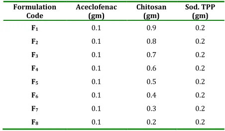
Chitosan microspheres were prepared by ionotropic gelation method. In this method chitosan stock solution (1% w/v) was prepared by dissolving chitosan in acetic acid (1% v/v) at room temperature. The drug was dissolved directly into the above prepared chitosan solution. 10 ml of this bubble free solution was dropped through a disposable syringe needle into a gently agitating 100ml of 2% (w/v) sodium tripolyphosphate solution. The dropping rate and falling distance were kept constant. The solution was magnetically stirred for half an hour followed by filtration and rinsing with distilled water. Gel like beads were obtained which was air dried for twenty four hours followed by oven drying for six hours at 40˚C. [6]
Table 1:Formulation table of Aceclofenac Chitosan
Formulation Code
Aceclofenac (gm)
Chitosan (gm)
Sod. TPP (gm)
F1 0.1 0.9 0.2
F2 0.1 0.8 0.2
F3 0.1 0.7 0.2
F4 0.1 0.6 0.2
F5 0.1 0.5 0.2
F6 0.1 0.4 0.2
F7 0.1 0.3 0.2
F8 0.1 0.2 0.2
Characterization of Aceclofenac Chitosan
Microspheres
Particle size analysis
weight size of formulation to % total weight of chitosan microspheres. [7]
Drug Entrapment
The various formulations of the chitosan microspheres were subjected for drug content. 50 mg of chitosan microspheres from all batches were accurately weighed and crushed. The powdered of microspheres were dissolved with 10ml ethanol in 100ml volumetric flask and makeup the volume with 0.1 N HCl. This resulting solution is than filtered through whatmann filter paper No. 44. After filtration, from this solution 10 ml was taken out and diluted up to 100 ml with 0.1 N HCl. Again from this solution 2 ml was taken out and diluted up to 10 m1 with 0.1 N HCI and the absorbance was measured at 275 nm against 0.1 N HCI as a blank. The percentage drug entrapment was calculated as follows. [8]
Calculated drug concentration % Drug entrapment = x 100 Theoretical drug concentration
Shape and Surface Characterization of
Chitosan Microspheres by Scanning Electron
Microscopy
From the formulated batches of chitosan microspheres, formulation (F4) which showed an appropriate balance between the buoyancy and the percentage release were examined for surface morphology and shape using scanning electron microscope Hitachi, Japan, Trichy. Sample was fixed on carbon tape and fine gold sputtering was applied in a high vacuum evaporator. The acceleration voltage was set at 20KV during scanning. Microphotographs were taken on different magnification and higher magnification (200X) was used for surface morphology. [9]
In vitro Release Studies:
The drug release rate from chitosan micro spheres was carried out using the USP dissolution paddle assembly. A weighed amount of micro spheres equivalent to 100 mg drug were dispersed in 900 ml of phosphate buffer 6.8 maintained at 37 ± 0.5°C and stirred at 100 RPM. At preselected time intervals one ml sample was withdrawn and replaced with equal amount of phosphate buffer 6.8. The collected samples were suitably diluted with phosphate buffer 6.8 and analyzed spectrophotometrically at 275 nm to determine the concentration of drug present in the dissolution medium. The dissolution studies were repeated using phosphate buffer pH 6.8 as dissolution medium. [10]
In-Vivo Anti - Inflammatory Study
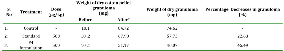
weighing 10 ± 1 mg were prepared and sterilized in hot air oven at 120°C for 3 hours. The abdomen was shaved cleanly, swabbed with 70% (v/v) ethanol and small incision was made in the lower abdomen of the rat. Using a blunt forceps, one sterile cotton pellet of known weight was placed in each aexilla and groin region and then incision was closed with sutures. On 8th day albino rat were sacrificed and four pellets were removed. The pellets were dried overnight at 55°C and weighed. The difference between the final weight of the pellet after drying and its initial weight was taken as the granuloma tissue weight. Results were expressed as percentage inhibition of granuloma in drug treated groups compared with the control group. [11]
Stability Study
Stability study was carried out for the F4 formulation by exposing it to different temperature 5-8°C, 27°C and 42°C for 45 days. The sample was analyzed for drug content at the regular intervals. It was found that no remarkable change in the drug content of F4 formulation. This indicates that F4 was stable for following temperature. [12]
RESULT AND DISCUSSION
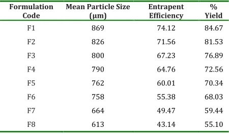
Controlled release microspheres were prepared using chitosan. The mean particle size of the microspheres significantly increased with increasing the concentration of chitosan and was in the range 613 µm to 869 µm as shown in Table 2. The viscosity of the medium increases at a higher polymer concentration resulting in enhanced interfacial tension with diminished shearing efficiency and increased particle size. As the stirring rate was increased, the mean particle
size decreased. The SEM photographs showed
that the fabricated microspheres were spherical
with a smooth surface and exhibited a range of
sizes within each batch as shown in Fig. 1.
Table 2: Evaluation characteristics of Aceclofenac Chitosan Microsphere
Formulation Code
Mean Particle Size (µm)
Entrapent Efficiency
% Yield
F1 869 74.12 84.67
F2 826 71.56 81.53
F3 800 67.23 76.89
F4 790 64.76 72.56
F5 762 60.01 70.34
F6 758 55.38 68.03
F7 664 49.47 59.44
F8 613 43.14 55.10
Table 3: Result Of Anti Inflammatory Activity Measurement
S.
No Treatment Dose (µµµµg/kg)
Weight of dry cotton pellet granuloma
(mg) Weight of dry granuloma (mg) Percentage Decreases in granuloma (%)
Before After*
1. Control - 10.1 84.72 74.62 -
2. Standard 500 10 .2 67.98 57.73 22.63
3. formulation F4 500 10 .1 51.17 40.07 45.49
Table 4: Stability study data of the optimized aceclofenac chitosan microsphere
S. No Days % Drug Remaining 5-8°°°°C % Drug Remaining 27 ±±±± 2°°°°C % Drug Remaining 42 ±±±± 2°°°°C
1. 0 100 ± 00 100 ± 00 100 ± 00
2. 14 99.6 ± 0.015 99.9 ± 0.003 99.4 ± 0.041
3. 28 99.5 ± 0.013 99.8 ± 0.027 99.2 ± 0.036
4. 45 99.4 ± 0.15 99.6 ± 0.012 99.1 ± 0.02
Fig. 1: SEM of Aceclofenac Chitosan Microsphere
0 10 20 30 40 50 60 70 80 90 100
0 2 4 6 8 10 12
Time (hrs)
C
u
m
u
la
ti
v
e
%
d
ru
g
r
e
le
a
s
e
F1 F2 F3 F4
F5 F6 F7 F8
Fig. 2: Release rate for Aceclofenac Chitosan Microsphere
REFERENCE
1. J.K. Patel, R.P. Patel, A.F. Amin, M.M. Patel (2005). Formulation and evaluation of mucoadhesive glipizide microspheres. AAPS Pharm Sci Tech. 6, E49-55.
2. K.V. Ranga Rao, K.P. Devi (1988). Swelling controlled release systems: recent developments and application. Int J Pharm. 48, 1-16.
3. Jayakrishnan A, Latha MS. Biodegradable polymeric microspheres as drug carriers. In: Jain NK, Editor. Controlled and Novel drug delivery. New Delhi: CBS publishers. 1997. pp 236-255. 4. Kreuter J., Evaluation of nanoparticles as
drug-delivery systems, Pharm. Acta. Helv, 1983, 58, 196-208.
5. Rasool D, Elham R, Efat F. Gelatin microspheres for controlled release of All trans Retinoic acid topical formulation and Drug Delivery Evaluation. Iranian Journal of Pharmaceutical Research 2003; 47-50.
6. Rossler B, Kreuter J, Scherer D. Collagen microparticles: Preparation and properties. J. Microencapsul.1995; 49-57.
7. Veerareddy PR, Tedla S, Banda SR, Bandari S, Jukanti R. Preparation and evaluation of mucoadhesive cefdinir microcapsules. J Adv Pharm Tech Res 2011;2:115-20
9. Mankala SK, Korla AC, Gade S. Development and evaluation of aceclofenac-loaded mucoadhesive microcapsules. J Adv Pharm Tech Res 2011;2:245-54
10.Shilpa A, Agrawal SS Ray AR, Controlled Delivery of Drugs from Alginate Matrix, Journal of Macromolecular science Part C-Polymer reviews, 43, 2003, 187.
11.Arora S, Budhiraja RD. Chitosan-alginate microcapsules of amoxicillin for gastric stability and mucoadhesion. J Adv Pharm Tech Res 2012;3:68-74.
12.Grellier M, Granja P L, Fricain J C, Bidarra S J, Renard M, Bareille R, et al. The effect of the co-immobilization of human osteoprogenitors and endothelial cells within alginate microspheres on mineralization in a bone defect. Biomaterials 2009; 30: 3271e8.
Bhavya Rastogi, Amit Chaudhary, Upendra Nagaich,; Formulation Development and Evaluation of Aceclofenac Chitosan Microspheres; J. Adv. Pharm. Edu. & Res. 2012: 4: 215-220