ARTIGO ORIGINAL
Multidimensional Strategy Regarding the Reduction of
Central-Line Associated Infection in Pediatric Intensive
Care
Estratégia Multidimensional na Redução de Infeção
Associada a Cateter Venoso Central em Pediatria
1. Faculdade de Medicina. Universidade de Coimbra. Coimbra. Portugal.
2. Serviço de Cuidados Intensivos Pediátricos. Hospital Pediátrico. Centro Hospitalar e Universitário de Coimbra. Coimbra. Portugal.
3. Unidade de Neurodesenvolvimento e Autismo. Serviço do Centro de Desenvolvimento da Criança. Centro de Investigação e Formação Clínica. Hospital Pediátrico. Centro Hospitalar e Universitário de Coimbra. Coimbra. Portugal.
Autor correspondente: Andrea Dias. [email protected]
Recebido: 24 de outubro de 2015 - Aceite: 21 de março de 2016 | Copyright © Ordem dos Médicos 2016
Jorge RODRIGUES1, Andrea DIAS1,2, Guiomar OLIVEIRA1,3, José Farela NEVES1,2
Acta Med Port 2016 Jun;29(6):373-380 ▪ http://dx.doi.org/10.20344/amp.5558
RESUMO
Introdução: Determinar a incidência de infeções da corrente sanguínea associadas ao uso de cateter venoso central, após reforço de medidas multidisciplinares de boa prática e a sua comparação com a taxa de incidência de infeções da corrente sanguínea associadas ao uso de cateter venoso central prévia.
Material e Métodos: Estudo observacional descritivo, com colheita prospetiva de dados, durante cinco meses, após implementação de medidas multidisciplinares. Foram incluídas todas as crianças admitidas na unidade de Cuidados Intensivos Pediátricos, submeti-das à colocação de cateter venoso central e foi efetuada comparação com controlos históricos.
Resultados: Incluíram-se 75 doentes com idade mediana de 23 meses: 22 (29,3%) recém-nascidos, 28 (37,3%) submetidos a cirurgia e 32 (43,8%) com patologia subjacente. Foram colocados 105 cateteres venosos centrais, com tempo médio de permanência de 6,8 ± 6,7 dias. O tipo de cateter venoso central mais comum foi de curta duração (45,7%), sendo os locais de inserção mais frequentes a veia subclávia e da flexura braquial (ambos 25,7%). Não ocorreu nenhuma infeção da corrente sanguínea associada ao uso de cateter venoso central durante o período do estudo. Comparando com os controlos históricos, ambos os grupos eram semelhantes relativamente à idade, género, proveniência dos doentes e local de colocação de cateter venoso central. No estudo atual, a duração mediana de internamento foi superior, com tempo de permanência de cateter venoso central (excluindo epicutâneo-cava) semelhante. Não se verificou diferença em relação ao calibre e número de lumens do cateter venoso central utilizado. A percentagem de crianças que colocou cateter venoso central em relação ao total de crianças admitidas no serviço no mesmo período foi menor no estudo atual, não existindo diferença significativa entre colocação de cateter venoso central único ou múltiplo.
Discussão: Após implementação da estratégia multidimensional não se registou nos Cuidados Intensivos Pediátricos ocorrência de infeções da corrente sanguínea associadas ao uso de cateter venoso central.
Conclusões: Devem ser encetados esforços para preservar o mesmo grau de prevenção multidimensional, para que se confirme a redução efetiva da taxa de incidência de infeções da corrente sanguínea associadas ao uso de cateter venoso central.
Palavras-chave: Cateteres Venosos Centrais; Infecções Relacionadas a Cateter; Infecção Hospitalar; Unidades de Cuidados Inten-sivos Pediátricos.
ABSTRACT
Introduction: To determine the central-line associated bloodstream infection rate after implementation of central venous catheter-care practice bundles and guidelines and to compare it with the previous central-line associated bloodstream infection rate.
Material and Methods: A prospective, longitudinal, observational descriptive study with an exploratory component was performed in a Pediatric Intensive Care Unit during five months. The universe was composed of every child admitted to Pediatric Intensive Care Unit who inserted a central venous catheter. A comparative study with historical controls was performed to evaluate the result of the intervention (group 1 versus group 2).
Results: Seventy five children were included, with a median age of 23 months: 22 (29.3%) newborns; 28 (37.3%) with recent surgery and 32 (43.8%) with underlying illness. A total of 105 central venous catheter were inserted, the majority a single central venous catheter (69.3%), with a mean duration of 6.8 ± 6.7 days. The most common type of central venous catheter was the short-term, non-tunneled central venous catheter (45.7%), while the subclavian and brachial flexure veins were the most frequent insertion sites (both 25.7%). There were no cases of central-line associated bloodstream infection reported during this study. Comparing with historical controls (group 1), both groups were similar regarding age, gender, department of origin and place of central venous catheter insertion. In the current study (group 2), the median length of stay was higher, while the mean duration of central venous catheter (excluding peripherally inserted central line) was similar in both groups. There were no statistical differences regarding central venous catheter caliber and number of lumens. Fewer children admitted to Pediatric Intensive Care Unit had central venous catheter inserted in group 2, with no significant difference between single or multiple central venous catheter.
Discussion: After multidimensional strategy implementation there was no reported central-line associated bloodstream infection
Conclusions: Efforts must be made to preserve the same degree of multidimensional prevention, in order to confirm the effective reduction of the central-line associated bloodstream infection rate and to allow its maintenance.
ARTIGO ORIGINAL INTRODUCTIONSafe and reliable central venous access is one of the
most crucial aspects in modern clinical practice, mainly in neonatal and paediatric intensive care, used to meet pa-tient’s energy, fluid and electrolyte needs, as well as for blood sampling and the administration of therapy.1-10 Central line-associated bloodstream infections (CLABSI) are includ-ed as the most frequent healthcare-associatinclud-ed infections, leading to longer hospital stay, high morbidity and mortal-ity as well as to increased hospital costs.1-5,8,10-12 In order to produce a CLABSI, causative microorganisms must gain access to the indwelling catheter surface (intra or extralumi-nal) and produce a biofilm, allowing for a sustained infection with subsequent haematogenous spread,1,12,19 through four different routes: migration of skin microflora at the insertion site into the cutaneous catheter tract and along the surface of the catheter, with colonisation of the CVC tip; contamina-tion of the catheter or catheter hub; haematogenous cath-eter colonisation from another focus of infection and rarely, by infusate contamination.1,12,19 Extraluminal is the most common route of infection for short-term catheters, while intraluminal is more frequent for long-term CVCs.1,12
Cath-eter size, diamCath-eter and number, CVC type and material, insertion site, hygiene and aseptic technique, duration of catheterisation, frequency of manipulation and virulence of causative microorganisms all are factors that may con-tribute to CLABSIs.1,12,19 Coagulase-negative staphylococci (CoNS), Staphylococcus aureus and Escherichia coli are most frequently involved.9
When any CLABSI is suspected, CVC removal is recommended, based on clinical examination of the patient, using systemic antibiotic therapy with which removal of microbial biofilm is not always achieved and different closure devices have been used to obtain the destruction of the biofilm, with variable efficacy and safety.9
A rate of CLABSI of 1.4 per 1,000 catheter days in medical and surgical paediatric intensive care units has been estimated by the most recent report of the National Healthcare Safety Network (NHSN), which has gradually been reduced over the last decade7,14 due to implemented good-practice recommendations and multidimensional infection control strategies.7,13 The rate of CLABSI can vary
widely, depending on the hospital, the department and the device used. In addition, it may also be influenced by risk factors inherent to patients12. Most of the data is based
on studies with adult patients and, considering paediatric heterogeneous and specific risk factors, we reach the conclusion that further studies are needed in order to know and improve the impact of these interventions.
Reducing the rate of CLABSI has always been an objective for the Intensive Care Unit (CIPE) of the Hospital Pediátrico, Centro Hospitalar e Universitário de Coimbra (CIPE-HP/CHUC) and a rate of 1.7 per 1,000 catheter days has been described in the latest study regarding the 2010-2011 period.15 The introduction and improvement of educational, monitoring and preventive multidisciplinary actions is crucial for the improvement of
healthcare delivery.4,6 Our study aimed to determine the incidence of CLABSI upon the implementation of different multidisciplinary measures, when compared to traditional control measures.15
MATERIAL AND METHODS
The CIPE-HP/CHUC is a referral 12-bed unit for the Central Region of Portugal included in a highly differentiated medical centre, including intermediate and intensive care medical and surgical units, serving a population of approximately 450,000 children and adolescents aged under 18,20 with an average 400 inpatient hospital admissions per year.
This was a cross-sectional descriptive and observational study with prospective data obtained from patients admitted to the Unit from 6 September 2013 to 5 February 2014 (five-month duration study) and having undergone one or more CVC placements or having presented on admission with a previously inserted in situ CVC. The following types of CVC were considered: short-term, term tunnelled, long-term with subcutaneous reservoir, haemodiafiltration CVC, umbilical venous catheter (UVC) and peripherally inserted central catheter (PICC).
Healthcare professionals previously attended a training and information meeting with the authors, including the analysis of CVC insertion and maintenance policy and CLABSI prevention guidelines.4,16-18 At the same time, the ‘Information Sheet on CVC insertion’ ‘Ficha de Colocação de CVC’ (Appendix 1 - http://www.actamedicaportuguesa. com/revista/index.php/amp/article/view/5558/373-380_ Apendice01.pdf) and the ‘Information Sheet on CVC maintenance’ ‘Ficha de Manutenção de CVC’ (Appendix 2 - http://www.actamedicaportuguesa.com/revista/index.php/ amp/article/view/5558/ 373-380_Apendice02.pdf) adapted from Wheeler D, et al. were presented to healthcare professionals.2
The following demographic variables were assessed: patient’s age, gender, unit of origin, reason for admission, clinical history, CLABSI on admission and length of stay. Data from the ‘Insertion sheet’ and ‘Maintenance sheet’ were obtained.
ARTIGO ORIGINAL and chemotherapy.
CDC-NHSN definitions were used for diagnosis and CLABSI was defined with a laboratory-confirmed BSI (LC-BSI) occurring 48 hours upon placement and maintenance of a CVC, in the absence of any other infectious focus and in the presence of at least one LC-BSI criteria.21 When
the patient is transferred from another department or healthcare institution with a previously inserted in situ CVC, a CLABSI was only considered as our unit’s responsibility when occurring 48 hours upon patient’s admission to the unit. As regards patients that were discharged from our unit with a previously inserted in situ CVC, the presence of a CLABSI was checked over the 48-hour period upon patient’s discharge.
Upon removal, catheter tips were always sent for culture. Whenever a microorganism was isolated, even in the absence of any clinical symptoms or signs of infection, this was considered as a catheter colonisation.
The rate of CLABSI was obtained by the ratio between the number of CLABSI events and the number of CVC days multiplied by 1,000.26 According to NHSN guidelines, the presence of at least one CVC per patient was considered as one CVC day.22
The results of our study were compared to those obtained in a previous study carried out between 1 July and 31 December 2010 (six-month duration).15 Two groups were therefore considered: group 1 (controls) and group 2 (current study).
IBM’s SPSS Statistics Software, version 20 software was used in statistical analysis. The determination of absolute and relative frequencies for qualitative variables and of central tendency and dispersion measures for quantitative variables was used for group characterisation. A comparative analysis (group 1 versus group 2) was carried out and a significance level of p = 0.05 was considered and Chi-square and Fisher’s exact test were used for the comparison between nominal qualitative variables and Mann-Whitney’s and Student’s t-test for the comparison between quantitative and ordinal qualitative variables.
RESULTS
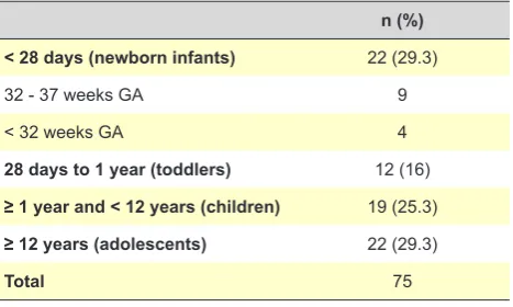
In total, 75 patients with a CVC were admitted to the CIPE over study period (75/173; 43.4%; 58.7% male). Median age of the patients was of 23 months (Q1 - 4 days; Q3 - 12.5 years) and 45.3% (34/75) were under one year of age. Twenty-two of the 75 patients (29.3%) were newborn infants (under 28 days of life); from these, thirteen (59.1%) were premature (Table 1).
Median length of stay at the CIPE was of seven days (Q1 - two days; Q3 - 15 days), ranging from one to 77 days. Median length of stay in the hospital was of 19 days (Q1 - nine days; Q3 - 36 days). Twenty eight from the 75 patients (37.3%) underwent surgery, four of whom (14.3%) were newborn infants. Forty-eight patients (64%) presented with underlying pathology: 15 (20%) presented with cancer, five (6.7%) had been transplanted (four underwent a liver transplant and one bone marrow transplant) and 28 (37.3%)
presented with other pathologies. Three patients (4%) underwent haemodiafiltration, three (4%) were admitted with severe burn injuries and 10 (13.3%) underwent parenteral nutrition.
Patients mostly originated from the operating room (33 patients; 44%), followed by other departments (24 patients; 32%). Fifty five patients (73.3%) were admitted to the CIPE with a previously inserted in situ CVC. In total, 50 patients (66.7%) were discharged to other departments with a previously inserted in situ CVC, while 25 (33.3%) underwent a CVC removal while staying at the CIPE. A new CVC was reinserted in 14 patients (18.7%).
Median Pediatric Index of Mortality II (PIM2) was of 2.4% in the subgroup of patients in whom a CVC was inserted (Q1 - 0.59%; Q3 - 6.5%). Overall median PIM2 at the CIPE for the same period was of 0.95% (Q1 - 0.45%; Q3 - 4.2%), while a 2.3% (4/173) overall mortality rate was found. Three patients included in the study passed away (4%), none of them due to any healthcare-associated infection.
In total, 105 CVC devices were inserted (75 patients included in the study), corresponding to 613 catheter days. In most patients (69.3%) only one CVC was inserted; in 23 patients (30.7%) a second catheter was required and a third one was required in seven (9.3%). Short-term CVC was the most frequently inserted type of CVC (48/105, 45.7%), followed by PICC (28/105, 26.7%) (Table 2). When more than one CVC was inserted (30/105, 28.6%), PICC was the most frequently used (17/30, 56.7%). Brachial and subclavian veins were the preferred sites for CVC insertion (27; 25.7%) followed by femoral vein (25; 23.8%).
When CVC insertion occurred at the CIPE, femoral vein was the preferred site of insertion (9/19; 47.4%), while subclavian vein was preferred in patients admitted to the unit with a previously inserted in situ CVC (22/56; 39.3%). Two was the median number of catheter lumens (1 to 3 lumens) and 4 Fr (4 to 8 Fr) was median catheter diameter. The average maintenance time of CVC (mean ± SD) was of 6.8 ± 6.7 days. In total, 96 (91.4%) polyurethane and nine silicone (8.6%) CVCs were used.
Most CVC insertions were completed by a specialist physician (34.3%, 36/105) and were mostly removed by a nursing professional (10/105, 9.5%), followed by a specialist physician (8/105, 7.6%) and an internal medicine physician Table 1 - Distribution of patients according to their age group
n (%)
< 28 days (newborn infants) 22 (29.3)
32 - 37 weeks GA 9
< 32 weeks GA 4
28 days to 1 year (toddlers) 12 (16)
≥ 1 year and < 12 years (children) 19 (25.3) ≥ 12 years (adolescents) 22 (29.3)
Total 75
ARTIGO ORIGINAL
(7/105, 6.7%); this information was not available in 24.8% (26/105) of the patients. Most patients (54/105, 51.4%) were discharged from the CIPE with a previously inserted in situ CVC. Median time until completion of CVC insertion was of five minutes (Q1 - 5 minutes; Q3 - 10 minutes). A median three blood samplings through the CVC were found (39/75, 52%) (Q1 - 1; Q3 - 6).
CVC insertion guidelines complied with the ‘Information Sheet on CVC Insertion’ as regards hand hygiene, aseptic maintenance, maximal sterile barrier precautions and adequate aseptic technique prior to insertion in 100% of CVC placed at the CIPE. One ultrasound-guided CVC placement into the internal jugular vein was performed. Povidone iodine was used for skin disinfection in all the cases and no CVC was placed under an emergency situation. In total, 55 out of the 75 patients included in the study presented with a previously inserted in situ CVC on admission to the CIPE and therefore the information regarding who had inserted it and the compliance with insertion guidelines could not be confirmed.
All CVC insertions complied with good-practice guidelines included into the ‘Information Sheet of CVC Maintenance’, namely regarding the daily assessment on continued need for the CVC, CVC integrity and compliance with the guidelines regarding its maintenance and manipulation.
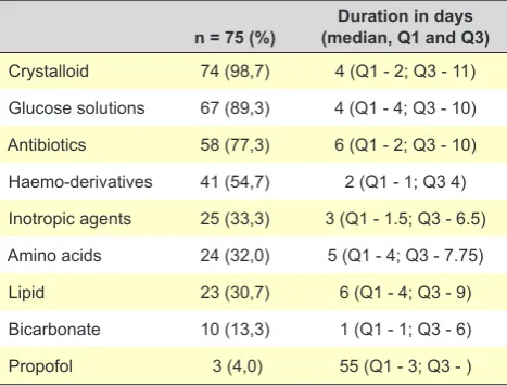
Glucose (89.3%) and crystalloid (98.6%) solutions were most frequently used (Table 3). Haemo-derivative infusion was used in 41 patients (54.7%), mainly of erythrocyte concentrate (21.3%). Antibiotic therapy was used in 58 (77.3%) patients for an infection as reason for admission,
for pre and post-operative prophylaxis and for infectious complications acquired while in hospital. No antibiotic therapy was started for any suspected CLABSI.
No infections at the insertion site were found and no CLABSI occurred over the study period. CVC tip colonisation was found in 12 patients and Staphylococcus epidermidis was most frequently isolated. Eight patients presented with an infection on admission to the CIPE (four patients with pulmonary, one with urinary, one with sepsis and two patients with CLABSI onset prior to admission to the CIPE and therefore not considered for the study).
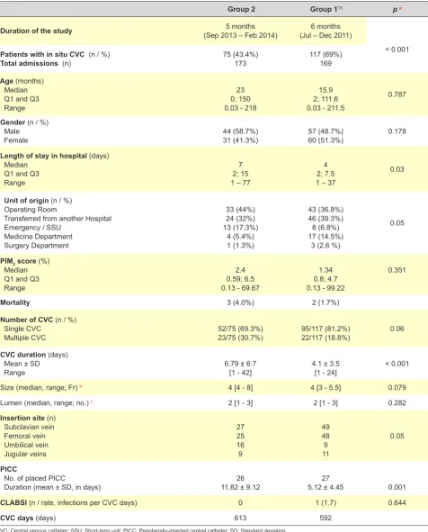
When comparing both groups of patients15 (Table 4), no significant difference was found regarding patient’s median age (15.9 months in group 1 vs. 23 months in group 2; Mann-Whitney’s U-test, p = 0.787), gender (Chi-Square, p = 0.178), unit of origin (Chi-Square, p = 0.05) and clinical severity on admission (median PIM2 score of 1.34% in group 1 vs. 2.4% in group 2; Mann-Whitney’s U-test, p = 0.351).
Median length of stay was higher in group 2 (4 days in group 1 versus 7 days in group 2; Mann-Whitney’s U-test, p = 0.03) and a CVC was inserted in a lower percentage of admitted patients in group 2 (69% in group 1 vs. 43.4% in group 2; Chi-Square, p < 0.001). No significant differences were found between both groups as regards the percentage of patients in whom a single or multiple CVC were inserted (Chi-Square, p = 0.06) and as regards CVC insertion site (Chi-Square, p = 0.05).
Considering all types of CVC and PICC compared to the other types, a longer length of stay in hospital was found in group 2 (4.1 ± 3.5 days in group 1 vs. 6.8 ± 6.7 days Table 2 - Type and CVC insertion site
CVC1
n = 75 n = 23CVC2 CVCn = 73
Catheter type
Short-term CVC 40 (53.3%) 7 (30.4%) 1 (14.3%)
UVC 13 (17.3%) 3 (13.0%) 0
PICC 11 (14.7%) 11 (47.8%) 6 (85.7%)
Long-term CVC w/ subcutaneous reservoir 6 (8.0%) 1 (4.3%) 0
Haemodiafiltration CVC 4 (5.3%) 0 0
Long-term tunnelled CVC 1 (1.3%) 1 (4.3%) 0
Insertion site
Subclavian vein 24 (32.0%) 3 (13.0%) 0
Brachial veins 10 (13.3%) 11 (47.8%) 6 (85.7%)
Femoral vein 20 (26.7%) 4 (17.4%) 1 (14.3%)
Umbilical vein 13 (17.3%) 3 (13.0%) 0
Internal jugular vein 7 (9.3%) 1 (4.3%) 0
External jugular vein 1 (1.3%) 0 0
Saphenous vein 0 1 (4.3%) 0
ARTIGO ORIGINAL
in group 2, Student’s t-test p < 0.001 for all types of CVC and 5.1 ± 4.5 days in group 1 vs. 11.8 ± 9.1 days in group 2, Student’s t-test p = 0.001 for PICC alone). Considering the average duration of CVC use, except for the PICC, no significant differences were found between both groups (3.95 ± 4.04 days in group 1 vs. 4.97 ± 4.25 days in group 2; Student’s t-test, p = 0.075). No significant differences were found regarding the number of catheter lumens – except for PICC, UVC and long-term CVC with subcutaneous reservoir – (Mann-Whitney’s U-test, p = 0.282) and regarding catheter size – except for PICC and long-term CVC with subcutaneous reservoir– (Mann-Whitney’s U-test, p = 0.079) in both groups.
DISCUSSION
CLABSI are associated to significantly increased morbidity and mortality, namely in paediatric intensive care units, where most patients are critically ill and frequently in need for central vascular access.1-11
No CLABSI events were described in patients admitted to the CIPE of the HP-CHUC over the study period, upon implemented interventions. Such a positive result supports a possible correlation between the implemented actions and the reduction in the incidence of CLABSI. A comparative analysis of the demographic characteristics of both populations has been made in order to enhance this conclusion. When comparing the data of the present study with the control population, no significant differences were found between both groups as regards patient’s gender, age, unit of origin and clinical severity on admission, with similar characteristics and therefore comparable. The presence of two CLABSI events, with its onset prior to being admitted to the CIPE, should be mentioned. There is no clear association between the risk factors found in these two patients and the CLABSI in paediatric population, although some studies have described patient’s age under two and the presence of cancer as risk factors.9,23,24
The presence of CVC tip colonisation was found in 12 patients. Even though the current recommendations (2016) do not recommend CVC tip culture28 (unlike what was
recommended in latest 2011 guidelines, at the time when the study took place),21 the fact that this was carried out
allowed for the recognition of the main causative agents of CVC colonisation in our unit as well as its sensitivity profile. Even though no CLABSI were found, our population presented with different conditions that may be associated to an increased risk for infection. The risk factors for CLABSI in children include (i) under two years of age (corresponding to almost half of our population and approximately one third of the patients were newborn infants); (ii) low body weight (< 8 kg); (iii) haematopoietic stem-cell transplant (one patient), (iv) recent associated surgery and other pathology (around 30% of the patients); (v) exposure to invasive procedures apart from CVC insertion – including the use of mechanical ventilation, intubation, placement of arterial catheter, placement of thoracic drain or parenteral nutrition (used in around 20% of the patients); (vi) longer CVC use and the use of multiple CVC (around 30% of our patients).9,25 A longer median length of stay in hospital was found in our group of patients when compared to controls, mainly due to the fact that both were small groups and therefore a small number of admitted patients with longer length of stay being enough to significantly change overall length of stay, which was found in group 2. Despite a longer length of stay being directly related to the risk for CLABSI,23 this was not found
in our study. A longer average CVC duration was found in our study, which may also represent a risk factor.6,24,25 This was mainly due to the longer duration of PICC, which are recommended for long-term use, with lower risk for infection than short-term catheters; however, keeping those beyond thirty-five days increases that risk.6,19 Except for PICC, a shorter CVC duration was found in our study and may have had a contribution for reducing the infection rate.
Polyurethane CVCs were mostly used, complying with the latest good-practice guidelines.19,23,25
No reduction in the number of catheter lumens was found, when compared to controls. No causal relation was found in current literature between the presence of CLABSI and higher number of catheter lumens, except for long-term tunnelled CVC; however, the lowest possible number of lumens is recommended by good-practice guidelines.19,23,25 The implementation of a systematic completion of the ‘Information Sheet on CVC insertion’ and the ‘Information Sheet on CVC maintenance’ with a high compliance level in most cases required a higher daily CVC care, improving overall healthcare delivery within the unit. According to previous reports, the introduction of this set of actions, standardising CVC insertion, maintenance and removal procedures, aimed to a daily assessment by healthcare professionals regarding the integrity and continued need for a vascular access, led to a significant reduction in the incidence of CLABSI.2,3,9-11,13,15,16-19,25,27
Some limitations should be mentioned though. In first place, data sampling was influenced by the collaboration of healthcare professionals in completion of daily task forms and sometimes sampling errors may have occurred and have interfered with final result accuracy. In addition, Table 3 - CVC infusions
n = 75 (%) (median, Q1 and Q3)Duration in days
Crystalloid 74 (98,7) 4 (Q1 - 2; Q3 - 11) Glucose solutions 67 (89,3) 4 (Q1 - 4; Q3 - 10) Antibiotics 58 (77,3) 6 (Q1 - 2; Q3 - 10) Haemo-derivatives 41 (54,7) 2 (Q1 - 1; Q3 4) Inotropic agents 25 (33,3) 3 (Q1 - 1.5; Q3 - 6.5) Amino acids 24 (32,0) 5 (Q1 - 4; Q3 - 7.75)
Lipid 23 (30,7) 6 (Q1 - 4; Q3 - 9)
Bicarbonate 10 (13,3) 1 (Q1 - 1; Q3 - 6)
ARTIGO ORIGINAL
as checking of individual compliance with CVC insertion, maintenance and removal guidelines, the study was mostly based on data reported by who performed the CVC placement, which may have biased the results.
In second place, the study was only restricted to one department – the CIPE – which is a specialized paediatric department and therefore the results should not be extrapolated to other hospital units, including non-paediatric Table 4 -Characteristics of our group of patients and the control group
Group 2 Group 115 p a
Duration of the study (Sep 2013 – Feb 2014)5 months (Jul – Dec 2011)6 months
< 0.001
Patients with in situ CVC (n / %)
Total admissions (n) 75 (43.4%)173 117 (69%)169
Age (months)
Median Q1 and Q3 Range
23 0; 150 0.03 - 218
15.9 2; 111.6 0.03 - 211.5
0.787
Gender (n / %) Male
Female 44 (58.7%)31 (41.3%) 57 (48.7%)60 (51.3%) 0.178
Length of stay in hospital (days) Median
Q1 and Q3 Range
7 2; 15 1 – 77
4 2; 7.5 1 – 37
0.03
Unit of origin (n / %) Operating Room
Transferred from another Hospital Emergency / SSU
Medicine Department Surgery Department
33 (44%) 24 (32%) 13 (17.3%)
4 (5.4%) 1 (1.3%)
43 (36.8%) 46 (39.3%) 8 (6.8%) 17 (14.5%)
3 (2.6 %)
0.05
PIM2 score (%)
Median Q1 and Q3 Range
2.4 0.59; 6.5 0.13 - 69.67
1.34 0.8; 4.7 0.13 - 99.22
0.351
Mortality 3 (4.0%) 2 (1.7%)
Number of CVC (n / %) Single CVC
Multiple CVC 52/75 (69.3%)23/75 (30.7%) 95/117 (81.2%)22/117 (18.8%) 0.06
CVC duration (days) Mean ± SD
Range 6.79 ± 6.7[1 - 42] 4.1 ± 3.5[1 - 24] < 0.001
Size (median, range; Fr) b 4 [4 - 8] 4 [3 - 5.5] 0.079
Lumen (median, range; no.) c 2 [1 - 3] 2 [1 - 3] 0.282
Insertion site (n) Subclavian vein Femoral vein Umbilical vein Jugular veins
27 25 16
9
49 48
9 11
0.05
PICC
No. of placed PICC
Duration (mean ± SD, in days) 11.82 ± 9.1226 5.12 ± 4.4527 0.001
CLABSI (n / rate, infections per CVC days) 0 1 (1.7) 0.644
CVC days (days) 613 592
CVC: Central venous catheter; SSU: Short-term unit; PICC: Peripherally-inserted central catheter; SD: Standard deviation.
a Comparison using Mann-Whitney’s and Student’s t-test for quantitative and ordinal or interval qualitative variables and Chi-square test for nominal qualitative variables.
b All CVC types, except PICC and long-term CVC with subcutaneous reservoir were considered for the calculations involving the number of catheter lumens.
ARTIGO ORIGINAL intensive care units.
In third place, some of the recommendations included in latest CDC guidelines for the prevention of CLABSI could not be complied with, namely regarding the preference for the use of chlorhexidine for skin disinfection prior to CVC insertion, for ultrasound-guided CVC insertion and for the preference for CVC insertion into subclavian rather than femoral vein.19 A 2% chlorhexidine solution was not available over the study period and 70% ethanol or povidone iodine were alternatively used. Only some studies have described an association between the use of chlorhexidine and a reduced incidence of CLABSI; therefore, no precise indications are provided by current recommendations as regards any of those substances.11,13,19 When comparing the patients admitted to the unit with a previously inserted in situ CVC and the patients in whom a CVC was inserted during their stay in the unit, the preference for CVC placement into the femoral vein was found in our department, due to the higher experience with this type of placement, even though against the recommendations suggesting the use of this site should be reduced, giving preference to the placement into the subclavian vein.19 These items should be re-addressed in future interventions.
Finally, despite the results, as no CLABSI events occurred, we were not able to determine any risk factors for CLABSI in our population, as in our initial aim. The study was a contribution for improving our knowledge on the subject as well as for improving healthcare delivery in this area. Further monitoring of the outcomes of these measures is recommended in order to confirm a real CLABSI reduction and to assess the impact and sustained benefit of multidisciplinary actions.
CONCLUSION
A reduction in CLABSI and healthcare-associated infections should be considered as one of the main aims of any hospital department, as these are mostly preventable.1-11 No CLABSI events were described in our unit over these five-month period, corresponding to a reduction in current incidence of CLABSI, considering the rate of 1.7 infections per 1,000 CVC days obtained in the study previously carried
out in our unit.15 Even though a short study period has been considered, compliance with good-practice guidelines and multidisciplinary actions showed a very positive impact, confirming the evidences supported by similar studies carried out by other hospital units worldwide.
The results allowed for the confirmation of the important role of this type of strategy in hospital infection prevention, being systematically recommended nationwide.
High standards of demand in healthcare delivery, namely using updated protocols, are crucial to reach a medium and long-term reduction in CLABSI. A systematic record of healthcare-associated infections and its regular analysis using pre-defined platforms (such as those made available by the Portuguese DGS (http://www.insa-rios.net/) should be encouraged.
ACKNOWLEDGEMENTS
The authors wish to acknowledge for the willingness and cooperation of all the professionals at the Serviço de Cuidados Intensivos Pediátricos do Hospital Pediátrico de Coimbra – namely the physicians, nurses and administrative staff – in writing this manuscript.
HUMAN AND ANIMAL PROTECTION
The authors declare that the followed procedures were according to regulations established by the Ethics and Clinical Research Committee and according to the Helsinki Declaration of the World Medical Association.
DATA CONFIDENTIALITY
The authors declare that they have followed the protocols of their work centre on the publication of patient data.
CONFLICTS OF INTEREST
The authors declare that there were no conflicts of interest in writing this manuscript.
FINANCIAL SUPPORT
The authors declare that there was no financial support in writing this manuscript.
REFERENCES
1. Safdar N, Maki D. The pathogenesis of catheter-related bloodstream infection with noncuffed short-term central venous catheters. Intensive Care Med. 2004;30:62-7.
2. Wheeler D, Giaccone MJ, Hutchinson N, Haygood M, Bondurant P, Demmel K et al. A hospital-wide quality improvement collaborative to reduce catheter-associated bloodstream infections. Pediatrics. 2011;128;e995.
3. Provonost P, Needham D, Berenholtz S, Sinopoli D, Chu H, Cosgrove S et al. An intervention to decrease catheter-related bloodstream infections in the ICU. N Eng J Med. 2006;355:2725-32.
4. Exline M, Ali NA, Zikri N, Mangino JE, Torrence K, Vermillion B et al. Beyond the bundle – journey of a tertiary care medical intensive care unit to zero central-line associated bloodstream infections. Critical Care. 2013;17:R41.
5. Niedner MD, Huskins C, Colantuoni E, Muschelli J, Harris M, Rice T et al. Epidemiology of central line-associated bloodstream infections in the Pediatric Intensive Care Unit. Infec Control Hosp Epidemiol.
2011;32:1200-8.
6. Almeida A, Pereira O, Neto MT, Casella P. Cateteres vasculares centrais no recém-nascido: recomendações para prevenção de infeção relacionada com ou associada a cateteres vasculares centrais. Lisboa: Sociedade Portuguesa de Neonatologia; 2012.
7. Edwards JR, Peterson KD, Banerhee S, Allen-Bridson K, Morrell G, Dudeck MA et al. National Healthcare Safety Network report: Data summary for 2006 through 2008. Am J Infect Control. 2009;37:783-805. 8. Miller M, Niedner MF, Huskins WC, Colantuoni E, Yenokvan G, Moss
M et al. Reducing PICU central line-associated bloodstream infections: 3-year results. Pediatrics. 2011;128:1077-83.
9. Janum S, Zingg W, Afshari A, Classen V. Bench-to-bedside review: challenges of diagnosis, care and prevention of central catheter-related bloodstream infections in children. Critical Care. 2013;17:238. 10. Marra A, Rodrigues RG, Durão MS, Correa L, Guastelli LR, Faria
ARTIGO ORIGINAL
2010;38:434-9.
11. Hatler C, Buckwald L, Salas-Allison Z, Murphy-Taylor C. Evaluating central venous catheter care in a pediatric intensive care unit. Am J Crit Care. 2009;18:514-20.
12. Pina E, Silva EG, Costa VM, Neto T, Estrada J, Estrada H. et al. Recomendações para a prevenção da infeção nosocomial associada aos dispositivos intravasculares. Lisboa: Plano Nacional para o Controlo de Infeção; 2006.
13. Galpern D, Guerrero A, Tu A, Fahoum B, Wise, L. Effectiveness of a central line bundle campaign on line-associated infections in the intensive care unit. Surgery. 2008;144:492-5.
14. Dudeck MA, Weiner LM, Allen-Bridson K, Malpiedi PJ, Peterson KD, Pollock DA et al. National Healthcare Safety Network report: data summary for 2012, device-associated module. Am J Infect Control. 2013;41:1148-66.
15. Caldeira M. Infeções da Corrente Sanguínea Associadas aos Cuidados de Saúde numa Unidade de Cuidados Intensivos Pediátricos [trabalho final de mestrado]. Coimbra: Faculdade de Medicina da Universidade de Coimbra; 2011.
16. Band JD, Gaynes R. Prevention of intravascular catheter-related infections. UpToDate. 2013. [consultado 2014 mar 30] Disponível em: http://www.uptodate.com/home
17. Li S, Faustino EV, Golombek S. Reducing central line infections in pediatric and neonatal patients. Curr Infect Dis Resp. 2013;15:269-77. 18. Li S, Bizzarro MJ. Prevention of central line associated bloodstream
infections in critical care units. Curr Opin Pediatr. 2011;23:85-90. 19. Centers for Disease Control and Prevention. Guidelines for the
Prevention of Intravascular Catheter-Related Infections, 2011. [consultado 2014 mar 30]. Disponível em: http://www.cdc.gov/hicpac/ pdf/guidelines/bsi-guidelines-2011.pdf.
20. Instituto Nacional de Estatística, I.P. – Censos 2011, Resultados
definitivos – Portugal. Lisboa: INE; 2012.
21. Centers for Disease Control and Prevention. Central line-associated bloodstream infection (CLABSI) event. [consultado 2014 mar 30]. Disponível em: http://www.cdc.gov/nhsn/pscmanual/4psc clabscurrent. pdf.
22. Horan TC, Andrus M, Dudeck MA. CDC/NHSN surveillance definition of health care-associated infection and criteria for specific types of infections in the acute care setting. Am J Infect Control. 2008;36:309-32. 23. Yogaraj JS, Elward AM, Fraser VJ. Rate, risk factors and outcomes of
nosocomial primary bloodstream infection in Pediatric Intensive Care Unit patients. Pediatrics. 2002;110:481-5.
24. Urrea M, Iriondo M, Thio M, Krauel X, Serra M, LaTorre C, et al. A prospective incidence study of nosocomial infections in a neonatal care unit. Am J Infect Control. 2003;31:505-7.
25. Odetola FO, Moller FW, Dechert RE, Van der Elzen K, Chenoweth C. Nosocomial catheter-related bloodstream infections in a pediatric intensive care unit: risks and rates associated with various intravascular technologies. Pediatr Crit Care Med. 2003;4:432-6.
26. Warren DK, Zack JE, Elward AM, Cox MJ, Fraser VJ. Nosocomial primary bloodstream infections in intensive care unit patients in a nonteaching community medical center: a 21-month prospective study. Clin Infect Dis. 2001;33:1329-35.
27. Berenholtz SM, Provonost PJ, Lipsett PA, Hobson D, Earsing K, Farley JE et al. Eliminating catheter-related bloodstream infections in the intensive care unit. Crit Care Med. 2004;32:2014-20.