Chemical shift imprint of intersubunit communication
in a symmetric homodimer
Bradley T. Falka, Paul J. Sapienzab, and Andrew L. Leea,b,1
aDepartment of Biochemistry and Biophysics, School of Medicine, University of North Carolina at Chapel Hill, Chapel Hill, NC 27599; andbDivision of Chemical Biology and Medicinal Chemistry, UNC Eshelman School of Pharmacy, University of North Carolina at Chapel Hill, Chapel Hill, NC 27599
Edited by Adriaan Bax, National Institutes of Health, Bethesda, MD, and approved June 21, 2016 (received for review March 24, 2016)
Allosteric communication is critical for protein function and cellular homeostasis, and it can be exploited as a strategy for drug design. However, unlike many protein–ligand interactions, the structural ba-sis for the long-range communication that underlies allostery is not well understood. This lack of understanding is most evident in the case of classical allostery, in which a binding event in one protomer is sensed by a second symmetric protomer. A primary reason why study of interdomain signaling is challenging in oligomeric proteins is the difficulty in characterizing intermediate, singly bound species. Here, we use an NMR approach to isolate and characterize a singly ligated state (“lig1”) of a homodimeric enzyme that is otherwise obscured by rapid exchange with apo and saturated forms. Mixed labeled dimers were prepared that simultaneously permit full population of the lig1 state and isotopic labeling of either protomer. Direct visualization of peaks from lig1yielded site-specific ligand-state multiplets that provide a convenient format for assessing mechanisms of intersubunit commu-nication from a variety of NMR measurements. We demonstrate this approach on thymidylate synthase fromEscherichia coli, a homodimeric enzyme known to be half-the-sites reactive. Resolving the dUMP1state shows that active site communication occurs not upon the first dUMP binding, but upon the second. Surprisingly, for many sites, dUMP1 peaks are found beyond the limits set by apo and dUMP2peaks, in-dicating that binding the first dUMP pushes the enzyme ensemble to further conformational extremes than the apo or saturated forms. The approach used here should be generally applicable to homodimers.
homodimer
|
subunit communication|
allostery|
NMR|
thymidylate synthaseA
llosteric regulation in proteins is a ubiquitous mechanism for controlling cellular behavior and an attractive strategy for therapeutic development. Even though broadly recognized, long-range communication is not well understood mechanistically (1–4). Although there have been numerous strategies to reveal the struc-tural and dynamic underpinnings of allostery, oddly these strategies have largely focused on complex oligomeric or, alternatively, on small monomeric allosteric proteins. A likely more straightforward approach is to study allosteric mechanisms using simple symmetric homodimeric proteins, which would allow for answering the basic and general question of how the occurrence of an event in one subunit is communicated to another subunit, as occurs in classical multisubunit allosteric proteins. Given the large number of homo-dimeric proteins involved in cellular regulation (such as growth factors, cytokines, kinases, G protein-coupled receptors, transcrip-tion factors, and metabolic proteins), insights into intersubunit communication should be widely beneficial (5).As a key step toward a broad understanding of allosteric mechanisms, it will be important to observe how binding a ligand in one subunit is communicated to a second subunit, even in the absence of conformational change. Although this communica-tion is straightforward in specific cases of two differing neigh-boring domains or heterodimers in which domains have distinct ligands (6–8), it is more elusive for the common case of sym-metric homodimers. Homodimers present a challenge because it is difficult to either observe individual protomers or study states with a single ligand bound (referred to here as“lig1”) because of
dynamic binding equilibria. Having a method to isolate lig1
homo-dimers would facilitate detailed study of intersubunit communication and allostery by high resolution structural methods. A well-established approach for mapping communication networks in proteins is NMR spectroscopy, most commonly using so-called chemical shift pertur-bation (CSP) (9, 10). In the case of homodimers, however, tracking CSPs or making additional measurements on lig1peaks (or
reso-nances) becomes problematic because (i) resonances from sym-metric protomers can overlap, (ii) resonances are frequently in fast or intermediate exchange on the NMR timescale, especially in di-meric enzymes where substrate affinities are low to moderate, and, most importantly, (iii) unless ligand binding is highly negatively co-operative, there will be additional resonances from apo (lig0) and
doubly bound (lig2) states. In principle, monitoring lig1states is most
easily carried out in highly negatively cooperative systems although, even in the few reported cases, most lig1resonances were not well
resolved (11, 12). A general, experimental format for monitoring specific peaks (or sites) in both bound and empty protomers for lig1states will therefore help advance our understanding of
inter-subunit communication and allostery in homodimers (and poten-tially higher order oligomers).
Escherichia colithymidylate synthase (TS), a 62-kDa symmetric homodimer, presents a favorable example for lig1studies by NMR.
TS catalyzes the synthesis of the sole source of 2′ -deoxythymidine-5′-monophosphate (dTMP) via a multistep mechanism involving reductive methylation of dUMP using N5,N10-methylene-5,6,7,8-tetrahydrofolate (mTHF) as both a methylene and hydride donor. In addition, TS is half-the-sites reactive (13–15), with substrate binding sites separated by 35 Å, leading to an expectation for
Significance
Regulatory signals between protein subunits depend on commu-nication between sequential binding events. How such long-range communication, or allostery, operates at the subdomain level has been elusive, especially for homooligomeric proteins. To address this problem, homodimers of thymidylate synthase were gener-ated for optimized study of individual protomers of the singly bound dimer. Mixed15N-labeled dimers were created with a sin-gle functional active site, allowing site-specific, protomer-specific chemical shifts to report on step-wise binding effects. Long-range intersubunit communication was observed although this com-munication was apparent only in the second ligand-binding step, in which changes were in the first ligand-bound region. Visuali-zation of up to four peaks for each residue amide provides a unique way to assess the allosteric mechanism.
Author contributions: B.T.F., P.J.S., and A.L.L. designed research; B.T.F. and P.J.S. per-formed research; B.T.F., P.J.S., and A.L.L. analyzed data; and B.T.F., P.J.S., and A.L.L. wrote the paper.
The authors declare no conflict of interest.
This article is a PNAS Direct Submission.
See Commentary on page 9407.
1To whom correspondence should be addressed. Email: [email protected].
This article contains supporting information online atwww.pnas.org/lookup/suppl/doi:10. 1073/pnas.1604748113/-/DCSupplemental.
BIOPHYSICS
AND
COMPUTATIONAL
BIOLOGY
SEE
COM
negative substrate binding cooperativity between protomers. Al-though dUMP was recently shown to bind with minimal coopera-tivity for the E. colienzyme at 25 °C, there are signs of unequal thermodynamics between the two protomers at lower temperatures (16), and indeed data herein show clear intersubunit communica-tion. Moreover, other TS enzymes, and in particular human, seem to show more dramatic cooperativity, suggesting that intersubunit communication is an intrinsic feature of TS (13, 17–21). To over-come the difficulties of studying symmetric proteins by NMR, we generated a pair of mixed labeled dimers of TS that each have a single functional active site and a single protomer labeled for NMR studies. These complementary mixed dimers allow for determining protomer-specific responses to a single dUMP binding event by isolating the dUMP1 state. In the presence of dUMP, the mixed
dimers revealed dUMP1 peak positions normally hidden in WT
(wild-type) dimer titrations and highlighted the important differ-ences between the two dUMP binding events. These data also allow construction of complete“ligand state peak multiplets”that reflect the responses of residues on both sides of the interface. Most no-tably, we show that there is communication between the two active sites primarily upon binding the second dUMP because this binding event causes perturbations in the already-bound first site.
Materials and Methods
Protein Expression and Purification.WT and double mutant R126E, R127E (RREE) thymidylate synthase fromE. coliwas expressed and purified as de-scribed inSI Materials and Methods.
Generation of TS Mixed Dimers.Preparation of specific labeled mixed dimers was accomplished in vitro by mixing purified WT and RREE homodimers, in 2 M urea at pH 9, to reapportion the monomers yielding three species: WT and RREE homodimers and a mixed dimer with one WT and one RREE monomer. The mixed dimer was then separated from the parent homodimers by anion exchange chromatography. For NMR studies, two different preparations of mixing were made to isolate both species of mixed dimers: one where the binding subunit is U-[2H,15N]–labeled (mix RREE-labeled with WT-unlabeled) and one where the nonbinding subunit is U-[2H,15N]–labeled (mix WT-labeled with RREE-unlabeled). For full details, seeSI Materials and Methods.
NMR Spectroscopy.Standard transverse relaxation-optimized spectroscopy (TROSY) triple resonance and 1H-15N heteronuclear single quantum co-herence (HSQC) experiments were used for backbone resonance assignment experiments and ligand titrations, respectively, as described inSI Materials and Methods.
Results
Generation and Characterization of Mixed Dimers. To study inter-subunit communication in a homodimeric protein, we sought to use NMR to investigate the effect that binding of the first ligand has on both subunits of thymidylate synthase. Study of lig1states, however,
requires overcoming two primary degeneracies. The first is that addition of ligand to populate the lig1state is typically accompanied
by population of lig0and lig2states, and accordingly, more complex
spectra. The second is that, for symmetric protein dimers, it can be difficult to spectroscopically distinguish between the bound and empty protomers (or subunits). Our strategy was to break these degeneracies by creation of mixed15N-labeled dimers that (i) can
bind substrate only in one protomer, and (ii) have only a single protomer labeled for NMR detection, allowing for two comple-mentary lig1dimer samples, one with the bound subunit15N-labeled
and a second lig1sample with the empty subunit15N-labeled.
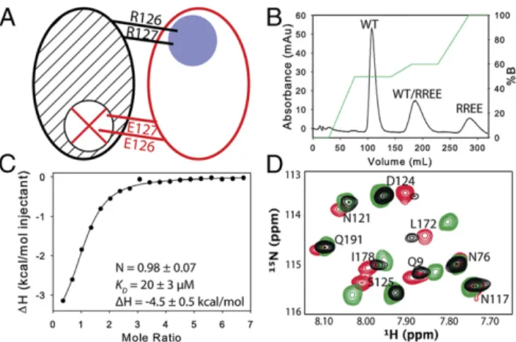
Mixed labeled dimers with a single functional active site were prepared by first abolishing substrate binding with an active site mutation (R126E, R127E). Mixing this inactive, purified homo-dimer with purified 15N-labeled WT homodimer yielded three
species, one of which is the mixed dimer with the nonfunctional subunit (the WT)15N-labeled (Fig. 1A) (note that the mutation at positions 126–127 abolishes binding to the opposite subunit because the loop bearing the arginine mutations forms critical
interactions with dUMP in the opposite subunit). Labeling of the functional subunit is achieved by15N-labeling the mutant enzyme and subsequent mixing with unlabeled WT (and purification from the two homodimers) (Fig. 1B). Binding of dUMP to the mixed dimer, measured by isothermal titration calorimetry (Fig. 1C), was similar to the WT (ΔH1of−4.5 kcal/mol,ΔH2of−4.4 kcal/mol and KD1=KD2of 16μM) (16). HSQCs of the apo mixed dimers showed that the mutation primarily affects nearby residues, with minimal effects on distal sites (Fig. 1D). With this pair of mixed labeled dimers, addition of dUMP yielded lig1samples with15N chemical
shift probes distributed throughout the bound or empty subunit, enabling subunit-specific tracking of ligand-binding effects without interference from dUMP0or dUMP2.
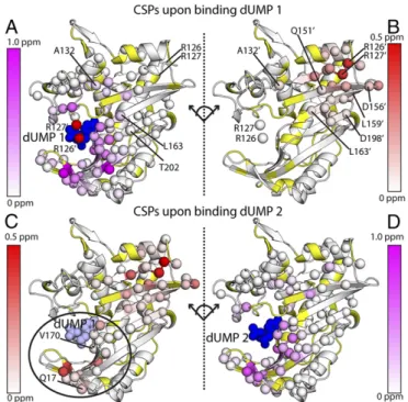
Imbalanced Chemical Shift Response to dUMP Binding.With the two mixed dimers, we characterized the subunit-specific effects of the first and second dUMP binding events using standard chemical shift perturbations (CSPs).1H-15N CSPs due to the first dUMP binding event were calculated directly from the mixed labeled dimers to monitor perturbations in each subunit. The bound subunit of dUMP1showed large CSPs in the binding site, dropping off∼20 Å
from dUMP, with smaller perturbations extending along the in-terface, out to∼28 Å from dUMP (Fig. 2A). The empty subunit was largely unaffected, dropping off∼15 Å from dUMP, with all of the CSPs in the dUMP binding loop (residues 123′–128′, where prime indicates the empty subunit) and the backside of the binding site (residues 150′–163′) (Fig. 2B). Overall, the effects of binding the first dUMP were highly localized, primarily to the binding region, with small perturbations to the dimer interface (Fig. S1A).
Although CSPs for the second dUMP binding event (dUMP1
to dUMP2) cannot be directly calculated, they can be obtained
indirectly from the mixed dimers by reconstructing the WT dUMP1chemical shifts, which was accomplished using a
vector-based correction to account for the effects of the RREE mutation (Fig. 3 and SI Materials and Methods). This correction allows generation of WT dUMP1chemical shifts from the mixed dimer
dUMP1chemical shifts (open circles in Figs. 3 and 4). The correction
dUMP had more pronounced effects throughout the entire protein. The effects of the second dUMP on the binding subunit resembled the effects of the first dUMP, with the largest and majority of the perturbations localized around the binding site (Fig. 2D). Most no-tably, however, unlike the first dUMP, the second dUMP caused significant perturbations to the other binding site: in this case, the site bound by the first dUMP (e.g., Q17 and V170) (Fig. 2C). The widespread perturbations in both subunits upon the second binding dUMP indicated subunit communication between the two binding sites. Overall, the CSP analysis showed an imbalance in the subunits’ responses to the two dUMP binding events: CSPs were limited to the binding site for the first dUMP but covered the interface and both binding sites for the second dUMP.
Ligand State Multiplets Reveal That dUMP1Is an Extreme State.
Al-though standard CSP analysis is effective at revealing overall perturbations of large magnitude, it can also obscure, especially in the case of a homodimer, interesting chemical shift behavior. To view the complete WT chemical shift responses, we sought to visualize relative peak positions of the dUMP0, dUMP1
(recon-structed), and dUMP2states for each residue amide. In general,
these overlays yielded four-peak ligand state multiplets (Fig. 4) because the dUMP0and dUMP2states yielded single peaks due
to dimer symmetry, and the dUMP1state can yield two distinct
peaks, one from each of the mixed dimer samples. For clarity, we
used superscripts“apo,” “empty,” “bound,”and“doub”to refer to peak positions of the dUMP0state, the empty subunit of the
dUMP1 state, the bound subunit of the dUMP1 state, and the
dUMP2state, respectively.
One interesting feature of the multiplets is that, in some cases, the peak positions of the dUMP1 state actually extended further
than the dUMP2 peaks, which we refer to as “supershifting.”
Supershifting can be seen for V170 and Q17 (Fig. 4AandB), where V170bound (Q17bound) shifts in the same direction as V170doub (Q17doub), but actually shifts beyond V170doub (Q17doub). The simple case where supershifting is along the apo–doub chemical shift change vector suggests a fast equilibrium of free and bound states, and, oddly, binding the first dUMP pushes the equilibrium further than the second dUMP. Alternatively, it could suggest that protomers may not simply snap into a free or bound conformation, but rather that there are additional states that protomers can adopt and that binding the first dUMP induces a more extreme state. Much of the dUMP1supershifting occurs around the binding site
(Fig. S3 A and B), which, remarkably, explains the long-range intersubunit communication observed upon binding the second dUMP (Fig. 2C). Because the first dUMP seems to induce an ex-treme state beyond what is observed in the dUMP2state, and one
that cannot be supported with both subunits bound, a response is set up in which binding the second dUMP partially reverses the initial shift (e.g., V170 in Fig. 4A). This result leads to intersubunit com-munication upon binding the second dUMP by making corrections to supershifting caused by the first dUMP.
More complex shifting behavior was seen in additional residues. For example, A132 exhibited not only supershifting, but also an A132empty and A132bound shift in a direction orthogonal to the A132apo–A132doubvector, termed“orthogonal shifting,”indicating that such sites are not simply in a fast, two-state equilibrium (Fig. 4C). This result, again, points to the existence of an additional state that becomes significantly populated upon binding the first dUMP. Another multiplet pattern we observed was “reverse shifting,” where dUMP1 peaks shift in the opposite direction of the apo–
doub chemical shift vector (Fig. 4DandE). It is not immediately clear why reverse shifting was observed although, along with supershifting, it suggests that there are compensatory behaviors occurring in the protomers upon binding the first dUMP. Overall, the observations of supershifting, orthogonal shifting, and reverse shifting suggest that a single dUMP binding induces sampling of extreme conformations relative to apo and dUMP2states.
Using ligand state multiplets from spectral overlays to assess specific residue chemical shift behaviors is generally not practical because the four peaks from each residue render the spectra too crowded. Thus, to enhance the analysis of multiplet behavior, we Fig. 2. Chemical shift perturbations of the two dUMP binding events. The
ef-fects of binding the first dUMP to the binding (A) and nonbinding (B) subunits of the dimer are shown at theTop, using the CSP scheme shown in Fig. 3A. The effects of binding the second dUMP to the nonbinding (C) and binding (D) sub-units are shown at theBottom, using the reconstructed WT CSPs shown in Fig. 3B. Viewing the dimer interface region is enhanced by separating the two subunits, where the subunits on the right underwent a hinge-type rotation (dotted line) to yield the same viewing angle as those on the left. As a reference point for the rotation, the red (A) and white (B) spheres show the locations of R126 and R127 from the other subunit. Residues with significant CSPs are shown as spheres. Residues that are missing or unassigned are in yellow. Annotated residues are discussed in Figs. 4 and 5, where residues denoted prime (e.g., R126′) correspond to the empty subunit of the dUMP1state. The first bound (dUMP 1) and second bound (dUMP 2) dUMPs are shown in dark blue; for binding of dUMP 2 (Bottom), the previously bound dUMP 1 is shown in light blue. InC, the circle highlights the major difference in the distant subunit response for binding of dUMP 1 and dUMP 2. The CSPs in context of the full dimer are shown inFig. S1.
Fig. 3. Vector correction to determine WT dUMP1peak positions. The two schematic diagrams show how CSPs were calculated for each dUMP binding event (SI Materials and Methods). (A) The CSPs for binding the first dUMP, CSP1, were calculated directly from the apo and dUMP1peak positions of the two mixed labeled dimer (MD) samples (thick arrows). Dashed arrows show the CSP due to the mutation (mut). (B) CSPs for binding the second dUMP, CSP2, were calculated as the vectors connecting the WT dUMP2peak (in blue) and the dUMP1peaks for WT (empty red and green circles), which were recon-structed by applying the CSP1vectors to the apo WT peak (dashed arrows).
BIOPHYSICS
AND
COMPUTATIONAL
BIOLOGY
SEE
COM
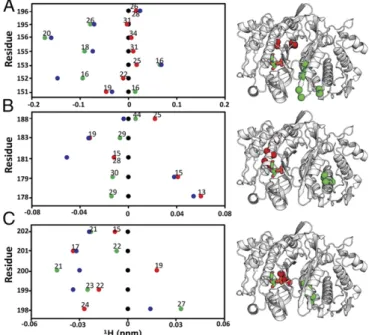
condensed the information into “line plots” of 1H and 15N chemical shifts, which allowed for simpler viewing of chemical shifts to more conveniently inspect individual residues in terms of the spatial response to dUMP binding (Fig. 5 andFigs. S4
and S5). For simplicity,1H line plots are shown although
cor-responding 15N line plots are also easily viewed (Fig. S4B).
From the line plots, we identified clusters of residues with similar behaviors, indicating regions of ligand sensitivity throughout the protein. Residues 155, 156, and 195 (Fig. 5A) are spatially clustered and exhibit similar supershifting behavior, indicating an extreme state (or skewed population) in this region. In the case of E195, the two sites are nearly equidistant from dUMP,
∼28 Å, yet E195empty supershifts and E195bound hardly shifts, showing how communication can be more effective across the in-terface. Residues in the central helix, in particular I178 and A179 (Fig. 5B), show that ligand binding can be sensed at distal sites even with no direct contact to, or across, the interface. Additionally, these regions show a symmetric pattern in which supershifting in one subunit is linked with reverse shifting in the other. This pattern was observed frequently throughout TS, indicating extensive compensa-tory behavior between subunits. Residues along the interface (Fig. 5C) showed the largest variety, as well as the most symmetric, of behaviors. These residues in particular reflect the quasi-symmetrical, compensatory shifts that occur where a partial shift or supershift in one subunit is coupled with a partial or reverse shift in the other subunit: e.g., V199, and D198. In summary, using mixed labeled dimers coupled with viewing ligand state multiplets via line plots facilitates thorough inspection of shifting from binding of both dUMP molecules, and extensive supershifting and reverse shifting indicate significant population of extreme states (or a further shifting of the equilibrium) beyond the known apo and dUMP2conformations.
Subunits’Response to Diligand Binding Is Equally Balanced.Although the mixed dimers are required to visualize the dUMP1states, they
are not required for the cofactor binding step of the reaction. Binding of a substrate analog, 5-FdUMP, along with cofactor, to-gether referred to as the“diligand,”forms a covalent bond to the enzyme, leading to a stable ternary complex (22, 23). Because of the covalent nature of this complex, the resonances from the diligand-bound TS are in slow exchange, and, thus, the chemical shifts of the diligand1states can be more easily measured (SI Materials and
Methods) (16). Together, dUMP and diligand will allow us to com-pare the proteins’response, not only to an additional binding event but also to a conformational change, because, unlike dUMP binding, diligand binding causes significant conformational changes in the vi-cinity of the binding site to form the closed ternary complex (22, 24). CSP analysis highlighted a number of interesting differences be-tween dUMP and diligand binding (Fig. 2 andFig. S6). Binding of the first diligand had overall larger magnitude and more extensive CSPs in both the binding and empty subunits than did dUMP, likely due to the combined effects of the conformational change and in-creased size of the diligand relative to dUMP (Fig. S6 AandB). Most notably, the effects of the first diligand propagated further in both subunits,∼30 Å from diligand, extending almost all the way to the second binding site. Thus, unlike with dUMP binding, there was clearly communication between the two sites upon binding the first diligand. Additionally, similar to dUMP binding, the effects of the second diligand on the binding subunit resembled those of the first (Fig. S6D). Most strikingly, there were no significant perturbations to the other binding site, as with dUMP (Fig. S6C). Overall, the similarities in the CSPs for the two diligand binding events indicate a balanced response to diligand binding, in contrast to dUMP binding. This balanced response to diligand is also evident in the diligand line plots (Fig. S7). Not surprisingly, the diligand line plots show more symmetrical and fewer extreme features, with the vast
Fig. 5. Line plots of1H chemical shifts. These line plots show how the chemical shift behaviors observed from ligand state multiplets are distrib-uted throughout the protein.1H chemical shift changes (relative to apo) are plotted for apo (black), dUMP2 (blue), dUMP1bound (red), and dUMP1empty (green). Each of the dUMP1points is labeled with the distance (in Å) of that residue (red and green spheres) from the bound dUMP (sticks). Distances are measured from the amide N to the centroid of the bound dUMP. (A) Behaviors of residues at opposite ends of the dimer interface. (B) Behaviors of interior residues. (C) Behaviors of residues at the center of the dimer interface. Fig. 4. dUMP ligand state multiplets. Spectral overlays of apo, dUMP1, and
majority of the diligand1peaks either coinciding with, or being
equally displaced from, the diligand0 and diligand2 peaks.
Ac-cordingly, there were very few diligand residues that showed supershifting (Fig. S3). However, as with dUMP, when super-shifiting did occur, it was often coupled with reverse shifting, yielding symmetrical patterns.
Discussion
In this study, we used mixed labeled dimers to investigate, by NMR, intersubunit signaling in response to single ligand (dUMP or diligand) binding events in the homodimeric enzyme thymidylate synthase (TS). The mixed dimer approach allows for isolation of singly ligated (lig1) states and breaks the symmetry degeneracy in
the NMR signals. This approach yields a rare example of step-wise progression of chemical shifts upon binding identical ligands in a homodimer and prompted the use of visualization strategies beyond simple CSPs. Standard CSP analysis revealed that only the binding of the second dUMP “signals” to the other binding site. This modulation of second ligand binding due to changes at the first site represents a nonintuitive yet valid potential allosteric mechanism. More generally, a distribution of ligand state peak multiplets were observed that point to regional behaviors, including the surprising observation of shifting of lig1 peaks beyond lig2 peaks, termed
“supershifting”here. There was also a surprising degree of quasi-symmetrical responses in the two protomers, especially in the dili-gand complex, indicating substantial compensatory behavior coupled across the dimer interface. One caveat of the RREE mixed dimers used here is that, without binding of the second ligand, there can technically be no functional allostery. The primary goal, how-ever, was to observe the mechanistic preparations that the homo-dimer makes before the second ligand binding event that enable the allosteric effect. Although the RREE mutation may have altered some of these preparations, the reconstructed binding steps yielded much useful information about allosteric communication. Although the focus of this study was on chemical shifts, clearly this strategy lends itself to protomer-specific NMR measurements, such as spin relaxation for the characterization of dynamics, on specific ligation states. Lastly, although the strategy used here applies primarily to tight dimers that do not readily dissociate, it can potentially be applied to other systems by covalently linking the monomers.
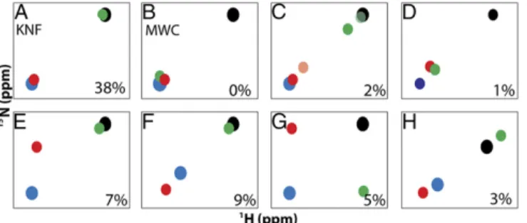
Insights into Allostery from Ligand State Peak Multiplets.The mixed labeled dimers allow for simple viewing of complete ligand state NMR peak multiplets, which, to our knowledge, have not been previously reported. The patterns observed in the multiplets can provide a format for evaluating allosteric models. For example, the most easily observed patterns are the doublets that would arise from a Koshland–Némethy–Filmer (KNF) or Monod–Wyman–Changeux (MWC) type system, and their expected intensities (12, 25), with only two possible states (Fig. 6AandB). In the KNF system, where only the binding subunit responds to ligand, the lig1bound and
lig1empty peaks would coincide with the lig2 and lig0 peaks,
re-spectively. In the MWC system, ligand binding causes a concerted shift in both subunits, where both lig1peaks would coincide with the
lig2peak. Alternatively, one might expect to observe two possible
linear triplet patterns, one where one of the lig1 peaks coincides
with either the lig2or lig0peaks and the other is partially shifted
toward lig2, or one where both lig1peaks are partially shifted toward
lig2(Fig. 6CandD) (12). We observed such triplet patterns in our
dUMP data as did others previously in studies of half-titrated, negatively cooperative dimers that are in the slow exchange regime (12, 26). Interestingly, we also observed many nonlinear triplets, and quartets (Fig. 6EandG), that indicate behaviors beyond simple population or exchange between lig0and lig2states. It is currently
not clear precisely what structural changes produce these nonlinear multiplets although it must involve at least a third conformation distinct from the lig0and lig2conformations. Given this diverse set
of multiplet patterns, it seems that there is not a consistent response
throughout the protein, but, rather, TS has a mixture of intersubunit responses to binding of dUMP. Evaluation of the ligand state multiplets makes this response clear, and it is possible to do so outside of the slow exchange condition. In general, the evaluation of NMR ligand state multiplets in oligomeric proteins is a powerful approach to characterize allosteric mechanisms in proteins (12, 25). Perhaps the most surprising multiplet pattern evident was the observation of supershifting (and reverse shifting) in the lig1state,
which was unexpected under the assumption that lig0and lig2peak
positions represent end states. However, as is apparent here and from previous studies (27, 28), this assumption is not always a good one because peak positions of specific mutants point to a shift in the equilibrium beyond the assumed end points of apo and ligand bound, which suggests that, for those sites, the apo and ligand saturated states (29) both represent dynamic equilibria between two extreme states that are not readily detected. It is interesting that, although supershifting and reverse shifting have been observed from com-parisons of mutant and WT peak positions, here they are observed from lig1peak positions. It is also interesting that these behaviors are
highly dependent on the residue (Fig. S3). Although changes in two-state equilibria can explain NMR peaks moving in a linear fashion, they cannot explain orthogonal peak movement. Therefore, a general explanation for the various behaviors observed here is that the lig1
state peak positions may reflect a range of different local confor-mations that TS samples upon binding the first ligand. These con-formations may represent different sets of interactions (hydrogen bond geometries, for example) that lead to particular lig1chemical
shifts. Thus, TS may reside in a relatively shallow conformational basin on the energy landscape that allows it to modulate various interactions by conformational adjustment, yielding different chem-ical shifts, upon binding one or two ligands. This scenario provides a more flexible model for interpreting chemical shifts and is funda-mentally distinct from two-state switching.
Lig1Asymmetry from X-Ray and NMR Chemical Shifts.Although the
functional significance of these multiplet behaviors remains to be determined, there is a precedent for the asymmetric effects of dUMP binding to TS. A crystal structure ofPneumocystis carinii
TS bound to dUMP and a cofactor analog, CB3717, has an asymmetric ternary complex wherein one active site has both dUMP and cofactor bound whereas the other has only dUMP bound (17). A key observation from this structure is that, in addition to the global changes that occur upon cofactor binding, there are subtle, yet significant conformational differences be-tween the two monomers for a number of residues along the otherwise rigid dimer interface. It was proposed that these Fig. 6. Observable patterns for ligand state multiplets. Schematic HSQC peak patterns according to allosteric model (AandB) or observed for dUMP binding in TS as resolved triplets (C–F) or quartets (GandH). Peaks are shown for apo/lig0(black), lig1bound(red), lig1empty(green), and lig2(blue). In C, two partial shifting behaviors are shown in which lig1partial peak shifting is observed in the binding subunit (light red) or in the empty subunit (dark green). Behaviors are observed for supershifting (F), orthogonal shifting (G), and reverse shifting (H). In each panel, the percentages indicate the abun-dance of the peak pattern for dUMP binding to TS.
BIOPHYSICS
AND
COMPUTATIONAL
BIOLOGY
SEE
COM
residues are the primary candidates for signaling between the active sites and could provide the basis for cooperativity. Of these residues, 75% exhibited nonlinear or supershifted triplets and quartets here for dUMP binding. The fact that we see such extensive overlap could suggest that these subtle structural re-arrangements are also occurring in the dUMP1state. The
non-linear shifts observed for these residues point to an additional state outside the apo-bound equilibrium, rather than simply an equilibrium shift. This extreme state could be due to strain in-duced by binding the first dUMP, leading to an asymmetric lig1
state. This strained conformational state may be the result of differential perturbations to the hydrogen-bonding network across the interface or to differences in dynamics between the two sub-units in the lig1state. The fact that we see nonlinear behavior for
these residues suggests that, if in fact these residues form a communication pathway, this extreme state may be one that is involved in TS cooperativity.
Given the long-range, intersubunit impact of binding the second dUMP, it is surprising that dUMP binding is thermodynamically noncooperative at 25 °C, at which the NMR chemical shifts were investigated. However, at lower temperatures, theΔH°bindvalues
for the first and second dUMP molecules are nonequivalent, reflecting intrinsically different ΔCp° values for the two binding
events. Thus, the underlying thermodynamics can be considered nonidentical for the two binding events, which could be reflected in the observed chemical shift behaviors. Furthermore, although dUMP binding does not trigger functional intersubunit allo-stery, it may still reveal the intrinsic communication mecha-nisms that could potentially lead to functional allostery during subsequent reaction steps. Overall, the presence of commu-nication upon dUMP binding could indicate that TS is poised for intersubunit allostery.
Potential Origin of Symmetrical Chemical Shift Response.Based on the observations made here, we propose that binding of the first dUMP or diligand to the structurally symmetric dimer imparts compensatory effects between the two protomers. In the case of dUMP binding, presuming there is no significant conformational change, the symmetrical chemical shift changes arise from either propagation of changes in hydrogen bonding strengths in a symmetrical fashion, or from compensatory dynamical responses between the two protomers, or a combination of both. In the case of diligand binding, given that there is likely a conforma-tional change in the bound subunit (22, 24), the symmetrical chemical shift changes are even more surprising because the shift in the bound subunit from the structural change cannot be rep-licated in the empty subunit. In either case, the propagation of chemical shift changes likely represents a form of structural or dynamic strain that, remarkably, has opposite manifestations in the two protomers for many residues. These considerations of quasi-symmetrical multiplets provide a unique view into how intersubunit allostery can be achieved for the simple example of symmetric homodimers. It seems that, at least for TS, symmetric cross-dimer interactions are“built in,”such that quasi-symmetrical strain is introduced by the binding of the first ligand. The magni-tude of symmetric chemical shift multiplet patterns and their extent throughout the protein indicate that TS is incredibly sensitive to substrate binding at sites throughout its structure, but particularly at the dimer interface. A fundamental question for understanding allostery is whether the intrinsic compensatory effects observed here will be observed in other homodimeric proteins, especially those that are allosteric with regard to binding two ligands.
ACKNOWLEDGMENTS.This work was supported by National Institutes of Health Grant GM083059 (to A.L.L.).
1. Cui Q, Karplus M (2008) Allostery and cooperativity revisited.Protein Sci17(8): 1295–1307.
2. Lisi GP, Loria JP (2016) Solution NMR spectroscopy for the study of enzyme allostery. Chem Rev116(11):6323–6369.
3. Nussinov R, Tsai CJ (2015) Allostery without a conformational change? Revisiting the paradigm.Curr Opin Struct Biol30:17–24.
4. Tsai CJ, Del Sol A, Nussinov R (2009) Protein allostery, signal transmission and dy-namics: A classification scheme of allosteric mechanisms.Mol Biosyst5(3):207–216. 5. Klemm JD, Schreiber SL, Crabtree GR (1998) Dimerization as a regulatory mechanism
in signal transduction.Annu Rev Immunol16:569–592.
6. Lipchock JM, Loria JP (2010) Nanometer propagation of millisecond motions in V-type allostery.Structure18(12):1596–1607.
7. Zhuravleva A, Clerico EM, Gierasch LM (2012) An interdomain energetic tug-of-war creates the allosterically active state in Hsp70 molecular chaperones.Cell151(6):1296–1307. 8. Akimoto M, et al. (2015) Mapping the free energy landscape of PKA inhibition and
activation: A double-conformational selection model for the tandem cAMP-binding domains of PKA RIα.PLoS Biol13(11):e1002305.
9. Williamson MP (2013) Using chemical shift perturbation to characterise ligand bind-ing.Prog Nucl Magn Reson Spectrosc73:1–16.
10. Selvaratnam R, Chowdhury S, VanSchouwen B, Melacini G (2011) Mapping allostery through the covariance analysis of NMR chemical shifts.Proc Natl Acad Sci USA 108(15):6133–6138.
11. Popovych N, Sun S, Ebright RH, Kalodimos CG (2006) Dynamically driven protein al-lostery.Nat Struct Mol Biol13(9):831–838.
12. Stevens SY, Sanker S, Kent C, Zuiderweg ER (2001) Delineation of the allosteric mechanism of a cytidylyltransferase exhibiting negative cooperativity.Nat Struct Biol 8(11):947–952.
13. Johnson EF, Hinz W, Atreya CE, Maley F, Anderson KS (2002) Mechanistic character-ization of Toxoplasma gondii thymidylate synthase (TS-DHFR)-dihydrofolate re-ductase: Evidence for a TS intermediate and TS half-sites reactivity.J Biol Chem 277(45):43126–43136.
14. Maley F, Pedersen-Lane J, Changchien L (1995) Complete restoration of activity to inactive mutants of Escherichia coli thymidylate synthase: Evidence that E. coli thy-midylate synthase is a half-the-sites activity enzyme.Biochemistry34(5):1469–1474. 15. Saxl RL, Changchien LM, Hardy LW, Maley F (2001) Parameters affecting the
resto-ration of activity to inactive mutants of thymidylate synthase via subunit exchange: Further evidence that thymidylate synthase is a half-of-the-sites activity enzyme. Biochemistry40(17):5275–5282.
16. Sapienza PJ, Falk BT, Lee AL (2015) Bacterial thymidylate synthase binds two molecules of substrate and cofactor without cooperativity.J Am Chem Soc137(45):14260–14263. 17. Anderson AC, O’Neil RH, DeLano WL, Stroud RM (1999) The structural mechanism for
half-the-sites reactivity in an enzyme, thymidylate synthase, involves a relay of changes between subunits.Biochemistry38(42):13829–13836.
18. Dev IK, et al. (1994) Mode of binding of folate analogs to thymidylate synthase: Ev-idence for two asymmetric but interactive substrate binding sites.J Biol Chem269(3): 1873–1882.
19. Lovelace LL, Gibson LM, Lebioda L (2007) Cooperative inhibition of human thymidylate synthase by mixtures of active site binding and allosteric inhibitors.Biochemistry46(10): 2823–2830.
20. Reilly RT, Barbour KW, Dunlap RB, Berger FG (1995) Biphasic binding of 5-fluoro-2′ -deoxyuridylate to human thymidylate synthase.Mol Pharmacol48(1):72–79. 21. Swiniarska M, et al. (2010) Segmental motions of rat thymidylate synthase leading to
half-the-sites behavior.Biopolymers93(6):549–559.
22. Hyatt DC, Maley F, Montfort WR (1997) Use of strain in a stereospecific catalytic mechanism: Crystal structures of Escherichia coli thymidylate synthase bound to FdUMP and methylenetetrahydrofolate.Biochemistry36(15):4585–4594.
23. Santi DV, McHenry CS, Perriard ER (1974) A filter assay for thymidylate synthetase using 5-fluoro-2′-deoxyuridylate as an active site titrant.Biochemistry13(3):467–470. 24. Stroud RM, Finer-Moore JS (2003) Conformational dynamics along an enzymatic
re-action pathway: Thymidylate synthase,“the movie”.Biochemistry42(2):239–247. 25. Freiburger LA, et al. (2011) Competing allosteric mechanisms modulate substrate
binding in a dimeric enzyme.Nat Struct Mol Biol18(3):288–294.
26. Tzeng SR, Kalodimos CG (2009) Dynamic activation of an allosteric regulatory protein. Nature462(7271):368–372.
27. Gardino AK, et al. (2009) Transient non-native hydrogen bonds promote activation of a signaling protein.Cell139(6):1109–1118.
28. McDonald LR, Boyer JA, Lee AL (2012) Segmental motions, not a two-state concerted switch, underlie allostery in CheY.Structure20(8):1363–1373.
29. Beach H, Cole R, Gill ML, Loria JP (2005) Conservation of mus-ms enzyme motions in the apo- and substrate-mimicked state.J Am Chem Soc127(25):9167–9176. 30. Changchien LM, et al. (2000) High-level expression of Escherichia coli and Bacillus
subtilis thymidylate synthases.Protein Expr Purif19(2):265–270.
31. Agrawal N, Hong B, Mihai C, Kohen A (2004) Vibrationally enhanced hydrogen tunneling in the Escherichia coli thymidylate synthase catalyzed reaction.Biochemistry43(7): 1998–2006.
32. Hardy LW, Pacitti DF, Nalivaika E (1994) Use of a purified heterodimer to test negative cooperativity as the basis of substrate inactivation of Escherichia coli thymidylate synthase (Asn177–>Asp).Structure2(9):833–838.
33. Kang X, Frey DD (2003) High-performance cation-exchange chromatofocusing of proteins.J Chromatogr A991(1):117–128.
34. Schmidt M, Hafner M, Frech C (2014) Modeling of salt and pH gradient elution in ion-exchange chromatography.J Sep Sci37(1-2):5–13.