Morphine paradoxically prolongs neuropathic pain in rats
by amplifying spinal NLRP3 inflammasome activation
Peter M. Gracea,b,c,1, Keith A. Stranda,b, Erika L. Galera,b, Daniel J. Urband, Xiaohui Wanga,b,e,f,g,h, Michael V. Barattaa,b, Timothy J. Fabisiaka,b, Nathan D. Andersona,b, Kejun Chengi, Lisa I. Greenea,b, Debra Berkelhammera,b,
Yingning Zhanga,b, Amanda L. Ellisa,b, Hang Hubert Yinf,g,h,j, Serge Campeaua,b, Kenner C. Ricei, Bryan L. Rothd, Steven F. Maiera,b, and Linda R. Watkinsa,b
aDepartment of Psychology and Neuroscience, University of Colorado, Boulder, CO 80309;bThe Center for Neuroscience, University of Colorado, Boulder, CO 80309; cDiscipline of Pharmacology, School of Medicine, University of Adelaide, Adelaide, SA 5005, Australia;dDepartment of Pharmacology, University of North Carolina,
Chapel Hill, NC 27599;eChemical Biology Laboratory, Changchun Institute of Applied Chemistry, Chinese Academy of Sciences, Changchun, Jilin 130022, China; fDepartment of Chemistry and Biochemistry, University of Colorado, Boulder, CO 80309;gBioFrontiers Institute, University of Colorado, Boulder, CO 80309;hThe
Center for Neuroscience, University of Colorado, Boulder, CO 80309;iChemical Biology Research Branch, National Institute on Drug Abuse and National Institute on
Alcohol Abuse and Alcoholism, Bethesda, MD 20892; andjCenter of Basic Molecular Science, Department of Chemistry, Tsinghua University, Beijing 100082, China
Edited by David Julius, University of California, San Francisco, CA, and approved April 19, 2016 (received for review February 16, 2016)
Opioid use for pain management has dramatically increased, with little assessment of potential pathophysiological consequences for the primary pain condition. Here, a short course of morphine, starting 10 d after injury in male rats, paradoxically and remarkably doubled the duration of chronic constriction injury (CCI)-allodynia, months after morphine ceased. No such effect of opioids on neuropathic pain has previously been reported. Using pharmacologic and genetic ap-proaches, we discovered that the initiation and maintenance of this multimonth prolongation of neuropathic pain was mediated by a previously unidentified mechanism for spinal cord and pain—namely, morphine-induced spinal NOD-like receptor protein 3 (NLRP3) inflam-masomes and associated release of interleukin-1β(IL-1β). As spinal dorsal horn microglia expressed this signaling platform, these cells were selectively inhibited in vivo after transfection with a novel De-signer Receptor Exclusively Activated by DeDe-signer Drugs (DREADD). Multiday treatment with the DREADD-specific ligand clozapine-N -oxide prevented and enduringly reversed morphine-induced persis-tent sensitization for weeks to months after cessation of clozapine-N-oxide. These data demonstrate both the critical importance of microglia and that maintenance of chronic pain created by early ex-posure to opioids can be disrupted, resetting pain to normal. These data also provide strong support for the recent“two-hit hypothesis” of microglial priming, leading to exaggerated reactivity after the sec-ond challenge, documented here in the context of nerve injury fol-lowed by morphine. This study predicts that prolonged pain is an unrealized and clinically concerning consequence of the abundant use of opioids in chronic pain.
TLR4
|
P2X7R|
danger signals|
DAMP|
opioid-induced hyperalgesiaR
ecent reports are critical of the lack of controlled, long-termstudies to support the dramatic escalation of opioid
treat-ment for chronic pain over the past decade (1–5). Although one
long-term concern is that there may be no benefit, another is that opioid treatment could have negative consequences for pain. For example, opioids are documented to paradoxically induce nociceptive sensitization [opioid-induced hyperalgesia (OIH)], both in the presence and absence of a pain condition (6, 7). With only one exception (8), OIH has been observed in chronic pain populations and is amplified by the preexisting
pain condition (9–16). However, the mechanistic interactions
between OIH and the pathophysiology of chronic pain are enigmatic, in part due to the absence of preclinical studies. Furthermore, the duration of OIH in either chronic pain pop-ulations or laboratory animals has never been assessed after discontinuation of opioid treatment; rather, pain was only as-sessed concurrently with, or within a few hours after, opioid administration. There would be major implications for how pain transitions to a chronic state if opioid treatment were to prolong the course of pain long after opioid cessation.
We predicted that opioid treatment would increase the magni-tude and/or duration of long-term neuropathic pain, based on three
interrelated lines of evidence: (i) Spinal microglial reactivity is
triggered after peripheral nerve injury, in part via spinal release of danger-associated molecular patterns (DAMPs) that initiate glial Toll-like receptor 4 (TLR4) signaling (17). Chronic pain is gated by TLR4 in preclinical models, as the ensuing production of neuro-excitatory, immune mediators amplify nociceptive signaling in the
spinal dorsal horn (17, 18); (ii) spinal microglial reactivity is also
triggered by nonstereoselective opioid activation of TLR4 that promotes spinal release of neuroexcitatory immune mediators (7,
19, 20); and (iii) an immunological phenomenon termed glial
“priming”has been described (21, 22), wherein a primary immune
challenge (hit 1) confers a heightened neuroinflammatory response to secondary challenge (hit 2). It therefore follows that neuropathic pain after peripheral nerve injury (hit 1) may be exacerbated and prolonged by opioid treatment (hit 2). However, it has not been previously anticipated that opioids could contribute to chronic pain. In addition, the superimposition of peripheral nerve injury and opioid treatment may activate a unique mechanism never pre-viously implicated in spinal cord, in opioid treatment, or for
path-ological pain—namely, activation of the NOD-like receptor protein
3 (NLRP3) inflammasome, a protein complex that activates
Significance
Pain after disease/damage of the nervous system is predominantly treated with opioids, but without exploration of the long-term consequences. We demonstrate that a short course of morphine after nerve injury doubles the duration of neuropathic pain. Using genetic and pharmacological interventions, and innovative De-signer Receptor Exclusively Activated by DeDe-signer Drugs disrup-tion of microglia reactivity, we demonstrate that opioid-prolonged neuropathic pain arises from spinal microglia and NOD-like re-ceptor protein 3 inflammasome formation/activation. Inhibiting these processes permanently resets amplified pain to basal levels, an effect not previously reported. These data support the“two-hit hypothesis”of amplification of microglial activation—nerve injury being the first“hit,”morphine the second. The implications of such potent microglial“priming”has fundamental clinical implications for pain and may extend to many chronic neurological disorders.
Author contributions: P.M.G., X.W., M.V.B., H.H.Y., S.F.M., and L.R.W. designed research; P.M.G., K.A.S., E.L.G., X.W., M.V.B., T.J.F., N.D.A., L.I.G., D.B., Y.Z., A.L.E., and S.C. performed research; D.J.U., K.C., S.C., K.C.R., and B.L.R. contributed new reagents/analytic tools; P.M.G. analyzed data; and P.M.G., X.W., S.C., S.F.M., and L.R.W. wrote the paper.
The authors declare no conflict of interest.
This article is a PNAS Direct Submission.
1To whom correspondence should be addressed. Email: [email protected].
This article contains supporting information online atwww.pnas.org/lookup/suppl/doi:10.
1073/pnas.1602070113/-/DCSupplemental.
NEUR
OSCIENCE
PNAS
interleukin-1β (IL-1β), a “gatekeeper of inflammation”
(summa-rized in Fig. S1) (23, 24). TLR4 signaling primes the
inflam-masome by increasing the expression of NLRP3 and pro–IL-1β(25).
A second signal, such as the purinergic receptor P2X7R—engaged by
morphine and after peripheral nerve injury (7, 17, 26)—leads to the
association of NLRP3, the adaptor protein apoptosis-associated speck-like protein containing a CARD (ASC), and caspase-1,
allowing proteolytic activation of IL-1β(25, 27). Therefore, the aim
of the present study was to test whether morphine treatment after peripheral nerve injury prolonged neuropathic pain in rats and whether the prolonged pain was mediated by spinal NLRP3 inflammasomes. Our data implicate the two superimposed challenges as both immunological in nature and as contributors to persistent neuropathic pain.
Results
Morphine Induces Persistent Nociceptive Sensitization After Peripheral
Nerve Injury. To assess whether morphine could induce persistent
sensitization under conditions of established neuropathic pain, morphine or saline was administered for 5 d (5 mg/kg, twice daily), beginning 10 d after sciatic chronic constriction injury (CCI) or sham surgery.* Morphine treatment significantly prolonged CCI-allodynia
in the Fischer 344 (F344) strain (Fig. 1A) and increased the magnitude
of CCI-allodynia in the Sprague–Dawley (SD) rat strain (Fig. 1B). The
5-d morphine regimen induced only mild and transient mechanical
allodynia in sham-operated rats (Fig. 1AandB),a recognized feature
of opioid abstinence (30). The empirical observation that morphine increased the vigor and speed of hindpaw withdrawal to the von Frey filaments in SD rats was supported by increased startle (converted to
force; N) to a 0.2-mA shock (Fig. 1C). These data implicate morphine
in the prolongation and amplification of neuropathic pain.
Morphine-Induced Persistent Nociceptive Sensitization Is Independent of
Opioid Receptors.To determine whether opioid receptors mediated
persistent sensitization, the μ-, κ-, andδ-opioid receptor-inactive
stereoisomer (+)-morphine (31) was administered in lieu of
(−)-morphine. (+)-morphine recapitulated persistent sensitization
(Fig. 2A), demonstrating that this effect can occur independently of
classical opioid receptors. In support, knockdown of spinalOprm1
(encoding for the μ-opioid receptor) failed to prevent the
devel-opment of morphine-induced persistent sensitization (Fig. 2B),
despite knockdown of the target mRNA and protein sufficient to
impair (−)-morphine analgesia (Fig. S2 A andB). Because both
morphine isomers are TLR4 agonists (7, 19, 20), the role of this
innate immune receptor was assessed by substituting (−)-morphine
with the structurally distinct TLR4 agonist disulfide high mobility group box-1 (ds-HMGB1) (32). Persistent sensitization was
re-capitulated with ds-HMGB1 (Fig. 2C). Therefore, mechanisms of
central immune signaling were investigated to explain morphine-induced persistent sensitization.
Central Immune Signaling Mediates Morphine-Induced Persistent
Nociceptive Sensitization.Morphine nonstereoselectively activates
innate immunity, inducing production of the“gatekeeper of
in-flammation”and neuroexcitatory cytokine IL-1β(7, 20, 23, 33, 34).
Therefore, IL-1 receptor antagonist (IL-1ra) was intrathecally ad-ministered to test whether spinal IL-1 mediated morphine-induced persistent sensitization. Such a result would be congruent with the
results using (+)-morphine described above. Intrathecal IL-1ra
in-fusion during morphine administration prevented persistent
sensi-tization (Fig. 3A), whereas acute intrathecal IL-1ra during the
period of persistent sensitization significantly attenuated mechanical
allodynia, in F344 rats (Fig. 3B) (for parallel data in SD rats, seeFig.
S3). Inhibition of TNF and IL-6, cytokines that can be regulated by
IL-1β(23), also attenuated morphine-induced persistent
sensitiza-tion in F344 rats (Fig. 3H) (for parallel SD data, seeFig. S1). These
data indicate that the initiation and maintenance of morphine-induced persistent sensitization are dependent on proinflammatory cytokine signaling.
There are several known mechanisms by which IL-1β may
in-crease the excitability of second-order nociceptive projection neu-rons, including phosphorylation of postsynaptic NR1 NMDA receptor subunits (35), and down-regulation of both the astrocyte glutamate transporter GLT-1 (36) and neuronal G protein-coupled receptor kinase 2 (GRK2; an enzymatic regulator of the homolo-gous desensitization of many G protein-coupled receptors that protects against overstimulation) (37). The respective levels of these proteins were assessed in the ipsilateral lumbar dorsal horn during the period of persistent sensitization in F344 rats (5 wk after the conclusion of morphine or saline administration). Phospho-NR1 was elevated, whereas GRK2 and GLT-1 were decreased by the
superimposition of CCI and morphine (Fig. 3 D–F). These data
provide biochemical validation of the prolonged allodynia
pre-sented in Fig. 1Aand additional supportive evidence that
morphine-induced persistent sensitization was dependent on IL-1βsignaling.
Morphine-Induced Persistent Sensitization Is Associated with Spinal
Cord Inflammasome Activation in Microglia.Inflammasomes regulate
IL-1βactivation in peripheral immune cells (Fig. S1), yet it is not
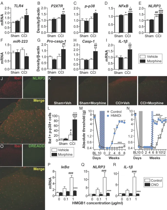
known whether parallel mechanisms exist in the spinal cord (24). Thus, expression of inflammasomes was quantified in the ipsilateral lumbar dorsal horn during the period of persistent sensitization in F344 rats (5 wk after the conclusion of morphine or saline ad-ministration). TLR4 mRNA and P2X7R protein levels, which represent the respective first (priming) and second (activation) signals, were elevated by the combination of CCI and morphine,
relative to sham and saline control (Fig. 4AandB). Phosphorylated
p38 and the p65 subunit of NF-κB [which are responsible for
NLRP3 and IL-1βtranscription (25)], as well as NLRP3, were
el-evated by the combination of CCI and morphine, relative to sham
and saline control (Fig. 4C–E). Expression of a negative regulator
of NLRP3, microRNA-223 (miR-223) (38), was decreased by the combination of CCI and morphine, relative to sham and saline saline (5 d; shaded area) was administered 10 d after CCI/sham surgery, and absolute thresholds for me-chanical allodynia were quantified in F344 (A) and SD (B) rats. (C) Startle force to 0.2-mA foot shocks at baseline (BL), after CCI but before morphine (predose), and 5 wk after the conclusion of morphine dosing (5 wk). *P<0.05; **P<0.01; ***P<0.001 (relative to CCI+saline); ###P < 0.001 (relative to sham+saline).
Data are presented as mean±SEM;n=6 or 7 per group.
control (Fig. 4F). The precursor enzyme procaspase-1, its active
form caspase-1, and the product IL-1βmRNA were elevated by the
combination of CCI and morphine, relative to sham and saline
control (Fig. 4G–I). These biochemical data support the behavioral
attenuation of morphine-induced persistent sensitization by IL-1ra and demonstrate that expression of the NLRP3 inflammasome by microglia is associated with such persistent sensitization.
Lumbar dorsal spinal NLRP3 was colocalized with the microglia
marker Iba1 (Fig. 4J), but not GFAP (astrocytes) or NeuN (neurons)
(Fig. S4A). Furthermore, the combination of CCI and morphine
increased the number of reactive lumbar dorsal spinal microglia
(Iba1+and phospho-p38+), relative to all other conditions, when
assessed 5 wk after the conclusion of morphine or saline
adminis-tration (Fig. 4K). Therefore, the role of microglia in mediating
morphine-induced persistent sensitization was functionally assessed. Current pharmacological methods to attenuate microglial reactivity lack selectivity, whereas the introduction of cellular debris to the local environment by depletion methods may present an immune stimulus in the central nervous system (CNS) (17). Therefore, we
developed an inhibitory (Gi) Designer Receptor Exclusively
Acti-vated by a Designer Drug (DREADD) (39) under a CD68 promoter that was intrathecally transfected via an AAV9 vector. Transfection
of the Gior control constructs occurred before experimental
ma-nipulation, to ensure that microglia would form the majority of
CD68+ cells in the spinal cord (40, 41). Gi-linked signaling was
predicted to attenuate microglial reactivity because activation of the
M4 muscarinic receptor [the GiDREADD progenitor (39)] inhibits
Ca2+influx in parasympathetic neurons (42), a process associated
with decreased proinflammatory cytokine production in microglia
(43, 44). DREADD expression was restricted to Iba1+cells in the
lumbar dorsal spinal cord (Fig. 4L), and not those expressing GFAP
or NeuN (Fig. S4B). DREADDs were activated with the selective,
biologically inert ligand clozapine-N-oxide (CNO). Intrathecal CNO
infusion during morphine administration prevented
morphine-induced persistent sensitization in F344 rats expressing the Gi
DREADD (Fig. 4M). Intrathecal infusion of CNO at 5 wk after the
conclusion of morphine administration [which is within the period of persistent sensitization induced by morphine, because mechanical
allodynia resolved in saline-treated CCI rats by this time (Fig. 1A)]
reversed morphine-induced persistent sensitization in F344 rats
expressing the GiDREADD (Fig. 4N) (for parallel SD data, seeFig.
S4C). Inhibition of proinflammatory signaling by Gi DREADDs
was confirmed in vitro by using a GiDREADD-transfected BV-2
microglia cell line. HMGB1—a DAMP released spinally in chronic
pain models (17, 45)—increased the expression of gene transcripts
encoding IκBα(a negative regulator induced by NF-κB), NLRP3,
and IL-1βin a concentration-dependent manner (Fig. 4O–Q). Such
increases in gene expression were attenuated by coincubation with 50μM CNO (Fig. 4O–Q). Similar results were found for expression
of gene transcripts encoding TNF and IL-6 (Fig. S4D). These data
demonstrate that expression of the NLRP3 inflammasome by microglia is associated with morphine-induced persistent sensitization
and that the initiation and maintenance of such persistent sensiti-zation is dependent on microglial reactivity.
Spinal Cord Inflammasomes Mediate Initiation of Morphine-Induced
Persistent Sensitization.The following experiments were designed
to test whether spinal NLRP3 inflammasome activation was causal to the induction of morphine-induced persistent sensitization. Thus, the inflammasome platform was pharmacologically inhibited at several levels during morphine administration and followed by assessment of the behavioral and biochemical consequences for opioid-induced persistent sensitization.
The role of spinal TLR4—activated by both morphine (20) and
DAMPs (17)—was explored as the first signal for inflammasome
activation. Intrathecal infusion of the TLR4 antagonist (+)-naloxone
(46) during morphine administration prevented the development of
morphine-induced persistent sensitization in F344 rats (Fig. 5A) (SD
data are in Fig. S5A). In support of the pharmacological data,
knockdown of spinalTlr4(Fig. S5B), as well as TLR2/4 inhibition by
oxidized 1-palmitoyl-2-arachidonyl-sn-3-glycero-phosphorylcholine
(OxPAPC) (Fig. S5C), also prevented the development of
mor-phine-induced persistent sensitization. Next, the role of spinal
P2X7R—also activated by DAMPs (17)—was explored as the
sec-ond signal for inflammasome activation. Intrathecal infusion of A438079 (47), a selective P2X7R antagonist, during morphine ad-ministration prevented the development of morphine-induced
per-sistent sensitization in F344 rats (Fig. 5B) (SD data are inFig. S5D).
In support of the A438079 results, P2X7R inhibition by Brilliant Blue G (48) likewise prevented the development of morphine-induced persistent sensitization in F344 rats and SD rats under
identical experimental designs (Fig. S5E). The role of spinal
caspase-1 was then explored, because this is the enzyme responsible for the
proteolytic activation of IL-1β(25). Intrathecal infusion ofN
-Ac-Tyr-Val-Ala-Asp-chloromethyl ketone (ac-YVAD-cmk) (49) during morphine administration prevented the development of
morphine-induced persistent sensitization in F344 rats (Fig. 5C) (SD data are
inFig. S5F). These data provide evidence that initiation of mor-phine-induced persistent sensitization is dependent on TLR4, P2X7R, and caspase-1 signaling during morphine administration.
Markers of IL-1β–induced neuroexcitation were quantified in
the ipsilateral lumbar dorsal quadrant after coadministration of
(+)-naloxone, A438079, or ac-YVAD-cmk with morphine (within
the period of persistent sensitization in F344 rats; 5 wk after the conclusion of morphine administration). Each inhibitor decreased expression of phospho-NR1, and increased expression of GRK2
and GLT-1, relative to vehicle controls (Fig. 5D–F). These data
provide biochemical support for the prevented allodynia presented
in Fig. 5A–Cand of attenuated IL-1βsignaling.
Expression of inflammasomes was quantified in the ipsilateral lumbar dorsal quadrant within the period of persistent sensitization in F344 rats (5 wk after the conclusion of morphine administration).
(+)-naloxone, A438079 and ac-YVAD-cmk each decreased
ex-pression of receptors mediating inflammasome priming (TLR4)
and activation (P2X7R ) (Fig. 5G–I). Furthermore, each inhibitor
Fig. 2. Opioid receptors do not mediate morphine-induced persistent sensitization. (A) The opioid-receptor inactive (+)-morphine or saline (5 d; shaded area) was administered 10 d after CCI, and absolute thresholds for mechanical allodynia were quantified in F344 rats. (B)Oprm1siRNA (7 d, beginning 8 d after CCI; green hatched bar) and morphine (5 d, beginning 10 d after CCI; shaded area) were ad-ministered, and absolute thresholds for mechanical allodynia were quantified in F344 rats. (C) The TLR4 agonist ds-HMGB1 or saline (5 d; shaded area) was administered 10 d after CCI, and absolute thresholds for mechanical allodynia were quantified in F344 rats. *P<0.05; ***P<0.001 (relative to CCI+saline). Data are presented as mean±SEM;n=6 per group.
NEUR
OSCIENCE
PNAS
decreased expression of phospho-p38 and p65 NF-κB, and,
con-sequently, NLRP3 (Fig. 5J–L). Each inhibitor decreased expression
of procaspase-1, caspase-1, and IL-1βmRNA (with the exception of
procaspase-1 expression, which was not altered by (+)-naloxone at
this timepoint) (Fig. 5M–O). In support of a role for microglia in
morphine-induced persistent sensitization, the number of reactive
lumbar dorsal spinal microglia (Iba1+and phospho-p38+) was
at-tenuated by (+)-naloxone, A438079, and ac-YVAD-cmk, relative to
vehicle controls (Fig. 4P-R). Together, these data demonstrate that
activation of microglia and spinal cord inflammasomes is de-pendent on TLR4, P2X7R, and caspase-1 signaling during mor-phine administration and reveal underlying biochemical and molecular changes likely responsible for the behavioral effects.
Finally, the role of NLRP3 activation in the initiation of morphine-induced persistent sensitization was confirmed by knockdown of
spinalNlrp3, which prevented prolonged allodynia in F344 rats (Fig.
5S). Knockdown of the target mRNA and protein was verified (Fig.
S5G). By intrathecally inhibiting the first (TLR4) and second
(P2X7R) signals, as well as NLRP3 and caspase-1, during morphine administration, these affirmative data demonstrate a causal role for spinal NLRP3 inflammasomes in the initiation of morphine-induced persistent sensitization.
Spinal Cord Inflammasomes Mediate the Maintenance of Persistent
Sensitization.Because NLRP3 inflammasome expression remained
elevated within the period of morphine-induced persistent sensiti-zation (5 wk after the conclusion of morphine administration) (Fig. 4), we tested whether such expression was causal to the mainte-nance of persistent sensitization. Thus, the inflammasome platform was pharmacologically inhibited within the period of persistent sensitization (5 wk after the conclusion of morphine administration for F344 rats). Inhibition was accompanied by assessment of the behavioral and biochemical consequences for opioid-induced persistent sensitization.
The role of TLR4 was explored as the first signal for
inflamma-some activation. Intrathecal infusion of (+)-naloxone starting 5 wk
after morphine administration enduringly reversed established
mor-phine-induced persistent sensitization in F344 rats (Fig. 6A) (SD data
are inFig. S6A). The role of P2X7R was explored as the second
signal for inflammasome activation. Intrathecal infusion of A438079 starting 5 wk after morphine administration enduringly reversed established morphine-induced persistent sensitization in F344 rats
(Fig. 6B) (SD data are inFig. S6B). In support, Brilliant Blue G also
reversed morphine-induced persistent sensitization in F344 rats and
SD rats under identical experimental designs (Fig. S6C). The role of
caspase-1 was then explored, because it is the enzyme that is re-sponsible for the proteolytic activation of IL-1β. Intrathecal infusion of ac-YVAD-cmk beginning 5 wk after morphine administration reversed morphine-induced persistent sensitization in F344 rats (Fig.
6C) (SD data are inFig. S6D). These data demonstrate that
main-tenance of morphine-induced persistent sensitization is dependent on sustained TLR4, P2X7R, and caspase-1 signaling.
Markers of IL-1–induced neuroexcitation were quantified in the
ipsilateral lumbar dorsal quadrant after reversal of
morphine-induced persistent sensitization by (+)-naloxone, A438079, or
ac-YVAD-cmk. Each inhibitor decreased expression of phospho-NR1, and increased expression of GRK2 and GLT-1, relative to vehicle
controls (Fig. 6D–F). These data provide biochemical support for
the reversed allodynia presented in Fig. 6A–Cand of attenuated
IL-1βsignaling.
Expression of inflammasomes was quantified in the ipsilateral lumbar dorsal quadrant 1 d after the conclusion of inhibitor infusion (43 d after the conclusion of morphine administration) in F344 rats.
(+)-naloxone, A438079, and ac-YVAD-cmk each decreased
expres-sion of receptors mediating inflammasome priming and activation
TLR4 and P2X7R (Fig. 6G–I). Furthermore, each inhibitor decreased
expression of phospho-p38 and p65 NF-κB, and, consequently, NLRP3
(Fig. 6J–L). Each inhibitor decreased expression of procaspase-1,
caspase-1, and IL-1β mRNA (Fig. 6 M–O). There were three
exceptions, where (+)-naloxone did not decrease expression of
P2X7R or procaspase-1, and ac-YVAD-cmk did not decrease ex-pression of P2X7R or procaspase-1 at this time point. These data demonstrate that the sustained activation of inflammasomes is dependent on TLR4, P2X7R, and caspase-1 signaling after mor-phine administration. Furthermore, this affirmative dataset dem-onstrates a causal role for spinal inflammasomes in the maintenance of morphine-induced persistent sensitization.
Discussion
We discovered that a brief course of morphine treatment, adminis-tered upon expression of neuropathic pain, drives persistent sensiti-zation for months after cessation of morphine. This persistent
sensitization is (i) not dependent on opioid receptor signaling; (ii)
correlated with increased expression of the ipsilateral spinal lumbar
dorsal inflammasome and localized to microglia; (iii) initiated by
morphine-induced spinal NLRP3 inflammasome activation, a protein structure that had not previously been identified in the spinal cord or
linked to pain; and (iv) maintained by spinal inflammasome activation.
(blue hatch; 5 d) was coadministered with morphine (5 d; shaded area), 10 d after CCI surgery, and abso-lute thresholds for mechanical allodynia were quan-tified in F344 rats. Morphine (5 d; shaded area) was administered 10 d after CCI surgery, (b) IL-1ra, (c) etanercept or TB-2–081 were intrathecally adminis-tered 5 wk after morphine conclusion, and absolute thresholds for mechanical allodynia quantified in F344 rats. Ipsilateral lumbar dorsal spinal cords were collected from CCI/sham F344 rats, 5 wk after mor-phine/saline administration and phospho-NR1 (D), GRK2 (E), and GLT-1 (F) protein levels were quanti-fied. *P<0.05; **P<0.01; ***P<0.001 [relative to vehicle (A–C) and relative to sham+saline (D–F)];#P<
0.05;###P<0.001 [TB-2-081 vs. vehicle (C) and relative
to sham+morphine (D–F);^^P< 0.01;^^^P <0.001
Fig. 4. Repeated morphine after CCI amplifies inflammasome activation in microglia. (A–I) Ipsilateral lumbar dorsal spinal cords were collected from F344 rats that had undergone sham or CCI surgery, 5 wk after morphine/saline administration, and respective levels of P2X7R (A), TLR4 (B), phospho-p38/total ERK ratio (C), NF-κB (p65 subunit) (D), NLRP3 (E), miR-223 (F), procaspase-1 (G), caspase-1 (H), and IL-1β(I) quantified. (J) NLRP3 colocalization with Iba1 in the ipsilateral lumbar dorsal horn. (K) Phospho-p38 colocalization with Iba1 in the ipsilateral lumbar dorsal horn.(L)DREADD colocalization with Iba1 in the lumbar dorsal horn. (MandN) F344 rats were transfected with intrathecal inhibitory Gior control DREADDs, and morphine (5 d; shaded area) was administered 10 d after CCI and
absolute thresholds for mechanical allodynia were quantified in F344 rats. CNO (blue hatched bar) was coadministered with morphine (5 d) (M) or 5 wk after morphine dosing had concluded (CNO dosed for 7 d) (N), and absolute thresholds for mechanical allodynia were quantified. (O–Q) Gene expression in BV-2 cells expressing the GiDREADD after 4 h incubation with a concentration range of HMGB1, and 0μM (control) or 50μM CNO. *P<0.05; **P<0.01; ***P<0.001
[relative to sham+saline (A–IandK), relative to vehicle (MandN), and relative to control (O–Q)];#P<0.05;##P<0.01;###P<0.001 [relative to sham+morphine
(A–IandK) and relative to 0μg (O–Q)];^P<0.05;^^P<0.01;^^^P<0.001 (relative to CCI+saline). Data are presented as mean±SEM;n=6 or 7 per group.
NEUR
OSCIENCE
PNAS
Fig. 6. Maintenance of persistent sensitization is dependent on inflammasome signaling. (A–C) The TLR4 antagonist (+)-naloxone (blue hatch; 5 d) (A), the P2X7R antagonist A438079 (purple hatch; 5 d) (B), or the caspase-1 inhibitor ac-YVAD-cmk (green hatch; 5 d) (C) was administered 5 wk after morphine (5 d, administered 10 d after CCI; shaded area), and absolute thresholds for mechanical allodynia were quantified in F344 rats. Ipsilateral lumbar dorsal spinal cords were collected from F344 rats, 1 d after the conclusion of inhibitor treatment. (D–F) Respective levels of phospho-NR1, GRK2, and GLT-1 were quantified after treatment with (+)-naloxone (D), A438079 (E), or ac-YVAD-cmk (F). (G–I) Respective levels of P2X7R and TLR4 were quantified after treatment with (+)-naloxone (G), A438079 (H), or ac-YVAD-cmk (I). (J–L) Respective levels of phospho-p38/total ERK ratio, NF-κB (p65 subunit), and NLRP3 were quantified after treatment with (+)-naloxone (J), A438079 (K), or ac-YVAD-cmk (L). (M–O) Respective levels of procaspase-1, caspase-1, and IL-1βwere quantified after treatment with (+)-naloxone (M), A438079 (N), or (ac-YVAD-cmk (O). *P<0.05; **P<0.01; ***P<0.001 (inhibitor vs. control). Data are presented as mean±
SEM;n=6 or 7 per group.
NEUR
OSCIENCE
PNAS
oratory animal studies (6, 7). However, we discovered that mor-phine interacts with neuropathic pain pathophysiology to potently prolong this allodynia. We implicated the dorsal spinal NLRP3 inflammasome in morphine-induced persistent sensitization, dis-covering that this signaling platform has a triumvirate of previously undocumented roles in: the spinal cord, a neuropathic pain model, and enhancement of its activity by morphine (24). Dorsal spinal NLRP3 inflammasomes mediate the initiation of morphine-induced persistent sensitization, because inhibition of TLR4, P2X7R, caspase-1, or IL-1 during morphine administration prevents pro-longed allodynia. Maintenance of morphine-induced persistent sensitization is also dependent on this pathway, because inhibition of TLR4, P2X7R, caspase-1, or IL-1 reversed prolonged allodynia, an effect that was sustained after TLR4 or P2X7R antagonism. It should be noted that the role of TLR4 in OIH has been challenged (50, 51), although these data do not preclude a role for this receptor in morphine-induced persistent sensitization. Furthermore, TLR4 is posited to exclusively regulate male pain behaviors (26, 52). How-ever, ongoing studies indicate that morphine-induced persistent sensitization also occurs in female rodents.
Expression of NLRP3 induced by persistent sensitization was lo-calized to microglia, cells that also express TLR4 and P2X7R (17). The contribution of microglia to the induction and maintenance of morphine-induced persistent sensitization was confirmed by
selec-tively inhibiting these cells with a GiDREADD (Fig. 4). The novel
application of DREADD technology represents an important tech-nical advance, because putative microglial inhibitors (e.g., minocycline, ibudilast, or propentofylline) have activity at other CNS cells, including neurons (17, 53). Expression of DREADDs before neu-ropathic pain induction prevented injury-induced recruitment of monocyte-derived cells from contributing to the observed effects.
Although we predict that Gi-linked signaling inhibits Ca2+influx in
microglia to attenuate proinflammatory cytokine production (43, 44), the precise mechanisms are the subject of ongoing investigation. Because microglial activity has not been selectively manipulated in any prior study, these data, to our knowledge, are the first to un-equivocally implicate microglia in a pathological pain state.
The mechanism(s) by which inflammasomes remained activated after cessation of morphine is an avenue for further investigation. Initial activation of inflammasomes may have induced several ad-aptations that create a positive feedback loop at TLR4 and P2X7R. One adaptation may be disrupted glutamate homeostasis, due to
IL-1β–mediated down-regulation of GLT-1 (Fig. 3F). Elevated
glutamate may trigger ATP release from glia (54, 55), as well as excitotoxicity and subsequent DAMP release (17). ATP and re-active oxygen species released after glial P2X7R activation (56, 57), as well as additional DAMPs released as a consequence of HMGB1-induced excitotoxicity (58), may also maintain inflam-masome signaling. However, whether spinal cord inflaminflam-masomes remain activated in the absence of morphine by reactive oxygen species and/or DAMP signaling at TLR4 and P2X7R, as part of a positive feedback loop, requires future examination.
The implications of the present study are striking in light of the
“two-hit”model of glial priming and exaggerated neuroinflammation.
Firstly, this model may provide a basis for understanding how opioids
Secondly, opioids superimposed on CNS neuroinflammation may have far-ranging consequences beyond pain. For example, opioids may also serve as a second hit for glia primed by aging or in-flammation/trauma and may lead to cognitive decline in the elderly (63), postoperative cognitive decline (64), and impaired recovery of motor function after spinal cord injury (65, 66). Whether the mechanistic underpinnings revealed in the current series of studies will prove to generalize to such opioid-related phenomena remains to be defined. Finally, the implications of the present studies may extend beyond opioids as the second hit. A broad range of repeated neuroinflammatory challenges not only induce a transition from acute to persistent pain (60, 67, 68), but also induce behaviors that are comorbid with pain, including cognitive impairment (69), de-pression (70), and anxiety (71). Therefore, our data provide a rationale to examine whether the ubiquitous management of chronic pain with opioids contributes to the incidence of such pain,
and potentially pain comorbidities—a hypothesis not previously
considered or tested.
In summary, the mechanisms underlying the transition from acute to chronic pain are poorly understood (17, 72, 73). We discovered that a short course of morphine administered upon expression of neuropathic pain remarkably doubled the duration of CCI-allodynia. This process was dependent upon dorsal spinal microglial re-activity and NLRP3 inflammasomes. These findings comport with prior demonstrations that repeated immune challenges induce a transition from acute to chronic pain (60, 67, 68), which may also
underpin pain comorbidities (69–71). An evaluation of the
long-term consequences of opioid treatment for chronic pain will identify whether this phenomenon manifests clinically. Our data suggest a unique strategy to prevent and reverse the deleterious long-term effects of opioid treatment without compromising
mor-phine analgesia;μ-opioid receptor-mediated analgesia can be
main-tained, while simultaneously eliminating inflammasome-mediated persistent sensitization.
Materials and Methods
SI Materials and Methodsprovides complete experimental methods. It includes subjects, drugs, RNA interference, surgery, catheter implantation, mechanical allodynia, shock sensitivity, and thermal analgesia testing, in vitro GiDREADD
transfection and stimulation, RT-PCR, Western blotting, and immunohisto-chemistry. Methods for statistical analysis are also included.
All animal procedures were approved by the Institutional Animal Care and Use Committee of the University of Colorado Boulder.
ACKNOWLEDGMENTS. This work was supported by the American Pain Society Future Leaders in Pain Research Grants Program (P.M.G.); National Health and Medical Research Council CJ Martin Fellowship ID 1054091 (to P.M.G.); American Australian Association Sir Keith Murdoch Fellowship (P.M.G.); National Natural Science Foundation of China Grant 21543013 (to X.W.); Natural Science Foundation of Jilin Province Grant 20160101211JC (to X.W.); and NIH Grants DE021966, DA023132 (to L.R.W.), DA017204 (to D.J.U. and B.L.R.), U01MH105892 (to B.L.R.), and GM101279 (to H.H.Y.). The work of the Chemical Biology Research Branch was supported by the NIH Intramural Research Programs of the National Institute on Drug Abuse and the National Institute of Alcohol Abuse and Alcoholism.
1. Manchikanti L, Fellows B, Ailinani H, Pampati V (2010) Therapeutic use, abuse, and nonmedical use of opioids: A ten-year perspective.Pain Physician13(5):401–435. 2. Manchikanti L, et al. (2012) Opioid epidemic in the United States.Pain Physician15(3,
Suppl):ES9–ES38.
3. Daubresse M, et al. (2013) Ambulatory diagnosis and treatment of nonmalignant pain in the United States, 2000-2010.Med Care51(10):870–878.
4. Chou R, et al. (2014) The Effectiveness and Risks of Long-Term Opioid Treatment of Chronic Pain. (Agency for Healthcare Research and Quality, Rockville, MD). Available at www.effectivehealthcare.ahrq.gov/ehc/products/557/1971/chronic-pain-opioid-treatment-report-141007.pdf).
5. Franklin GM; American Academy of Neurology (2014) Opioids for chronic noncancer pain: A position paper of the American Academy of Neurology.Neurology83(14): 1277–1284.
6. Chu LF, Angst MS, Clark D (2008) Opioid-induced hyperalgesia in humans: Molecular mechanisms and clinical considerations.Clin J Pain24(6):479–496.
7. Grace PM, Maier SF, Watkins LR (2015) Opioid-induced central immune signaling: implications for opioid analgesia.Headache55(4):475–489.
8. Chu LF, et al. (2012) Analgesic tolerance without demonstrable opioid-induced hyperalgesia: A double-blinded, randomized, placebo-controlled trial of sustained-release morphine for treatment of chronic nonradicular low-back pain.Pain153(8):1583–1592.
9. Hooten WM, Lamer TJ, Twyner C (2015) Opioid-induced hyperalgesia in community-dwelling adults with chronic pain.Pain156(6):1145–1152.
10. Hina N, Fletcher D, Poindessous-Jazat F, Martinez V (2015) Hyperalgesia induced by low-dose opioid treatment before orthopaedic surgery: An observational case-control study.Eur J Anaesthesiol32(4):255–261.
11. Basaria S, et al. (2015) Effects of testosterone replacement in men with opioid-induced androgen deficiency: A randomized controlled trial.Pain156(2):280–288. 12. Suzan E, Eisenberg E, Treister R, Haddad M, Pud D (2013) A negative correlation
13. Doehring A, Oertel BG, Sittl R, Lötsch J (2013) Chronic opioid use is associated with increased DNA methylation correlating with increased clinical pain.Pain154(1): 15–23.
14. Chen L, et al. (2009) Altered quantitative sensory testing outcome in subjects with opioid therapy.Pain143(1-2):65–70.
15. Ram KC, Eisenberg E, Haddad M, Pud D (2008) Oral opioid use alters DNIC but not cold pain perception in patients with chronic pain - new perspective of opioid-induced hyperalgesia.Pain139(2):431–438.
16. Chu LF, Clark DJ, Angst MS (2006) Opioid tolerance and hyperalgesia in chronic pain patients after one month of oral morphine therapy: A preliminary prospective study. J Pain7(1):43–48.
17. Grace PM, Hutchinson MR, Maier SF, Watkins LR (2014) Pathological pain and the neuroimmune interface.Nat Rev Immunol14(4):217–231.
18. Nicotra L, Loram LC, Watkins LR, Hutchinson MR (2012) Toll-like receptors in chronic pain.Exp Neurol234(2):316–329.
19. Hutchinson MR, et al. (2011) Exploring the neuroimmunopharmacology of opioids: An integrative review of mechanisms of central immune signaling and their impli-cations for opioid analgesia.Pharmacol Rev63(3):772–810.
20. Wang X, et al. (2012) Morphine activates neuroinflammation in a manner parallel to endotoxin.Proc Natl Acad Sci USA109(16):6325–6330.
21. Frank MG, Baratta MV, Sprunger DB, Watkins LR, Maier SF (2007) Microglia serve as a neuroimmune substrate for stress-induced potentiation of CNS pro-inflammatory cytokine responses.Brain Behav Immun21(1):47–59.
22. Combrinck MI, Perry VH, Cunningham C (2002) Peripheral infection evokes exaggerated sickness behaviour in pre-clinical murine prion disease.Neuroscience112(1):7–11. 23. Dinarello CA (2011) A clinical perspective of IL-1βas the gatekeeper of inflammation.
Eur J Immunol41(5):1203–1217.
24. de Rivero Vaccari JP, Dietrich WD, Keane RW (2014) Activation and regulation of cellular inflammasomes: Gaps in our knowledge for central nervous system injury. J Cereb Blood Flow Metab34(3):369–375.
25. Latz E, Xiao TS, Stutz A (2013) Activation and regulation of the inflammasomes.Nat Rev Immunol13(6):397–411.
26. Sorge RE, et al. (2012) Genetically determined P2X7 receptor pore formation regu-lates variability in chronic pain sensitivity.Nat Med18(4):595–599.
27. Franceschini A, et al. (2015) The P2X7 receptor directly interacts with the NLRP3 in-flammasome scaffold protein.FASEB J29(6):2450–2461.
28. Grace PM, Hutchinson MR, Manavis J, Somogyi AA, Rolan PE (2010) A novel animal model of graded neuropathic pain: Utility to investigate mechanisms of population heterogeneity.J Neurosci Methods193(1):47–53.
29. Bennett GJ, Xie YK (1988) A peripheral mononeuropathy in rat that produces disor-ders of pain sensation like those seen in man.Pain33(1):87–107.
30. Li X, Angst MS, Clark JD (2001) A murine model of opioid-induced hyperalgesia.Brain Res Mol Brain Res86(1-2):56–62.
31. Pert CB, Snyder SH (1973) Opiate receptor: Demonstration in nervous tissue.Science 179(4077):1011–1014.
32. Yang H, et al. (2015) MD-2 is required for disulfide HMGB1-dependent TLR4 signaling. J Exp Med212(1):5–14.
33. Hutchinson MR, et al. (2010) Possible involvement of toll-like receptor 4/myeloid differentiation factor-2 activity of opioid inactive isomers causes spinal proin-flammation and related behavioral consequences.Neuroscience167(3):880–893. 34. Hutchinson MR, et al. (2007) Opioid-induced glial activation: Mechanisms of
ac-tivation and implications for opioid analgesia, dependence, and reward. ScientificWorldJournal7:98–111.
35. Zhang RX, et al. (2008) IL-1ra alleviates inflammatory hyperalgesia through pre-venting phosphorylation of NMDA receptor NR-1 subunit in rats.Pain135(3): 232–239.
36. Yan X, Yadav R, Gao M, Weng HR (2014) Interleukin-1 beta enhances endocytosis of glial glutamate transporters in the spinal dorsal horn through activating protein ki-nase C.Glia62(7):1093–1109.
37. Kleibeuker W, et al. (2008) IL-1 beta signaling is required for mechanical allodynia induced by nerve injury and for the ensuing reduction in spinal cord neuronal GRK2. Brain Behav Immun22(2):200–208.
38. Bauernfeind F, et al. (2012) NLRP3 inflammasome activity is negatively controlled by miR-223.J Immunol189(8):4175–4181.
39. Armbruster BN, Li X, Pausch MH, Herlitze S, Roth BL (2007) Evolving the lock to fit the key to create a family of G protein-coupled receptors potently activated by an inert ligand.Proc Natl Acad Sci USA104(12):5163–5168.
40. Ginhoux F, Lim S, Hoeffel G, Low D, Huber T (2013) Origin and differentiation of microglia.Front Cell Neurosci7:45.
41. Zhang J, et al. (2007) Expression of CCR2 in both resident and bone marrow-derived microglia plays a critical role in neuropathic pain.J Neurosci27(45):12396–12406. 42. Cuevas J, Adams DJ (1997) M4 muscarinic receptor activation modulates calcium
channel currents in rat intracardiac neurons.J Neurophysiol78(4):1903–1912. 43. Hayashi Y, et al. (2011) Microglial Ca(2+)-activated K(+) channels are possible
mo-lecular targets for the analgesic effects of S-ketamine on neuropathic pain.J Neurosci 31(48):17370–17382.
44. Hoffmann A, Kann O, Ohlemeyer C, Hanisch UK, Kettenmann H (2003) Elevation of basal intracellular calcium as a central element in the activation of brain macrophages (microglia): Suppression of receptor-evoked calcium signaling and control of release function.J Neurosci23(11):4410–4419.
45. Agalave NM, et al. (2014) Spinal HMGB1 induces TLR4-mediated long-lasting hyper-sensitivity and glial activation and regulates pain-like behavior in experimental ar-thritis.Pain155(9):1802–1813.
46. Hutchinson MR, et al. (2008) Non-stereoselective reversal of neuropathic pain by naloxone and naltrexone: Involvement of toll-like receptor 4 (TLR4).Eur J Neurosci 28(1):20–29.
47. Nelson DW, et al. (2006) Structure-activity relationship studies on a series of novel, substituted 1-benzyl-5-phenyltetrazole P2X7 antagonists. J Med Chem 49(12): 3659–3666.
48. Jiang LH, Mackenzie AB, North RA, Surprenant A (2000) Brilliant blue G selectively blocks ATP-gated rat P2X(7) receptors.Mol Pharmacol58(1):82–88.
49. Rabuffetti M, et al. (2000) Inhibition of caspase-1-like activity by Ac-Tyr-Val-Ala-Asp-chloromethyl ketone induces long-lasting neuroprotection in cerebral ischemia through apoptosis reduction and decrease of proinflammatory cytokines.J Neurosci 20(12):4398–4404.
50. Ferrini F, et al. (2013) Morphine hyperalgesia gated through microglia-mediated disruption of neuronal Cl⁻homeostasis.Nat Neurosci16(2):183–192.
51. Mattioli TA, et al. (2014) Toll-like receptor 4 mutant and null mice retain morphine-induced tolerance, hyperalgesia, and physical dependence.PLoS One9(5):e97361. 52. Stokes JA, Cheung J, Eddinger K, Corr M, Yaksh TL (2013) Toll-like receptor signaling
adapter proteins govern spread of neuropathic pain and recovery following nerve injury in male mice.J Neuroinflammation10:148.
53. Grace PM, et al. (2014) Activation of adult rat CNS endothelial cells by opioid-induced toll-like receptor 4 (TLR4) signaling induces proinflammatory, biochemical, morpho-logical, and behavioral sequelae.Neuroscience280:299–317.
54. Liu GJ, Kalous A, Werry EL, Bennett MR (2006) Purine release from spinal cord mi-croglia after elevation of calcium by glutamate.Mol Pharmacol70(3):851–859. 55. Queiroz G, Meyer DK, Meyer A, Starke K, von Kügelgen I (1999) A study of the
mechanism of the release of ATP from rat cortical astroglial cells evoked by activation of glutamate receptors.Neuroscience91(3):1171–1181.
56. Ficker C, et al. (2014) Astrocyte-neuron interaction in the substantia gelatinosa of the spinal cord dorsal horn via P2X7 receptor-mediated release of glutamate and reactive oxygen species.Glia62(10):1671–1686.
57. Suadicani SO, Brosnan CF, Scemes E (2006) P2X7 receptors mediate ATP release and amplification of astrocytic intercellular Ca2+signaling.J Neurosci26(5): 1378–1385.
58. Balosso S, Liu J, Bianchi ME, Vezzani A (2014) Disulfide-containing high mobility group box-1 promotes N-methyl-D-aspartate receptor function and excitotoxicity by activating Toll-like receptor 4-dependent signaling in hippocampal neurons.Antioxid Redox Signal21(12):1726–1740.
59. Célérier E, González JR, Maldonado R, Cabañero D, Puig MM (2006) Opioid-induced hyperalgesia in a murine model of postoperative pain: Role of nitric oxide generated from the inducible nitric oxide synthase.Anesthesiology104(3):546–555. 60. Loram LC, et al. (2012) Prior exposure to repeated morphine potentiates mechanical
allodynia induced by peripheral inflammation and neuropathy.Brain Behav Immun 26(8):1256–1264.
61. van Gulik L, et al. (2012) Remifentanil during cardiac surgery is associated with chronic thoracic pain 1 yr after sternotomy.Br J Anaesth109(4):616–622. 62. Salengros JC, et al. (2010) Different anesthetic techniques associated with different
incidences of chronic post-thoracotomy pain: Low-dose remifentanil plus presurgical epidural analgesia is preferable to high-dose remifentanil with postsurgical epidural analgesia.J Cardiothorac Vasc Anesth24(4):608–616.
63. Puustinen J, et al. (2011) Use of CNS medications and cognitive decline in the aged: a longitudinal population-based study.BMC Geriatr11:70.
64. Wang Y, Sands LP, Vaurio L, Mullen EA, Leung JM (2007) The effects of postoperative pain and its management on postoperative cognitive dysfunction.Am J Geriatr Psychiatry15(1):50–59.
65. Hook MA, et al. (2007) The impact of morphine after a spinal cord injury.Behav Brain Res179(2):281–293.
66. Hook MA, et al. (2009) Intrathecal morphine attenuates recovery of function after a spinal cord injury.J Neurotrauma26(5):741–752.
67. Hains LE, et al. (2011) Prior laparotomy or corticosterone potentiates lipopolysac-charide-induced fever and sickness behaviors.J Neuroimmunol239(1-2):53–60. 68. Hains LE, et al. (2010) Pain intensity and duration can be enhanced by prior
chal-lenge: Initial evidence suggestive of a role of microglial priming.J Pain11(10): 1004–1014.
69. Barrientos RM, et al. (2006) Peripheral infection and aging interact to impair hippo-campal memory consolidation.Neurobiol Aging27(5):723–732.
70. Fenn AM, et al. (2014) Immune activation promotes depression 1 month after diffuse brain injury: A role for primed microglia.Biol Psychiatry76(7):575–584.
71. Spencer SJ, Heida JG, Pittman QJ (2005) Early life immune challenge—effects on behavioural indices of adult rat fear and anxiety.Behav Brain Res164(2):231–238. 72. Voscopoulos C, Lema M (2010) When does acute pain become chronic?Br J Anaesth
105(Suppl 1):i69–i85.
73. Apkarian AV, Baliki MN, Farmer MA (2013) Predicting transition to chronic pain.Curr Opin Neurol26(4):360–367.
74. Milligan ED, Hinde JL, Mehmert KK, Maier SF, Watkins LR (1999) A method for in-creasing the viability of the external portion of lumbar catheters placed in the spinal subarachnoid space of rats.J Neurosci Methods90(1):81–86.
75. Reagan-Shaw S, Nihal M, Ahmad N (2008) Dose translation from animal to human studies revisited.FASEB J22(3):659–661.
76. APS-AAPM (2009) Clinical Guideline for the Use of Chronic Opioid Therapy in Chronic Noncancer Pain. Available at americanpainsociety.org/uploads/education/guidelines/ chronic-opioid-therapy-cncp.pdf. Accessed April 4, 2016.
77. Chaplan SR, Bach FW, Pogrel JW, Chung JM, Yaksh TL (1994) Quantitative assessment of tactile allodynia in the rat paw.J Neurosci Methods53(1):55–63.
NEUR
OSCIENCE
PNAS
79. Milligan ED, et al. (2001) Intrathecal HIV-1 envelope glycoprotein gp120 induces enhanced pain states mediated by spinal cord proinflammatory cytokines.J Neurosci 21(8):2808–2819.
80. Harvey LO, Jr (1986) Efficient estimation of sensory thresholds.Behav Res Methods Instrum Comput18:623–632.
81. Treutwein B, Strasburger H (1999) Fitting the psychometric function.Percept Psychophys61(1):87–106.
83. Hargreaves K, Dubner R, Brown F, Flores C, Joris J (1988) A new and sensitive method for measuring thermal nociception in cutaneous hyperalgesia.Pain32(1):77–88. 84. Chomczynski P, Sacchi N (1987) Single-step method of RNA isolation by acid
guani-dinium thiocyanate-phenol-chloroform extraction.Anal Biochem162(1):156–159. 85. Livak KJ, Schmittgen TD (2001) Analysis of relative gene expression data using