EVALUATING THE THERAPEUTIC POTENTIAL OF THE PAK1 AND TBK1 KINASES IN PANCREATIC DUCTAL ADENOCARCINOMA
Nicole Marie Baker
A dissertation submitted to the faculty of the University of North Carolina at Chapel Hill in partial fulfillment of the requirements for the degree of Doctor of Philosophy in the
Department of Pharmacology
Chapel Hill 2016
ABSTRACT
Nicole Marie Baker: Evaluating the therapeutic potential of the PAK1 and TBK1 kinases in pancreatic ductal adenocarcinoma
(Under the direction of Channing J. Der)
Pancreatic ductal adenocarcinoma (PDAC) is an extremely lethal cancer characterized by a high frequency (>95%) of activating mutations in the KRAS oncogene,
which is a well-validated driver of PDAC growth. However, to date, no successful anti-KRAS therapies have been developed. Inhibitors targeting components of KRAS downstream signaling pathways, when used as monotherapy or in combination, have been ineffective for long-term treatment of KRAS-mutant cancers. Decidedly, the most studied and most
targeted KRAS effector pathways have been the RAF-MEK-ERK mitogen-activated protein kinase (MAPK) cascade and the PI3K-AKT-mTOR lipid kinase pathway. The apparent lack of success exhibited by inhibitors of these pathways is due, in part, to an underestimation of the importance of other effectors in KRAS-dependent cancer growth. Additionally, compensatory mechanisms reprogram these signaling networks to overcome the action of inhibitors of the ERK MAPK and PI3K pathways. Consequently, the central hypothesis of my dissertation research is that a better understanding of the role of less studied KRAS effector signaling pathways may lead to more effective therapeutic strategies to block KRAS effector signaling and PDAC growth.
1 (PAK1) in this effector pathway. In support of this, I found that PAK1 protein levels are overexpressed both in a subset of pancreatic cancer cell lines and in primary patient tumor samples. Moreover, I determined that stable shRNA-mediated suppression of PAK1 protein expression inhibited the anchorage-dependent and -independent growth of PDAC cell lines
in vitro. I also observed that a pharmacologic inhibitor of PAK1 recapitulated the reduced
growth phenotypes observed upon genetic ablation of PAK1.
As KRAS-mutant tumors are known to upregulate certain cellular processes in order
to support the increased metabolic demands of uncontrolled cellular proliferation, I sought to determine whether PAK1 signaling was partially accountable for ensuring that these metabolic needs were met. My studies confirmed a role for PAK1 in regulating macropinocytosis, a mechanism by which PDAC cells acquire macromolecules (e.g., proteins, polysaccharides, and lipids) from the extracellular environment as a source of nutrients. I found that both pharmacologic inhibition and genetic ablation of PAK1 resulted in markedly decreased macropinocytosis in PDAC cells. These data suggest inhibition of PAK1 in KRAS-mutant PDAC could interfere with PDAC metabolism and reduce tumor cell growth. I observed a further reduction in macropinocytosis upon inhibition of PAK1 together with concurrent ERK1/2 or PI3K inhibition. In summary, my results support PAK1 as a promising therapeutic target for pancreatic cancer.
TBK1 may be a critical mediator of RalGEF-RAL effector-driven cancer growth. However, a subsequent study questioned the role of TBK1 in the growth of KRAS-mutant cancers.
When my lab obtained a novel pharmacologic inhibitor of TBK1, I embarked on studies to determine whether inhibition of TBK1 kinase activity could be an efficacious treatment strategy for PDAC. My studies revealed that inhibition of TBK1 alone led to limited growth inhibition in PDAC cell lines. Furthermore, I found that concurrent inhibition of TBK1 did not enhance the growth inhibitory activity of an ERK inhibitor. However, loss of TBK1 protein via shRNA or pharmacologic inhibition prompted the development of large, intracellular vesicles that appeared to be swollen autolysosomes and the product of non-productive autophagy. This work suggests that TBK1 may play a role in PDAC autophagic flux and provides a rationale for pairing a TBK1 inhibitor with other targeted therapies or chemotherapies to drive these tumor cells towards death.
ACKNOWLEDGEMENTS
This all started with my parents, aunt, and maternal grandmother. Instead of giving me dolls, Easy Bake Ovens, and other nonsense, they bought me Legos and books about dinosaurs. Instead of watching TRL on MTV, I was watching documentaries on outer space and blue whales. Somewhere in my genetics, I was also given the attitude that I wanted to be as intelligent as possible to better understand the world around me. I learned to eschew religion as a means of explanation, and sought my own answers through a deeper understanding of the natural world. So this really all starts with my parents, Bill and Joanne Baker, my aunt, Jean Dryden, and my grandmother, Grace St. Dennis, who gave me more than they ever had for themselves, and for whose influences I will be forever grateful.
I urgently need to acknowledge and thank my elementary, junior high, and high school teachers, who saw enough of something in me to encourage me to reach a potential beyond that of most other children from my slice of the world. Specifically, I’d like to thank Mrs. Denise Cozzolino, Mr. Ken Bell, Ms. Barbara Samara, Mr. Dan Skidmore, Ms. Kathy Roumell, Mr. Gary Scheff, Mrs. Michelle Rollinger-Kaulfield, and Mr. Dan Chesher. Thank you for being excellent teachers and mentors, and for being a positive force of knowledge in my life.
At Pfizer, I’d like to thank John Ceglarek for granting me the opportunity to work in industry and helping me early on to realize my dream of being employed by the best pharmaceutical company in the world… and then promptly convincing me that, yes, I needed to go to graduate school.
I have no proper words at my disposal to thoroughly express my gratitude to my mentor, Dr. Channing J. Der. Graduate school is a time of struggle for everyone, and I think especially so for me. I appreciate your understanding and guidance through this process and I’m immeasurably proud to be a part of your research lab and the truly wonderful group of people in it. Thank you for taking a gamble on me and helping me find the wherewithal to be successful, especially when times were tough for me mentally, which was nearly the entirety of graduate school. Also, please keep sending challenging recipes my way and exotic, spherical plants.
I am eternally beholden to Dr. Adrienne D. Cox. From scientific suggestions to insight about life and the encouragement to just get out and find the career I really want, not the one that I think I’m supposed to have, I’m forever grateful. Thank you for being my free-of-charge psychoanalyst and therapist, a great theater date, and a cherished role model of incalculable value to my life.
pancreatic cancer. Samuel George: Jimminy Christmas, I appreciate you for being just so much gosh-dang fun, and I thank you in advance for your cessation of daily “Dad Jokes.” To everyone else currently in the Der Lab, thank you for not being a bag of you-know-what. I truly adore each and every one of you.
As for my friends, I quite literally would not be here without Kyle Bradford and Audrey Clemens. You’ll never know exactly how much you helped me. Saundra Mason, thank you for being my Vanilla Bear and a general, all-around badass who I look up to for being such a cool, intelligent, and well-adjusted adult. Ashley Schaeffer, thank you for worrying with me and commiserating with me. Frank J. Hampton, thanks for setting the bar tremendously high a long time ago. To my graduate school friends, Dr. Jessica Nesmith, Dr. Leanna Gentry, Tigist Tamir, and Dr. Kathleen Mulvaney, you were a necessary (and fun!) support system while I slogged through this mess. Of these people, I’d especially like to express my gratitude to Dr. Leanna Gentry for being the ultimate pillar of support and an exceptionally loyal friend through all of the good times and bad in graduate school. I know that I would not have made it without you and I’m grateful that you and your family are a part of my life.
I’d like to give special thanks to the Science Writing and Communication Club (SWAC) that saw my vision and helped me to realize it. Especially Chris Givens, my current accomplice in spreading the joy, frivolity, and importance of science to the masses in a slightly-less-than-outright-vulgar way. You’ve kept me sane.
To my feline children, Bumble Dinklage and Gordon Shumway, thank you for being lazy, snuggly, totally useless creatures. You put life in perspective for me.
TABLE OF CONTENTS
ABSTRACT ...iii
ACKNOWLEDGEMENTS ...vi
LIST OF FIGURES...xi
LIST OF ABBREVIATIONS AND SYMBOLS ... xiii
CHAPTER 1: INTRODUCTION...18
RAS small GTPases in cancer ...18
Therapeutically targeting mutant RAS in human cancer ...20
Targeting RAC-PAK signaling in RAS-driven cancers ...23
RAC and RAS in cancer ...24
PAK activation in RAS mutant cancer ...25
PAK effector signaling in human cancer ...25
Clinical-translational advances ...28
The RalGEF-RAL pathway in RAS-mutant cancer ...30
Targeting the RAL effector TBK1 in human cancer ...31
Pancreatic ductal adenocarcinoma as a model to study KRAS effector signaling ...33
Rationale and objectives for the studies described in this document ...35
CHAPTER 2: PAK1 REGULATES MACROPINOCYTOSIS IN PANCREATIC CANCER ...39
Overview...39
Introduction ...40
Materials and Methods ...42
PAK1 protein levels are elevated in PDAC cell lines and tissue ...46
Genetic suppression of PAK1 impairs anchorage-independent but not anchorage-dependent growth ...47
AZ13705339 effectively impairs anchorage-independent growth in PDAC cells ...47
PAK1 inhibition reduces macropinocytosis ...49
KRAS and RAC1 drive macropinocytosis in only a subset of PDAC cell lines ...50
Dual inhibition of PAK1 and ERK or PI3K further reduces macropinocytic uptake in PDAC ...51
Discussion ...52
CHAPTER 3: TARGETING THE TBK1 PROTEIN KINASE IN KRAS-MUTANT PANCREACTIC DUCTAL ADENOCARCINOMA ...71
Overview...71
Introduction ...72
Materials and Methods ...75
Results ...77
LSN3090279 is a selective TBK1 inhibitor ...77
LSN3090279 has a minimal effect on cancer cell proliferation in vitro and in vivo ...78
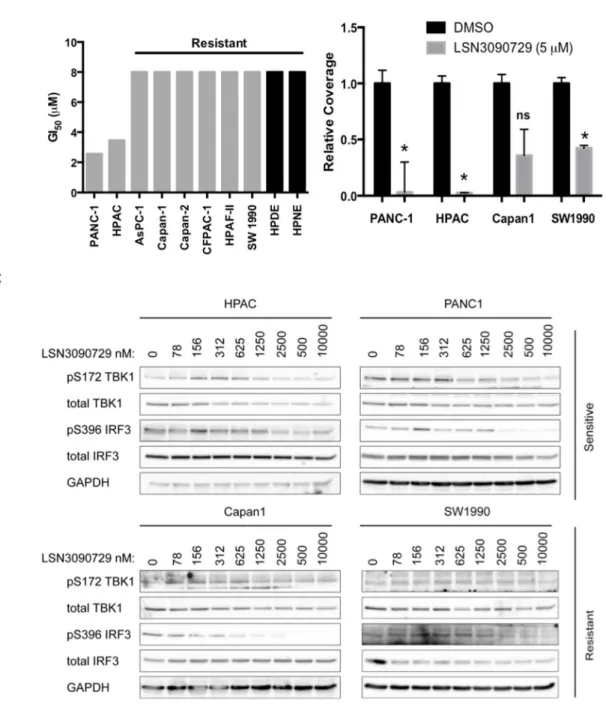
PDAC cell lines show varying sensitivity to pharmacologic TBK1 inhibition ...79
TBK1 inhibition leads to non-productive autophagy ...81
TBK1 inhibition has no effect on tumor growth in vivo ...83
Combined TBK1 and ERK inhibition do not synergize to reduce PDAC cell proliferation ...83
Discussion ...84
CHAPTER 4: CONCLUDING REMARKS AND FUTURE DIRECTIONS ...97
LIST OF FIGURES
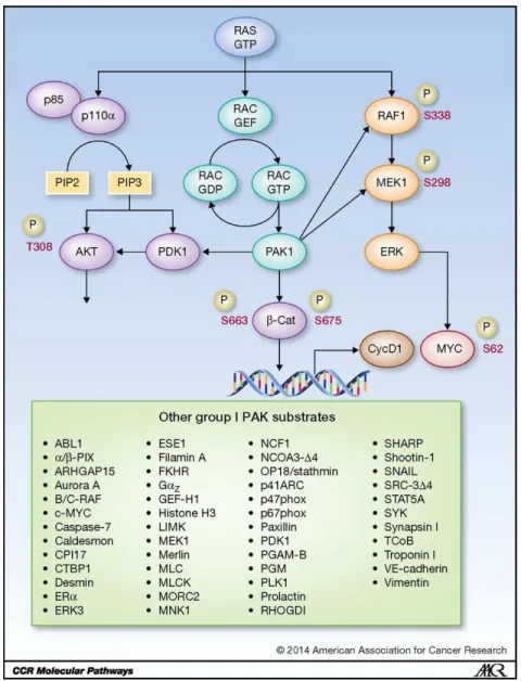
Figure 1.1: RAS-RAC-PAK1 effector signaling ...37 Figure 1.2: TBK1 is activated downstream of both TLR signaling and RAS-mediated RALB/Sec5 signaling ...38 Figure 2.1: PAK1 is overexpressed in a subset of PDAC cell lines ...57 Figure 2.2: Genetic suppression of PAK1 reduces cellular proliferation in KRAS-mutant PDAC cell lines ...58 Figure 2.3: Pharmacologic inhibition of PAK1 phenocopies the proliferative defect seen upon genetic suppression ...60 Figure 2.4: Genetic suppression and pharmacologic inhibition of PAK1
significantly reduce macropinocytotic uptake in PDAC cells ...62 Figure 2.5: A KRAS-RAC1-PAK1 signaling axis does not drive
macropinocytosis in most PDAC cell lines ...63 Figure 2.6: Combinatorial inhibition of PAK1 with ERK1/2i or PI3Ki further
reduces macropinocytosis in some PDAC cell lines ...65 Supplemental Figure 2.1: Levels of phospho-MEK1 at S298 do not correlate with levels of PAK1 in pancreatic cell lines ...67 Supplemental Figure 2.2: Phospho-MEK1 levels are not reduced following stable knockdown of PAK1 in PDAC cell lines. ...68 Supplemental Figure 2.3: PAK activity, but not SRC family kinase activity, is inhibited at sub-micromolar concentrations of AZ13705339 ...69 Supplemental Figure 2.4: RAC1 activity is not dependent on KRAS in all PDAC cell lines. ...70 Figure 3.1: LSN3090729 is an inhibitor of TBK1 in vitro and in vivo ...87
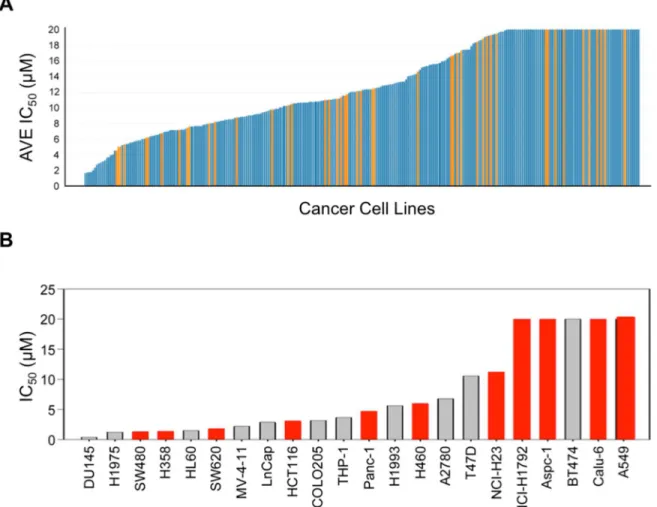
Figure 3.2: Sensitivity of human tumor cell lines to LSN3090729 treatment does not correlate with RAS mutation status ...89 Figure 3.3: KRAS-mutant PDAC cell lines are differentially sensitive to LSN3090279 treatment in a manner dependend on the growth endpoint ...90 Figure 3.4: TBK1 depletion reduces clonogenic single cell colony formation ...91 Figure 3.5: LSN3090729 increases autophagic flux and autolysosome
Figure 3.7: Combined treatment with TBK1 inhibitor LSN3090729 and
ERK1/2 inhibitor SCH772984 does not efficiently reduce PDAC growth ...95 Supplemental Figure 3.1: Western blot analysis of tumor cell lysates
LIST OF ABBREVIATIONS AND SYMBOLS
ARF ADP-ribosylation factor
AID Autoinhibitory domain
AKT Protein kinase B
ATCC American Type Culture Collection
BAD Bcl-2-associated death promoter
Bcl-2 B-cell lymphoma 2
CDC42 Cell division cycle 42 small GTPase
cDNA Complementary deoxyribonucleic acid
C-terminus Carboxyl-terminus
DMEM Dulbecco’s modified Eagle medium
DMSO Dimethyl sulfoxide
DNA Deoxyribonucleic acid
EGF Epidermal growth factor
EGFR Epidermal growth factor receptor
Exo84 Exocyst complex 84 kDa subunit
ERK Extracellular signal-regulated kinase
FAK Focal Adhesion Kinase
FBS Fetal bovine serum
FITC Fluorescein isothiocyanate
FTI Farnesyltransferase inhibitor
GAP GTPase-activating protein
GDP Guanine diphosphate
GFP Green fluorescent protein
GI50 Growth inhibitory 50, concentration of drug to cause 50% reduction in proliferation
GPCR G-protein coupled receptor
GST Glutathione S-transferase
GTP Guanine triphosphate
GTPase Guanosine triphosphatase
h Hour
HRAS Harvey rat sarcoma viral oncogene homolog
IC50 Inhibitory concentration 50, concentration of drug to cause 50% reduction in protein signaling activity
IFR3 Interferon regulatory factor 3
IHC Immunohistochemistry
IKK IκB kinase
IPA-3 (2,2’-dihydroxy-1,1’dinaphthyldisulfide)
kDa Kilodalton
KRAS Kirsten rat sarcoma viral oncogene homolog
LCCC Lineberger Comprehensive Cancer Center
LPS Lipopolysaccharide
MAPK Mitogen-activated protein kinase
MEK Mitogen-activated protein kinase kinase
MET mesenchymal-epithelial transition factor receptor
mL Milliliter
MM Mis-match siRNA
mRNA Messenger ribonucleic acid
mTOR Mammalian target of rapamycin
MTT 3-[4,5-dimethylthiazol-2-yl]-2,5-diphenyltetrazolium bromide
MYC v-Myc avian myelocytomatosis viral oncogene homolog
NCI National Cancer Institute
NF1 Neurofibromin 1
NF-κB Nuclear factor kappa-light-chain-enhancer of activated B cells
NIH National Institutes of Health
nM Nanomolar
NRAS Neuroblastoma RAS viral oncogene homolog
NSCLC Non-small cell lung cancer
NS Non-specific shRNA
N-terminus Amino-terminus
p90RSK p90 ribosomal S6 kinase
PDAC Pancreatic ductal adenocarcinoma
PAK p21-activated kinase
PBD p21-binding domain
PBS Phosphate-buffered saline
PDEδ Phosphodiesterase-delta
PDGF Platelet-derived growth factor
PDK1 Phosphoinositide-dependent protein kinase 1
PI3K Phosphatidylinositol 2-kinase
PIP3 Phosphatidylinositol-3,4,5-triphosphate
PREX2 Phosphatidylinositol-3,4,5-triphosphate-dependent RAC exchanger 2
PTEN Phosphatase and tensin homolog
PVDF Polyvinylidene difluoride
RAB Rat brain small GTPase
RAC Ras-related C3 botulinum toxin substrate
RAF Rapidly accelerated fibrosarcoma kinase
RAL RAS-like small GTPase
RalBP1 Ral binding protein 1
RalGAP Ral GTPase-activating protein
RalGDS Ral guanine nucleotide dissociation stimulator RalGEF Ral guanine nucleotide exchange factor
RAN RAS-related nuclear protein
RAS Rat sarcoma viral oncogene homolog
RBD RAS-binding domain
RHO Ras homologous small GTPase
RNA Ribonucleic acid
RNAi RNA interference
RPPA Reverse phase protein array
RTK Receptor tyrosine kinase
Sec5 Exocyst complex component 2
SEM Standard error of the mean
SDS Sodium dodecyl sulfate
SOS1 Son of sevenless homolog 1
shRNA Short hairpin ribonucleic acid
siRNA Short interfering ribionucleic acid
TANK TRAF-associated NF-κB activator
TBK1 TANK-binding kinase 1
TBST Tris-buffered saline containing Tween-20
TCoB Tubulin cofactor B
Tiam1 T-cell invasion and metastasis gene 1
TLR Toll-like receptor
TMA Tissue microarray
TNF Tumor necrosis factor
TRAF TNF receptor-associated factor
TRC The RNAi Consortium
WT Wild-type
μm Micron
μM Micromolar
CHAPTER 1: INTRODUCTION1
RAS small GTPases in cancer
The RAS (rat sarcoma viral oncogene homolog) small GTPases comprise a family of proteins that are involved, to some degree, in nearly every known cellular process (1). The members of this monomeric G-protein family are bound to guanine triphosphate (GTP) when in an activated state and are able to bind to and regulate downstream effectors (e.g., RAF) (2). GTP is then hydrolyzed to guanine diphosphate (GDP), either by slow intrinsic hydrolysis within the RAS protein, or by interaction with GTPase activating proteins (GAPs; e.g., NF1), which provide critical amino acid residues and assist the GTPase in adopting a more favorable conformation to catalyze the hydrolysis of GTP to GDP. Subsequently, guanine nucleotide exchange factors (GEFs; e.g, SOS1) facilitate the exchange of GDP for GTP, thus continuing the GTPase activation-deactivation cycle (2).
The RAS superfamily is comprised of five distinct groups: the ARF, RAB, RAN, RHO, and RAS small GTPases (3). Of these families, the two with the strongest association with cancer are the RAS and RHO families (1, 4-6). In addition to RAS, the RAS family includes the RAL small GTPases (7, 8). Among the best-studied RHO family members is RAC1 (9, 10). In addition to sharing strong structural and biochemical similarities with RAS, as described below, RAL and RAC1 also function downstream of RAS as key effectors in
1
driving RAS biology (11-13). RAC1 mutations have also recently been identified in human cancer (14).
There are three genes that encode four isoforms of the founding members of the RAS protein family: HRAS, NRAS, KRAS4A, and KRAS4B. KRAS4A and KRAS4B are formed by alternative KRAS gene splicing (1). The sequence of these proteins is highly conserved, save for a C-terminal hypervariable domain, which contains a tetrapeptide CAAX-motif (Cysteine-Alanine-Alanine-terminal amino acid) that is differentially prenylated to facilitate proper RAS subcellular localization (15, 16). In 1982, multiple groups discovered that these proteins were mutationally activated in human cancers and act as oncogenes (17-22). Subsequently, extensive sequencing of many human cancers determined that approximately 25% of all human cancers contain activating mutations in one of the three RAS isoforms (COSMIC). KRAS mutations comprise 86% of all RASmutations, followed by NRAS (10%), with HRAS mutations rarely seen in cancer (2). Though over 130 missense mutations of RAS proteins have been identified in human tumors, 98% of these mutations are found at three specific residues: G12, G13, and Q61 (2, 23, 24). Missense mutations at these residues leaves RAS proteins in a state that is GAP insensitive and, due to the low intrinsic rate of GTP hydrolysis, this results in RAS proteins that are essentially constitutively bound to GTP and actively signaling to downstream effectors.
proteins as an important target for therapeutic intervention. With RAS mutations found in cancers that comprise the three top causes of cancer deaths in the US (lung, colorectal and pancreatic cancer), an effective anti-RAS therapy will have a significant impact on cancer deaths.
Therapeutically targeting mutant RAS in human cancer
Over three decades of intense effort have gone into attempting to drug RAS (1). Though directly targeting mutationally activated RAS for the treatment of human cancer sounds promising, and while impressive progress has been made, it remains to be determined whether direct inhibitors can be developed into clinically active and effective drugs. Since mutant RAS is persistently GTP-bound, by analogy to ATP-competitive inhibitors of protein kinases, one logical approach is the development of GTP antagonists. The primary reason these efforts have been confounded is due to the picomolar affinity of RAS for GTP (26). The especially high affinity of RAS for its natural activator leaves little room for pharmacologic intervention in the GTP-binding pocket. Therefore, attempts to directly target mutant RAS have been difficult. However, in recent years, moderate success has been seen with small molecules that directly bind RAS. For example, targeting specific mutant RAS proteins, such as KRAS-G12C, has been reported (27).
motif, restores RAS plasma membrane association. Another recent method for targeting KRAS membrane association involves inhibition of PDEδ, a RAS-binding protein that chaperones RAS to the plasma membrane (31, 32). However, more research is required to determine whether PDEδ inhibitors are a promising direction for therapeutic intervention (33).
A currently exciting alternative approach to inhibit RAS activity involves exploiting the roles RAS plays in cellular metabolism (34-36). Tumor cells often exist in a hypoxic, nutrient depleted environment, which directly opposes the elevated metabolic needs of these cells. Many studies have demonstrated that RAS mutant tumors exhibit increased rates of glycolysis and non-oxidative phosphorylation (37). Additionally, nutrient scavenging from both internal and external resource pools, via autophagy and macropinocytosis, respectively, has been validated as a critical component necessary to support RAS-mutant tumor growth. Autophagy is a process of “self-eating” whereas macropinocytosis is a process of taking up extracellular materials (38-40). Both processes culminate in the lysosomal degradation of captured cargo and the creation of free nutrients that tumor cells use to sustain their unremitting growth.
Given the complex nature of RAS signaling, whether targeting one or multiple RAS effector signaling pathways will be required for effective and long-term therapy is unresolved (41, 43). Currently, most efforts have centered on the two canonical RAS effectors, the RAF serine/threonine protein kinases and the class I lipid kinases, the phosphatidylinositol 3-kinases (PI3Ks) (44). Numerous inhibitors of each of these effector pathways are currently under clinical evaluation. Most attention has been focused on the RAF serine/threonine kinases. One RAF isoform, BRAF, is often mutationally activated in human cancers (45). Activated RAF phosphorylates and activates MEK1 and MEK2, which then go on to phosphorylate and activate the ERK1/2 mitogen-activated protein kinases (Figure 1.1).
PI3Ks comprise the second most studied RAS effector class. PI3K activation causes increased conversion of phosphatidylinositol-4,5-bisphosphate (PIP2) to phosphatidylinositol-3,4,5-trisphosphate (PIP3). Membrane-bound PIP3 can then regulate a diversity of signaling proteins, including the AKT1-3 serine/threonine kinases. One PI3K isoform, p110 alpha is frequently mutated in cancer and PI3K is considered essential for
RAS-driven cancer development (46, 47).
propose that the TBK1 serine/threonine kinase may be a promising direction for targeting the RAL effector pathway.
Yet another method for indirectly targeting mutant RAS activity has involved the search for synthetic lethal partners of mutant, but not wild-type, RAS (52, 53). A gene is a synthetic lethal partner of mutant KRAS when its function is vital to the growth of only cancer cells containing mutant RAS, whereas its loss in RAS wild-type cells is inconsequential to cellular viability and function. Many unbiased functional genetic screens using large RNAi libraries against a multitude of gene products have sought to identify proteins whose loss sensitizes cells to loss of KRAS. Chapter 3 of my thesis includes the description of one such identified synthetic lethal partner of mutant KRAS, the TBK1 serine/threonine kinase (54). However, enthusiasm for the data generated in synthetic lethal studies is mixed due to some misgivings about the reproducibility of results from these reports (55).
Targeting RAC-PAK signaling in RAS-driven cancers
A somewhat overlooked RAS effector network results in activation of a RHO family protein, the RAC1 small GTPase. One mechanism that this can be mediated through is via RAS interaction with a RAS-GTP binding domain (RBD)-containing RAC-selective GEF, TIAM1 (56, 57) (Figure 1.1). Another mechanism involves PI3K-mediated formation of PIP3, which then activates other RAC-selective GEFs (e.g., PREX1/2, Vav) (58, 59).
The three RAC isoforms are members of the RHO branch of the RAS superfamily (3). They are best known for their regulation of actin organization, in particular to regulate lamellipodia induction and promotion of cell migration and pinocytosis. RAC also regulates the formation of reactive oxygen species (60). The recent identification of activated RAC1 mutants in melanoma supports an important driver role for RAC in cancer growth (61, 62).
growth remain to be determined, the PAK protein kinases are intriguing candidates. Below, I summarize the evidence for the importance of the RAC-PAK effector signaling pathway in RAS-driven cancer development and growth.
RAC and RAS in cancer
Early studies identified upregulated RAC activation in HRAS-transformed rodent fibroblasts (57, 64, 65). These were followed by studies where dominant negative RAC1 mutants that sequester and inactivate RAC-GEFs, impaired the growth of HRAS-transformed rodent fibroblasts (13, 66, 67). Subsequent genetically engineered mouse model studies found that tissue-restricted genetic loss of Rac1 impaired mutant Kras-driven
lung (68) and pancreatic (69) cancer development. Furthermore, in a mutant Kras-driven
model of papilloma development, tumor tissue exhibited increased levels of RAC-GTP, and loss of one Rac1 copy alone was sufficient to reduce tumor growth and increase survival
(70).
PAK activation in RAS mutant cancer
PAKs comprise a family of six proteins divided into two sub-groups: group I comprises PAK1-3 and group II contains PAK4-6 (75-77). Since group I PAKs are RAC and CDC42 effectors, whereas group II PAKs are CDC42 only, my work will primarily focus on the group I PAKs. Although the group I PAKs share strong sequence identity in their kinase domains (92-95%), PAK1 is thus far the most studied family member, so I have focused primarily on PAK1 in my studies.
Though PAK1 activity can be deregulated by a diversity of mechanisms in cancer that include gene amplification and increased gene transcription (76, 78), here I focus on activation of PAK downstream of RAS, RACGEFs, and RAC. While in the inactive conformation within the cytosol, PAK1-3 form head-to-tail homodimers with the N-terminal autoinhibitory domain (AID) of one monomer inserted within the C-terminal kinase domain of another. Upon binding of RAC1-GTP to the GTPase binding domain of group I PAKs, a conformational change releases the AID from the kinase domain leading to autophosphorylation at multiple serine/threonines and activation of PAK catalytic activity, allowing phosphorylation of substrates (79). Additionally, plasma membrane-associated RAC binding facilitates PAK plasma membrane recruitment, where PAKs can interact with effectors.
PAK effector signaling in human cancer
scaffolding function to facilitate PDK1-mediated recruitment of AKT to the plasma membrane to facilitate AKT activation (85). The physiologic relevance of PAK1 cross-talk with ERK and AKT signaling is supported by the observation that genetic or pharmacologic ablation of PAK1 impaired both ERK and AKT activation in Kras-driven skin tumors (86).
Pharmacologic inhibitors of the RAF and PI3K pathways have been ineffective in RAS
mutant cancer cells, in part, due to kinome reprogramming mechanisms that stimulate signaling activities that overcome inhibitor action (87-90). Consequently, combined targeting of PAKs and members of these pathways, such as MEK, ERK, PI3K or AKT, may help overcome these resistance mechanisms. However, PAK1 cross-talk with these RAS effector pathways can be context-dependent as PAK1 suppression in KRAS-mutant colon
carcinoma cells impaired anchorage-dependent and -independent proliferation, but not ERK or AKT activation (91).
PAKs are also capable of influencing transcription of genes that promote cell cycle progression and cell survival. In breast cancer and colon cancer cell lines, PAK1 can phosphorylate β-catenin on S663 and S675, stabilizing it and promoting its nuclear translocation and transcriptional stimulation of TCF-responsive genes, including CCND1 and MYC (92, 93).
PAKs enhance cell survival by phosphorylating proteins associated with apoptosis. PAK1 phosphorylates BAD on S111 to prevent Bcl-2 binding and induction of apoptosis (94). Additionally, PAK1 can phosphorylate and induce relocalization of RAF-1 to the mitochondria where it also inhibits BAD by phosphorylating it on S112 (94).
responsible for assembling tubulin heterodimers (99), and through the inactivation of stathmin, which is normally responsible for destabilizing microtubules at the leading edge of cells (100-102).
Metabolism is a critically important factor to the survival of cancer cells because of their high energy demands, and PAKs play a role in driving several metabolic processes that aid tumor cell growth and survival. Elevated macropinocytosis to facilitate increased extracellular protein and lipid uptake is one consequence of the high metabolic requirements of cancer cells (36). PAK1 was found to be necessary and sufficient for growth factor- and RAC-induced macropinocytosis in NIH 3T3 fibroblasts (103). RAC and PAK1 were found to be both necessary for bladder cancer cell macropinocytotic uptake of Bacille Calmette-Guerin (BCG), a strain of bacteria used in the treatment of bladder carcinoma (104). Additionally, bacterial uptake was also stimulated by activated KRAS or HRAS and this activity was blocked by pharmacologic inhibition of group I PAKs by IPA-3 (2,2’-dihydroxy-1,1’dinaphthyldisulfide). This study suggests that the activity state of PAKs in cancer cells could be a determinant of efficient uptake of cancer therapeutics. Similarly, in pancreatic cancer cells, KRAS-dependent stimulation of macropinocytosis and uptake of albumen (105) may provide a basis for the efficacy of albumen-bound (nab) paclitaxel for the treatment of this cancer. It will also be important to assess a role for RAC-PAK signaling in KRAS-dependent macropinocytosis to determine whether pharmacologic inhibition of PAK1 may be an effective approach to blocking cancer cell metabolism.
ligand-independent manner, leading to tamoxifen resistance (109). Finally, PAK1 can translocate to the nucleus to drive transcription of fibronectin, which is crucial for supporting pancreatic cancer cell growth and migration (110).
The first evidence for a role for PAK1 in RAS-dependent growth transformation came from studies in model cell systems. Ectopic expression of a kinase-dead PAK1 dominant negative mutant impaired HRAS and RAC1 growth transformation of rat 3Y1 fibroblasts (111) or HRAS transformation of Rat-1 rat fibroblasts, but not NIH3T3 mouse fibroblasts (112, 113). Similarly, dominant negative RAC1 and kinase-dead PAK1 inhibited KRAS transformation of MT4H1 rat Schwann cells (114). Recently, in a mouse model of Kras
-driven skin squamous cell carcinoma formation, genetic ablation of Pak1 strongly impaired
tumor initiation and progression (86). Together with the validated role of RAC1 in RAS-driven oncogenesis, these observations implicate the RAC-PAK effector pathway as a target for the development of anti-RAS therapeutic strategies. Like the RAS small GTPase, the RAC small GTPase is not considered a highly tractable drug target. Therefore, below we focus on the development of inhibitors of the PAK1 kinase for cancer treatment.
Clinical-translational advances
leading to Phase I evaluation in patients with solid tumors. Unfortunately, this trial was stopped in phase I due to pharmacokinetic issues. Subsequently, derivatives of PF-3758309 have been described with much improved pharmacologic properties, raising hope that this class of compound may yet have clinical utility (117).
More recently, Licciulli and colleagues described the discovery of a small molecule pyridopyrimidinone, FRAX59, that potently inhibits Group I PAKs by preventing ATP-binding and hydrolysis (118). FRAX597 exhibited high specificity and potency for Group I PAKs, although potent inhibition of other kinases was also seen. When evaluated in vivo, FRAX597
inhibited the tumorigenic growth of NF2-null Schwann cells. NF2 loss causes RAC1 and
PAK1 activation, indicating that this compound could be a viable therapeutic strategy for treating PAK-dependent tumors. FRAX597 treatment also phenocopied genetic loss of Pak1
and impaired Kras-driven skin tumorigenesis (86). Interestingly, in this mouse model, both
genetic and pharmacologic inhibition of PAK1 resulted in reduction of ERK and AKT activity, supporting the importance of PAK1 signaling cross-talk with these two RAS effector pathways.
Peterson and colleagues performed a screen to identify small molecule allosteric inhibitors of Cdc42 activation of Group I PAKs. The results of this screen led to the development of IPA-3, which interacts with the PBD/AID region of group I PAKs and prevents their activation by GTPase binding (119, 120). IPA-3 showed strong selectivity for Group I PAKs, with no inhibitory activity for Group II PAKs or more than 200 other protein kinases evaluated. However, the inability of IPA-3 to inhibit already activated PAK1, its micromolar IC50 and its rapid metabolism to a toxic compound due to the reduction of the disulfide bond it contains, limit the ability to transition IPA-3 as a clinically useful chemical platform.
defining genetic and/or biochemical markers that identify those cancers that will respond to anti-PAK therapy. The position of PAK downstream of mutant KRAS and RAC, in addition to PAK signaling cross-talk with the key RAS effector pathways, support PAK inhibitors as a therapeutic strategy for RAS-mutant cancers. Given the involvement of multiple effectors in
driving RAS-dependent cancer growth, PAK inhibition in combination with inhibitors of RAF or PI3K effector signaling will likely be required. Currently, pharmacologic inhibitors of PAK1 also inhibit other Group I PAKs; whether PAK1-selective inhibitors are more desirable and possible to develop are issues that remain to be resolved. Of the spectrum of PAK substrates, which substrate(s) will provide a reliable biomarker for PAK inhibitor anti-tumor activity also remains unclear. A survey of the patent literature indicates that more PAK inhibitors are in the pipeline (121, 122). As more potent and selective inhibitors become available, the answers to many of these unresolved questions will likely be addressed. In Chapter 2, I detail my studies with a novel, ATP-competitive and PAK1-selective inhibitor.
The RalGEF-RAL pathway in RAS-mutant cancer
Our lab and others have demonstrated that RALA and RALB have divergent roles in promoting various traits of cancer growth. For example, in pancreatic cancer RALA is necessary for anchorage-independent growth in vitro and tumorigenicity in vivo but RALB is
dispensable (128). In contrast, RALB, but not RALA, is required for invasion in vitro and
metastasis in vivo (129). Moreover, loss of RALB is lethal for tumor cells, but not normal
cells (128). These differential roles for two highly identical proteins, which diverge primarily in their C-terminal membrane targeting sequences and their posttranslational modifications (130), highlight the importance of subcellular localization and substrate engagement for promoting specific cellular outcomes in that RALA and RALB must engage with disparate downstream effectors in the correct time and place to regulate these RAL isoform-driven phenotypes.
Targeting the RAL effector TBK1 in human cancer
Like other RAS family members, RAL sits at the center of a diverse web of cellular effectors that mediate processes such as gene transcription, endocytosis, autophagy, and actin reorganization (8). The most well characterized effector signaling pathway of RAL proteins involves the exocyst complex, which facilitates cellular transport of vesicular cargo from the Golgi apparatus to the plasma membrane where it is released into the intercellular millieu (131). Two members of the exocyst complex that directly engage with RAL proteins are Exo84 AND Sec5. Interestingly, downstream of RALB, Sec5 is able to bind to and activate TANK-binding kinase 1 (TBK1) (132), a non-canonical IκB kinase (IKK), involved in regulation of NF-κB.
genes that encode proteins important for growth and cell survival. Typically, complexes of different NF-κB family members are held inactive in the cytoplasm by inhibitors of NF-κB, known as IκB proteins. In response to various stimuli, such as inflammatory signals, the canonical IKKs, IKKα and IKKβ, become activated and promote dissolution of the IκB-NF-κB complex through phosphorylation and ubiquitylation of IκB proteins (134).
There are other IKK proteins besides IKKα/β that are responsible for activating NF-κB signaling. These non-canonical IKKs act downstream of diverse stimuli that converge on Toll-like receptors (TLR) and TNF receptor-associated factor (TRAF) proteins. These receptors mediate TANK binding to TBK1, which is one such non-canonical IKK (135). TBK1 can then go on to promote both survival and inflammatory signaling. TBK1 is the focus of my studies in Chapter 3.
Additionally, TBK1 is capable of phosphorylating the interferon regulatory factor proteins, IRF3 and IRF7, and inducing transcription of pro-inflammatory genes (55, 136). Therefore, the ability of the exocyst component Sec5 to induce activation of TBK1 provides a critical linkage between RAS-mediated proliferative signaling in cancer and NF-κB responsive inflammation (132). TBK1 also plays a role in pro-survival signaling through direct phosphorylation of AKT at both T308 and S473 (Figure 1.2) (137).
A study searching for synthetic lethal interactors of mutant RAS utilized a systematic siRNA screen and reported that TBK1 was deleterious to cancer cell lines harboring mutationally activated but not wild-type KRAS (54). The results of this screen were validated in non-small cell lung cancer (NSCLC). However, subsequent studies in other cell types reported that the relationship between KRAS and TBK1 was far more complex than initially imagined, and that not all KRAS-mutant tissue was exclusively dependent on TBK1 signaling for tumor cell growth and survival (55, 137).
shRNA-mediated suppression of TBK1 expression had any measurable effect on the viability of the cell lines tested, though reduction in pIRF3 S386 was observed (55). Thus, conflicting observations have been made concerning whether TBK1 is a bona fide target for RAS-mutant cancers. In Chapter 3 I describe the application of a novel TBK1 inhibitor to focus on the study of TBK1 in KRAS-mutant PDAC. In my studies, I determined whether TBK1 inhibition could enhance the anti-tumor activity of an ERK1/2 inhibitor, as ERK1/2 is a vital member of the canonical RAF-MEK-ERK RAS effector pathway.
Pancreatic ductal adenocarcinoma as a model to study KRAS effector signaling
Pancreatic ductal adenocarcinoma (PDAC) is among the deadliest human cancers, with a 5-year survival rate of approximately 8% (138). In 2016, PDAC surpassed breast cancer and is now the 3rd leading killer among cancer deaths in the United States and is set to surpass colorectal cancer by 2020 (139, 140). The reasons for the dire and deadly state of PDAC are manifold. First of all, patients are often asymptomatic, affording the primary tumor time to disseminate and create extensive metastatic lesions in other areas of the body, such as the lymph nodes, lungs, liver and other abdominal compartments (141). Surgical resection of the primary tumor is the only option for long-term effective treatment, though most patients, at the time of diagnosis, are well past the stage where surgical resection is a viable option. This leaves radiation and chemotherapy as the only treatment strategies for the majority of PDAC patients.
the second treatment is the four-drug cocktail FOLFIRINOX, comprised of folinic acid, fluorouracil (5-FU), irinotecan and oxaliplatin. 5-FU is a pyrimidine analogue and oxaliplatin is a DNA alkylating agent; both disrupt DNA synthesis. Irinotecan is a topoisomerase inhibitor that introduces strand breaks into DNA, also disrupting DNA synthesis. Folinic acid is a vitamin B analog that decreases some of the toxic side effects of 5-FU.
Despite the approval of these therapies for PDAC, most patients still succumb to this disease in less than a year (139). While our knowledge of the genetic basis of pancreatic cancer is quite extensive (143-147), unlike in other cancers where such knowledge has led to the development of effective targeted therapies, no truly effective targeted therapies are available for PDAC. One targeted therapy that is approved for PDAC is erlotinib, an inhibitor of the EGFR receptor tyrosine kinase, which is responsible for driving activation of RASGEF proteins that stimulate RAS signaling (148). The clinical trial that led to the approval of erlotinib in combination with gemcitabine in 2005 showed a statistically significant but clinically insignificant two-week improvement in patient survival (149). That this minor improvement in survival was enough for erlotinib to be approved for PDAC therapy speaks volumes about the dire need for new therapeutic interventions for this disease. In fact, erlotinib is no longer used in the clinic for PDAC. Clearly, the need for more effective treatments will become even more acute in the coming years as PDAC overtakes colon cancer as the number two cancer killer in the US.
Aside from KRAS mutations, three other genes are mutated frequently in PDAC. These include the TP53, CDKN2A and SMAD4 tumor suppressors (141). Since pharmacologic inhibitors that antagonize an activated oncoprotein, such as KRAS, should be much easier to develop than small molecules that restore the function of lost tumor suppressors, KRAS is the most attractive target for PDAC drug discovery. While many other genes have been identified in PDAC, these occur at frequencies of less than 5% (1, 25, 43). Thus, for this and other reasons mentioned above, the development of anti-KRAS drugs is one of four major directions for the field identified recently by the National Cancer Institute. The high frequency of KRAS mutations in PDAC and their driver role in this cancer type make PDAC an exceptional model for studying KRAS signaling and for elucidating the downstream signaling mechanisms that KRAS employs to promote tumor development and maintenance.
Rationale and objectives for the studies described in this document
The aims of my thesis work described herein encompass evaluating the contribution and therapeutic potential of PAK1 and TBK1, which are downstream protein kinase components of two less studied KRAS effector pathways, the Tiam1-RAC and RalGEF-RAL pathways. In Chapter 2, I describe my studies of PAK1 in PDAC. I determined that PAK1 was necessary to maintain PDAC cell line proliferation and anchorage-independent growth. I show that PAK1 could be promoting the growth and survival of pancreatic cancer cells via macropinocytosis, wherein the cell non-specifically engulfs portions of the extracellular compartment as a means to scavenge nutrients and to fuel unrestrained growth. In Chapter 3, I demonstrate that, although TBK1 appears to be dispensable for maintaining PDAC cell viability, both in vitro and in vivo, TBK1 activity is intimately linked to autophagy, and this
CHAPTER 2: PAK1 REGULATES MACROPINOCYTOSIS IN PANCREATIC CANCER2
Overview
Despite attempts to directly target mutant KRAS and to design inhibitors of validated KRAS effector pathways, to date, no clinically successful anti-KRAS therapies have been developed. The lack of success of these inhibitors is due, in part, to the importance of other effectors in KRAS-dependent cancer growth and to the upregulation of compensatory signaling programs that overcome inhibitor activity. We have determined that PAK1 serine/threonine kinase protein expression is elevated in a subset of primary patient pancreatic tumors and in pancreatic ductal adenocarcinoma (PDAC) cell lines. Both genetic silencing of PAK1 and pharmacologic inhibition of PAK1 kinase activity by a novel,
highly-selective ATP-competitive inhibitor, AZ13705339, impaired PDAC anchorage-independent growth. Additionally, as mutant KRAS has been linked to upregulation of metabolic processes, such as macropinocytosis, we examined the ability of PAK1 to regulate macropinosome formation in PDAC. Inhibition or genetic ablation of PAK1 resulted in a marked decrease in macropinocytosis. Surprisingly, PAK1-driven macropinocytosis is independent of KRAS-RAC1 signaling. Finally, we determined that concurrent inhibition of PAK and either ERK or PI3K synergistically reduced macropinosome formation in a subset of PDAC lines. Our findings validate PAK1 as a therapeutic target in PDAC.
2
Introduction
Pancreatic cancer is a rapidly fatal disease with a 8% 5-year survival rate (138, 139). Despite the fact that nearly all pancreatic ductal adenocarcinomas (PDAC) are driven by activating mutations in the KRAS oncogene, no clinically effective anti-KRAS targeted
therapies have been developed. KRAS acts as a major cell signaling hub that promotes multiple cellular processes required to maintain tumorigenic growth (1). Most notably, a plethora of inhibitors have been generated against the two canonical KRAS effector pathways, the RAF-MEK-ERK mitogen-activated protein kinase (MAPK) and the PI3K-AKT-mTOR lipid kinase pathways, with many currently under clinical evaluation (43). Disappointingly, as monotherapy, these inhibitors have shown limited to no clinical benefit. One known basis for their limited clinical efficacy is the process of dynamic kinome reprogramming, whereby pharmacological inhibitors induce signaling changes that compensate for and overcome inhibitor actions (88).
A second likely basis for the limited efficacy of inhibitors of the canonical effector pathways is an unreallized requirement to concurrently inhibit other effector pathways that are also essential for KRAS-dependent cancer growth and that can compensate, at least in part, for impaired signaling through the canonical effectors. One such less studied pathway includes the Tiam1 guanine nucleotide exchange factor (GEF) and the RAC1 small GTPase.
Tiam1-deficient mice show impaired Hras-induced tumorigenesis (56). Other studies
demonstrated that Rac1 deficiency reduced mutant Kras-driven lung (68) and pancreatic
cancer growth (69). However, the key effector(s) that are critical for Tiam1-RAC1-dependent oncogenesis remain to be established.
epithelial-mesenchymal transition. PAK1 has been shown to act as a driver in breast (151, 152), colon (91, 93), lung (153, 154), and other cancers (79). A recent study implicated a requirement for PAK1 in initiating and driving tumor development in a mouse model of Kras
-mutant skin squamous cell carcinoma (86). More importantly, studies of PAK1 in PDAC reveal that PAK1 is a key regulator of MET-regulated PDAC cell migration (155) and that PAK1 promotes PDAC cell growth through transcriptional regulation of NF-κB and fibronectin (110).
Additionally, PAK1 is known to regulate macropinocytosis (103), an actin-driven process by which cells engulf extracellular protein as a source of amino acids to sustain the increased metabolic demands of tumor growth. Mutant KRAS has been shown to drive macropinocytosis in PDAC (64, 105, 156) and inhibition of macropinocytosis impaired PDAC tumor growth. However, the critical KRAS effector signaling pathways that promote macropinocytic activity in pancreatic cancer have not been determined. Elucidation of how mutant KRAS drives macropinocytosis may identify a therapeutic target, such as PAK1, to enable effective blockade of macropinocytosis and PDAC growth.
Materials and Methods
Cell lines, tumor tissue lysates, and tissue microarray
Authenticated PDAC cell lines were obtained from ATCC and maintained in either DMEM (HPAC, MIA PaCa-2, and PANC-1) or RPMI-1640 (AsPC-1, HPAF-II, and CFPAC-1) supplemented with 10% fetal bovine serum, and were not maintained in continuous culture for longer than two months after receipt from the source. Detergent buffer lysates generated from patient primary pancreatic tumor and adjacent non-tumor tissues were kindly provided by Dr. Jen Jen Yeh (UNC-Chapel Hill). Pancreatic tumor tissue was acquired and tissue microarrays (TMAs) were generated by the Translational Genomics Research Institute (TGen) (Phoenix, Arizona) from freshly cut sections of fixed, embedded, de-identified primary pancreatic cancer tumor tissues.
Plasmids and reagents
Lentiviral plasmids encoding shRNA against PAK1 were obtained from the University
of North Carolina Lenti-shRNA Core Facility. All shRNAs are in the pLKO.1 backbone and contain a puromycin resistance gene. All hairpin sequences are based on those deposited in the TRC RNAi Consortium of the Broad Institute, and are as follows: NS (non-specific): (5’-CCTCTTGATGAACCATCTATT-3’), shPAK1-1: (5’-CTTCTCCCATTTCCTGATCTA-3’), and shPAK1-2: (5’-GCTGTGGGTTGTTATGGAATA-3’).
Silencer Select siRNA targeting KRAS (#s s7939 and s7940) and RAC1 (#s s11711 and s11713) were obtained from Thermo-Fisher. Mismatch control siRNA was obtained from Dharmacon (#D-001210-05).
Millipore: KRAS (#OP24) and RAC1 (#05-389). Secondary HRP-conjugated anti-mouse (#31432) and anti-rabbit (#31460) antibodies were obtained from Thermo-Fisher.
Immunohistochemistry
Pancreatic TMAs were stained with anti-PAK1 antibody (Cell Signaling Technology, #2602). Samples were blinded and the intensity of PAK1 staining was scored by a pathologist (J. LoBello) and binned as none (+0), low (+1), medium (+2), or high (+3).
Lentiviral transduction to silence PAK1
Lentivirus was generated in HEK-293T cells transfected with the pLKO.1 shRNA expressing plasmids and the psPAX2 and pMD2.G packaging vectors (Addgene). Virus was harvested 48 h post-transfection. PDAC cell lines were seeded at a density such that they would reach approximately 70% confluence the following day, and then were transduced with NS or shPAK1 virus in the presence of 8 μg/μL polybrene. Stably transduced cells were selected for 2-3 days in puromycin at a concentration determined empirically for each cell line.
siRNA transfection to silence KRAS and RAC1
All siRNA knockdown experiments were performed via reverse transfection to maximize transfection efficiency. Briefly, 10 μM siRNA was complexed with Lipofectamine RNAiMax (Life Technologies) per the manufacturer’s instructions, and cells were seeded in 6 well dishes at a density of 3x10⁵ cells/well in the presence of this complex. Knockdown was allowed to proceed for 48 hours before cells were used in assays.
Growth assays
3-(4,5-dimethylthiazol-2-yl)-2,5-diphenyltetrazolium bromide (MTT) was added to each well at a final concentration of 5 μg/ml. Cells were incubated in the presence of MTT for 4 h, media was aspirated, cells were lysed in 100 μL DMSO per well, and the absorbance at 590 nM was measured.
Soft agar colony formation assays were performed as we have described previously (157). Briefly, 0.6% bacto-agar dissolved in complete growth medium was allowed to solidify in a 6- or 12-well plate. Cells were seeded on top at densities ranging from 5x10³ to 2x10⁴ cells per well, depending on the growth properties of each cell line, in 0.3% bacto-agar in complete growth medium. Cells were fed with complete growth medium supplemented with vehicle alone (DMSO) or with PAK inhibitor AZ13705339 for 7-14 days. Colonies were stained with 2 mg/mL MTT, visualized with a Typhoon FLA 7000 Scanner, and quantified with ImageJ (NIH) (158).
Inhibitor treatment
The PAK1-selective ATP-competitive inhibitor, AZ13705339 (manuscript in preparation, McCoull et al., 2016) and the PI3K inhibitor, AZD8186 (159), were synthesized and provided by AstraZeneca. The ERK1/ERK2-selective ATP-competitive and allosteric inhibitor SCH772984 (160) was provided by Merck. Inhibitors were dissolved in DMSO to yield stock concentrations of 10-100 mM, aliquotted, and stored at -80°C.
PDAC cell lines were seeded and allowed to adhere for 24 h before inhibitor treatment. Compounds were dissolved in DMSO and serially diluted to attain the desired treatment concentrations. The amount of DMSO was held constant in all samples. GI50 and IC50 values for PDAC cells treated with AZ13705339 were determined with Prism Graphpad 6 software.
Western blot analyses
inhibitors. Protein concentration was determined by Bradford Protein Assay (Bio-Rad). Lysates were resolved by SDS-PAGE and transferred to PVDF membranes that were then probed with the appropriate antibodies. Following application of Amersham ECL Prime Western Blotting Detection reagent (GE Healthcare Life Sciences #RON2232), chemiluminescence was visualized with a BioRad ChemiDoc, and images were analyzed for quantitation of bands with ImageJ.
Macropinocytosis assay
The macropinocytic index of cells was determined as described previously (161). Briefly, cell were seeded into glass-bottom MatTek dishes at densities such that they would reach approximately 30-40% confluence within 24 h. Cells were treated with vehicle or inhibitor at the indicated concentrations in complete growth medium for 24 h, then in serum-free medium for 16 h. The treated cells were then transferred to serum-serum-free medium containing 1 mg/mL 70 kDa dextran conjugated with FITC (Thermo-Fisher, #D1823), for 30 min, washed with PBS, and fixed in 4% paraformaldehyde. Nuclei were stained with DAPI and dishes were stored in the dark at 4°C in PBS containing 1 mg/mL BSA and 0.01% sodium azide until they were imaged. Cells were visualized on a Zeiss LSM 700 Confocal Laser Scanning Microscope at 63x magnification. Ten discrete fields were collected per sample and ImageJ was used to quantitate the area of FITC-dextran signal per total cell area of each image, as detailed previously (161).
RAC1 activation assay
which were kindly provided by Dr. Keith Burridge. Western blot analysis was performed on pulldown samples and whole cell lysates as described above.
Statistical analysis
All data were imported into GraphPad Prism 6 software and statistical analyses were performed using a One-way ANOVA using multiple comparisons. Significance values were designated as follows: *: p<0.05, **: p<0.01, ***: p<0.001, ****: p<0.0001.
Results
PAK1 protein levels are elevated in PDAC cell lines and tissue
Previous studies established a role for the RacGEF-RAC1 pathway in cancer growth (56, 57, 68, 69). However, the critical RAC1 effector(s) important for RAC1-dependent pancreatic cancer growth have not been established. Here we addressed a possible role for the RAC1-activated protein kinase, PAK1, in pancreatic cancer. We observed elevated levels of PAK1 protein in a subset of PDAC cell lines (8 of 11) compared to HPNE immortalized human pancreatic ductal epithelial cells (Figure 2.1A). Elevated protein levels did not correlate with KRAS-mutation status, since there were high levels of PAK1 in the KRAS-wild type PDAC cell line BxPC-3, but not in KRAS-transformed HPNE cells.
Additionally, PAK1 total protein did not always correlate with pMEK S298 levels, a validated marker of PAK1 activity (163) (Supplemental Figure 2.1). This could imply either that PAK1 protein is not necessarily hyperactive simply because it is overexpressed, or that pMEK1 S298 is not an ideal biomarker for PAK1 activity.
intense staining (Figure 2.1C-D). We found no correlation of PAK1 levels with patient survival rates (data not shown).
Genetic suppression of PAK1 impairs independent but not
anchorage-dependent growth
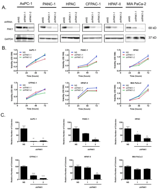
To determine whether PAK1 serves a driver role in PDAC, we evaluated the consequences of genetic suppression of PAK1 on PDAC growth. We utilized two shRNA sequences targeting PAK1 to stably suppress PAK1 protein expression (Figure 2-2A). We
then assayed the ability of cells to proliferate in anchorage-dependent and -independent growth assays. We observed limited reduction in anchorage-dependent proliferation (Figure 2-2B). In contrast, PAK1 suppression significantly reduced anchorage-independent growth as determined by colony formation in soft agar (Figure 2-2C). The level of growth suppression correlated with the level of knockdown achieved by each individual shRNA. We observed a reduction in soft agar colony formation in all six cell lines evaluated, albeit with a range of sensitivity to PAK1 knockdown. In 5 of 6 cell lines, we observed at least a 50-70% impairment of colony formation (p<0.01 to p<0.001). We conclude that PAK1 is critical for PDAC anchorage-independent cell proliferation. Unexpectedly, levels of pMEK1 S298, the best validated marker of PAK1 activity, were not reduced upon knockdown of PAK1 expression, possibly due to cellular reprogramming of compensatory signaling pathways (Supplemental Figure 2.2).
AZ13705339 effectively impairs anchorage-independent growth in PDAC cells
in phosphorylation of the well-validated PAK1 substrate, MEK1, at residue S298 (pMEK1) (163). We observed a dose-dependent decrease in pMEK1 S298 in all cell lines evaluated (Figure 2.3A-B), although the IC50 for pMEK1 reduction varied significantly among them. MIA PaCa-2 cells were the most sensitive to the PAK1 inhibitor with respect to target inhibition, displaying the lowest IC50 for pMEK1 reduction (2.2 μM), whereas CFPAC-1 cells were the most resistant (>10 μM). Additionally, although in vitro specificity analyses revealed that
AZ13705339 exhibits limited potency against SRC family kinases (SFKs) (manuscript in preparation, McCoull et al., 2016), we did not observe detectable inhibition of SFK activity at concentrations that fully suppressed MEK1 phosphorylation at S298. Instead, inhibition of SFKs, as measured by phosphorylation of focal adhesion kinase (FAK), was seen only at a concentration approximately 10-fold greater than that required for inhibition of PAK1 activity (Supplementary Figure 2.3). This result indicates that AZ13705339 displays good selectivity for PAK1 in cells.
We next determined if pharmacologic inhibition of PAK1 signaling impaired the growth of PDAC cell lines on plastic. We observed a range of sensitivity that correlated generally with reduction in pMEK1. However, the concentrations for inhibition of anchorage-dependent growth (GI50) were considerably higher than the IC50 values for inhibition of PAK1 signaling (Figure 2.3C). The GI50 values for sensitive cell lines ranged between 2 and 10 μM, with two cell lines, HPAF-II and CFPAC-1, demonstrating no sensitivity at 10 μM. Thus the growth inhibition seen at these high concentrations is likely due to off-target activities of AZ13705339. These results are consistent with our PAK1 shRNA analyses (Figure 2.2B), indicating that PAK1 is not essential for anchorage-dependent growth.
consequences supports a role for PAK1 kinase activity in driving anchorage-independent but not -dependent growth of PDC cells.
Interestingly, the two cell lines that demonstrated resistance to AZ13705339 when evaluated for viability on plastic, HPAF-II and CFPAC-1 (Figure 2.3C), exhibited enhanced colony formation with increasing doses of AZ13705339 (Figure 2.3D). This could be due to compensatory signaling pathways or due to the scaffolding function of PAK1 that couples PDK1 to AKT and induces phosphorylation of AKT at T308 to drive survival signaling, which treatment with AZ13705339 would not block.
PAK1 inhibition reduces macropinocytosis
We next addressed a possible mechanism for PAK1 support of PDAC growth. Recent studies found that PDAC cells exhibit increased macropinocytosis as one mechanism to fulfill their increased metabolic needs (105, 156). Since previous studies also described a role for PAK1 in macropinocytosis (103, 104, 164), we sought to determine whether PAK1 is important for macropinocytosis in KRAS-mutant PDAC cells. First, we used shRNA to suppress PAK1 expression in a panel of PDAC cell lines. Depletion of PAK1 markedly reduced macropinocytotic uptake in three out of four PDAC cell lines (AsPC-1, PANC-1, and HPAF-II, but not MIA PaCa-2) (Figure 2.4 A-C). In these lines, the impairment of macropinocytosis correlated with the level of PAK1 suppression.
KRAS and RAC1 drive macropinocytosis in only a subset of PDAC cell lines
PAK proteins are often activated downstream of the small GTPase RAC1 (165), and RAC1 has been previously implicated in driving macropinocytosis (166, 167). Additionally, both PAK1 and RAC1 are thought to be activated downstream of KRAS. We therefore determined whether KRAS and RAC1 regulate PAK1 activity and signaling. We used shRNA to stably suppress either KRAS or RAC1 expression (Figure 2.5A) and observed that
pMEK1 levels were not reduced. Thus, current evidence indicates that neither KRAS nor RAC1 activity regulate PAK1-dependent MEK1 signaling in PDAC cell lines, though it is possible that another marker of PAK1 activity may better reflect modulation of PAK1 activity downstream of KRAS or RAC1.
Next we performed macropinocytosis assays on our panel of PDAC cell lines following suppression of KRAS or RAC1 (Figure 2.5B). Loss of KRAS expression significantly reduced macropinocytosis in two PDAC cell lines, AsPC-1 and MIA PaCa-2, supporting previously published observations that KRAS is necessary for macropinocytic uptake in these and other PDAC cells (105) (Figure 2.5C). Two other PDAC lines, HPAF-II and PANC-1, displayed either no change in macropinocytosis or increased macropinocytotic uptake, respectively. This could be due to insufficient KRAS knockdown in the case of HPAF-II cells, or compensatory signaling mechanisms in PANC-1 cells. Alternatively, it is possible that KRAS regulates macropinocytosis in some but not all PDAC cell lines.
Dual inhibition of PAK1 and ERK or PI3K further reduces macropinocytic uptake in
PDAC
Although PAK inhibition alone caused a significant reduction in the macropinocytic index, we endeavored to determine whether combining inhibition of PAK1 with other targeted therapies could further reduce the ability of PDAC cells to undergo macropinocytosis. Furthermore, we sought to overcome the resistance to AZ13705339-mediated inhibition of macropinocytosis exhibited by MIA PaCa-2 cells that were nevertheless quite sensitive to AZ13705339-mediated inhibition of PAK1 kinase activity as measured by anchorage-independent growth. We propose that a blockade of extracellular nutrient scavenging could prove to be a viable strategy for the treatment of pancreatic cancer.
inhibition of PAK1 and other validated KRAS effectors could lead to a macropinocytic blockade.
Discussion
Despite more than three decades of effort, an effective anti-KRAS therapeutic strategy has not reached the clinic. Currently, the most comprehensive efforts being pursued involve inhibitors of KRAS effector signaling, with many compounds currently under clinical evaluation (43). Disappointingly, inhibitors of components of the two canonical KRAS effector pathways, the RAF-MEK-ERK and PI3K-AKT-mTOR cascades, have not been effective when applied as single agents. One likely basis for the ineffectiveness of these compounds is that KRAS utilizes additional effectors to drive cancer growth. In this study, we addressed the role of PAK1 as a component of KRAS effector signaling and as a key driver of PDAC growth. Although we found that PAK1 activity is not directly linked with KRAS and RAC1, we did find that PAK1 is critical for anchorage-independent growth and for elevated extracellular nutrient scavenging. We conclude that pharmacologic inhibition of PAK1, in combination with inhibitors of other KRAS effector pathway components, may be an effective therapeutic approach for PDAC treatment.