The role of Quantitative variations in Connective Tissue
Growth Factor gene expression in Cardiac Hypertrophy
and Fibrosis
Heather E Doherty
A dissertation submitted to the faculty of the University of North Carolina at Chapel Hill in partial fulfillment of the requirements for the degree of Doctor of Philosophy in the
Curriculum in Genetics and Molecular Biology.
Chapel Hill
2010
Approved by:
Nobuyo Maeda
David Threadgill
Susan Lord
Joan Taylor
©2010 Heather E Doherty ALL RIGHTS RESERVED
ABSTRACT
Heather E Doherty
The role of Quantitative variations in Connective Tissue Growth Factor gene expression in Cardiac Hypertrophy and Fibrosis
(Under the direction of Nobuyo Maeda)
Connective tissue growth factor (CTGF) is a secreted, extracellular matrix (ECM) bound signaling molecule. While it stimulates maintenance and repair of the ECM, increased Ctgf gene expression is strongly associated with excess deposition of ECM in conditions of chronic organ injury and fibrotic diseases. High expression of Ctgf is also associated with cardiac fibrosis following chronic hypertension,
myocardial infarction, and other cardiovascular diseases. Cardiac fibrosis is a major cause of heart dysfunction leading to heart failure, for which there is currently no effective treatments. The prevailing opinion in the field is that high expression of
Ctgf causes fibrotic disease, and it has been proposed that inhibiting Ctgf expression will reduce fibrosis. This hypothesis, however, has not been directly tested. To test whether genetic variations in Ctgf expression modify hypertrophy or fibrosis, I have generated seven strains of mice that differ in their basal Ctgf expression levels across a 30-fold range, including utilizing a novel method of swapping the 3’UTR to achieve altered gene expression. Mice were naturally aged or treated with
iv
Ctgf gene expression is increased only slightly or not at all, and mice with lower Ctgf
gene expression had equal degrees of cardiac fibrosis compared to wild type mice. In contrast, both increased and reduced Ctgf gene expression mildly enhanced cardiac hypertrophy. Furthermore, cardiac hypertrophy was not always increased in the presence of increased fibrosis, or visa versa, showing disconnects between these two phenotypes. Thus my experiments suggest that CTGF is not likely to be the master regulator of cardiac fibrosis, and that drug interventions to reduce Ctgf
ACKNOWLEDGEMENTS
I would like to gratefully acknowledge the help and support of my lab mates in the Oliver Smithies and Nobuyo Maeda lab who have made themselves available for endless questions, deep moral support, thoughtful academic debate and caring friendship. In particular I would like to thank the other graduate students, Avani Pendse, Lance Johnson, and Ray Fox, who have taken this journey with me. I would also like to thank my friends and family for understanding when holidays were missed and unpredictable schedules prevailed. My husband Kevan has been by my side through the last few busiest and sometimes painful years with his love and support. My best friend Dave, please finish faster than I did, a PhD should never take this long! You have been my closest support academically and I most
DEDICATION
I dedicate this dissertation to my father who passed before he could see me work with a Nobel Prize winner and finish my PhD. Dad this dissertation is for you, I know it would have meant so much.
TABLE OF CONTENTS
LIST OF TABLES... x
LIST OF FIGURES ...xii
ABBREVIATIONS ...xiv
Chapter I. BACKGROUND AND SIGNIFICANCE ... 1
CTGF protein and binding partners... 2
CTGF gene structure and regulation... 7
Models of CTGF in development and disease ... 16
Genetic variants of CTGF in human disease ... 19
CTGF protein levels as a noninvasive measurement of fibrotic disease in humans ... 22
Summary... 23
References... 24
II. GENERATION OF MICE WITH ALTERED CTGF GENE COPY NUMBER ... 33
Introduction ... 33
Experimental procedures and results... 34
Discussion... 43
Introduction ... 50
Materials and Methods... 52
Model 1. Angiotensin II Treatment ... 58
Model 2. Renin Mice ... 73
Model 3. Transverse Arotic Constriction ... 78
Discussion... 90
References... 96
IV. THE EFFECTS OF INCREASED CTGF GENE EXPRESSION ON AGING-ASSOCIATED CARDIAC HYPERTROPHY AND CARDIAC FIBROSIS ... 100
Introduction ... 100
Materials and Methods... 101
Results ... 102
Discussion... 117
References... 121
V. A MOUSE STRAIN WHERE BASAL CONNECTIVE TISSUE GROWTH FACTOR GENE EXPRESSION CAN BE SWITCHED FROM LOW TO HIGH ... 123
Abstract... 125
Introduction ... 126
Results ... 129
Discussion... 146
Methods ... 153
Acknowledgements... 162
References... 163
LIST OF TABLES Table
1.1 Location and description of the four non-synonymous coding
SNPs in the human CTGF gene ... 21 3.1 Description of models for cardiac phenotypes ... 55 3.2 Echocardiogram parameters for 1 copy mice prior to and after
AngII treatment ... 62 3.3 Echocardiogram parameters for 3 copy mice prior to and after
AngII treatment ... 63 3.4 Organ to body weight ratios for AngII treated mice ... 65 3.5 mRNA expression of genetic markers of cardiac hypertrophy
and fibrosis in AngII treated 1 copy and WT hearts ... 66 3.6 mRNA expression of genetic markers of cardiac hypertrophy
and fibrosis in AngII treated WT and 3 copy hearts ... 67 3.7 mRNA expression of genetic markers of cardiac hypertrophy
and fibrosis in WT and 3 copy hearts verses untreated controls
of the same genotype ... 68 3.8 Echocardiogram parameters of Ren or non-Ren mice... 75
3.9 Heart and Body weights at necropsy of Ren and non-Ren
mice ... 77 3.10 Echocardiogram parameters for 1 copy mice prior to, during
and after TAC treatment ... 80 3.11 Echocardiogram parameters for 3 copy mice prior to, during
and after TAC treatment ... 82 3.12 Heart and Body weight measurements for TAC treated mice ... 87 3.13 mRNA expression of genetic markers of cardiac hypertrophy
and fibrosis in TAC treated 1 copy and WT hearts ... 88 3.14 mRNA expression of genetic markers of cardiac hypertrophy
and fibrosis in TAC treated WT and 3 copy hearts ... 89
4.1 Echocardiogram parameters of mice aged to one year ... 104 4.2 Organ and body weight measurements at necropsy of mice
aged to one year ... 106 4.3 mRNA expression of genetic markers of cardiac hypertrophy
and fibrosis in mice aged to one year ... 110 4.4 Organ and body weight measurements at necropsy of young
and old WT and 3 copy mice ... 114 4.5 mRNA expression levels of genetic markers of cardiac
hypertrophy and fibrosis in aged WT and 3 copy hearts
verses young controls of the same genotype ... 116 5.1 DEXA and necropsy measurements of Ctgf Hi/+ survivors and
LIST OF FIGURES
Figure
1.1 Schematic structure of CTGF protein with location of motif ... 3
1.2 Schematic of the CTGF gene including upstream binding sites in the promoter... 8
1.3 CTGF, TGFβ, and PPARγ regulatory relationship ... 9
1.4 Angiotensin II signaling in blood pressure regulation and angiotensin II regulation of CTGF gene expression ... 12
1.5 Schematic of the CTGF CAESAR element ... 15
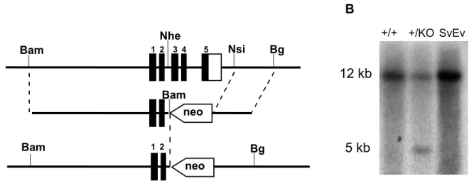
2.1 Ctgf knockout schematic and Southern blots confirming the presence of the knockout allele in animals ... 35
2.2 Ctgf duplication schematic and Southern blots confirming the presence of the duplication allele in animals... 37
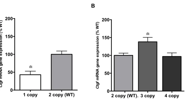
2.3 Ctgf gene expression levels in Ctgf mutant mice ... 39
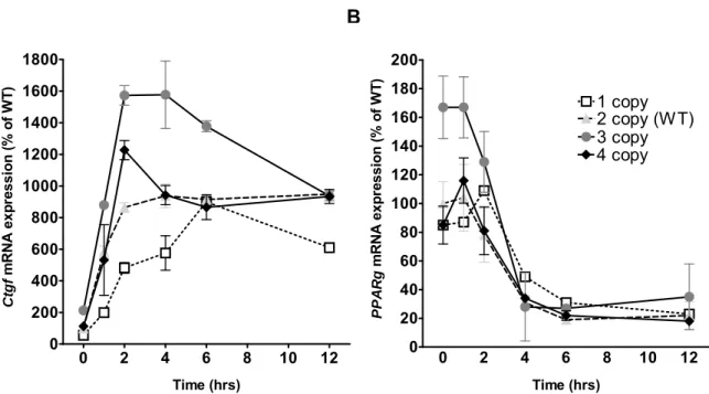
2.4 TGFβ-induced expression of Ctgf and its putative regulator PPARγ... 42
3.1 Blood pressure measurements by tail cuff before and after AngII Infusion ... 60
3.2 Masson’s Trichrome stained AngII treated hearts... 69
3.3 Left ventricle volume in systole from echocardiograms performed at zero, two and four weeks of TAC treatment ... 83
3.4 Frequency of histology score for TAC treated mice by copy Number ... 85
3.5 Organ morphology after five weeks of TAC treatment ... 86
4.1 Morphology of lung at one year of age of WT and 4 copy mice ... 108
4.2 Echocardiogram parameters in young verses old 3 copy mice ... 113
4.3 Organ to body weight ratios as mice age... 115
5.1 Ctgf KO and Ctgf Lo-Hi allele... 130
5.2 Ctgf mRNA levels by quantitative RT-PCR ... 132
5.3 Phenotype of E13.5 embryos... 134
5.4 E14.5 and E10.5 embryos... 136
5.5 Immunofluorescence of E11.5 Ctgf Lo and Hi embryos... 139
5.6 Survivors of Ctgf Hi/+ embryonic lethality ... 141
5.7 Ctgf mRNA levels in tamoxifen treated adults and CTGF protein abundance in plasma ... 144
ABBREVIATIONS
129S6/SvEvTac 129/SvEv or SvEv
αMHC alpha myosin heavy chain
βMHC beta myosin heavy chain
AngII Angiotensin II
ANOVA Analysis of Variances
ANP Atrial Natriuretic Protein
bGH bovine growth hormone
BMC bone mineral content
BMD bone mineral density
BW Body Weight
C57BL6/J B6
CAESAR cis-acting element of structure-anchored repression
CaM kinase Calcium-calmodulin–dependent protein kinases
CCN Cyr61, Ctgf, Nov
c-fos FBJ osteosarcoma oncogene
CHD Coronary Heart Disease
CO Cardiac Output
Col Collagen
Ctgf Connective Tissue Growth Factor (CCN2)
CVD Cardiovascular Disease
Dias Diastole
DEXA Dual Energy X-ray Absorptiometry
DNA deoxy-ribonucleic acid
ECM Extracellular Matrix
EIIa Elongation factor 2a
EM Electron Microscopy
ER Endoplasmic Reticulum
FAK Focal Adhesion kinase
Fig Figure
Fisp12 Fibroblast inducible secreted protein 12 (Ctgf)
FS Left ventricle Fractional Shortening
H & E Hematoxylin and Eosin
HBP High Blood Pressure
Hcs24 Hypertrophic chondrocyte specific gene 24 (Ctgf)
He Heart
IACUC Institutional Animal Care and Use Committee
IGF Insulin-like growth factor
IHC Immunohistochemistry
IL Interleukin
JNK c-jun N-terminal Kinase
Ki Kidney
KO Knockout
Lo-Hi Low High allele
Lu Lung
LV-EF Left ventricle Ejection Fraction
LVH Left ventricle hypertrophy
MAPKs Mitogen activated protein kinases
MEFs Mouse Embryonic Fibroblasts
MI Myocardial Infarction
miRNA micro RNA
MMPs Matrix metalloproteinases
mRNA messenger RNA
neo neomycin resistance gene
NO Nitric oxide
NF-AT3 Nuclear factor of activated T-cells 3
PBS Phosphate-buffered saline
PCR Polymerase Chain Reaction
PPARγ Peroxisome Proliferator Activated Receptor gamma
PWTh Posterior Wall Thickening
Racs Ras-related botulinum toxin substrates
RAS Renin-Angiotensin system
Ren RenTgMk allele
RenTgMK Renin transgenic Marilyn Kozak
RNA Ribonucleic acid
RT-PCR Real Time PCR
RWT Relative Wall Thickness
siRNA small interfering RNAs
SMA Smooth Muscle Actin
SMAD small (sma) mothers against decapentaplegic
SNP single nucleotide polymorphism
SV Stroke Volume
Syst Systole
TAC Trans-aortic constriction/ banding
TBS Tris-buffered saline
TBST TBS + tween
Tg Transgenic
TGFβ Transforming Growth Factor Beta
TNFα Tumor Necrosis Factor alpha
UTR Untranslated region
VECs Vascular Endothelial Cells
VEGF Vascular Endothelial Growth Factor
VSMCs Vascular Smooth Muscle Cells
CHAPTER 1
BACKGROUND AND SIGNIFICANCE
Coronary heart disease (CHD) is the leading killer in the United States,
accounting for 1 in every 5 deaths in 2003 [1, 2]. Forty percent of CHD incidents
result in death. Even for those who survive cardiac ischemia caused by
cardiovascular disease or high blood pressure, permanent scarring of the heart
tissue is a largely unavoidable consequence. For instance, following myocardial
infraction (MI), scarring of the myocardium occurs at the site of ischemia as well as
in regions of the heart where blood flow was not disrupted [3, 4]. Scarring of the
heart can cause reduced tissue elasticity and can disrupt electrical conductivity, both
of which are ultimately responsible for much of the long-term impairment in heart
function that follows cardiac damage [3, 5]. Reduction of tissue scarring is a likely
target for improving long-term outcomes in patients with an ischemic event. Despite
the therapeutic potential of anti-fibrotic treatment, it remains a largely neglected field.
Myocardial scarring is characterized by necrosis of myocardial tissue followed
by replacement with fibrillar collagen, primarily composed of collagen I and III [4].
Collagen buildup and scar tissue contraction at the site of injury often results in
ventricular diastolic dysfunction [3, 4]. In both humans and animal models, sites of
MI scarring have enhanced expression of transforming growth factor beta (TGFβ)
and angiotensin II (AngII). Both peptides are upstream inducers of connective tissue
signaling molecule [6, 7]. CTGF is widely expressed during development in all major organs, the vasculature, and the skeletal system [8]. In the adult, expression
continues to be nearly ubiquitous and functions in extracellular matrix (ECM)
maintenance and repair. Specifically, a marked induction of CTGF expression occurs in healthy wound healing and in pathological fibrosis [9]. Due to the
profibrotic profile of upstream inducers of CTGF expression, its known function in fibrosis, and its localization to cardiac scars [3, 5, 10], many researchers in the field
have suggested that CTGF has an essential role in cardiac fibrosis and other forms
of tissue scarring [6, 9, 11-14].
A. CTGF protein and binding partners
1. Protein
CTGF was first identified in mouse fibroblasts as a serum-inducible, secreted
protein encoded by an immediate-early gene [15]. The human ortholog, discovered
around the same time, was identified in the conditioned media of human umbilical
vein endothelial cells and thought to have mitogenic activity [16]. The CTGF protein
{a.k.a. CCN2, hypertrophic chondrocyte specific gene 24, (Hcs24), or
fibroblast-inducible secreted protein (Fisp12)} is a member of the CCN (Cyr61, CTGF, and
Nov) family [16, 17]. Each functional domain of a CCN family gene is self-contained
within a single exon, and it has been suggested that the CCN family of genes was
evolutionarily assembled via exon reshuffling [18]. Each exon contains a single
functional domain, originally from another gene, that were combined to make the first
divergence, has created a family of genes with a shared set of functional domains
and a modular structure. This family was originally characterized as growth factors
[16, 17], but has been collectively reclassified as extracellular signaling molecules
that principally modify the signaling of other molecules [20]. All six CCN proteins
(Cyr61, Ctgf, Nov and CCN4-6, which were previously called WISP1-3) are
well-conserved across bony vertebrate species and are grouped due to their shared
multimodular exon structure and similar cysteine-rich amino acid sequence [17, 19].
Exon: 1 2 3 4 5
von Willenbrand repeat domain
Thrombospondin binding domain
Cysteine knot domain Heparin binding domain IGF protein
binding domain Signal
sequence 1
Amino Acid #: -21 22-96 96-179 179-249 250-348 Figure 1.1. Schematic structure of CTGF protein with location of motifs. The boxes represent exons, with the related functional elements. Below each box is the number of the amino acids occurring in each portion of the protein. The signaling leader sequence in exon 1 is snipped off generating a mature protein including amino acids 22-348.
The Ctgf gene contains five exons that code for a 38kDa protein that contains one leader sequence and four functional domains (Fig 1.1). It is a secreted protein
with the N-terminal amino acid signal sequence required for Golgi-mediated
secretion encoded in Exon 1 [21]. Exon 2 encodes a domain with an insulin-like
growth factor (IGF) binding motif that mediates weak but significant IGF binding [22].
Exon 3 encodes a domain containing a von Willenbrand factor type C repeat motif
that mediates oligmerization to other proteins such as TGFβ [22]. Exon 4 encodes a
thrombospondin type 1 domain that mediates cell attachment through integrin α6β1
and possibly other integrins [7, 19, 22]. Finally, Exon 5 encodes a cysteine knot
motif common in many growth factors. The domain in Exon 5 also mediates binding
to heparin sulfate proteoglycans, low density lipoprotein receptor related protein
(LRP1), and several different integrins [17, 19, 22-24] (also reviewed in [7, 9] and
diagramed in Fig 1.1).
CTGF undergoes post translational modification including glycosylation, but
this is not required for secretion and no other function attributed to this modification
to date [21]. Studies have also documented that CTGF is proteolytically cleaved into
fragments ranging in size from 10-31 kDa which can be found in a number of
biological fluids (see more details in section E). The points of cleavage generally
occur between functional domains creating pieces of CTGF that contain subsets of
the functional domains [19]. Although the exact function of each of these fragments
is not known, it has been shown that the C-terminal 10 kDa fragment, containing a
very highly conserved region across mammalian species, is mitogenic (can stimulate
fibroblast proliferation), binds heparin, and is sufficient to mediate cell adhesion
through association with integrins [25, 26].
2. CTGF protein and cell adhesion
CTGF has a large number of binding partners, and most of these interactions
appear to be largely cell-type dependent and contextually determined. This
multi-functionality could be attributed to CTGF’s unusual localization as it can be found
anchored to ECM components as well as in cell culture medium and in bodily fluids
in vivo [19]. I will briefly mention a few of the better-studied of these binding
partners, but it is important to note that this is not an exhaustive list since reports of
The association of CTGF with integrins, whether a CTGF fragment or the
full-length protein, occurs in a variety of cell types and is thought to be at least partially
cell-type specific. For example, CTGF reportedly binds to integrins αvβ3 in
endothelial cells [27], αIIbβ3 in platelets [28], α6β1 in human foreskin fibroblasts [29],
αMβ2 in blood monocytes [7, 19, 30], and α5β1 in chondrocytes [31]. The binding of
CTGF to various integrins mediates cell adhesion and in some cases activates cell
signaling cascades. In other cases, the functional consequences of interactions
between CTGF and integrins are unknown. Most likely more integrins will be added
to this list in the future as other interactions are discovered.
Immobilized CTGF alone is sufficient to mediate cell adhesion of fibroblasts to
a untreated coverslips via a unique mechanism. CTGF bind directly to the coverslip
then binds both integrin α6β1 and heparin, which are bound to the cell surface, and
thus acts as adhesive bridge between the coverslip and the cell [29]. This
interaction is unusual in that it requires both integrin α6β1 and heparin for functional
binding. In addition, binding of CTGF to α6β1 integrin stimulates downstream
signaling and has functional consequences, including long-term activation of
mitogen activated protein kinases (MAPKs), Rho family GTPases {in this case one
of the Ras-related C3 botulinum toxin substrate (Racs)}, and matrix
metalloproteinases (MMP-1 and MMP-3). In addition, there is transient activation of
focal adhesion kinase (FAK) that results in actin cytoskeletal reorganization to
promote paxillin-containing focal adhesion formation, cell spreading, and active
formation of filopodia and lamellipodia [29]. Other studies have shown that CTGF is
a strong chemokinetic and chemotactic agent. Cells exposed to CTGF become
generally more mobile and migrate towards an area with concentrated CTGF protein
[27]. Evidence of CTGF as a chemotactic, pro-cell adhesion, and an activator of
FAK suggests that CTGF can stimulate migration to a region, function as a tether of
cells to the ECM, and influence cell signaling behavior. Altogether, the evidence
suggests that CTGF may be an important adhesive signaling molecule.
3. The CTGF receptor or lack thereof
A specific receptor for CTGF binding to the cell surface has yet to be
identified. CTGF does independently bind LRP1, leading to CTGF internalization
and endosomal degradation [21]. It is unclear, however, if any signal transduction
results from CTGF binding to LRP or if binding to LRP simply represents a
mechanism for CTGF degradation [9, 21].
CTGF may act primarily by altering the signaling of other molecules. For
example, as described above, CTGF binds to TGFβ at a binding site within the Von
Willenbrand factor motif. Abreu et al. have shown that CTGF increases TGFβ binding to the TGFβ type II receptor, consequently amplifying the effects of TGFβ
signaling [32]. In addition, CTGF binds to cell surface proteins such as heparin and
integrins. These proteins act as required co-receptors for CTGF’s role in cell
adhesion and angiogenesis and may, in fact, be the primary signaling receptors for
CTGF [19, 29]. Aside from heparin and integrins, other signaling molecules, such as
TGFβ, may act as co-factors and be another primary method by which CTGF binds
B. CTGF gene structure and regulation 1. CTGF promoter and gene regulation
The CTGF gene consists of five exons. Exon one encodes the signaling sequence for shuttling of the protein from the endoplasmic reticulum (ER) to the
Golgi and exons two through five code for each of the four functional domains as
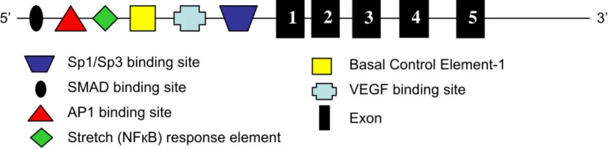
mentioned above [7, 9]. The promoter of the CTGF gene contains multiple response elements suggesting the existence of many upstream activators and repressors of
CTGF and that CTGF is part of a complex regulatory network (Fig 1.2). The known upstream signaling molecules that can increase or decrease Ctgf gene expression include, but are not limited to the following: peroxisome proliferators activated
receptor gamma (PPARγ), TGFβ, AngII, vascular endothelial growth factor (VEGF),
tumor necrosis factor alpha (TNFα), and nitric oxide (NO) [33-42]. In addition, Ctgf
gene expression is modulated by a number of environmental cues such as skin or
vascular wounding, high glucose, fluid flow sheer stress, and cell stretch [33-42].
Binding sites or response elements to some of these molecules and phenotypes
have been mapped to the CTGF 5’-UTR and upstream region for some regulators of
CTGF expression and/or different CTGF protein functions (Fig 1.2) (reviewed in [9]). For example, CTGF expression is increased in response to cell stretch in mesangial cells, smooth muscle cells and chondrocytes, but it is unknown if this response also
occurs in cardiomyocytes [42-44]. In smooth muscle cells increased CTGF
expression is mediated through NFκB binding to the stretch response element in the
CTGF promoter [43] (Fig 1.2), but the same mechanism may or may not be used in the other reported cell types. Thus, the current literature consists of many disparate
models of regulation of CTGF expression in different cell types with little cohesive sense of whether each stimulus is cell-type specific or universal.
5
3 4
1
5’ 2 3’
Sp1/Sp3 binding site Basal Control Element-1 SMAD binding site VEGF binding site AP1 binding site Exon
Stretch (NFкB) response element
Figure 1.2. Schematic of the CTGF gene including upstream binding sites in the promoter. The five exons are numbered. Not to scale.
2. CTGF and Transforming Growth Factor-Beta
TGFβ expression has long been linked to pathological fibrosis in diabetic
nephropathy, scleroderma, cardiac fibrosis, as well as fibrosis in other organs. The
interaction between TGFβ and CTGF is thought to be one of the most important in
the regulation of fibrosis [6, 45, 46]. In a mouse model of scleroderma, application of
either CTGF or TGFβ alone to the skin can induce transient fibrosis but both TGFβ
and CTGF is required to produce severe persistent fibrosis [11, 47]. This finding
suggests that a synergy of the signaling of both molecules is required to sustain a
fibrotic phenotype in cells. CTGF expression is induced by TGFβ receptor signaling, but the relationship between the two molecules is complicated [6]. TGFβ-induced
Ctgf gene expression occurs by way of the mitogen activated protein kinase/ extracellular signal-regulated kinase (MAPK/Erk) pathway with translocation of
SMAD3 and SMAD4 {named for a combination of the C. elegans gene family sma (small) and the Drosophila gene family MAD (mothers against decapentaplegic)} into
A
TGF
β
TGFβ Receptor 1
Smad4
Smad7
Collagen & Fibronectin
Inflammation & Pro-fibrotic signaling
Myofibroblast differentiation
Pro-hypertrophic signaling
CTGF
Smad3 Ras/MEK/ERK
CTGF expression
Nuclear membrane Cell membrane
B
Smad3/Smad4
↑
PPARγ3
TGF
β
↓
PPARγ 21
Smad3/Smad4
↑
CTGF↓
CTGF?
Smad 7 T
i m e
Figure 1.3. CTGF, TGFβ, and PPARγ regulatory relationship. A) Cyclical regulation of CTGF and TGFβ. B) Regulation of TGFβ-induced CTGF expression by PPARγ. Gene expression changes are in italics.
determined and may vary in different cell types with both the rat sarcoma protein
(Ras) and phospholipase C (PKC) also having been implicated in TGFβ-dependent
regulation of CTGF gene expression [9]. Downstream of TGFβ, and thought to be mediated by CTGF, is increased expression of procollagen I, III, and IV as well as
fibronectin expression, and possibly other ECM proteins (Fig 1.3a).
More recently, CTGF was shown to directly bind TGFβ in the extracellular
space in a protein to protein interaction that amplifies TGFβ signaling by increasing
the binding of TGFβ to the TGFβ receptor II at low ligand concentrations [32]. TGFβ
and CTGF appear to work together synergistically in a complex network to generate,
regulate, and maintain normal ECM as well as induce pathological profibrotic
signaling. As CTGF is at the top and bottom of the regulatory pathway with TGFβ, it
may have partial autocrine regulation. Without some brake on such a cyclic
regulatory pathway CTGF expression could continue to increase endlessly.
PPARγ is better known for its role in type II diabetes and adipogenesis [49],
but recent findings by Fu et al., report that PPARγ is a regulator of TGFβ-induced
CTGF gene expression [33]. Another study by Fu et al. showed that TGFβ, long thought to repress PPARγ expression, actually initially stimulates PPARγ expression
at one hour after TGFβ treatment then it represses PPARγ expression by 4 hours
[50]. While neither of these papers directly suggest the idea, the combination of the
two findings could suggest that PPARγ acts in conjunction with TGFβ to form a
cyclic regulatory feedback loop controlling CTGF gene expression. The timing of TGFβ’s induction of first CTGF expression then PPARγ expression is such that Ctgf
PPARγ. The loop is completed when PPARrγ expression is then repressed by TGFβ signaling through AP1 and SMAD3/4 [50]. Finally, repression of PPARγ
allows initiation of a new signal to induce CTGF expression (Fig 1.3b). In summary, the initial signal induces a primary result (increased CTGF gene expression) but also has a secondary signal (increased PPARγ gene expression) that ensures the
primary response is shut off in a timely manner. In chronic fibrotic disease, this
regulatory loop and/or other regulators of Ctgf may be overwhelmed and Ctgf
expression is not repressed in a timely manner, thus causing persistent and
pathological profibrotic signaling [9].
3. Angiotensin II and CTGF
AngII is the terminal signaling molecule in the Renin-Angiotensin System
(RAS), which is a primary signaling pathway that controls systemic blood pressure.
Increases in renin expression and renin levels result in increased circulating AngII
[51]. Increased AngII is a pro-hypertensive signal that increases blood pressure by
two mechanisms. First it acts in the kidney to cause retention of salts, which in turn
causes fluid retention and increased blood volume [52]. AngII also acts in the
vascular smooth muscle cells (VSMCs) as a potent vasoconstrictor [51-53] (Fig 1.4).
The general model is that chronic high blood pressure (a.k.a. hypertension) leads to
cardiac hypertrophy, which in turn is thought to lead to cardiac fibrosis. Buildup of
inflexible, non-conductive collagens reduces cardiac elasticity as well as disrupts
electrical signaling in the heart causing the majority of the heart dysfunction related
to chronic hypertension [54]. Recently, however, studies have shown that cardiac
fibrosis can occur in the absence of hypertrophy shedding doubt on this canonical
model of hypertrophy causing fibrosis [55].
Angiotensin II
AT1 receptor
MAPK
Figure 1.4.Angiotensin II signaling in blood pressure regulation and angiotensin II regulation of CTGF gene expression.
AngII-mediated induction of CTGF gene expression has been well described in a number of tissues [35, 56-59]. In fibroblasts, AngII signals through the AT1
receptor and requires p42/p44 MAPK and RhoA signaling to maximally induce
CTGF expression [56]. In cardiomyocytes, AngII induction of CTGF expression requires activation of PKC [58]. In fibroblasts and VSMCs, induction of CTGF
expression occurs in a TGFβ-independent manner [56, 60] (Fig 1.4). Induction of
CTGF expression by AngII in the heart has been shown to occur in rat and mouse models of myocardial infarction scarring and hypertension [3, 5, 10, 56]. Cell types
likely to produce CTGF in response to AngII include, but are not limited to, vascular
Kidney Vascular SMCs Fibroblasts
Salt retention Vasoconstriction
Systemic Blood Pressure ?
Myocardial Remodeling
(Hypertrophy?) CTGF
Collagen & Fibronectin ?
RhoA
? Cardiomyocytes
conditions myofibroblasts [5, 10, 61]. In addition, treatment with the AT1 receptor
inhibitor, losartan, attenuated CTGF expression in infarcted hearts showing a strong
connection between AngII and CTGF [10].
In addition to direct AngII-induced CTGF gene expression through the MAPK pathway, AngII is likely to indirectly increase CTGF expression. In VSMCs and vascular endothelial cells (VECs) hypertension related changes in fluid flow
dynamics in the blood, such as increased sheer stress and turbulent blood flow,
induce expression of CTGF [42, 62-64]. Multiple reports also show CTGF
expression is induced in response to mechanical stresses such as tension, cell
stretch, and static pressure [42, 62-64] such that CTGF is likely being expressed in one or more cell types in response to the mechanical stresses brought on by
hypertension. Therefore, AngII is inducing CTGF expression directly through the AT1 receptor and indirectly due to hypertension-related phenotypes which in
combination may further induce CTGF expression locally in the cardiac tissue and vasculature. Increased CTGF expression in hypertension is thought to be a key cause of hypertension-induced fibrosis in the heart and other tissues; however, no in vivo model has clearly tested this hypothesis.
4. The CTGF 3’-UTR and CTGF micro RNAs
In addition to regulation by response elements in the 5’UTR and regulation by
other proteins in signaling cascades, CTGF is also know to be regulated by 3’UTR elements and micro RNAs (miRNAs). Some of these elements are implicated by
sequence homology, while others have defined functions. The CTGF 3’UTR
includes multiple AU-rich elements that likely play a role in RNA stability. In addition,
several regions of the CTGF 3’-UTR are highly conserved between humans and mice, implying a possible regulatory role [65]. These sequences in the 3’UTR of the
CTGF gene are not known to have a defined regulatory function, but do require further scrutiny.
In chondrocyte differentiation, miRNA 18a has been shown to target the
CTGF 3’UTR and repress CTGF expression, thereby preventing chondrocyte
maturation [66]. In addition, two cardiac miRNAs that decrease in abundance during
left ventricle pathological hypertrophy, miR-133 and miR-30, bind to the CTGF
3’UTR and downregulate CTGF protein production [67]. The known decrease in
these two miRNAs in cardiac hypertrophy, and their regulatory role in CTGF
abundance, hint that there may be a role for CTGF in cardiac hypertrophy.
Another RNA element targeting the CTGF 3’UTR has been demonstrated by Kubota et al. is called cis-acting element of structure-anchored repression
(CAESAR). The transcript for CAESAR is within the 3’-UTR of CTGF gene and overlaps the coding sequence of exon five by 16 bp [68, 69]. The minimal CAESAR
element able to reduce CTGF protein levels is about 200 bp in length [69] (Fig 1.5).
Whether this element is transcribed only in conjunction with the CTGF gene transcript, or can be separately transcribed, is unknown. In addition, there are
conflicting reports about how CAESAR reduces CTGF protein abundance. One
report suggests that CAESAR acts to reduce translation efficiency by slowing the
Using a reporter assay, CAESAR has been shown to act as a repressor of CTGF
protein production in response to hypoxia [69, 71, 72], but the regulatory role of
CAESAR in other functions of CTGF is unclear.
0 -16
CAESAR element
+174
CTGF
Exon 5
Figure 1.5. Schematic of the CTGF CAESAR element. The CAESAR element overlaps the last
exon of the CTGF gene. The remaining portion of the element is within the CTGF 3’UTR.
The examples above are a subset of a larger number of molecules
responsible for inducing, repressing, and generally regulating CTGF. For a molecule
like CTGF, where acute expression is likely crucial to wound healing while chronic
expression could be pathological, proper regulation of gene expression, whether at
the RNA or protein level, maintains a balance between tissue repair and tissue
damage. In addition, a large number of biological inputs (hypoxia, cell stretch, NO
and glucose concentration, etc.) and outputs (cell adhesion, angiogenesis, ECM
breakdown and deposition, etc.) that CTGF coordinates indicate that CTGF is at the
hub of a regulatory nexus where it acts to coordinate response to a complex set of
signals. As a consequence, dysregulation of one or more of these pathways may be
responsible for chronic pathological expression of CTGF.
Taken together, all the evidence from cell culture supports a model of tight
and complex regulation of Ctgf. The list of Ctgf regulators contains over 100 different proteins, drugs, and environmental cues including stimuli as diverse as
hormones such as testosterone [73], growth factors such as TGFβ [6], and
mechanical stimuli such as cell stretch and fluid shear stress [43, 74]. Unfortunately,
most of these regulators of Ctgf have been discovered in different cell types and under different culture conditions leaving no unified sense of the in vivo function of CTGF.
C. Models of CTGF in development and disease
1. Animal models of altered Ctgf gene expression
Experiments in both animals and cells have shown that CTGF is important in
development. In a transgenic mouse model of Ctgf over-expression in bone (using the collagen XI promoter), animals were born, but were dwarfed, and X-rays showed
that the femur and hind limb bones were less dense than wild type littermates [75].
Another group reported similar findings in a bone/cartilage specific over-expression
of Ctgf using the osteocalcin promoter [76]. On the other end of the spectrum, Ctgf
null embryos are born but die shortly after birth. Ivkovic et al. suggested that improper ossification and general malformation of the skeleton, including the ribs,
ultimately caused the neonates to die due to respiratory failure [77]. In vitro models showed that over-expression of CTGF induces premature osteoblastogenesis [78].
Other reports suggest that CTGF is important in the proliferation and differentiation
of chondrocytes [79, 80]. Both in vivo and in vitro models, such as these, establish a role for Ctgf in bone development and growth.
CTGF has also been implicated in the differentiation of other cell types
preadipocytes differentiate into mature adipocytes, CTGF expression is considerably reduced. In addition, over-expression of CTGF prior to or during differentiation of preadipocytes was shown to reduce the ability of cells to become mature adipocytes
[81]. Osteocytes, chondrocytes and adipocytes are all derived from the
mesenchymal lineage [82]. Proper Ctgf expression and functional regulation in general may be an important factor in cell type determination or transdifferentiation
within the mesenchymal lineage [83].
2. Myocardial Infarction model of cardiac fibrosis and scarring
As discussed above, amongst the upstream regulators of CTGF expression,
TGFβ and AngII are of particular importance to fibrosis [6, 10]. As a method of
examining wound healing in cardiac tissue, a number of research groups have
induced MI in rats by left coronary artery ligation or similar methods [3-5, 10, 84, 85]
and found that following MI, TGFβ, AngII and CTGF are all highly expressed in both
infarcted and uninfarcted cardiac tissue [3, 5, 10]. In the area of scar formation
CTGF is expressed by fibroblast-like cells [3], while in the granulation tissue CTGF is
primarily expressed by endothelial cells [3, 85]. In the uninfarcted perivascular
region, both vascular endothelial cells and interstitial fibroblast-like cells are the
major expressers of CTGF [10]. The combination of TGFβ and CTGF is thought to
be required for fibroblast proliferation and myofibroblast differentiation [86], which
are both part of the fibrotic process. CTGF is expressed in a variety of cells in
response to injury and CTGF, in conjunction with TGFβ, is thought to have an
important profibrotic role in the post MI healing process [3, 5, 10, 85].
AngII also has a clear influence on myofibroblasts. Sun Y et al. showed that in infarcted rat myocardium, myofibroblasts are the primary cell type expressing the
AT1 receptor [4]. Myofibroblasts are a nearly ubiquitous marker of healing and
scarring tissue (reviewed in [87]). In MI scar tissue, myofibroblasts persist long after
wound healing is complete, as opposed to apoptosing, which they do in normal
wound healing in the skin [5]. Elevated TGFβ and CTGF levels in MI scars has also
been observed [3], implicating the two proteins as a cause of myofibroblast
persistence. Myofibroblasts not only promote tissue contraction, but also synthesize
elevated levels of ECM components, such as collagen I and III, as well as matrix
degrading proteases such as matrix metalloproteinase (MMP-2) [5, 88-90]. The
persistence of myofibroblasts within a healing tissue eventually leads to excessive
scarring and functional impairment of the tissue. Thus, persistence of
myofibroblasts in the healing tissue changes the properties of the resulting healed
wound [89].
Rat models of MI clearly show that CTGF, in conjunction with TGFβ and
AngII, acts throughout the process of wound healing and scar formation in cardiac
tissue. In MI, CTGF is expressed early and is thought to act as a profibrotic factor
that signals increased ECM production as well as myofibroblasts differentiation to
create a profibrotic environment that ultimately leads to cardiac scarring and
dysfunction [3-5, 10, 84, 85]. A role for CTGF in cardiac fibrosis is implied by its
induction by AngII, but it seems that the function of CTGF almost always overlaps
D. Genetic variants of CTGF in human disease
Like all human diseases, some people appear more prone to cardiac fibrosis
than others. The central position that CTGF occupies within fibrosis-related
signaling pathways suggests that natural allelic variation in the CTGF gene and resulting variations in the levels of CTGF expression may be a factor in determining fibrosis susceptibility. A recent study revealed a single nucleotide polymorphism
(SNP) in the 5’ upstream region of the CTGF gene that associated with risk and severity of systemic scleroderma (systemic sclerosis) [12]. The C to G transversion
located 945 bp upstream of the transcription start site is associated with increased
scleroderma risk. In particular, those subjects who are homozygous for the G allele
seem especially prone to having two of the more severe phenotypes associated with
systemic sclerosis, fibrosing aveolitis and the presence of anti-topoisomerase I
antibodies. The authors suggested that the SNP at -945 modulates the level of
CTGF expression. In vitro, the G allele caused both higher basal and induced levels of CTGF gene expression. In addition, differential binding of the stronger
transcription factor, Sp1, preferentially to the G risk allele increased expression and
preferential binding of the weaker transcription factor, Sp3, to the C allele, produced
lower CTGF expression levels [12]. This study establishes CTGF as a candidate gene for risk and severity of systemic sclerosis further strengthening the association
between CTGF and fibrotic disease.
Another report of a connection between CTGF variants and fibrotic disease
focuses on hepatic fibrosis following Schistosomiasis infection. In 1999, Dessein et al. examined 65 family pedigrees in a village in Sudan looking for genetic factors that
modulate the severity of liver damage following Schistosoma mansoni infection. They mapped a locus in human chromosome 6q22-23 which was associated with a
more severe hepatic fibrosis following infection [91]. The CTGF gene was within the candidate interval. The same group then followed up using a candidate gene
approach and genotyped SNPs within and nearby the CTGF gene. They found four SNPs upstream or downstream of the CTGF gene that associated with severity of
Schistosomiasis liver damage in a diverse sampling of populations (Chinese,
Sudanese, and Brazilians). Two of these SNPs showed independent association in
each of the populations [92]. Further study of these SNPs could reveal any role they
may have in CTGF expression levels or function.
In addition to the 5’-upstream SNP mentioned above, recent extensive SNP
discovery efforts in the human genome have uncovered 35 SNPs in the human
CTGF gene including two 5’-UTR or upstream SNPs, fifteen 3’-UTR SNPs, eleven intronic SNPs (including one in a splice site), three synonymous and four
non-synonymous protein coding SNPs [65]. This number of SNPs is a large for such a
small gene and is a particularly large number of non-synonymous amino acids
changes. All of these amino acid substitutions are likely to be non-conservative and,
therefore, functionally relevant (Table 1.1). The diversity of natural variation in the
CTGF gene suggests that functional variation exists within the human population,
and these changes may alter the likelihood of development or severity of cardiac
fibrosis. Unfortunately, little or no meaningful allele frequency data is available for
the four non-synonymous coding SNPs, and the importance of any phenotypic
change the amino acid sequence, a large number of intronic and UTR variations are
present in the human CTGF gene, which suggests variations in expression level, protein and mRNA stability, as well as binding site strength and specificity may exist
within human populations.
Amino Acid number (Exon)
Protein Domain Amino Acid Change
Functional Change
42 (2) IGF binding
domain
Cys → Arg neutral sulfur-containing to basic + charged
83 (2) IGF binding
domain
Asp → His acidic + charged to slightly basic - charged
118 (3) von Willebrand
repeat domain Asn → Ser highly hydrophilic to less hydrophilic
296 (5) Cysteine knot
domain Tyr → His weak aromatic acid to basic aromatic
Table 1.1. Location and description of the four non-synonymous coding SNPs in the human CTGF gene. IGF = Insulin-like growth factor
Another peculiarity of CTGF worth mentioning is the highly conserved
C-terminal amino acid sequence. In the last 150 amino acids of CTGF, there are less
than 10 amino acid differences in any pair-wise comparison between the human,
mouse, rat, bovine, pig, and rabbit sequences (my own observation). For instance,
there are only two differences between the human and bovine sequence in the last
150 amino acids. The reason for this very high level of amino acid sequence
conservation is unknown, but it suggests that there has been strong purifying
selection and thus it is likely that an essential conserved function resides in this
region. Strong conservation of the CTGF gene between species is an encouraging observation and suggests that the phenotypes and molecular relationships observed
in mouse models are likely to be conserved in humans.
E. CTGF protein levels as a noninvasive measurement of fibrotic disease in humans
CTGF has been identified at measurable levels in a wide variety of human
biological fluids including urine, serum/plasma, amniotic, follicular, peritoneal and
cerebrospinal fluids [93, 94]. In humans and mouse models of fibrotic disease both
plasma and urine levels of CTGF have become of particular interest. CTGF levels in
plasma and urine have been tested as a non-invasive biomarker for many fibrotic
diseases including scleroderma, kidney allograft fibrosis, liver fibrosis (viral and
non-viral), diabetic retinopathy, end-stage renal disease, and heart failure [93, 95-99]. In
all of these tests, CTGF levels in plasma or urine were positively correlated with
severity or disease progression [100].
Reports of basal CTGF concentrations in plasma have varied widely with
levels ranging from 3 ng/mL to 400 ng/mL [95-99]. Three studies, however, have
reported very similar values for control subjects, suggesting the plasma level of
CTGF for normal human adults is likely between 9 -15 ng/mL [95, 98, 99]. Under
disease conditions those levels can increase three to five-fold [95, 97, 98]. Similarly,
in mice the basal CTGF plasma level is about 20 ng/mL and can increase to 40-90
ng/mL during kidney allograft fibrosis [95]. The similarity in the basal levels and
responsiveness between humans and mice suggests that mouse models will be a
reliable surrogate for studying changes in plasma CTGF levels during fibrotic
disease. In addition, the correlation of plasma CTGF levels to severity of fibrotic
disease in humans suggests that even if CTGF in not causal of disease, it is likely an
Summary
Through a number of tissue culture models of fibrotic processes, many of the
molecular pathways that control Ctgf expression have been elucidated. This in vitro
work has put CTGF at the center of a complicated set of inputs surrounding ECM
maintenance and repair and implicated that it may have a function in fibrotic disease.
It, however, remains to be shown whether Ctgf over-expression or modulation is truly a cause of fibrotic disease. There have been a few models of altered Ctgf
expression in vivo, but a model of global over-expression of Ctgf has yet to be reported. My project will focus on generating mouse models with varying levels of
Ctgf expression and challenging those animals with treatments that induce cardiac fibrosis and hypertrophy to elucidate the role of varying Ctgf expression in
cardiovascular disease.
References
1. Thom, T., et al., Heart disease and stroke statistics--2006 update: a report from the American Heart Association Statistics Committee and Stroke Statistics Subcommittee. Circulation, 2006. 113(6): p. e85-151.
2. Tsai, Y.S., et al., Genetic variations in peroxisome proliferator-activated receptor gamma expression affect blood pressure. Proc Natl Acad Sci U S A, 2009. 106(45): p. 19084-9.
3. Sun, Y. and K.T. Weber, Infarct scar: a dynamic tissue. Cardiovasc Res, 2000. 46(2): p. 250-6.
4. Sun, Y., et al., Angiotensin II, transforming growth factor-beta1 and repair in the infarcted heart. J Mol Cell Cardiol, 1998. 30(8): p. 1559-69.
5. Sun, Y., et al., Cardiac remodeling by fibrous tissue after infarction in rats. J Lab Clin Med, 2000. 135(4): p. 316-23.
6. Chen, M.M., et al., CTGF expression is induced by TGF- beta in cardiac fibroblasts and cardiac myocytes: a potential role in heart fibrosis. J Mol Cell Cardiol, 2000. 32(10): p. 1805-19.
7. Leask, A. and D.J. Abraham, The role of connective tissue growth factor, a multifunctional matricellular protein, in fibroblast biology. Biochem Cell Biol, 2003. 81(6): p. 355-63.
8. Surveyor, G.A. and D.R. Brigstock, Immunohistochemical localization of connective tissue growth factor (CTGF) in the mouse embryo between days 7.5 and 14.5 of gestation. Growth Factors, 1999. 17(2): p. 115-24.
9. Blom, I.E., R. Goldschmeding, and A. Leask, Gene regulation of connective tissue growth factor: new targets for antifibrotic therapy? Matrix Biol, 2002.
21(6): p. 473-82.
10. Ahmed, M.S., et al., Connective tissue growth factor--a novel mediator of angiotensin II-stimulated cardiac fibroblast activation in heart failure in rats. J Mol Cell Cardiol, 2004. 36(3): p. 393-404.
11. Mori, T., et al., Role and interaction of connective tissue growth factor with transforming growth factor-beta in persistent fibrosis: A mouse fibrosis model.
J Cell Physiol, 1999. 181(1): p. 153-9.
13. Gressner, O.A. and A.M. Gressner, Connective tissue growth factor: a fibrogenic master switch in fibrotic liver diseases. Liver Int, 2008. 28(8): p. 1065-79.
14. Shi-Wen, X., A. Leask, and D. Abraham, Regulation and function of
connective tissue growth factor/CCN2 in tissue repair, scarring and fibrosis.
Cytokine Growth Factor Rev, 2008. 19(2): p. 133-44.
15. Ryseck, R.P., et al., Structure, mapping, and expression of fisp-12, a growth factor-inducible gene encoding a secreted cysteine-rich protein. Cell Growth Differ, 1991. 2(5): p. 225-33.
16. Bradham, D.M., et al., Connective tissue growth factor: a cysteine-rich mitogen secreted by human vascular endothelial cells is related to the SRC-induced immediate early gene product CEF-10. J Cell Biol, 1991. 114(6): p. 1285-94.
17. Bork, P., The modular architecture of a new family of growth regulators related to connective tissue growth factor. FEBS Lett, 1993. 327(2): p. 125-30.
18. Rodgers, B.D., E.H. Roalson, and C. Thompson, Phylogenetic analysis of the insulin-like growth factor binding protein (IGFBP) and IGFBP-related protein gene families. Gen Comp Endocrinol, 2008. 155(1): p. 201-7.
19. Lau, L.F. and S.C. Lam, The CCN family of angiogenic regulators: the integrin connection. Exp Cell Res, 1999. 248(1): p. 44-57.
20. Leask, A. and D.J. Abraham, All in the CCN family: essential matricellular signaling modulators emerge from the bunker. J Cell Sci, 2006. 119(Pt 23): p. 4803-10.
21. Chen, Y., et al., Connective tissue growth factor is secreted through the Golgi and is degraded in the endosome. Exp Cell Res, 2001. 271(1): p. 109-17. 22. Brigstock, D.R., The connective tissue growth factor/cysteine-rich
61/nephroblastoma overexpressed (CCN) family. Endocr Rev, 1999. 20(2): p. 189-206.
23. Moussad, E.E. and D.R. Brigstock, Connective tissue growth factor: what's in a name? Mol Genet Metab, 2000. 71(1-2): p. 276-92.
24. Perbal, B., NOV (nephroblastoma overexpressed) and the CCN family of genes: structural and functional issues. Mol Pathol, 2001. 54(2): p. 57-79. 25. Ball, D.K., et al., The heparin-binding 10 kDa fragment of connective tissue
growth factor (CTGF) containing module 4 alone stimulates cell adhesion. J Endocrinol, 2003. 176(2): p. R1-7.
26. Brigstock, D.R., et al., Purification and characterization of novel binding growth factors in uterine secretory fluids. Identification as heparin-regulated Mr 10,000 forms of connective tissue growth factor. J Biol Chem, 1997. 272(32): p. 20275-82.
27. Babic, A.M., C.C. Chen, and L.F. Lau, Fisp12/mouse connective tissue growth factor mediates endothelial cell adhesion and migration through integrin alphavbeta3, promotes endothelial cell survival, and induces angiogenesis in vivo. Mol Cell Biol, 1999. 19(4): p. 2958-66.
28. Jedsadayanmata, A., et al., Activation-dependent adhesion of human platelets to Cyr61 and Fisp12/mouse connective tissue growth factor is mediated through integrin alpha(IIb)beta(3). J Biol Chem, 1999. 274(34): p. 24321-7.
29. Chen, C.C., N. Chen, and L.F. Lau, The angiogenic factors Cyr61 and connective tissue growth factor induce adhesive signaling in primary human skin fibroblasts. J Biol Chem, 2001. 276(13): p. 10443-52.
30. Schober, J.M., et al., Identification of integrin alpha(M)beta(2) as an adhesion receptor on peripheral blood monocytes for Cyr61 (CCN1) and connective tissue growth factor (CCN2): immediate-early gene products expressed in atherosclerotic lesions. Blood, 2002. 99(12): p. 4457-65.
31. Hoshijima, M., et al., CT domain of CCN2/CTGF directly interacts with fibronectin and enhances cell adhesion of chondrocytes through integrin alpha5beta1. FEBS Lett, 2006. 580(5): p. 1376-82.
32. Abreu, J.G., et al., Connective-tissue growth factor (CTGF) modulates cell signalling by BMP and TGF-beta. Nat Cell Biol, 2002. 4(8): p. 599-604. 33. Fu, M., et al., Peroxisome proliferator-activated receptor gamma inhibits
transforming growth factor beta-induced connective tissue growth factor expression in human aortic smooth muscle cells by interfering with Smad3. J Biol Chem, 2001. 276(49): p. 45888-94.
34. Hishikawa, K., T. Nakaki, and T. Fujii, Transforming growth factor-beta(1) induces apoptosis via connective tissue growth factor in human aortic smooth muscle cells. Eur J Pharmacol, 1999. 385(2-3): p. 287-90.
35. Finckenberg, P., et al., Angiotensin II induces connective tissue growth factor gene expression via calcineurin-dependent pathways. Am J Pathol, 2003.
163(1): p. 355-66.
37. Abraham, D.J., et al., Tumor necrosis factor alpha suppresses the induction of connective tissue growth factor by transforming growth factor-beta in normal and scleroderma fibroblasts. J Biol Chem, 2000. 275(20): p. 15220-5. 38. Keil, A., et al., Nitric oxide down-regulates connective tissue growth factor in
rat mesangial cells. Kidney Int, 2002. 62(2): p. 401-11.
39. Igarashi, A., et al., Regulation of connective tissue growth factor gene
expression in human skin fibroblasts and during wound repair. Mol Biol Cell, 1993. 4(6): p. 637-45.
40. Murphy, M., et al., Suppression subtractive hybridization identifies high
glucose levels as a stimulus for expression of connective tissue growth factor and other genes in human mesangial cells. J Biol Chem, 1999. 274(9): p. 5830-4.
41. Yoshisue, H., et al., Large scale isolation of non-uniform shear stress-responsive genes from cultured human endothelial cells through the
preparation of a subtracted cDNA library. Atherosclerosis, 2002. 162(2): p. 323-34.
42. Hishikawa, K., B.S. Oemar, and T. Nakaki, Static pressure regulates
connective tissue growth factor expression in human mesangial cells. J Biol Chem, 2001. 276(20): p. 16797-803.
43. Chaqour, B., R. Yang, and Q. Sha, Mechanical stretch modulates the promoter activity of the profibrotic factor CCN2 through increased actin polymerization and NF-kappaB activation. J Biol Chem, 2006. 281(29): p. 20608-22.
44. Nishida, T., et al., Role of mechanical-stress inducible protein
Hcs24/CTGF/CCN2 in cartilage growth and regeneration: mechanical stress induces expression of Hcs24/CTGF/CCN2 in a human chondrocytic cell line HCS-2/8, rabbit costal chondrocytes and meniscus tissue cells. Biorheology, 2008. 45(3-4): p. 289-99.
45. Branton, M.H. and J.B. Kopp, TGF-beta and fibrosis. Microbes Infect, 1999.
1(15): p. 1349-65.
46. Smith, E.A. and E.C. LeRoy, A possible role for transforming growth factor-beta in systemic sclerosis. J Invest Dermatol, 1990. 95(6 Suppl): p. 125S-127S.
47. Chujo, S., et al., Connective tissue growth factor causes persistent
proalpha2(I) collagen gene expression induced by transforming growth factor-beta in a mouse fibrosis model. J Cell Physiol, 2005. 203(2): p. 447-56.
48. Chen, Y., et al., CTGF expression in mesangial cells: involvement of SMADs, MAP kinase, and PKC. Kidney Int, 2002. 62(4): p. 1149-59.
49. Tontonoz, P., E. Hu, and B.M. Spiegelman, Stimulation of adipogenesis in fibroblasts by PPAR gamma 2, a lipid-activated transcription factor. Cell, 1994. 79(7): p. 1147-56.
50. Fu, M., et al., Early stimulation and late inhibition of peroxisome proliferator-activated receptor gamma (PPAR gamma) gene expression by transforming growth factor beta in human aortic smooth muscle cells: role of early growth-response factor-1 (Egr-1), activator protein 1 (AP1) and Smads. Biochem J, 2003. 370(Pt 3): p. 1019-25.
51. Sealey, J.E. and J.H. Laragh, The Renin-Angiotensin-Aldosterone System for Normal Regulation of Blood Pressure and Sodium and Potassium
Homeostasis, in Hypertension: Pathophysiology, Diagnosis, and
Management, J.H. Laragh and B.M. Brenner, Editors. 1990, Raven Press Ltd: New York. p. 1287-1317.
52. Sealey, J.E., et al., On the Renal Basis for Essential Hypertension: Nephron Heterogeneity with Discordant Renin Secretion and Sodium Excretion Causing a Hypertensive Vasoconstriction - Volume Relationship, in
Hypertension: Pathophysiology, Diagnosis, and Management, J.H. Laragh and B.M. Brenner, Editors. 1990, Raven Press Ltd: New York. p. 1089-1104.
53. Marsden, P.A., B.M. Brenner, and B.J. Ballerman, Mechanisms of
Angiotensin Action on the Vascular Smooth Muscle, the Adrenal, and the Kidney, in Hypertension: Pathophysiology, Diagnosis, and Management, J.H. Laragh and B.M. Brenner, Editors. 1990, Raven Press Ltd: New York. p. 1247-1272.
54. Devereux, R.B. and M.J. Roman, Hypertensive Cardiac Hypertrophy: Pathophysiologic and clinical considerations, in Hypertension:
Pathophysiology, Diagnosis, and Treatment, J.H. Laragh and B.M. Brenner, Editors. 1995, Raven Press, Ltd: New York. p. 409-423.
55. Royer, A., et al., Mouse model of SCN5A-linked hereditary Lenegre's disease: age-related conduction slowing and myocardial fibrosis. Circulation, 2005.
111(14): p. 1738-46.
56. Iwanciw, D., et al., Induction of connective tissue growth factor by angiotensin II: integration of signaling pathways. Arterioscler Thromb Vasc Biol, 2003.
23(10): p. 1782-7.
severe left ventricular dysfunction]. Zhonghua Xin Xue Guan Bing Za Zhi, 2005. 33(4): p. 323-7.
58. He, Z., et al., Differential regulation of angiotensin II-induced expression of connective tissue growth factor by protein kinase C isoforms in the
myocardium. J Biol Chem, 2005. 280(16): p. 15719-26.
59. Tung, J.N., et al., Paraquat increases connective tissue growth factor and collagen expression via angiotensin signaling pathway in human lung fibroblasts. Toxicol In Vitro. 24(3): p. 803-8.
60. Rodriguez-Vita, J., et al., Angiotensin II activates the Smad pathway in vascular smooth muscle cells by a transforming growth factor-beta-independent mechanism. Circulation, 2005. 111(19): p. 2509-17.
61. Tiggelman, A.M., et al., Transforming growth factor-beta-induced collagen synthesis by human liver myofibroblasts is inhibited by alpha2-macroglobulin.
J Hepatol, 1997. 26(6): p. 1220-8.
62. Kessler, D., et al., Fibroblasts in mechanically stressed collagen lattices assume a "synthetic" phenotype. J Biol Chem, 2001. 276(39): p. 36575-85. 63. Riser, B.L., et al., Regulation of connective tissue growth factor activity in
cultured rat mesangial cells and its expression in experimental diabetic glomerulosclerosis. J Am Soc Nephrol, 2000. 11(1): p. 25-38.
64. Schild, C. and B. Trueb, Mechanical stress is required for high-level
expression of connective tissue growth factor. Exp Cell Res, 2002. 274(1): p. 83-91.
65. Sanger, I. Ensembl Genome Browser: Gene: Ctgf (ENSMUSG00000019997). 2010 [cited 2010 May 12]; Ensembl release 57 - Mar 2010:[Available from: http://uswest.ensembl.org/Mus_musculus/Gene/Summary?g=ENSMUSG000 00019997.
66. Ohgawara, T., et al., Regulation of chondrocytic phenotype by micro RNA 18a: involvement of Ccn2/Ctgf as a major target gene. FEBS Lett, 2009.
583(6): p. 1006-10.
67. Duisters, R.F., et al., miR-133 and miR-30 regulate connective tissue growth factor: implications for a role of microRNAs in myocardial matrix remodeling.
Circ Res, 2009. 104(2): p. 170-8, 6p following 178.
68. Kubota, S., et al., Involvement of cis-acting repressive element(s) in the 3'-untranslated region of human connective tissue growth factor gene. FEBS Lett, 1999. 450(1-2): p. 84-8.
69. Kubota, S., et al., Identification of an RNA element that confers post-transcriptional repression of connective tissue growth factor/hypertrophic chondrocyte specific 24 (ctgf/hcs24) gene: similarities to retroviral RNA-protein interactions. Oncogene, 2000. 19(41): p. 4773-86.
70. Kubota, S., et al., Novel intracellular effects of human connective tissue growth factor expressed in Cos-7 cells. FEBS Lett, 2000. 474(1): p. 58-62. 71. Kondo, S., et al., Hypoxic regulation of stability of connective tissue growth
factor/CCN2 mRNA by 3'-untranslated region interacting with a cellular
protein in human chondrosarcoma cells. Oncogene, 2006. 25(7): p. 1099-110. 72. Kubota, S., et al., Translational repression by the cis-acting element of
structure-anchored repression (CAESAR) of human ctgf/ccn2 mRNA. FEBS Lett, 2005. 579(17): p. 3751-8.
73. Swartz, C.D., et al., Estrogen-induced changes in IGF-I, Myb family and MAP kinase pathway genes in human uterine leiomyoma and normal uterine smooth muscle cell lines. Mol Hum Reprod, 2005. 11(6): p. 441-50.
74. McCormick, S.M., et al., DNA microarray reveals changes in gene expression of shear stressed human umbilical vein endothelial cells. Proc Natl Acad Sci U S A, 2001. 98(16): p. 8955-60.
75. Nakanishi, T., et al., Overexpression of connective tissue growth
factor/hypertrophic chondrocyte-specific gene product 24 decreases bone density in adult mice and induces dwarfism. Biochem Biophys Res Commun, 2001. 281(3): p. 678-81.
76. Smerdel-Ramoya, A., et al., Skeletal overexpression of connective tissue growth factor impairs bone formation and causes osteopenia. Endocrinology, 2008. 149(9): p. 4374-81.
77. Ivkovic, S., et al., Connective tissue growth factor coordinates chondrogenesis and angiogenesis during skeletal development.
Development, 2003. 130(12): p. 2779-91.
78. Smerdel-Ramoya, A., et al., Connective tissue growth factor enhances osteoblastogenesis in vitro. J Biol Chem, 2008. 283(33): p. 22690-9. 79. Fujisawa, T., et al., CCN family 2/connective tissue growth factor
(CCN2/CTGF) stimulates proliferation and differentiation of auricular chondrocytes. Osteoarthritis Cartilage, 2008. 16(7): p. 787-95.