Diverse Mechanisms of G Protein Regulation by Monoubiquitination
Rachael Ann Baker
A dissertation submitted to the faculty of the University of North Carolina at Chapel Hill in partial fulfillment of the requirements for the degree of Doctor of Philosophy in the
Department of Biochemistry and Biophysics
Chapel Hill 2013
ii ABSTRACT
RACHAEL BAKER: Diverse Mechanisms of G Protein Regulation by Monoubiquitination
(Under the direction of Henrik Dohlman and Sharon Campbell)
Cell signaling pathways convert information from the extracellular environment into an intracellular response. It is essential that these pathways be turned on and off on the appropriate timescales. Post-translational modifications are one essential mechanism used to maintain proper signaling. One post-translational modification that is emerging as a key regulator of cell signaling is monoubiquitination. Monoubiquitination is dynamic and reversible, making it ideal for temporal and spatial regulation. It has recently become evident that monoubiquitination regulates G proteins, which are the molecular switches that turn signaling pathways on and off. However, the mechanisms by which monoubiquitination acts on these enzymes is not known.
We used a chemical ubiquitination approach coupled with biochemical and biophysical assays to elucidate the mechanisms by which two G proteins, the small G protein Ras and the heterotrimeric G protein Gpa1, are regulated by monoubiquitination. Monoubiquitination at one position activates K-Ras by impeding regulator-mediated hydrolysis while monoubiquitination at a distinct site activates H-Ras by increasing intrinsic nucleotide exchange. Together, these results demonstrate that
iii
domain that is essential for trafficking but does not contribute to enzymatic activity. The G protein substrates we chose exhibited diverse mechanisms of regulation by
monoubiquitination including altering protein interactions (K-Ras), intrinsic activity (H-Ras), and localization (Gpa1). In summary, our results represent the first mechanistic study of G protein regulation by monoubiquitination and contribute to understanding Ras and Gpa1 regulation specifically as well as regulation of G proteins by
iv
v
ACKNOWLEDGEMENTS
First, I would like to thank my advisors, Henrik and Sharon. This project only came together because you were willing to take a risk and let me try something new. Thank you for teaching me not only how to be a good bench scientist, but how to ask good questions, write coherently (although not always succinctly, sorry Henrik), and to strive for balance in my life.
I would also like to thank my lab mates and friends. Much of my success and what I learned about science while I was here was because of you. I appreciate all the times you listened to practice presentations, offered new insights into my project when I was stuck, and were encouraging when I was having a hard day. Working around so many talented people was a large part of what made me successful. More importantly, you made grad school fun.
vi
To my husband Sean, your support during the first years of graduate school and frustration during the last one are the things that kept me moving. Thanks for giving me the confidence to try challenging and scary things. I appreciate all the nights you let me come back to work, just for a few minutes, and all the times you listened when I was frustrated or overwhelmed. After over five years of marriage, I still love you more every day, and I’m looking forward to many happy years together as Dr. and Mr. Baker.
vii
TABLE OF CONTENTS
LIST OF TABLES ... ix
LIST OF FIGURES ... x
LIST OF ABBREVIATIONS ... xii
CHAPTERS I. INTRODUCTION ... 1
Monoubiquitination... 2
Ubiquitination and Disease ... 17
New Approaches to Study Ubiquitination ... 19
Regulation of GTPase Signaling by Monoubiquitination ... 23
Thesis Summary... 33
II. SITE-SPECIFIC MONOUBIQUITINATION ACTIVATES RAS BY IMPEDING GTPASE ACTIVATING PROTEIN FUNCTION... 36
Introduction ... 37
Results ... 38
Discussion ... 65
Acknowledgements ... 67
Methods... 68
viii
Introduction ... 74
Results ... 79
Discussion ... 89
Methods... 92
IV. THE HELICAL DOMAIN INFLUENCES THE ENZYMATIC ACTIVITY OF THE YEAST G ALPHA PROTEIN ... 97
Introduction ... 98
Results ... 101
Discussion ... 113
Methods... 116
V. CONCLUSIONS AND FUTURE DIRECTIONS ... 119
Chemical Ubiquitination ... 121
Regulation of Ras by Monoubiquitination ... 129
Gpa1 as a Substrate for Monoubiquitination ... 145
Conclusions and Future Directions ... 151
Methods... 156
APPENDIX A ... 159
APPENDIX B ... 165
APPENDIX C ... 169
ix
LIST OF TABLES
Table 5.1. Summary of Disulfide Chemical Ubiquitination Approaches ... 128
Table 5.2. Proposed NMR Experiments for the Study of mUbRas ... 132
Table 5.3. Small GTPases Containing a Lysines in the Conserved SAX motif ... 153
x
LIST OF FIGURES
Figure 1.1. Monoubiquitination by the Ubiquitin Ligase Complex ... 6
Figure 1.2. Recognition and Outcomes of Monoubiquitination ... 12
Figure 1.3. Monoubiquitination Leads to Receptor Endocytosis ... 14
Figure 1.4. Domains of Small and Heterotrimeric GTPases ... 25
Figure 1.5. Small GTPase Enzymatic Cycle ... 28
Figure 1.6. Heterotrimeric Gα Protein Enzymatic Cycle... 31
Figure 2.1. Monoubiquitination of Ras ... 42
Figure 2.2. Raf-RBD Pull-Down From HEK293T Cells Indicates That Ubiquitination Increases the Fraction of GTP-Bound Ras ... 43
Figure 2.3. NMR Comparison of Ras and RasK147C ... 44
Figure 2.4. Monoubiquitinated Ras Retains Intrinsic Stability and Activity ... 45
Figure 2.5. Rosetta Model of Native and Chemical Ubiquitination of Ras ... 48
Figure 2.6. Surfaces of Ubiquitin Affected by Monoubiquitination ... 49
Figure 2.7. Surfaces of Ras Affected by Monoubiquitination ... 50
Figure 2.8. Increased Detection of mUbRas Backbone Amides at 500 MHz ... 54
Figure 2.9. Monoubiquitination Decreases the Sensitivity of Ras to Downregulation by GAPs ... 55
Figure 2.10. GAP-Mediated Hydrolysis of Monoubiquitinated K-Ras ... 56
Figure 2.11. Kinetic Modeling of Changes in Ras Activity Due to Monoubiquitination ... 57
Figure 2.12. Modification of Ras with PDZ2UL Resembles Modification with Ubiquitin ... 61
Figure 2.13. Modification of Ras with PDZ is Distinct from Modification with Ubiquitin ... 62
xi
Figure 2.15. Monoubiquitination Does not Sterically Occlude GAP Binding to Ras ... 64
Figure 3.1. Mutation of Ras at Lysine 104 ... 81
Figure 3.2. Monoubiquitination of Ras at Position 104 ... 82
Figure 3.3. Mutation of Ras at Lysine 117 ... 85
Figure 3.4. Monoubiquitination at Position 117 ... 86
Figure 3.5. Assays of H-Ras in Cell Lysate Indicate H-Ras is Primarily Modified at K117 ... 88
Figure 4.1. Gpa1 Activity at the Plasma Membrane and Endosome. ... 102
Figure 4.2. Optimization and Purification of Gpa1 ... 104
Figure 4.3. The Insert Domain of Gpa1 Does Not Alter Gpa1 Activity ... 110
Figure 4.4. Time Course of Gpa1 Ubiquitination ... 111
Figure 4.5. Changes in the α-Helical Domain of Gpa1 Influence its Enzymatic Activity ... 112
Figure 5.1. Native and Chemical Ubiquitination Linkages ... 126
Figure 5.2. Preliminary Molecular Dynamics Simulations of mUbRas ... 134
Figure 5.3. Proximity of Ubiquitination Sites to Switch I ... 142
Figure 5.4. Proximity of K104 to Surface of Sos Interface with Ras ... 143
xii
LIST OF ABBREVIATIONS
ABD-F 1 mM 4-fluoro-7-aminosulfonylbenzoflurazan
Ala or A Alanine
α Alpha
AlF3 Aluminum Fluoride
A. thaliana Arabidopsis thaliana Arg or R Arginine
ATP Adenosine Triphosphate
β Beta
CD Circular Dichroism
Cys or C Cysteine
CRD Cysteine Rich Domain
Δ Deletion
DUB Deubiquitinating Enzyme
D. melanogaster Drosophila melanogaster E. coli Escherichia coli E1 Ubiquitin Activating Enzyme
E2 Ubiquitin Conjugating Enzyme
E3 Ubiquitin Ligase
xiii
γ Gamma
GAP GTPase Activating Protein
GDP Guanosine Diphosphate
GEF Guanine Nucleotide Exchange Factor
Gly or G Glycine
GMPPNP Guanylyl Imidodiphosphate GPCR G Protein Coupled Receptor GST Glutathione S-Transferase
GTP Guanosine Triphosphate
HECT Homologous to E6-ap Carboxyl Terminus His or H Histidine
HSQC Heteronuclear Single Quantum Coherence HTPF Homogenous Time-Resolved Fluorescence
HVR Hypervariable Region
IKK IB Kinase
Ile or I Isoleucine
Leu or L Leucine
Lys or K Lysine
MANT-GDP 2'- / 3'- O- (N'- Methylanthraniloyl)- Guanosine Diphosphate
MD Molecular Dynamics
Mg Magnesium
mUbRas Monoubiquitinated Ras
xiv
PCNA Proliferating Nuclear Cell Antigen
PDB Protein Data Bank
PI3K Phosphatidyl-Inositol-3-Kinase
RBD Ras Binding Domain
RGS Regulator of G Protein Signaling RING Really Interesting New Gene RTK Receptor Tyrosine Kinase
S. cerevisiae Saccharomyces cerevisiae
SCF Skp1-Cullin-F-Box
Ser or S Serine
TCEP Tris (2-carboxyethyl) Phosphine Hydrochloride
TEV Tobacco Etch Virus
TLS Translesion Synthesis
Tm Melting Temperature
Ub Ubiquitin
UBA Ubiquitin Associated Domain UBD Ubiquitin Binding Domain
UD Ubiquitination Domain
UIM Ubiquitin Interacting Motif
UL Ubiquitin Linker
Val or V Valine
CHAPTER I
INTRODUCTION1
When cells receive a signal, it is crucial that they respond correctly and limit the time frame of their response. The regulation of all cellular events requires careful maintenance of proper protein levels and activity, a crucial balance that is determined by the control of protein synthesis, localization, activation, and degradation. Many of these levels of control are fine-tuned by post-translational modification of proteins by
mechanisms such as lipidation, glycosylation, phosphorylation, monoubiquitination, and polyubiquitination. While monoubiquitination is similar in name to polyubiquitination, it is more similar in function to conditional post-translational modifications like
phosphorylation. The dysregulation of ubiquitination is implicated in many diseases including cancer, autoimmune disorders, neurodegenerative diseases, and developmental disorders. However, particularly in the case of monoubiquitination, we have yet to understand the full extent of the mechanisms by which this modification is used to regulate proper cellular signaling and response. The potential functional diversity of this signal is staggering, and our ability to understand its mechanism of action is in many cases limited only by our ability to generate enough modified substrate to study by biochemical and biophysical methods. This thesis will focus specifically on the study of monoubiquitination of key components of signaling pathways, G proteins, using a
2
chemical approach to generate monoubiquitinated substrate. This approach allows us to couple the mechanistic understanding of a protein developed from biochemical and biophysical studies to data that demonstrate the importance of monoubiquitination for maintaining proper cellular signaling in vivo. This introductory chapter will specifically focus on what is known about regulation by monoubiquitination and limitations in our knowledge and available resources to study monoubiquitinated proteins.
Monoubiquitination
Monoubiquitination is a dynamic, reversible post-translational modification that involves attaching the 76 amino acid protein Ubiquitin to a targeted substrate. Even though monoubiquitination involves modifying a protein with a distinct protein, it is used in a manner similar to post-translational modification by phosphorylation.
Phosphorylation is central to the regulation of cell signaling pathways (1-4) and can even be required as a precursor for ubiquitination (5). Given the similarities between the ways these two post-translational modifications can be used and their effects on substrates, it is now evident that monoubiquitination is also emerging as a major player in cell signaling regulation.
Evidence for regulation by monoubiquitination is present in a number of key cell pathways, including regulation of DNA expression through modification of histones and processivity factors, regulation of signaling through endocytosis, and regulation of viruses (6). However, our understanding of the mechanisms by which
3
occur when a protein is monoubiquitinated and that could contribute to understanding the mechanisms through which substrates are regulated by monoubiquitination.
One challenge to advancing our understanding of the breadth of regulation by monoubiquitination is the lack of a resource that consolidates published information on substrates of monoubiquitination. As of April 2013 there have been 10,787
non-redundant substrates of ubiquitination (all species) published in the literature, and while this is less than the 207,569 non-redundant phosphorylation sites identified on 19,807 different proteins, it is a number that will continue to grow as detection methods improve and the diversity and importance of this modification is more fully appreciated (7). However, while there are databases that seek to document substrates of ubiquitination in general, as of the time of publication, there is no database that separately documents and categorizes substrates of monoubiquitination. In the future, considering
monoubiquitination as a post-translational modification distinct from polyubiquitination will be crucial for understanding and appreciating the complex and elegant way in which monoubiquitination aids in orchestrating proper cell function.
History of Discovery of Monoubiquitination
4
to protein degradation (8, 11, 12). It was polyubiquitination and degradation that dominated the next years of Ubiquitin research, eventually leading to the Nobel Prize in 2004 (12). While our understanding of the regulation of protein abundance by
polyubiquitination expanded, studies also began to show that monoubiquitination played a diverse role in the regulation of proper cellular function.
In 1986, a lymphocyte homing receptor was shown to be monoubiquitinated on its extracellular domain, suggesting a more general role of monoubiquitination in the non-degradive regulation of cell surface proteins (13, 14). There was also early evidence that plasma membrane proteins were modified with Ubiquitin and that this modification could direct proteins into the endocytotic pathway (15, 16). More recent reports have
demonstrated the functional importance of monoubiquitination for inhibiting the ability of endocytotic adaptor proteins to bind to other monoubiquitinated proteins and regulate endocytosis (17-19).
The early discoveries and knowledge of the first identified functions of ubiquitination shaped our perception of the way the monoubiquitination is used to regulate proteins in the cell. It is only more recently, as techniques for the detection and study of ubiquitinated substrates have improved, that it has become apparent that
5 How Substrates are Monoubiquitinated
Proteins are ubiquitinated by a three component enzyme cascade that includes a Ubiquitin activating enzyme (E1), Ubiquitin conjugating enzyme (E2), and Ubiquitin protein ligase (E3) (Figure 1.1) (20-24). Ubiquitin is first activated by the E1 enzyme in an ATP-dependent reaction in which a thioester bond is formed between the c-terminal glycine of Ubiquitin and a cysteine on the E1 (25). After activation by the E1 enzyme, Ubiquitin is transferred to a cysteine on the E2 enzyme through trans-esterification (26). The E3 ligase is then used to transfer the Ubiquitin either directly or indirectly to the substrate, depending on which type of E3 is used. The final result is a substrate modified with Ubiquitin through an isopeptide linkage between the lysine side chain of the
substrate and the c-terminal glycine of Ubiquitin (Figure 1.1).
E3 enzymes can be divided into two classes based on their mechanisms of
6
Figure 1.1. Monoubiquitination by the Ubiquitin Ligase Complex. Ubiquitin is activated by the E1 enzyme and then transferred to the E2 enzyme through
7
subunit RING E3s consist of multiple proteins, one of which contains the RING E3 and another that has the substrate binding specificity (29).
Ubiquitination is a specific and selective process. Monoubiquitination can target a single isoform of highly conserved proteins in a defined region of cell space. The specificity of substrate selection is primarily provided by the number of E3 ligases that exist in the cell (30, 31). There are 500 E3s in mammals, while there are 30 E2s and only a few E1s (6, 30, 32-34). Each E2 can therefore provide Ubiquitin for a number of different E3 ligases, and the E3 ligases can recognize distinct substrates, thus providing careful control over which substrates are ubiquitinated (35).
While the process of substrate ubiquitination has been extensively characterized, less is understood specifically about how monoubiquitinated signals (as opposed to polyubiquitinated signals) are generated. Cells adopt several strategies to ensure that a substrate is monoubiquitinated. The first mechanism involves using an E2 that only leads to monoubiquitination. For example, when the E2 Rad6 is used, histone H2B is only monoubiquitinated because this E2 does not remain associated with the E3, which is necessary for additional rounds of ubiquitination to occur (36). In a similar mechanism, Ubiquitin chain elongation could be restricted by coupling an E2 and E3 that do not strongly interact with each other. For example, when the E2 Cdc34 is coupled with the E3 Rag1, an unusual mode of interaction is used that does not favor re-association
8
example of this mechanism is the substrate Eps15. When Eps15 is monoubiquitinated, it undergoes a conformational change and folds back on itself, binding to Ubiquitin with its own ubiquitin-interacting motif (UIM). The conformational change that Eps15
undergoes inhibits the Ubiquitin ligase, Nedd4, from further interactions with Eps15, which are required for Ubiquitin chain elongation (39).
The complexity exhibited in the process of ubiquitination extends to a complex network of mechanisms that regulate ubiquitination. The activity of the Ubiquitin ligases themselves is carefully regulated. Some Ubiquitin ligases are constitutively active, but have an adaptor protein that must be recruited before the E3 can bind to the E2 (27). Other Ubiquitin ligase complexes are not active until they have been post-translationally modified (most often, phosphorylated). Phosphorylation often serves to release
inhibitory interactions between the domains of an E3 so it can bind E2 or substrate and transfer Ubiquitin (27). Regulation of ubiquitination can also occur through localization of the Ubiquitin ligase complex, which ensures that only specific pools of a protein or only specific isoforms are ubiquitinated. Finally, ubiquitination can be regulated by first requiring alternative post-translational modification of the substrate. For example, many substrates of Skp1-Cullin-F-box Ubiquitin ligase complex (SCF) must be phosphorylated before they are recognized as substrates for ubiquitination (29).
9
recycling and processing polyubiquitin chains (40, 41). The large number of DUBs suggest that the act of removing Ubiquitin, similar to adding Ubiquitin, is both tightly controlled and substrate specific. The DUBs themselves are regulated by mechanisms such as conformational changes that occur when the DUB binds to a substrate, a requirement for an adaptor protein to recognize and bind to a substrate, or
post-translational modification of the DUB itself, in some cases by monoubiquitination (42). The presence of DUBs highlights the exciting possibility that monoubiquitination can be used to transiently alter protein localization, binding partners, or even function or
activity.
Substrates of Monoubiquitination
Very little is known about what makes substrates amenable to
monoubiquitination. There is some general evidence that the amino acid composition and local structure surrounding the ubiquitination site on the substrate is crucial to the process of protein targeting. A recent analysis of almost 150 ubiquitination sites in yeast demonstrated that some Ubiquitin ligase complexes have a strong sequence bias for lysines surrounded by polar acidic and uncharged residues (43). However, the sites of ubiquitination identified in this study were primarily substrates of the HECT Ubiquitin ligase Rsp5, so this may not represent a universal observation about sites of
10
ubiquitination and can in some cases be used to predict where ubiquitination can occur (45).
Understanding what makes a good site for ubiquitination also requires consideration of structural elements of the substrate. A recent study by Hagai et al. showed that ubiquitination sites of a large number of substrates of monoubiquitination are targeted toward structured regions of proteins (46). Ubiquitination sites appear to favor a helix or a coil over a strand, and ubiquitination sites that exist on helices or strands are most often surrounded by ordered residues (46). Other studies focused on
polyubiquitination observe a preference for ubiquitination of lysines in stretches of amino acid sequence that are likely to be disordered (43, 45, 47-50). One reason it may be challenging to identify conserved patterns for sites of ubiquitination is that, as noted in a recent study of the evolutionary development of ubiquitination, in many cases it appears that Ubiquitin ligases evolve to modify existing lysines rather than lysines of substrates evolving to become favorable sites of ubiquitination (51). Thus, preference for targeting may be specific for a particular Ubiquitin ligase complex. It is also important to note that while the studies described give information about protein sites that are amenable to ubiquitination, they fail to provide information about whether those identified sites will actually become ubiquitinated in vivo.
Outcomes of Monoubiquitination
11
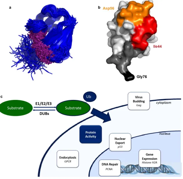
ligases, and DUBs being mutated or misregulated in disease and cancerous states. In many cases, a key determinant of the outcome of monoubiquitination is the presence of proteins or domains that recognize the post-translational modification (5, 52). Ubiquitin has also been shown to adopt distinct conformations depending on its binding partner, which can aid in recognition by distinct regulators of the monoubiquitinated substrate (Figure 1.2a) (53). In some cases, monoubiquitination alters interactions with pre-existing protein binding partners, but in other cases, the modified substrate is recognized by a new protein, often containing a Ubiquitin binding domain (UBD). There are sixteen different types of UBDs that mediate most of the interactions with ubiquitinated
substrates and that can form interactions with multiple surfaces of Ubiquitin (53, 54). Most UBDs interact with a hydrophobic patch on Ubiquitin (Leu8, Ile44, and Val70) (Figure 1.2b) (53).
There are numerous reviews on the well-characterized cellular functions of monoubiquitination, and these outcomes will be only briefly discussed here (52, 55). A summary of the known outcomes of monoubiquitination is shown in Figure 1.2c. Monoubiquitination is clearly involved in three distinct cellular functions: gene and protein expression through histone and transcription regulation, endocytosis, and retroviral budding. Monoubiquitination may also control the activity of the endocytotic machinery (15, 55, 56). Monoubiquitination can act on these systems through
12
13
Monoubiquitination is intricately involved in the regulation of gene expression in the nucleus. This post-translational modification is one of many (including methylation and acetylation) that can modify histones and alter chromatin structure, which directly alters gene expression (17, 59). Furthermore, monoubiquitination regulates gene expression by targeting enzymes involved in DNA repair and transcription. When the DNA processivity factor Proliferating Cell Nuclear Antigen (PCNA) is
monoubiquitinated, it recruits Translesion DNA synthesis (TLS)-specific DNA polymerases, which allow PCNA to bypass a DNA lesion (17, 61-63). In another example, the transcription factor NF-κB, which controls expression of genes involved in
cell growth and immunity (64, 65), is regulated by IB Kinase (IKK), which block NF-κB
inhibitors by marking them for polyubiquitination and degradation (66). For IKK to be activated, it must first be phosphorylated, which also makes it a substrate for
monoubiquitination in chronically activated cells (67, 68). IKK that cannot be monoubiquitinated is resistant to chronic activation (69).
Many of the best characterized substrates of monoubiquitination are involved in membrane protein trafficking and receptor internalization through endocytosis (Figure 1.3) (30, 58, 70). In endocytosis, monoubiquitination is a sorting signal for Receptor Tyrosine Kinases (RTKs), G Protein Coupled Receptors (GPCRs), transporters, and ion channels (17, 18, 58, 71). For example, the yeast GPCR, Ste2, is ubiquitinated after binding to its ligand, pheromone, and monoubiquitination promotes its entry into endocytotic vesicles and its rapid removal from the plasma membrane (15, 72).
14
15
proteins, like Ste2, can in some cases be degraded through the lysosomal system after endocytosis occurs (13). The Ubiquitin moiety fused to a membrane protein carries with it the necessary information for endocytosis facilitated by endocytotic adaptor proteins (73, 74). Ubiquitin binding endocytotic adaptor proteins are also themselves regulated by monoubiquitination (17). One example of this type of regulation is β-arrestin, which binds phospholipids and clathrin and is important for the endocytosis of activated GPCRs. β-arrestin itself is monoubiquitinated after the β2-adrenergic receptor is activated, and this monoubiquitination is required for the rapid internalization of the receptor (75, 76). In fact, monoubiquitination of β-arrestin is sufficient for β2-adrenergic
receptor endocytosis (77).
Monoubiquitination is also important in the process of virus budding. Enveloped viruses exit the cell by budding from the cell membrane, and it is known that reducing cellular levels of Ubiquitin inhibits budding (55). One example of this regulatory process is monoubiquitination of the protein Gag, which is an essential component of
retroviruses. The Gag protein has an embedded sequence, termed a late domain, which is essential for budding. This late domain is known to be an interaction motif for the Ubiquitin ligase Nedd4, leading to ubiquitination when Gag is properly localized to the membrane (78, 79). When Gag cannot be monoubiquitinated, viral budding does not occur (80, 81).
16
example, monoubiquitination has been shown to directly regulate the activity of the DUB ataxin-3, leading to an enhancement of its enzymatic activity (83). On the other hand, monoubiquitination of dihydrofolate reductase, an enzyme involved in DNA synthesis, suppresses its enzymatic activity (84). Finally, monoubiquitination of the tumor
suppressor p53 leads to a conformational change in the protein that exposes a previously buried nuclear export signal (85). Other substrates, like the small GTPase Rac1, have been identified as substrates of monoubiquitination, but the physiological role of the post-translational modification is not yet known (86). It is likely that as our ability to detect and study monoubiquitinated substrates improves, new cellular functions for the modification will be discovered.
One challenge that exists in understanding the role of monoubiquitination in cell regulation is that there is no online resource that consolidates information on all
monoubiquitinated substrates. There are databases that list all currently identified substrates of ubiquitination, but they do not distinguish between polyubiquitination and monoubiquitination. As we have previously discussed, the outcomes of these two types of post-translational modifications are clearly distinct. Mass spectrometry and other large scale approaches are being adapted to generate large databases of monoubiquitinated proteins (87, 88). However, it is important not just to document occurrences of this translational modification, but to understand and categorize the ways this
17 Ubiquitination and Disease
Understanding the diverse mechanisms by which monoubiquitination is used to regulate substrate localization, binding, and activity becomes more important as a role for ubiquitination is emerging in the study of cancer and developmental disorders. For example, sequence preferences surrounding the ubiquitination sites of Rsp5 have been identified, and many known protein mutations that lead to disease alter these potential ubiquitination sites (43). There is evidence that ubiquitination is also involved in
sensing of neuropathic pain and its misregulation in disease (89). Mutations of Ubiquitin, Ubiquitin ligases, and DUBs are all found in human diseases and disorders. For example, many Ubiquitin ligases are proto-oncogenes (90, 91). Receptor Tyrosine Kinases that are not ubiquitinated or lack proper ubiquitination can lead to constitutive receptor signaling and carcinogenesis (92).
There is already some precedence for targeting similar post-translational
modification for disease treatment. Targeting the post-translational phosphorylation for drug development has been successful, and there are currently over 150 drugs in various stages of clinical trials (93). There are many parallels between the phosphorylation and ubiquitination systems suggesting the ubiquitination may prove to be just as important a pathway to target for drug development. For example, the human genome contains more E3 Ubiquitin ligases than protein kinases (93). Furthermore, there is significant interplay between phosphorylation and ubiquitination that is critical for cell regulation. For
18
In fact, there are already drugs that successfully target the ubiquitination system. There are some therapies involving monocolonal antibodies that act by promoting Ubiquitin-dependent receptor degradation (94). However, a detailed knowledge of the mechanisms by which ubiquitination regulates these pathways is required for the design of effective inhibitors (95). Some of the most promising targets in the ubiquitination pathway are the E3 Ubiquitin ligases, which are crucial for ubiquitination and are also the critical point for substrate specificity (96). Currently, there has been some success in finding small molecule inhibitors of E3 substrate interactions, for example the interaction of the E3 MDM2 with its substrate p53 (96). There are also drugs already on the market that block the downstream effects of substrate ubiquitination downstream. For example, Bortezomib is a small molecule inhibitor of the 20S proteasome (97, 98). Bortezomib is used to treat multiple myeloma, likely by limiting cell immortality by blocking the degradation of pro-apoptotic proteins (97, 98). Another promising target for drug development is deubiquitinases; there are known small molecule inhibitors for some deubiquitinases already, but it remains to be seen whether they will become successful new anticancer therapies in the near future (99). In fact, deubiquitinases, which have a clear protein binding pocket and enzymatic activity, may represent the best targets for future drug development studies (99).
19
ubiquitinated substrates, requires knowledge of the structures and interactions that are formed between E3s, Ubiquitin, and substrates. As our understanding of the structural aspects of ubiquitination improves, drug development should prove more fruitful. Success in this regard will require information about protein-protein interactions, the impact of Ubiquitin on substrate structure, and the mechanisms of deubiquitination.
However, despite extensive characterization of the outcomes of ubiquitination in vivo, questions remain about how specific lysines on a substrate are ubiquitinated, how ubiquitination directly affects the structure and properties of the substrate, and how changes to the structure or dynamics of the substrate may contribute to the function of different substrates (100, 101). The answers to these questions are essential to fully understanding the roles ubiquitination plays in proper (normal signaling) and improper (disease and cancer) cellular functions.
New Approaches to Study Ubiquitination
As discussed in the previous section, an advance in our ability to understand and target the process of ubiquitination requires a clearer structural and mechanistic
knowledge of monoubiquitination, including how it may lead to changes in the structure and activity of the substrates that it modifies. While significant effort has been focused on characterizing the outcomes of monoubiquitination in vivo, very little has been done to understand what this modification does to the biochemical and biophysical properties of its substrates. There is, however, precedence for the value of information obtained from asking these types of questions. For example, the charge introduced by another
20
perturbation of the biophysical properties of a protein structure, which can lead to a conformation change that alters activity and protein-protein interactions (52, 102, 103).
There are some recent studies that focus on the structural and biophysical aspects of ubiquitination. Computational modeling of the ubiquitinated substrate Ubc7 suggests that ubiquitination changes the thermodynamic stability of a protein in a site-specific and modification-specific manner (104). Furthermore, Ubc7 was most thermodynamically destabilized by ubiquitination at the known site of polyubiquitination in vivo (104). Studies of the interaction between Ubiquitin and the Ubiquitin binding domains (UBDs) of proteins that recognize monoubiquitinated substrates suggest that different UBDs recognize and stabilize slightly different conformations of Ubiquitin (53). In solution, Ubiquitin is a dynamic molecule, and it is possible that when a substrate is ubiquitinated it stabilizes a conformation of Ubiquitin that is recognized by UBDs. These studies illustrate that knowledge of how structure and dynamics change lead to insight into the mechanism by which a monoubiquitinated substrate is recognized. Other structural studies have shown how Ubiquitin associated (UBA) domains recognize and bind to specific hydrophobic patches on Ubiquitin (105). These biochemical and structural analyses of interactions between Ubiquitin and Ubiquitin-binding proteins have helped develop a mechanistic understanding of the link between the modification, the process that it regulates, and the proteins that recognize the modified substrate (17).
21
methods are highly technical and may not be accessible to a molecular and cellular biologist. This is a problem not only for the study of monoubiquitination, but for other translational modifications as well. There are now over 200 documented post-translational modifications, a number of which involve modifying a substrate with another protein (like Sumoylation) that have the same constraints on the ability to perform mechanistic studies (17, 52, 63).
Use of Chemical Modification to Study Monoubiquitinated Substrates
One particularly promising approach to gaining a mechanistic understanding of regulation by monoubiquitination is through the use of synthetic methods to generate monoubiquitinated substrates. There are three approaches to generate ubiquitinated substrate suitable for study by biophysical methods: non-natural amino acids coupled with organic synthesis, semi-synthesis, and chemical modification that takes advantage of amino acid chemistry. The simplest approach to chemical modification is to form a disulfide bond between Ubiquitin and the substrate (106). The advantages and disadvantages, especially relating to ease of use, of these approaches are discussed in Chapter V of this dissertation.
Currently, there are a few examples of using chemical modification to study monoubiquitinated substrates by biophysical methods. Many of the successful studies of monoubiquitinated substrates have come from the use of either isopeptide bond
22
methylation of histone H3, demonstrating the importance of cross-talk between post-translational modifications. Studies of PCNA were also performed using multiple chemical approaches to modification (109, 110). By solving the crystal structure of monoubiquitinated PCNA, the authors found that monoubiquitination does not change the structure of PCNA itself, suggesting ubiquitination recruits alternative binding partners to PCNA, but that Ubiquitin does display limited conformational flexibility relative to PCNA, constraining the ways in which binding partners can interact with the protein (111-113). Finally, a semi-synthesis approach was also used to study
α-synuclein, a protein central in the development of Parkinson’s disease. Using
monoubiquitinated α-synuclein, the authors directly demonstrated that ubiquitination led to the inhibition of fibril formation (114), which was consistent with previous in vivo studies suggesting that N-terminal monoubiquitination stabilizes the monomeric form of the protein. Furthermore, additional studies of this protein using cysteine mutations showed that different ubiquitination sites had different effects on the formation of fibrils (115).
We are currently at an exciting time in the study of monoubiquitinated substrates. New approaches to study these substrates are available as well as evidence suggesting that monoubiquitination regulates substrates through mechanisms more diverse than the three primary categories described previously. In the future, it will be important to continue to systematically study and characterize the mechanisms by which
23
The recent advances in chemical ubiquitination approaches described above provide the opportunity to study monoubiquitinated substrates. Chemical ubiquitination will be especially important in the cases where the population of monoubiquitinated substrate might be too small to purify efficiently from cells or the Ubiquitin ligase is not known or cannot be reconstituted in vitro.
Regulation of GTPase Signaling by Monoubiquitination
One prominent family of proteins that promises to be particularly interesting for mechanistic studies of signaling regulation by monoubiquitination is GTPases. There is evidence in the literature for diverse mechanisms of GTPase regulation by ubiquitination and there are also a number of instances where monoubiquitination can regulate protein function by mechanisms other than protein localization. GTPases regulate cell signaling pathways, and are enzymes with a well-characterized guanine nucleotide binding and hydrolysis activity. These proteins are also integral in driving many types of cancer and developmental diseases. The remainder of this thesis will focus on the regulation of particular GTPases by monoubiquitination. The implications from these studies for the larger field of monoubiquitination will be considered in Chapter V.
GTPases as Regulators of Signaling Pathways
K-24
Ras, which were first discovered due to their oncogenic potential in retroviruses (119). Ras-like GTPases are divided into five subfamilies: Ras, Rho, Rab, Arf, and Ran (120). Each subfamily has different localization in the cell and different downstream effectors, leading to much of their observed functional specificity. Ras superfamily GTPases regulate a number of pathways, including cell cycle progression and gene expression (Ras), cytoskeletal rearrangement (Rho), nuclear import (Ran) and cellular trafficking (Rab and Arf) (120, 121). Small GTPases also have significant roles in driving cancer and, in some cases, developmental disorders. In particular, Ras is activated in over 30% of all human cancers. Germline mutations of Ras are found in Noonan syndrome, Costello syndrome, and Cardiofacio-cutaneous syndrome (122).
Heterotrimeric Gα proteins are coupled directly to cell-surface receptors and are responsible for receptor-mediated communication between the exterior and interior of the cell (121). There are four classes of heterotrimeric Gαs that are based on their homology
and downstream effectors: Gαs, Gαi/o, Gαq, and Gα12/13 (123). Gαs proteins activate
adenylyl cyclase, while Gαi are known to inhibit adenylyl cyclase and act in opposition to
Gαs (124). Gαi GTPases are also coupled to taste and odor receptors, and facilitate vision
through phototransduction. Gαq proteins activate phosphoinositide-specific
phospholipase C isozymes. This leads to the generation of the second messenger signals inositol 1,4,5-trisphosphate and diacylglycerol (124). Finally, Gα12/13 proteins regulate
25
26
The primary mechanism by which GTPases signal is by switching between GDP- and GTP-bound states, which results in conformational changes that allow interactions with downstream effectors when the GTPase is in the GTP-bound state (127-133). Small GTPases bind nucleotide with a Kd in the picomolar to nanomolar range. While there are variations in their c-terminal targeting sequences, the core Ras domain is highly
conserved (119). The Ras domain consists of an α/β Rossman fold of about 20 KDa and contains the basic function of guanine nucleotide binding and hydrolysis (118). There are four regions in the Ras domain that are directly involved in guanine nucleotide binding (Figure 1.4c) (119). The NKXD motif forms interactions with the nucleotide base and is crucial for nucleotide binding affinity. The other most important interaction for
nucleotide binding is the GXXXXGKS motif, which forms interactions with the α, β, and
γ phosphate of GDP and GTP and provides a serine or threonine for coordination with the
27
key residues (G60 and T35 on Ras) and GTP (118). Downstream effectors recognize and bind preferentially to the GTP-bound state of the GTPases through the switch regions.
On their own, GTPases are not very good enzymes; their enzymatic activity does not occur on a timescale fast enough to allow them to respond appropriately to
extracellular signals. Therefore, signaling is regulated both by Guanine Nucleotide Exchange Factors (GEFs) and GTPase Activating Proteins (GAPs) (Figure 1.5). GEFs facilitate GDP release by stabilizing the nucleotide-free state of the GTPase (118). The reaction is driven in the forward direction by the presence of excess GTP over GDP in the cell (136). The GEFs interact with the switch regions of the GTPase and residues close to the p-loop and magnesium binding region, leading to structural changes in the GTPases that do not favor binding to phosphates and magnesium. The release of magnesium and additional structural disturbances in the p-loop region account for the increased rate of GDP release in the presence of the GEF (118, 137). The mechanism of GAP-mediated hydrolysis depends on a conserved glutamine residue located near the γ
28
29
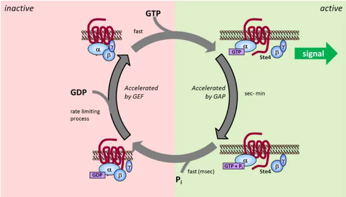
While in many ways the activity of heterotrimeric Gα proteins is similar to Ras-like GTPases, there are some differences (Figure 1.6). Nucleotide binding still occurs primarily within the Ras-like domain, but recent data have demonstrated that the
α-helical domain also contributes to the activity of Gα proteins. Evidence for α-α-helical
domain involvement includes the observation of a major displacement of the α-helical
domain during receptor activation (140). Furthermore, recent structural analysis has also shown that the α-helical domain is highly flexible in the absence of nucleotide,
suggesting that this domain undergoes a nucleotide-dependent transition to a stabilized state (141). Gα proteins, along with the canonical switch I and switch II regions, also have an extra structural element called switch III (116, 118). The switch I region also serves as one of the two connections between the Ras-like and α-helical domains (142).
Conformational changes occur in all three switch regions upon changes in the nucleotide binding state, similar to the mechanism of small GTPases (143). However, the
mechanism for hydrolysis is slightly different. The amino acid sequence of the Gα protein already contains the arginine finger which is provided by the GAPs for small GTPases, leading to faster observed rates of intrinsic hydrolysis (almost 100 times faster than small GTPases) (144). Heterotrimeric G proteins are regulated by GAPs, commonly referred to as regulators of G protein signaling (RGS) proteins (145). While RGS proteins do not contain an arginine finger, they do stabilize the active conformation of the
30
coupled to Gβγ when the Gα is GDP-bound. Upon GTP binding, the βγ interaction is released, allowing signaling through both Gα and Gβγ to occur.
GTPases as Substrates of Monoubiquitination
GTPases are known to undergo a number of post-translational modifications that are crucial for their localization and proper signaling. Some examples of these include phosphorylation (147), myristoylation (148), prenylation, and palmitoylation (147). Ubiquitination is also important for the regulation of many GTPases, not only to control total substrate levels but to target and regulate these proteins in a temporal and spatial manner (149, 150). Two isoforms of Ras, H-Ras and K-Ras, are substrates of
monoubiquitination, but monoubiquitination has distinct outcomes. Monoubiquitination of H-Ras in CHOK-1 cells is necessary to stabilize its association with the endosome and allow signaling to occur (151, 152). Monoubiquitination of K-Ras in HEK293T cells activates K-Ras and contributes to Ras-mediated tumorigenesis (153). Ubiquitination of Rap1B, another Ras-like GTPase, induces relocalization of the protein from the plasma membrane to a subcellular compartment, which is required for the establishment of neuronal polarity (154). It was also shown that Rap2A is monoubiquitinated by Nedd4, which acts as a positive regulator of dendrite development (155). In this case,
31
32
The small GTPases RalA and RalB are also substrates of
monoubiquitination(156). RalA is important for anchorage-independent proliferation as well as tumor growth while RalB contributes to cancer cell survival. In this case, activation of RalA does not affect the ubiquitination state of the protein, however, monoubiquitinated RalA was significantly enriched in the GTP-bound state (156). RalA ubiquitination increases when a cell is detached from its substrate (156). No single lysine of the 21 lysines within RalA appear key for ubiquitination based on systematic single and multiple mutations of the lysines to arginines, suggesting RalA can be ubiquitinated on multiple lysines (156). Monoubiquitination selectively modulates RalA and RalB localization, which is critical for their differences in the roles they play in cell signaling regulation. Finally, Rac1 monoubiquitination has been observed to occur at a single lysine, K147 (86, 157, 158) that lies within an insert region that is conserved in the Rho family of small GTPases. Rac1 is also polyubiquitinated, but when Cav1, which
regulates Rac1 polyubiquitination, is lost, a monoubiquitinated species appears. The data suggests that monoubiquitination may have a distinct role to play in Rac1 regulation, but this mechanism has not yet been pursued (86).
While there are multiple examples of monoubiquitination of small GTPases, less is known about how monoubiquitination regulates Gα proteins. A number of Gα proteins are known to be regulated by polyubiquitination and degradation, including Gαo (159), Gαi3 (160), Gαi2 (161), andGαs (162). However, currently, the only Gα currently known
33
pathways in more complex systems, suggesting that as detection techniques are refined, other examples of Gα monoubiquitination may come to light.
This brief summary focuses primarily on monoubiquitination of the GTPases themselves, when in fact regulators of GTPase signaling are also known to be
monoubiquitinated (163, 164). There are many examples of monoubiquitination and subsequent endocytosis of GPCRs being critical in a number of pathways (16, 58, 70, 77). Targeting the ubiquitination or deubiquitination of these GTPases and their regulators may represent an exciting new possibility for drug development, but first requires that the mechanisms of regulation by monoubiquitination be clearly understood (163).
Thesis Summary
GTPases undergo multiple forms of ubiquitination that lead to a variety of different outcomes. G proteins should serve as an excellent model system to study the effect of ubiquitination on substrate structure, dynamics, and thermodynamic stability. GTPases are of interest particularly because of the key role they play in cell signaling, which makes them good candidates for drug targeting. However, while we have
34
post-translational modification may act. In this thesis, we have employed biochemical and biophysical methods to understand the molecular basis through which GTPases can be regulated by monoubiquitination.
To be able to perform our studies of monoubiquitinated substrates in vitro, we first had to optimize a method to generate enough monoubiquitinated substrate. As described previously, chemical ubiquitination was the most promising approach to suit our needs. We used a simplified and optimized version of a disulfide chemistry
ubiquitination approach present in the literature. Our new approach gave us the ability to drive modification of our substrate to completion, as seen in Chapters II and III. One limitation of this type of approach is that, until recently, it has not been applied to studying other ubiquitinated substrates, in part because it was not known if the differences in the linkage type would alter the behavior of the monoubiquitinated
substrate. As discussed in Chapter II, we employed computational modeling to show that chemically monoubiquitinated protein accurately mimics natively ubiquitinated protein.
35
monoubiquitination at a different lysine than K-Ras results in protein activation through a distinct mechanism.
Finally, in Chapter IV, we turn our attention to a heterotrimeric GTPase, the yeast Gα Gpa1. Gpa1 is an interesting protein because it is both monoubiquitinated and
polyubiquitinated at the same site. There are very few known substrates where this occurs, but they include PCNA (63), a processivity factor, and p53, a well-known tumor suppressor (165). Structural studies of a substrate like this will likely offer insights into structural determinants and outcomes of monoubiquitination versus polyubiquitination. Choosing a yeast protein afforded us the ability to couple our biochemical and
biophysical approach to a system where we can also perform genetic studies. Gpa1 was a challenging substrate to study, and much of our effort was focused on optimizing
methods to obtain pure, stable substrate. While we have not completed the studies of ubiquitinated Gpa1, we have gained insight into the ways in which this protein has evolved to allow it be targeted for monoubiquitination
CHAPTER II
SITE-SPECIFIC MONOUBIQUITINATION ACTIVATES RAS BY IMPEDING GTPASE ACTIVATING PROTEIN FUNCTION1,2
Cell growth and differentiation are controlled by growth factor receptors coupled to the GTPase Ras. Oncogenic mutations disrupt GTPase activity leading to persistent Ras signaling and cancer progression. Recent evidence indicates that monoubiquitination of Ras leads to Ras activation. Mutation of the primary site of monoubiquitination impairs the ability of activated K–Ras to promote tumor growth. To determine the mechanism of human Ras activation we chemically ubiquitinated the protein and analyzed its function by NMR, computational modeling, and biochemical activity measurements. We established that monoubiquitination has little effect on Ras GTP binding, GTP hydrolysis, or exchange factor activation, but severely abrogates the response to GTPase activating proteins in a site–specific manner. These findings reveal a new mechanism by which Ras can trigger persistent signaling in the absence of receptor activation or an oncogenic mutation.
1
Elements of the work referenced in this chapter have been published in:
Baker, R., Lewis, S. M., Sasaki, A. T., Wilkerson, E. M., Locasale, J. W., Cantley, L. C., Kuhlman, B., Dohlman, H. G., and Campbell, S. L. (2013) Site-Specific Monoubiquitination Activates Ras by Impeding GTPase-Activating Protein Function, Nature Strucutural and Molecular Biology 20, 46-52.
2Figures contributed by:
Rachael A. Baker: 2.1, 2.3, 2.4, 2.6, 2.7, 2.8, 2.9a-b, 2.9d-f, 2.10, 2.12, 2.13b-d, 2.14a Steven M. Lewis: 2.5, 2.9c, 2.13a, 2.15
37 Introduction
Ras plays a central role in cell growth, differentiation, and apoptosis and is a member of a large superfamily of guanine nucleotide binding proteins whose activity is regulated by cycling between inactive GDP–bound and active GTP–bound states (166). Conformational changes associated with the GDP– and GTP–bound states are localized primarily to two regions, Switch I (residues 30–37) and Switch II (60–76), and these conformational changes direct specific interactions with regulators and effectors (167, 168). Ras effectors recognize the GTP–bound state of Ras with higher affinity than the GDP–bound state, and these effectors serve to initiate downstream signaling events. Ras has weak intrinsic GTPase activity, but it does not act alone (138). The guanine
nucleotide state of Ras is regulated by two distinct types of protein modulators, which act in opposition to one another. Guanine nucleotide exchange factors (GEFs) facilitate exchange of GDP with GTP to promote Ras activation (169) whereas GTPase–activating proteins (GAPs) stimulate the hydrolysis of GTP and Ras deactivation (121). Ras is the most prevalent oncogene found in human cancer; about 30% of human tumors contain an activating Ras mutation (170, 171). Most commonly, transforming Ras mutations
decrease the sensitivity of the protein to GAP–mediated regulation (172).
38
Ras at position 147 has been shown to promote tumorigenesis (153); mutation of oncogenic K–Ras to prevent monoubiquitination (RasK147L) impaired its ability to
promote tumor growth when ectopically expressed in NIH 3T3 mouse fibroblasts. These findings suggest that Ras activity and signaling are modulated by monoubiquitination, in the manner of an oncogenic mutation or receptor stimulus. Left unresolved is the
mechanism by which monoubiquitination leads to activation of Ras.
Here, we set out to identify the molecular mechanism through which Ras activity is regulated by monoubiquitination. We first developed a method to chemically
ubiquitinate Ras using conditions that drove post-translational modification to completion. Furthermore, we used computational modeling to validate the chemical ubiquitination approach. Using our system, we show that monoubiquitination at position 147 does not alter the intrinsic biochemical properties of Ras, but severely disrupts regulation of Ras by GAPs. This effect is specific to monoubiquitination at position 147 and is not observed when Ras is monoubiquitinated at other adjacent lysines. The loss of GAP–mediated hydrolysis accounts for the accumulation of Ras–GTP in vivo. Thus monoubiquitination reversibly renders the protein resistant to GAP–mediated regulation.
Results
Monoubiquitination of Ras
39
ubiquitinating enzymes employed multiple methods of direct chemical ligation to
generate the protein–Ubiquitin linkage (109, 110, 174-177). In our approach, we replaced the native Ubiquitin linkage with a disulfide bond between a substituted cysteine at position 147 of Ras (RasK147C) and a cysteine at the carboxyl–terminus (c–terminus) of Ubiquitin (UbiquitinG76C). A surface accessible cysteine (Cys118) in Ras was replaced with serine to avoid unwanted modification (RasC118S, hereafter “Ras”). We previously showed that the C118S mutation did not alter Ras structure or biochemical properties (178). The chemical ligation method does not require complicated intermediate chemical or enzymatic steps but instead provides a simple, specific approach to ubiquitination. The disulfide ligation strategy, using a more complicated cysteamine intermediate, was validated in previous studies of Proliferating Cell Nuclear Antigen (PCNA), where it was shown that chemically and enzymatically monoubiquitinated PCNA exhibit identical catalytic properties (109). As seen in Figure 2.1a, we drove Ubiquitin modification of Ras at position 147 to completion by the addition of a ten–fold excess of UbiquitinG76C at pH 8.0. We conducted our experiments using H–Ras (1–166), for which the biochemical and structural (NMR and X–Ray) properties are best established, but corroborated the results using K–Ras (1–166) as indicated. All three mammalian isoforms H–, K–, and N– Ras show similar biochemical properties in the absence of the hypervariable c–terminus (128, 179). Furthermore, we used immunoprecipitation assays to show that, in the absence of c–terminal modification, monoubiquitination still leads to an increase in the GTP–bound population of H–Ras or K–Ras in HEK293T cells (Figure 2.2).
40
end, we used NMR in the presence of free unlabeled Ubiquitin to determine whether Ubiquitin altered spectral features associated with Ras backbone amides. A 1H–15N 2D HSQC overlay of 15N–enriched H–RasK147C in the absence and presence of Ubiquitin is shown in Figure 2.1b. The assignments for H–Ras (1–166) were previously determined (181) and we verified the shifted backbone amide resonances of H–RasK147C using 3D HNCACB data (Figure 2.3). Comparison of the position and intensity of the backbone amide resonances indicates that Ras is not altered by the presence of free Ubiquitin. In support of these observations, as shown in Figure 2.4a–b, we found that the intrinsic rate of GDP dissociation and GTP hydrolysis were unaffected by the presence of Ubiquitin.
Furthermore, the presence of Ubiquitin dimers in solutions also had no effect on measuring thermal stability, intrinsic GDP dissociation, or GTP hydrolysis, as shown in Figure 2.4d-f. These results indicated that Ras did not specifically interact with
Ubiquitin. Therefore, for subsequent analyses, we did not separate monoubiquitinated Ras (mUbRas) from free Ubiquitin.
Monoubiquitinated Ras Retains Intrinsic GTPase Activity
41
39.2 ± 0.3 oC, and 39.6 ± 0.2 oC for Ras, RasK147A, and mUbRas, respectively), a change that is not likely to have a substantial effect on this otherwise highly stable protein in vivo. These data suggest that, despite the size of Ubiquitin, monoubiquitination at position 147 does not lead to thermal destabilization of Ras.
While monoubiquitination of Ras does not substantially affect thermal stability, it could alter intrinsic activity. For example, it is possible that ubiquitination impairs guanine nucleotide binding, similar to mutations at the adjacent residue, Ala146 (183, 184). To measure rates of nucleotide dissociation, we equilibrated Ras and mUbRas with the fluorescent analog N–methylanthraniloyl (MANT)–GDP and measured fluorescence over time in the presence of excess unlabeled GDP. We observed a slight increase (2–3 fold) in the intrinsic rate of nucleotide dissociation for RasK147C as compared to native Ras, while the rate for mUbRas was unaltered (Figure 2.4a). This result suggests that ubiquitination of Ras does not have the same impact on nucleotide binding as a point mutation at the same residue.
42
43
44
45
Figure 2.4. Monoubiquitinated Ras Retains Intrinsic Stability and Activity. (a) Intrinsic nucleotide dissociation rates for Ras, RasK147C, and mUbRas loaded with MANT–GDP. Dissociation was monitored following the addition of unlabeled GDP by the decrease in fluorescence emission over time. Data were fit to an exponential dissociation curve, and the results are the mean ± s.d. (n=4). (b) Intrinsic single–turnover GTP hydrolysis for Ras, RasK147C, and mUbRas. Hydrolysis was initiated by the addition of Mg2+ and monitored by the change in fluorescence of Flippi when bound to free phosphate. Data were converted to a phosphate concentration using a standard curve. The concentration of phosphate equal to 100% GTP hydrolyzed was determined in the presence of GAP. Results are the mean ± s.d. (n=6). (c) Thermal stability of Ras, RasK147A, and mUbRas measured by ABD–F incorporation as a function of temperature. The data were
46
Chemical Ubiquitination Mimics Native Ubiquitination
We found that monoubiquitination does not alter the intrinsic biochemical activity of Ras, even though mUbRas accumulates in the GTP–bound state in vivo. Previous studies have shown that chemically ubiquitinated PCNA functions similarly to the enzymatically ubiquitinated protein (109). To further establish that chemical
ubiquitination of Ras is a good mimic of native ubiquitination, we built computational models of Ubiquitin ligated to Ras. To create the model, we used a recently developed module of the Rosetta protein modeling software suite (186, 187) that samples the conformational space available to ligated proteins. We modified Rosetta to consider disulfide and native isopeptide ubiquitination linkages and generated model structures of mUbRas using both linkages. We generated these models without the use of
experimentally–derived constraints.
47
Monoubiquitination Affects the Switch Regions of Ras
Results obtained from computational modeling suggest that there is no single preferred interaction between Ubiquitin and Ras. To test this prediction experimentally, we used NMR to examine spectral differences between Ubiquitin and Ras upon
monoubiquitination. First, we 15N–enriched UbiquitinG76C and examined the 1H–15N 2D HSQC spectrum of Ubiquitin when ligated to RasK147C. By this method we observed partial to complete resonance broadening of eleven backbone amides, but no substantial chemical shifts within Ubiquitin (Figure 2.6a). By mapping these spectral changes onto the structure of Ubiquitin in Figure 2.6b, it is evident that one face of Ubiquitin is primarily altered upon ligation with Ras. A possible explanation for the inability to detect a subset of Ubiquitin amide resonances is that Ras ligation restricts conformational sampling, leading to exchange broadening.
48
49
50
51
The observed broadening in the GDP–bound state of mUbRas suggests a change in backbone dynamics, possibly due to conformational exchange or dynamic sampling of the switch regions. This hypothesis is supported by the observation that when an HSQC of mUbRas is collected on a 500 MHz spectrometer, some of the broadened residues in the switch regions and p-loop become visible, as seen in Figure 2.8. This suggests that the switch regions are beginning to shift from intermediate exchange back to fast
exchange on the NMR timescale. Since Ras regulators and effectors interact through the switch regions, monoubiquitination could alter the population of active Ras by changing how mUbRas interacts with regulators.
Monoubiquitination of Ras Inhibits GAP–Mediated Hydrolysis
In cells, the nucleotide–bound state of Ras is regulated both by GEFs, which increase the rate of GDP dissociation, and GAPs, which enhance the rate of GTP
hydrolysis. Our NMR data suggest that monoubiquitination affects the switch regions of Ras, which in turn could alter interactions with GEFs and GAPs. Thus the increased GTP–bound population of mUbRas in vivo could be caused by either an increased sensitivity to GEFs or decreased sensitivity to GAPs.
52
However, we observed a decrease in the rate of GEF–mediated nucleotide dissociation for mUbRas compared to unmodified Ras (Figure 2.9a).
We next considered the effect of Ras monoubiquitination on GAP–mediated hydrolysis. To this end we compared the rate of GTP hydrolysis for Ras and mUbRas in the presence of the catalytic domains of two GAPs, NF1 (NF1333) and p120GAP(GAP– 334)(138, 191). At a GAP–to–Ras ratio of 1:500, we observed an order of magnitude increase in the rate of GTP hydrolysis for unmodified Ras relative to the intrinsic rate of GTP hydrolysis. No increase in the rate of GTP hydrolysis was observed for mUbRas in the presence of the same GAP–to–Ras ratio (Figure 2.9b). Therefore, mUbRas is
insensitive to GAP–mediated regulation, similar to an oncogenic RasG12V mutation (172). We obtained similar results using K–Ras (Figure 2.10), indicating that the effects of monoubiquitination on Ras are not isoform–specific when the proteins are modified at the same lysine.
To validate the use of an in vitro system to dissect the mechanism of Ras regulation, we measured the sensitivity of mUbRas to GAP–mediated hydrolysis in a cellular reconstitution system. We immunoprecipitated Ras from HEK293T cells and compared the sensitivity of the monoubiquitinated and unmodified fractions of Ras to regulation by GAP. As seen in Figure 2.9c, monoubiquitinated K–Ras is less sensitive than the unmodified protein to GAP–mediated GTP hydrolysis. These data support our in vitro findings that monoubiquitination increases the population of active, GTP–bound Ras through a defect in sensitivity to GAP–mediated regulation.
53
the interaction between Ras and Soscat (Figure 2.9d). Results from these analyses indicated that the binding affinity between mUbRas and Soscat is 8.3 ± 0.9 μM, which is half the observed binding affinity between Ras and Soscat (4.2 ± 0.4 μM), consistent with the small reduction in the rate of GDP dissociation observed. However, a decrease in the rate of GDP dissociation would favor the GDP–bound state of Ras. Thus, the minor differences in GEF binding do not account for the accumulation of Ras–GTP in vivo.
To determine whether ubiquitination also leads to a reduction in GAP binding affinity, we compared the ability of Ras and mUbRas to bind to NF1333 in the presence of AlF4–. As seen in Figure 2.9e, in the presence of AlF4– almost 100% of NF1333 bound to Ras, which was present in slight excess. In contrast, about 50% of the NF1333 bound to mUbRas under the same conditions (Figure 2.9f), which suggests that the binding affinity between GAP and mUbRas is reduced relative to unmodified Ras.
54
Figure 2.9. Monoubiquitination Decreases the Sensitivity of Ras to Downregulation by GAPs. (a) Nucleotide dissociation
reaction for Ras, RasK147C, and mUbRas loaded with MANT–GDP in the presence of a 1:1 molar ratio of Ras to Soscat. Data were fit to an exponential dissociation curve, and the results are the mean ± s.d. (n=4). (b) Single–turnover GTP hydrolysis for
Ras, RasK147C, mUbRas, and RasG12V in the presence of NF1333 or GAP–334 at a molar ratio of 1:500 GAP:Ras. Results are the mean ± s.d. (n=6). (c) Immunoblotting of GTP-bound Ras and GTP-bound mUbRas in cell extract in the presence of
increasing concentrations of RasGAP. Anti–Flag and anti–HA antibodies reveal the relative fraction of total Ras and mUbRas, respectively. (d) Titration of Ras with Soscat. Experiments were performed as described panel a, except the concentration of
Soscat was varied while Ras was held constant at 0.2 μM. Data plotted as a function of the Soscat concentration. Results are the mean ± s.d. (n=3). (e) Gel filtration of Ras and NF1333 in the absence (dotted line) and presence (solid line) of AlF4–. (f) Gel
filtration of mUbRas and NF1333 in the absence (dotted line) and presence (solid line) of AlF 4–.
56
Figure 2.10. GAP-Mediated Hydrolysis of Monoubiquitinated K-Ras. Intrinsic and GAP-mediated single-turnover GTP hydrolysis of Ras in the absence and presence of GAP-334 (intrinsic, 1:500 GAP:Ras, and 1:200 GAP:Ras). Rates of GTP hydrolysis were measured for K-Ras, K-Ras with free Ubiquitin (K-Ras+Ub), and