Transcriptional regulatory control of
mammalian nephron progenitors revealed by
multi-factor cistromic analysis and genetic
studies
Lori L. O’Brien1☯¤a, Qiuyu Guo1,2☯, Emad Bahrami-Samani3, Joo-Seop Park4, Sean M. Hasso1¤b, Young-Jin Lee1¤c, Alan Fang1, Albert D. Kim1, Jinjin Guo1, Trudy M. Hong5, Kevin A. Peterson6, Scott Lozanoff5, Ramya Raviram7, Bing Ren7, Ben Fogelgren5, Andrew D. Smith3, Anton Valouev2, Andrew P. McMahon1
*
1 Department of Stem Cell Biology and Regenerative Medicine, Broad-CIRM Center, Keck School of Medicine, University of Southern California, Los Angeles, California, United States of America, 2 Department of Preventative Medicine, Division of Bioinformatics, Keck School of Medicine, University of Southern California, Los Angeles, California, United States of America, 3 Department of Molecular and Computational Biology, University of Southern California, Los Angeles, California, United States of America, 4 Division of Pediatric Urology and Division of Developmental Biology, Cincinnati Children’s Hospital Medical Center, Cincinnati, Ohio, United States of America, 5 Department of Anatomy, Biochemistry, and Physiology, University of Hawaii at Manoa, Honolulu, Hawaii, United States of America, 6 The Jackson Laboratory, Bar Harbor, Maine, United States of America, 7 Ludwig Institute for Cancer Research, Department of Cellular and Molecular Medicine, Institute of Genomic Medicine, Moores Cancer Center, University of California San Diego La Jolla, California, United States of America
☯These authors contributed equally to this work.
¤a Current address: Department of Cell Biology and Physiology, University of North Carolina at Chapel Hill, Chapel Hill, North Carolina, United States of America
¤b Current address: Cancer Genetics Incorporated, Morrisville, North Carolina, United States of America ¤c Current address: iDream Research Center, Mizmedi Women’s Hospital, Seoul, Republic of Korea
Abstract
Nephron progenitor number determines nephron endowment; a reduced nephron count is linked to the onset of kidney disease. Several transcriptional regulators including Six2, Wt1, Osr1, Sall1, Eya1, Pax2, and Hox11 paralogues are required for specification and/or main-tenance of nephron progenitors. However, little is known about the regulatory intersection of these players. Here, we have mapped nephron progenitor-specific transcriptional networks of Six2, Hoxd11, Osr1, and Wt1. We identified 373 multi-factor associated ‘regulatory hot-spots’ around genes closely associated with progenitor programs. To examine their func-tional significance, we deleted ‘hotspot’ enhancer elements for Six2 and Wnt4. Removal of the distal enhancer for Six2 leads to a ~40% reduction in Six2 expression. When combined with a Six2 null allele, progeny display a premature depletion of nephron progenitors. Loss of the Wnt4 enhancer led to a significant reduction of Wnt4 expression in renal vesicles and a mildly hypoplastic kidney, a phenotype also enhanced in combination with a Wnt4 null mutation. To explore the regulatory landscape that supports proper target gene expression, we performed CTCF ChIP-seq to identify insulator-boundary regions. One such putative boundary lies between the Six2 and Six3 loci. Evidence for the functional significance of this a1111111111 a1111111111 a1111111111 a1111111111 a1111111111 OPEN ACCESS
Citation: O’Brien LL, Guo Q, Bahrami-Samani E,
Park J-S, Hasso SM, Lee Y-J, et al. (2018) Transcriptional regulatory control of mammalian nephron progenitors revealed by multi-factor cistromic analysis and genetic studies. PLoS Genet 14(1): e1007181.https://doi.org/10.1371/journal. pgen.1007181
Editor: David R. Beier, Seattle Children’s Research
Institute, UNITED STATES
Received: January 18, 2017
Accepted: January 1, 2018
Published: January 29, 2018
Copyright:©2018 O’Brien et al. This is an open access article distributed under the terms of the Creative Commons Attribution License, which permits unrestricted use, distribution, and reproduction in any medium, provided the original author and source are credited.
Data Availability Statement: All relevant data are
within the paper and its Supporting Information files except sequencing data. All sequencing files are available from the GEO database, accession number GSE90017.
Funding: This work was supported by grants from
boundary was obtained by deep sequencing of the radiation-induced Brachyrrhine (Br) mutant allele. We identified an inversion of the Six2/Six3 locus around the CTCF-bound boundary, removing Six2 from its distal enhancer regulation, but placed next to Six3
enhancer elements which support ectopic Six2 expression in the lens where Six3 is normally expressed. Six3 is now predicted to fall under control of the Six2 distal enhancer. Consistent with this view, we observed ectopic Six3 in nephron progenitors. 4C-seq supports the model for Six2 distal enhancer interactions in wild-type and Br/+ mouse kidneys. Together, these data expand our view of the regulatory genome and regulatory landscape underpinning mammalian nephrogenesis.
Author summary
Nephrons, the filtering units of the kidney, derive from nephron progenitors. Deficiencies in nephron number increases the risk of kidney disease. An understanding of the regula-tory programs governing progenitor actions has important translational potential. Several transcription factors regulate the nephron progenitor population. However, their target interactions are largely unknown. Here, we mapped and intersected the genome-wide binding sites for four such factors in mouse nephron progenitor cells in the developing kidney: Six2, Hoxd11, Osr1, and Wt1. The intersectional data highlight a high-value set of putative enhancer elements linked to genes regulating nephron progenitor properties. We
validate the function of two such enhancer elements regulating the levels ofSix2, a key
transcriptional regulatory factor in nephron progenitor maintenance, andWnt4, a critical
signaling factor controlling the mesenchyme to epithelial transition of induced nephron
progenitors. Further characterization of theSix2regulatory landscape identified higher
order regulatory interactions that ensure appropriate enhancer-promoter specificity.
CTCF-bound sites betweenSix2and the adjacentSix3locus likely act as boundary
ele-ments to define topological interactions domains separating enhancer eleele-ments thereby providing distinct tissue specificity to each gene’s expression. An inversion of this region
in theBrachyrrhine(Br) mutant mouse reversesSix2andSix3expression domains, placing
Six3under control of theSix2enhancer element above resulting in kidney-specific
expres-sion, whileSix2expression shifts to the lens, a normal expression domain forSix3.
Together, these data expand our view of the regulatory genome and regulatory landscape underpinning mammalian nephrogenesis.
Introduction
The mammalian metanephric kidney maintains fluid homeostasis. The number of individuals afflicted with kidney disease is on the rise, and reduced nephron number has been associated with disease outcome [1]. In the mouse, genetic studies have demonstrated that nephrons are generated from a Six2+ progenitor pool in a regulatory process requiring the transcriptional action of Six2 for progenitor maintenance [2]. Human SIX2 shows an expression and activity similar to its murine counterpart suggesting that mouse Six2 and human SIX2 likely have
simi-lar functions [3]. Consistent with this view, human mutations inSIX2are associated with renal
hypodysplasia and the malignant transformation of progenitor cells in Wilms’ tumor, a pediat-ric nephroblastoma [4–6]. There is an increasing interest in the relationship between nephron
progenitors, their output, and congenital and acquired kidney disease [1,7]. Further, new
a March of Dimes (http://www.marchofdimes.org/) grant to BF (#5-FY14-56), a Broad Postdoctoral Fellowship from the University of Southern California to LLO, and a graduate student fellowship from the California Institute for Regenerative Medicine to QG. The funders had no role in study design, data collection and analysis, decision to publish, or preparation of the manuscript.
Competing interests: The authors have declared
approaches to modulate nephron progenitor outputs to generate kidney structuresin vitrocall
for a better understanding of regulatory processes at playin vivo[8–10]. Nephron progenitor
specification and nephron progenitor maintenance are dependent on a number of additional transcriptional regulatory factors including Hoxa/c/d11, Osr1, Wt1, Sall1, Eya1, Pax2, and Six1. Previous studies of mouse mutants in these genes suggest complex hierarchical interac-tions amongst these factors [11–27]. Identification of their genomic targets and target regula-tory mechanisms are essential to determine the nephrogenic regularegula-tory network.
Direct nephron progenitor ChIP-seq studies have identified a broad range of potential tran-scriptional targets of Six2/SIX2 action in the mouse and human kidney, respectively, and
veri-fied predicted enhancer modules for several of these targets [3,28,29]. Six2 interacts at
cis-regulatory modules of genes expressed both in the nephron progenitors and their committed nephron-forming descendants through enhancers co-engaged by differentiation-inducing
transcriptional complexes formed in response to canonical Wnt signaling [28,29].
Interest-ingly, a potential role for Hox11 paralogs within Six2-predicted cis-regulatory modules is sug-gested by the strong enrichment of AT-rich homeobox motifs in Six2 ChIP-seq peaks [28]. The genomic targets of Wt1 have also been analyzed by ChIP experiments of embryonic mouse kidneys [30–32]. Though the approach was not specific to nephron progenitors, these studies revealed the interplay with many genes expressed in, and critical for, nephron progeni-tors, including Fgf and Bmp family members [30–32]. Sall1 ChIP-seq has also shed light on its active roles in nephron progenitors and repressive actions on development of nascent neph-rons, respectively [29]. Interestingly, a subset of Six2- and Sall1-bound regions overlap suggest-ing these factors co-associate and target analysis predicts genes regulatsuggest-ing the nephron progenitor population [29].
With a working model that multi-factor binding will highlight key regulatory nodes of the nephron progenitor pathway [33–37], we utilized ChIP-seq analysis to identify a subset of putative regulatory elements associated with multiple transcription factors.
gRNA/Cas9-me-diated ablation of ‘regulatory hotspots’ adjacent toSix2andWnt4highlight the significance of
these enhancer elements in regulating target gene expression. Additional analyses of the
regu-latory landscape surroundingSix2identified insulator-bound elements which constrain
enhancer function. In support of this finding, deep sequencing of theBrmutant mouse
identi-fied an inversion ofSix2andSix3loci altering enhancer specificity. These studies highlight the
critical role of multi-factor input and proper enhancer context for directing appropriate target gene expression.
Results
Identification of nephron progenitor-specific transcription factor
interaction sites using a novel transgenic strategy
To extend our understanding of the transcriptional regulatory networks operating within mouse nephron progenitors, we developed a transgenic approach to overcome the limited availability and inconsistency of working antibodies for key transcriptional components, and complications that arise from the diverse expression of regulatory factors elsewhere in the kid-ney. In this transgenic strategy, an epitope-tagged transcription factor-of-interest is expressed
exclusively within the nephron progenitor compartment using aSix2distal enhancer (DE)
pre-viously shown to recapitulateSix2-like, nephron progenitor restricted expression (Fig 1A
complexes[38–41]. Each transcription factor-of-interest is appended with a BioTag-FLAG (BF) epitope at the C-terminus of the target protein. Co-production of an EGFP-BirA enzyme on the transgene through an IRES element also allows both ready visualization of transgenic kidneys and biotinylation of the biotin-recognition motif (BioTag) enabling an additional
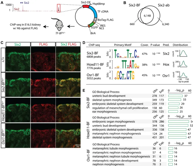
Fig 1. Identification of Six2, Hoxd11 and Osr1 binding sites in nephron progenitors by ChIP-seq. (A) Schematics shows characteristics of the transgenic mice used to generate nephron progenitor-specific ChIP-seq data: the Six2 distal enhancer (Six2-DE) drives nephron progenitor-specific expression of a FLAG-tagged transcription factor in the embryonic kidney. The IRES allows co-expression of GFP-BirA. Transgenic founders are utilized for FLAG ChIP-seq to identify progenitor specific programs. (B) Venn diagram shows overlap of peaks from FLAG ChIP-seq generated fromSix2-BFtg/+(Six2-BioTagFLAG) mouse kidneys and from the Six2 antibody (Six2-ab) ChIP-seq. (C) Immunostain for Six2 and FLAG at E16.5 shows the overlap of the two proteins for each transgenic line. (D) Motifs identified from Six2-BF, Hoxd11-BF, and Osr1-BF peaks with MEME (Multiple EM for Motif Elicitation; +/- 50 bp window). Coverage and p-values were calculated with FIMO (Find Individual Motif Occurrences) results. Smoothened histogram indicates distribution of motif-peak distance. Predicted
TF = predicted transcription factor binding the discovered motif. (E) Functional annotation of Six2-BF, Hoxd11-BF, and Osr1-BF peaks performed using GREAT (Genomic Regions Enrichment of Annotations Tool). The top over-represented gene ontology terms belonging to the two categories are shown. ‘Obs.’, number of peaks associated with genes annotated with corresponding term; ‘Exp.’, number of peaks expected to be associated with genes annotated with corresponding term by chance. The barplot indicates the binomial p-values measuring over-representation of the corresponding terms.
mode of isolation of factor-associated DNA or protein complexes through streptavidin affinity purification (Fig 1A). Though the biotin tagging strategy proved successful (S1C Fig) and pro-vided a secondary purification option, we did not utilize it for any ChIP experiments as anti-FLAG antibodies were sufficient for all of the studies presented here.
To rigorously assess the efficacy of this strategy and to develop a protocol for whole kidney
ChIP, we first generatedSix2-BFtgmice to determine whether Six2 ChIP-seq generated with
the transgenic line (Six2-BF) replicates Six2 ChIP-seq using a Six2-specific antibody (Six2-ab) [3]. Six2-BF was restricted to the Six2+ nephron progenitors as indicated by specific detection of the anti-FLAG epitope (Fig 1C). FLAG ChIP-seq from Six2-BF+ kidneys identified 6808 Six2-associated regions in the Six2-BF data, with 90% of these peaks overlapping with Six2-ab
peaks (Fig 1B). The two datasets were relatively correlated (R2= 0.69) and, as expected,
over-lapping peaks were ranked higher than Six2-BF unique peaks indicating the variability in the data reflects marginal peak calls (S1A Fig). The most enriched motif discovered from the top
1000 peaks in the Six2-BF ChIP-seq dataset (‘TCANGTTTCA’, 47%, p-value = 10−1190,Fig
1D) matched and agreed with the identified Six2 motif from our previous ChIP studies (S1B Fig; [3,28,29]). The motif was relatively centered within the peaks suggesting direct binding of Six2 to the motif (Fig 1D). Additionally, electrophoretic mobility shift assays (EMSA)
utiliz-ing recombinant Six2 and a Six2 motif identified within theSix2-DEshowed a strong
interac-tion of Six2 protein with its DNA target (S2A Fig). Mutainterac-tional analysis on this Six2-binding site demonstrated that the most conserved bases in the consensus (1T, 6T and 9C) were critical individually for effective protein-DNA interaction (S2A Fig). Wt1 and bHLH recognition motifs were also significantly enriched in Six2 binding regions (S1F Fig) consistent with an
expected role for Wt1 within the progenitor compartment [31,42], and an unidentified role
for a bHLH factor. To interrogate the regulatory functions of Six2-BF, we performed GREAT Gene Ontology (GO) analysis [43] on Six2-BF peaks. Six2-BF peaks were highly enriched near genes associated with kidney development as reflected by the top GO term ‘ureteric bud devel-opment’ (Fig 1E).
In summary, the FLAG transgenic strategy robustly reproduced Six2-ab ChIP-seq data gen-erated from wild-type kidneys identifying expected Six2 target and gene associations. These whole kidney-derived datasets significantly extend the depth of Six2 ChIP-seq peaks identified from earlier reports ([28]: 3907 peaks, [29]: 4306 peaks). While our transgenic strategy is useful for targets for which there are no working antibodies or when a progenitor-specific ChIP is desired, expression levels of the tagged protein or affinities of the FLAG antibody versus pro-tein-specific antibodies (if one exists) may affect the number of relevant peaks discovered. Interestingly, although peaks identified uniquely with the Six2-ab showed lower levels of enrichment, these peaks still enriched for the Six2 motif at a similar level (46%) and were linked to kidney development GO terms suggesting a biological relevance to the interactions (S1A Fig). As nearly all Six2-BF peaks are contained within the larger Six2-ab dataset (~90%, Fig 1B), for a more complete analysis of Six2 bound target regions we used the latter dataset for subsequent analyses.
Having validated the transgenic strategy for generation of nephron progenitor specific ChIP-seq data, we established additional transgenic mouse lines to identify regulatory interac-tions mediated by other transcriptional regulators in nephron progenitors. Viable and
pheno-typically normal founders were generated forHoxd11(Hoxd11-BFtg) andOsr1(Osr1-BFtg).
founder. These observations suggest transgene and/or transgenic line dependent lethality (see Discussion).
To map Hoxd11- and Osr1-associated genomic regions within nephron progenitors, we
performed FLAG ChIP-seq on E16.5Hoxd11-BFtg/+andOsr1-BFtg/+kidneys identifying 7776
Hoxd11-BF and 5032 Osr1-BF associated regions (Fig 1D). Osr1-BF protein levels were markedly lower and this may account for the lower number of target sites identified (Fig 1C). Both Hoxd11-BF and Osr1-BF peaks showed typical enhancer features: similar to the Six2-BF
dataset the majority of the peaks were located>5kb from the transcription start site (TSS)
within intronic (Six2-BF:46.6%, Hoxd11-BF: 46.2%, Osr1-BF: 44.7%) or intergenic regions (Six2-BF: 46.5%, Hoxd11-BF: 48.7%, Osr1-BF: 43.6%) (S1D and S1E Fig). GREAT analysis identified an enrichment for both factors near genes associated with processes related to meta-nephric kidney development (Fig 1E).
Using the same workflow adopted above for analysis of Six2 interactions, we identified the
top DNA motif enriched in Hoxd11-BF (‘TTTATGG’, 38%, p-value = 10−1033,Fig 1D) and
Osr1-BF datasets (‘GCTNCTG’, 45%, p-value = 10−1438,Fig 1D). Both motifs were
well-cen-tered within each peak dataset (Fig 1D). Multiple Hox factors are expressed in nephron pro-genitors and each may exhibit distinct binding preferences. While the predicted Hoxd11 motif has a prominent AT-rich Hox factor consensus feature, the motif differs from that identified
through protein-DNA binding microarray (PBM) studiesin vitro(‘TTTACGA’, [44],S2B Fig).
EMSA analysis confirmed Hoxd11 binding and the relative importance of the bases 2T and 4A which are conserved in both the PBM and ChIP-seq based predictions, while the 1T and the 5T/C positions, which differed between the two predicted motifs, were not important for bind-ingin vitro(S2B Fig). The Osr1 motif identified from our ChIP-seq data closely resembled that
predicted from PBM studies (‘GCTACTG’, [44]) though no strong preference for the 4th
nucleotide position was seen in thein vivomotif. EMSA demonstrated Osr1 bound to the
pre-dicted Osr1 binding site within theSix2-DE(GCTGCTG). Interestingly, substituting an A in
the 4G position to more closely reflect the PBM motif enhanced the Osr1 interaction (S2C Fig). These findings suggest thatin vivoregulatory processes may prefer weaker binding, potentially adding greater flexibility to transcriptional interactions. Wt1 and bHLH motifs were also enriched in each peak dataset, as was observed for Six2-BF peaks (S1F Fig).
Six2, Hoxd11, Osr1 and Wt1 co-bound sites predict key enhancers and
targets of the nephron progenitors
A Wt1-like binding motif was predicted within all three datasets suggesting Wt1 co-regulation within Six2, Hoxd11 and Osr1 transcriptional networks. Other groups have published Wt1 ChIP from the whole embryonic kidney or glomerulus [30–32] but no nephron progenitor-specific Wt1 data has been generated. We attempted to generate a viable Wt1-BF transgenic line but failed, so we adopted a recently developed protocol for enriching nephron progenitors by magnetic-activated cell sorting (MACS) [45], and performed ChIP-seq with a Wt1-specific
antibody on E16.5 nephron progenitors (Wt1-NP,S3A Fig).
Compared to a Wt1 ChIP from the whole kidney (Wt1-kidney) which we generated from the same stage (S3A Fig), the recovered motif from the Wt1-NP dataset, ‘CCTCCCCCNC’, closely matches the motif identified in our own whole kidney dataset, and published non-nephron progenitor-restricted Wt1 kidney ChIP data (S3B Fig, [30–32]). The motif also matched the predicted Wt1 motif that was highly enriched in the earlier Six2, Osr1, and Hoxd11 datasets (S1F Fig). The motif was centered in the ChIP peak dataset supporting direct
DNA binding (S3B Fig). The nephron progenitor-specific Wt1 ChIP shared>50% of peaks
focused on genes involved nephron progenitor maintenance and differentiation, while those unique to the whole kidney also targeted genes associated with podocytes (S3F Fig). This sug-gests that our Wt1-NP ChIP is representative of regulatory functions for Wt1 within nephron progenitors. The majority of peaks showed an intergenic (35%) and intronic (33%) distribu-tion (S3D Fig). However, Wt1 showed significant enrichment near promoters within 5kb of
the TSS (25%,S3C and S3D Fig), significantly more promoter enrichment than observed with
the other factors (between 3.1 and 5.3%,S1E Fig), potentially reflecting Wt1’s binding
prefer-ence to a cytosine-rich motif and GC enrichment at promoters. This result is in contrast to the Wt1 ChIP-seq performed by Motamedi et al., who found peaks to be enriched more distally [31]. However, if we performed GREAT analysis with the ‘basal plus extension’ parameter which includes larger regulatory domains compared to the more restricted ‘single nearest gene’ parameter which was utilized in all of our analyses, we observe a greater enrichment for Wt1-NP peaks 50-500kb from the TSS (S3C Fig). Importantly, the GREAT parameters recov-ered ‘ureteric bud development’ and ‘metanephric nephron morphogenesis’ terms which are consistent with Wt1 kidney functions (S3E Fig).
To investigate potential co-operative actions of Six2, Hoxd11, Osr1, and Wt1 in nephron progenitors, we analyzed all pairwise overlaps of transcription factor binding sites, and evalu-ated the statistical significance of such two-factor overlap. Not all genome fractions are accessi-ble to transcription factor binding, and binding of many transcription factors correlates with open chromatin [46]. For simplicity, our statistical analysis is built on the assumption that only open chromatin, identified by utilizing the Assay for Transposase Accessible Chromatin
with high-throughput sequencing (ATAC-seq) within nephron progenitors (seeMethodsfor
details of the approach and access to data), is accessible to any of the DNA binding factors ana-lyzed in the current study. We found that the greatest significance of co-binding is observed
between Six2 and Hoxd11 (-log10p = 320 at all Six2 sites where Hoxd11 is bound and
-log10p = 361 at all Hoxd11 sites where Six2 is bound), and Six2 and Wt1 (-log10p = 106 at all
Six2 sites where Wt1 is bound and -log10p = 123 at all Wt1 sites where Six2 is bound)
interact-ing regions. The weakest co-association is between Wt1 and Hoxd11 (-log10p = 6 for each
pair-wise association), although still significant (Fig 2A). Potential target genes for each factor (based on GREAT analysis, [43]) were also subjected to pairwise comparisons. Hoxd11, Osr1,
and Wt1 share the majority of their target genes with Six2 (ratio greater than 0.60 or 60%,Fig
2A and 2B). Hoxd11 shows the greatest overlap with Six2 (0.80 or 80%), although all pairwise overlaps showed that nearly half of the comparators target genes are shared with any one fac-tor. These results suggest that these factors likely cooperate in regulatory actions within neph-ron progenitors.
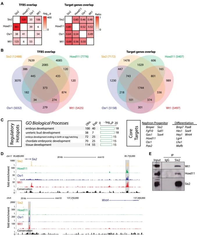
Next, we overlapped all four datasets to identify sites where all factors converge in the potential regulation of target genes. We recovered 373 putative cis-regulatory modules where
Six2, Hoxd11, Osr1, and Wt1 associated within 1kb of each other (Fig 2B,S1 Table). Regions
co-bound by all four factors displayed the strongest Six2 binding. In addition, Six2 peaks bound by any three-factor combination were on average stronger than two-factor combina-tions, while Six2 peaks bound by any factor in combination with Six2 were stronger than Six2-only peaks (S1H Fig).
We refer to regions co-bound by all four factors as ‘regulatory hotspots’ hypothesizing that these may play a key role in nephron progenitor programming. Consistent with this view, reg-ulatory hotspots were enriched around genes annotated to developmental processes such as ‘ureteric bud development’ (Fig 2C). Further, two regulatory hotspots are known from pub-lished studies to drive transgenic reporters with expression profiles reflecting the putative
tar-get genes: a region ~60kb upstream ofSix2which corresponds to theSix2-DEused in our
Fig 2. Regulatory hotspots in nephron progenitors defined by co-binding of Six2, Hoxd11, Osr1 and Wt1. (A) Heatmap shows significance of pairwise overlap between transcription factor binding sites (left, represented by binomial -log10 p-value) or between assigned target genes (right, represented by ratio). TFBS = transcription factor binding site. (B) Venn diagram shows the overlap of Six2, Hoxd11, Osr1, and Wt1 binding sites (left) and target genes (right). The 4-way overlapping sites were defined as the ‘regulatory hotspots’. The 4-way overlapping target genes were defined as ‘core targets’. (C) Barplots show result of gene ontology (GO) analysis on the ‘regulatory hotspots’ (left). Examples of ‘core targets’ known to have roles in the nephron progenitors and their differentiation are listed (right). (D) Genome browser view of Six2, Hoxd11, Osr1, and Wt1 ChIP-seq signals at the ‘regulatory hotspots’ (shadow area) nearSix2andWnt4. (E) Six2 immunoprecipitation from E16.5 kidney nuclear extracts. Western blot was probed with antibodies to Six2, Hoxd11, and Wt1 to identify protein complexes.
bound at theSix2distal enhancer but not at theWnt4enhancer site[29].Six2is largely
restricted to the nephron progenitors whileWnt4expression is absent from nephron
progeni-tors but activated on progenitor induction in the formation of differentiating renal vesicles [2, 47]. Thus, engagement of the four factors can occur on target genes for nephron progenitors or genes activated shortly after the onset of nephrogenesis. Other putative targets of regulatory
hotspots includeFgf9which is expressed by nephron progenitors and is involved in regulating
their maintenance [48], andPax8which regulates nephron progenitor differentiation [49] (S1
Table).Tsc22d1andMgat5also represent putative targets and knockouts of these genes are
reported to generate kidney phenotypes [50,51] (S1 Table).
Regulatory information may also converge on a common target through alternative enhancer usage. To examine this possibility, we intersected the predicted target gene sets for each factor and identified 1744 genes sharing Six2, Hoxd11, Osr1, and Wt1 associated peaks
(Fig 2B,S2 Table). The set of genes identified as having all four factors co-associated at one
putative cis-regulatory module or dispersed through multiple interactions sites are predicted to define a set of genes with a significant role in nephron progenitors or their derivatives; we
termed this group ‘core targets’ (Fig 2C,S2 Table). This set includes genes expressed in
neph-ron progenitors and implicated in progenitor maintenance and self-renewal includingSix2,
Pax2,Sall1,Sox4, andGas1[2,19,52–54]. However, the ‘core targets’ also included genes
nor-mally activated downstream in the induced/developing nephron such asWnt4,Lhx1,Pax8,
Hes1, andIrx1/2[47,49,55–57].
To determine whether interactions amongst these transcription factors existin vivo, we
per-formed immunoprecipitations with Six2 antibodies from E16.5 kidney nuclear lysates. Six2 was able to co-immunoprecipitate Hoxd11 and Wt1 (Fig 2E); however, the absence of a work-ing Osr1 antibody precluded analysis of this factor although recent studies show Six2 and Osr1
complexin vitro[17]. Six2 is also purported to complex with Sall1 [29] though we could not
replicate this interaction with available antibodies in our assay. Taken together, these data pro-vide epro-vidence for endogenous, multi-protein complexes among three of the four factors.
Transcription factor co-binding is preferentially associated with genes
active in differentiating structures and reveals novel targets
We sought to identify whether Six2, Hoxd11, Osr1, and Wt1 are each involved in activating or repressing gene expression in nephron progenitors. First, we generated RNA-seq expression
profiles of E16.5Six2TGCtg/+kidney cortex preparations FAC-sorted for GFP+(Six2+) or
GFP-(Six2-) cells. Six2+ cells would represent the nephron progenitor population (both self-renewing and recently induced) and Six2- cells would largely represent stromal cells as well as ureteric bud tip cells and endothelial cells. Genes with a TPM (Transcripts Per Kilobase
Mil-lion) value>5 and a fold difference>3 between the two cell types were identified: 246 genes
were enriched in the Six2+ fraction and 545 genes were enriched in the Six2- cortex fraction
(Fig 3A,S3 Table). We asked whether ChIP-seq peaks of any of the transcription factors or the
regulatory hotspots are preferentially located adjacent to differentially expressed genes. The results show that peaks from all ChIP-seq datasets occur significantly more often around genes enriched in the Six2+ cells (Fig 3C) consistent with a specific role in regulating the nephron progenitor cell versus other cell types of the kidney cortex. However, regulatory hotspots near Foxd1, a marker of self-renewing stromal progenitors [58], andWnt11, a ureteric tip marker required for normal kidney development [59] (S1 Table), raises the possibility that the four fac-tors may also work together to repress these genes within nephron progenifac-tors.
regulatory hotspots and programs of progenitor maintenance or commitment, we performed RNA-seq analysis to identify progenitor-specific and early induction gene sets. For the former,
a transcriptional profile was generated for E16.5 Cited1+; RFP+ cells from
Cited1-nuc-TagRFP-Ttg/+kidneys while Six2+; GFP+ cells fromSix2TGCtg/+P2 kidneys were used to
gen-erate the latter dataset (S4 Table, [61]). As expected,Cited1levels were appreciably lower in the
P2 Six2+ cells (200.9 TPM in E16.5 Cited1+ cells vs. 3.8 TPM in P2 Six2+ cells) whileWnt4
transcripts were markedly increased (9.0 TPM in E16.5 Cited1+ cells vs. 219.1 TPM in P2 Six2 + cells) supporting our classification of these datasets (S4 Table).
As expected, a comparison of the genes with a TPM>5 and a fold difference>3 between
the two cell types showed self-renewing nephron progenitor-specific genes such asCited1and
Osr1enriched in the E16.5 Cited1+ cell dataset whereas genes involved in progenitor
differen-tiation such asPax8andWnt4were enriched in the P2 Six2+ cell dataset (Fig 3B,S4 Table).
Six2 and Hoxd11 displayed similar enrichment near genes up-regulated in either self-renewing
nephron progenitors or in differentiating progenitors (1.4–1.5 fold;Fig 3C) consistent with
roles in promoting the progenitor state, and either preventing or priming nephron forming programs. Osr1 and Wt1 interactions were slightly enriched near genes associated with
self-renewing nephron progenitors (1.4-fold vs 1.0-fold for Osr1, 1.2-fold vs 1.0-fold for Wt1;Fig
3C). Interestingly, the regulatory hotspot associated gene lists showed a higher enrichment around genes upregulated in differentiating cells versus self-renewing progenitors (1.6-fold vs.
1.3-fold;Fig 3C).
Next, for each single factor or combination of factors we compared the percent of target genes in distinct transcriptional categories: nephron progenitor enriched (E16.5 Six2+ cells), self-renewing nephron progenitor enriched (E16.5 Cited1+ cells), or differentiating nephron progenitor enriched (P2 Six2+ cells) relative to the whole transcriptome. Target genes unique to any single factor were not enriched in any of these categories (1.6% for each) compared to the whole transcriptome (1.6% for each) suggesting that single factor input has no particular relevance to nephron progenitor function. Similar observations hold when Hoxd11 co-target-ing is examined with Osr1 and Wt1, (1.8%). but not with a Six2 binary combination
(2.5%) suggesting that Hoxd11 has a strong preference for co-regulation of target genes with Six2 (Fig 3D). Generally, the greatest enrichments are observed when all four factors are bound near the target gene in any category (2.4–7.2%) consistent with co-regulatory input by multiple factors impacting target gene regulation to the greatest extent. In agreement with our earlier analyses, the four-factor overlap has a preference for genes expressed upon
differentia-tion rather than in self-renewing progenitors (5.9% compared to 2.4%;Fig 3D).
While we have described target genes with known functions in kidney development, we wanted to identify potentially novel candidate genes which are targets of co-regulation either by one cis-regulatory module or dispersed through multiple interactions sites. Target genes of
interest includeShisa2andShisa3which are enriched in self-renewing nephron progenitors
Fig 3. Six2, Hoxd11, Osr1, and Wt1 binding sites are enriched near nephron progenitor specific genes and those associated with differentiation programs. (A) Scatter plots show gene expression profiles and correlation of the Six2GFP+ versus the Six2GFP- RNA-seq from E16.5 mouse kidney cortex. Specific genes for each category are highlighted in orange (Six2+) or blue (Six2-). TPM = Transcripts Per Kilobase Million. (B) Scatter plots show gene expression profiles and correlation of RNA-seq from E16.5 Cited1RFP+ cells versus P2 Six2GFP+ cells. Genes specific to each population are highlighted in red (Cited1RFP+) or green (Six2GFP+). Examples of specific genes are listed and highlighted on the plot. (C) Barplots show p-values indicating enrichment of Six2, Hoxd11, Osr1, and Wt1 binding sites, as well as regulatory hotspots (Six2/Hoxd11/Osr1/Wt1 overlapping sites) in genes that are specific to the Six2 + cortex fraction, specific to the Six2- cortex, enriched in self-renewing nephron progenitors, or enriched in differentiating nephron progenitors, respectively. The regulatory domain was defined as +/-500 kb from transcription start site. TFBS = transcription factor binding site. ‘Obs.’, number of peaks associated with genes annotated with corresponding term; ‘Exp.’, number of peaks expected to be associated with genes annotated with corresponding term by chance. Fold represents the fold enrichment or expected values. (D) Bar plots showing the percentage of total genes for each condition (x-axis) that falls into each category of 1) nephron progenitor (NP) enriched, 2) enriched in self-renewing nephron progenitors, or 3) enriched in differentiating progenitors.
(S2 Table).Shisa2is a modulator of Wnt and Fgf signaling, specifically attenuating such
sig-nals. The majority of mutant mice exhibit dwarfism and half die postnatally.Shisa3is a related
family member although no overt phenotype was observed for the null allele [62].Pdgfcand
Pdgfaare enriched in the self-renewing and differentiating progenitors, respectively (S2 Table). Their conserved expression in these cell populations of developing mouse and human
kidneys have been reported [63–65].PdgfaandPdgfcdouble mutants have a reported
defi-ciency in cortical renal mesenchyme, however, the mutant kidney phenotype was not analyzed
in detail [66].Ccnd1(cyclin D1) is a putative target that shows a nearly 7-fold increase in
expression in P2 Six2+ cells versus E16.5 Cited1+ cells (S2 Table).In situhybridization
con-firms strongCcnd1in E15.5 pretubular aggregates and early differentiating nephrons (www.
gudmap.org, [67,68]). This suggests that the regulatory networks may directly modulate cell cycle dynamics and balance progenitor proliferation or alternatively may prime putative
enhancers ofCcnd1for rapid activation upon nephron progenitor induction.Sema5aand
Epha4are predicted targets with a similar ~6-7-fold increase in expression in differentiating
progenitors confirmed byin situ studies(S2 Table;www.gudmap.org, [67,68]) suggesting
fac-tor regulation of targets genes controlling local cell interactions.
Deletion of the
Six2 and Wnt4 distal enhancers reveals their roles in
modulating target gene expression
To examine the functional significance of ‘regulatory hotspots”, we focused onSix2-DE(chr17:
85747271–85749534;Fig 4A) andWnt-4 DE(chr4:137216986–137217756;Fig 5A) elements
previously verified in transgenic reporter assays [28]. To examine the requirement for each enhancer, we used CRISPR/Cas9 gene editing technology to delete each enhancer in B6SJLF1/J
mice. TheSix2-DEdeletion andWnt4-DEdeletion were confirmed in founder lines by PCR
and Sanger sequencing of products (Six2ΔDE: chr17:85747284–85749542;Wnt4ΔDE: chr4:13721
6991–137217771). For theSix2-DEknockout, we examined kidneys at E16.5 and observed no
obvious difference in the size of wildtype,Six2ΔDE/+andSix2ΔDE/ΔDEkidneys (Fig 4B). Six2+
and Wt1+ nephron progenitors were present inSix2ΔDE/ΔDEkidneys though Six2 levels appear
reduced relative to wild-type embryos (Fig 4C and 4D). Nephron structures were formed as reflected by the presence of podocytes and proximal tubules, labeled by Wt1 and LTL (Lotus tet-ragonolobuslectin), respectively (Fig 4C). TheSix2ΔDE/ΔDEmice were viable; no phenotype was observed.
To more accurately assess the effect of the distal enhancer deletion onSix2expression, we
used qPCR to measure relativeSix2levels in nephron progenitors of E16.5 kidneys. A 40%
reduction ofSix2mRNA was measured inSix2ΔDE/ΔDEnephron progenitors compared to
wild-type (Fig 4E, p-value = 0.006); higher levels than in mice heterozygous for aSix2null allele
(Six2CE/+, [69]) whereSix2transcripts were reduced approximately 50% relative to wild-type as
expected (Fig 4E). The levels ofPax2mRNA, which is not dependent on Six2 [2], were
rela-tively similar across all genotypes showing a Six2-specificity for theSix2-DEdeletion.
Strik-ingly, whenSix2levels were further reduced by combining aSix2ΔDEallele with aSix2null
allele (eitherSix2CE/+orSix2GCE/+[69]), the resultantSix2ΔDE/CEembryos exhibited severely
hypoplastic kidneys at E16.5, with a complete absence of Six2+ nephron progenitors,
mirror-ing the phenotype of complete removal ofSix2activity (Fig 4B and 4D, [2]) where only a few
glomeruli (Wt1+) and tubules (LTL+) have formed by E18.5 (S4 Fig). As early as E11.5, at the
outset of active kidney morphogenesis,Six2ΔDE/GCEkidneys were devoid of Six2+ nephron
progenitor cells but filled with Pax8+ differentiating nephron progenitors as in Six2 protein
null mutant kidneys (Fig 4G, [2]). Taken together these results demonstrate thatSix2-DE
with aSix2null allele, the remainingSix2mRNA levels (predicted to be 30% of wild-type lev-els) were insufficient for Six2-mediated maintenance of the nephron progenitor state.
Next, we investigated a ‘regulatory hotspot’ predicted to function in progenitor
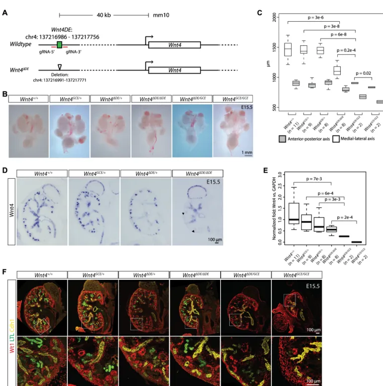
differentia-tion. Deletion of theWnt4distal enhancer resulted in mutant kidneys that are ~25% smaller
than those from wildtype animals (p = 0.2e-4;Fig 5B and 5C). Nephrons developed in
Wnt4ΔDE/ΔDEkidneys as reflected by presence of both LTL+ proximal tubules and Wt1+
podo-cytes (Fig 5D) andWnt4ΔDE/ΔDEmice are viable. Interestingly,in situhybridization revealed
that expression ofWnt4is significantly reduced in renal vesicles but remained largely
unch-anged in the renal medulla ofWnt4ΔDE/ΔDEkidneys consistent with an overall reduction of
Wnt4mRNA levels inWnt4ΔDE/ΔDEkidneys measured by qPCR (Fig 5F). Thus, theWnt4-DE
plays a functional role in regulatingWnt4mRNA levels in forming nephrons (Fig 5E). The
Wnt4ΔDE/ΔDEphenotype was less severe than Wnt4 protein null mutants where the severely
hypoplastic kidney lacks nephron tubules and glomeruli [47]; indeed, low levels ofWnt4RNA
were detected inWnt4ΔDE/ΔDEkidneys (Fig 5D; arrows inFig 5E). When theWnt4ΔDEallele
was combined with aWnt4GCEprotein null allele [69],Wnt4ΔDE/GCEkidney size and nephron
structures were further reduced, though kidneys were still larger thanWnt4null kidneys (Fig
5B–5D) andWnt4mRNA levels were markedly reduced in whole kidney PCR (Fig 5E and 5F). Taken together these results indicate a dose-dependent reduction in kidney size through
reduced nephrogenesis upon decreasingWnt4activity. Further, residual levels ofWnt4activity
inWnt4ΔDE/GCEkidneys were sufficient to drive low levels of nephrogenesis. Clearly, the Wnt4-DEplays a role in maintaining appropriate levels ofWnt4transcripts in the nephrogenic program to ensure a normal program of kidney development.
The
Br mouse is the result of an inversion altering the Six2 regulatory
landscape
Six2lies ~60 kb from a related family memberSix3, although significantSix3expression is not
observed in the self-renewing nephron progenitors (S4 Table) indicating a specificity inSix2-DE
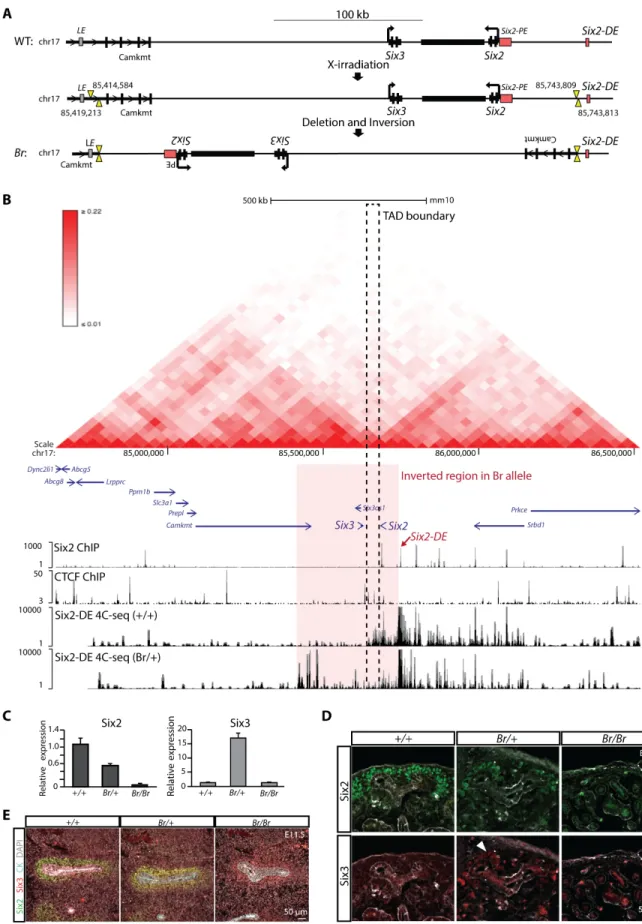
interactions. Topologically associating domain (TAD) boundaries have been described as CTCF-enriched sites which serve as insulators and prevent promiscuous enhancer activity [70]. We per-formed CTCF ChIP-seq using E16.5 purified nephron progenitors to identify CTCF-bound
regions of the genome (Fig 6BandS3G and S3H Fig). We identified strongly bound CTCF sites
between theSix2andSix3locus, most of which are consistent with ENCODE data analyzing
whole P0 kidney samples (S5B Fig, ENCODE experiment ENCSR143WOK, submitted by Rich-ard Myers, HAIB, [71]) and predictions from Hi-C on mouse ES cells (TAD: chr17:85640660–
85680660,S5A Fig, [70]). We hypothesize that this region serves as a TAD boundary to prevent
theSix2-DEfrom engaging theSix3promoter. Consistent with this view, there is a marked bias
in the engagement of regulatory factors in nephron progenitors to theSix2side of this putative
boundary, 5’ to theSix2transcriptional start site (Fig 6B).
Cas9 and the resulting Cas9-mediated deletion of theSix2-DEis shown. (B) Brightfield images of whole urogenital systems from E16.5 embryos resulting fromSix2ΔDE/+matings orSix2CE/+xSix2ΔDE/+
crosses. (C) Immunostaining for Wt1 to identify nephron progenitors and podocytes, LTL (Lotus
tetragonolobuslectin) to mark proximal tubules, and Cdh1 to show the collecting duct network of kidneys associated with (B). (D) Immunostaining for Six2 to identify nephron progenitors in kidneys associated with (B). (E) Box plots showing results of qPCR forSix2andPax2(normalized toGAPDH) from nephron progenitors (NP) and nephron progenitor-depleted cortex. Genotypes and number of samples analyzed are shown. (F) Samples fromSix2GCE/GCE were compared toSix2ΔDE/GCEcollected at early stages of kidney development and immunostained with Six2 to mark the nephron progenitors, Pax8 to identify differentiating structures (Pax8 antibody appears to cross react with Pax2 as seen by expression in Ecad+ collecting duct), and Ecad to mark epithelial structures.
With these insights into regulation ofSix2, our attention was drawn to theBrachyrrhine (Br) mouse, an X-irradiation induced mutant that displays kidney hypoplasia and frontonasal
Fig 5. Deletion of the distal enhancer forWnt4 results in reduced expression of Wnt4 specifically in renal vesicles and smaller kidneys. (A) Schematic of the Wnt4locus showing the location of the distal enhancer (DE) element. The DE was targeted for deletion using CRISPR/Cas9 and the resulting Cas9-mediated deletion of theWnt4-DEis shown. (B) Brightfield images of whole urogenital systems from E15.5 embryos. (C) Measurements of the anterior to posterior axis and medial to lateral axis of kidneys representing the genotypes shown in (B). (D) Sectionin situhybridization forWnt4on kidneys fromWnt4ΔDE/+,Wnt4ΔDE/ΔDEand Wnt4GCE/+embryos. (E) Box plots showing results of qPCR forWnt4(normalized toGAPDH) from whole kidneys at E15.5. Genotypes and number of samples analyzed are shown. (F) Immunostaining for Wt1 to identify nephron progenitors and podocytes, LTL to mark proximal tubules, and Cdh1 to show the collecting duct network of kidneys associated with (B).
Fig 6. TheSix2 regulatory landscape is altered in the Br mouse leading to reduced Six2 expression and ectopic Six3 expression in the
dysplasia, and maps to theSix2region of chromosome 17 [72]. ThoughBrmutants have
signif-icantly reducedSix2expression in the kidney and craniofacial tissues, no mutation has been
found in theSix2transcription unit or within 1.8 kb upstream of the start codon which
includes theSix2-PEelements [72]. Interestingly,Six2is ectopically expressed in the
develop-ing lens ofBrheterozygous and homozygous animals, a normal site ofSix3expression [72].
Given that irradiation induces large-scale genomic rearrangements, we speculated that theBr
mutation led to a chromosomal rearrangement that removedSix2from enhancer(s) directing
normal regulatory input to the nephron progenitor population, placingSix2under the control
ofSix3regulatory elements normally inaccessible the other side of a CTCF-dependent
bound-ary element.
Next generation sequencing and sequence alignment identified the underlying sequence
change in theBrmutant (S1 Supplemental Material and Methods). The main feature was a
large inversion of 324,596bp including both theSix2andSix3loci. The inversion movesSix3
~206kb from theSix2-DE, actually further than in the wild-type organization, but importantly
the inversion removes the intervening TAD boundary (Fig 6B). In contrast,Six2is
reposi-tioned on the other side of this boundary element withinSix3’s unchartered regulatory
terri-tory (Fig 6B). In addition to the inversion, two small deletions were detected: a 4,630bp
deletion (chr17:85414584–85419213) 5’ to theSix3TSS in the intron ofCamkmt, and a 5bp
deletion between theSix2-PEandSix2-DE(chr17:85743809–85743813,Fig 6A and 6B). The
results from the sequencing and computational analysis were confirmed by allele-specific
diag-nostic PCR assays (S5C and S5D Fig). The inversion also separates the last 4 exons ofCamkmt
from the rest of the transcription unit. However,Camkmthas a TPM of only 2.26 in the E16.5
Cited1+ nephron progenitor cells and homozygous mutant mice are viable (International
Mouse Phenotyping Consortium,http://www.mousephenotype.org/, Release 5.0 [73]), so
Camkmtis unlikely to contribute to the kidney phenotype. The rearrangement predicts: i)
ectopicSix2expression inSix3’snormal expression domain, the lens, asSix3enhancers can
now targetSix2, and ii) an abnormal interaction between theSix2-DEandSix3promoter
resulting in ectopicSix3expression in nephron progenitors.
To directly examine interaction ofSix2-DEwithSix2andSix3promoters, we performed
4C-seq [74] usingSix2-DEas the view point. As expected, in wildtype kidneysSix2-DE
inter-acts with a broad region that includesSix2transcription start site (TSS) (Fig 6B), with the local
maxima 7.2 kb upstream of Six2TSS. Noticeably,Six2-DEinteraction was restricted by the
TAD boundary betweenSix2andSix3(Fig 6BandS5B Fig) and no interaction was observed
around theSix3TSS. In kidneys fromBr/+embryos, strongSix2-DEcontacts were now
observed in the segment of the inverted region that was repositioned between theSix2-DEand
CTCF-bound TAD boundary element (Fig 6BandS5B Fig). As expected, with the loss of one
wildtype allele inBr/+embryos,Six2-DEinteractions withSix2TSS, and in general with the
region onSix2side of the TAD boundary, were significantly reduced. A relatively strong,de
novointeraction of theSix2-DEwas observed ~15 kb upstream ofSix3TSS (Fig 6BandS5B
Fig) consistent with the model ofSix3expression driven, at least in part, by theSix2-DEin the
Brallele. Importantly, the predictedSix2-DE/Six3upstream contact in theBrallele occurs over
LE = Lens enhancer (putative), PE = proximal enhancer, DE = distal enhancer. Black box between theSix2andSix3loci represents the predicted boundary. (B) Interaction matrix (top) generated by Hi-C data (Hardison lab hESC Hi-C data,http://promoter.bx.psu.edu/hi-c/ view.php). Genomic view showing Six2 ChIP-seq, CTCF-NP ChIP-seq, and 4C-seq data (bottom). The region inverted in theBrallele is highlighted. Dashed square indicates a predicted TAD boundary element that lies betweenSix3andSix2loci [70]. (C) qPCR showing the relative expression levels ofSix2andSix3in E13.5 kidneys of the indicated genotype. (D) E13.5 kidneys of the indicated genotype were sectioned and immunostained for Six2 and Six3. Arrow points to the low level Six3 expression in nephron progenitors. (E) Immunostaining for Six2, Six3, cytokeratin (CK), and DAPI in E11.5 kidneys of the indicated genotype.
a distance of 200 kb from theSix2-DEto theSix3TSS, a longer interval than the ~130 kb that separates these non-interacting elements in the wild-type allele (S5B Fig). Therefore, the
differ-ential interaction ofSix2-DEwithSix2TSS andSix3TSS between wildtype andBr/+cannot be
attributed to shortened distance fromSix2-DEtoSix3TSS. Rather, this data supports specific
regulation bySix2-DEtoSix2andSix3that is defined by the TAD boundary.
As a result of the altered chromatin architecture introduce by the genomic inversion,
ectopicSix2expression has been reported in the lens ofBr/Brmutants [72], andSix3
expres-sion was reduced in this structure (S5E Fig). Quantitative PCR detectedSix3expression in the
kidneys ofBr/+mice at E13.5 (Fig 6C) and Six3 protein was detected in Six2+ nephron
pro-genitors (Fig 6D).Br/Brmutants resembleSix2null mutants and have no nephron progenitors
at this stage [2] (Fig 6C and 6D). WhenBr/Brmutants were examined at E11.5, they showed a
similar loss and premature differentiation of nephron progenitors as inSix2null mutants but
interestingly low-levels of Six3 were detected in differentiating progenitors (Fig 6E). Ectopic
Six3 was also observed in the cranial base ofBrmutants at E14.5 concomitant with decreased
Six2 levels (S5F Fig). Taken together these data lend additional weight to the importance of the Six2-DEin directingSix2expression and reveal higher order principles of topological organi-zation acting in conjunction with this enhancer to provide target gene specificity to the regula-tory landscape.
Discussion
In this study, we utilized novel transgenic mouse strains to map the nephron progenitor-spe-cific interactions of Six2, Hoxd11, and Osr1, and incorporated nephron progenitor-speprogenitor-spe-cific ChIP-seq profiling of Wt1, to identify the regulatory genome controlled by these four factors in the developing mouse kidney. Our data identifies a subset of binding sites, or ‘regulatory hotspots’ where the engagement of all four factors occurs in close proximity. The putative tar-get genes of their combinatorial action are largely associated with kidney function. Deletion of
two of these hotspots forSix2andWnt4highlight their roles in target gene regulation and
their significance to kidney development. These data suggest that ‘hotspots’ with multi-factor
input play significant roles in target gene regulation. Our analysis on theBrmutant
demon-strated that re-arrangement of the regulatory scenario ofSix2andSix3genes can causes
dra-matic, predictable effects on their expression and the resulting developmental phenotypes highlighting the importance of appropriate regulatory context to proper gene regulation and biological function.
Transcriptional hierarchy of nephron progenitors
Our ChIP studies reveal a complex regulatory architecture of the nephron progenitors. Examining co-binding of the four factors suggests each of these genes is itself a target of their combined actions through auto and cross-regulatory inputs, as are a number of other tran-scriptional regulatory components important for kidney development and nephron progenitor
maintenance such asSall1andPax2(S2 Table). By combining our data with insight from
pre-vious studies, a hierarchical network starts to emerge. For example, mutational analyses have
demonstrated a requirement for theHox11paralogues to activateSix2expression in
meta-nephric mesenchyme [12]. Hox11 members complex with Pax2 and Eya1 binding to an
enhancer that lies within ~1kb of theSix2TSS, in theSix2-PE[75]. Hox11 acts as an activator
ofSix2activity and mutations in Hox motifs results in loss of reporter activity in transgenic
assays [76]. We have also shown that the Hox motif within theSix2-DEis required for reporter
activity [28]. Consistent with this data, Hoxd11 is bound at theSix2-DE(Fig 2D). However,
discrepancy may result from preferential enhancer usage at different developmental stages.
Two previous studies assayed reporter activity of the ~1kbSix2-PEat E11.5 [75,76] while our
studies assayedSix2-DEactivity at E15.5 [28]. Hox11 may be required at theSix2-PEto help
initiate Six2 expression, but maintenance of expression may then rely, at least partially, with theSix2-DEwhere Hoxd11 is engaged at E15.5. Additionally, Osr1 and Wt1 are enriched at theSix2-DEcompared to thePE(Fig 2D), as is Sall1 [29], supporting multifactor input at the
DEas an important mechanism ofSix2regulation. However, Six2 is bound at both thePEand
DE, thoughPEassociation is weaker (Fig 2D), suggesting both may contribute at some level to the maintenance and autoregulation by Six2 itself. Unfortunately, technical difficulties pre-clude detailed temporal analysis of engagement in the small numbers of cells that are the foun-dation of the nephron progenitor pool.
When assessing the targets unique to any transcription factor combination, the greatest enrichment for genes with expression within the nephron progenitors, either in self-renewing or differentiating cells, generally occurred when they were complexed with Six2 (Fig 3D). Hoxd11 showed the lowest levels of enrichment for these targets when engaged with Osr1 or Wt1 in the absence of Six2, suggesting that its primary regulatory functions rely on engaging with Six2. Taken together, these data suggest Six2 acts as a master regulator: co-engagement with Six2 predicts a higher probability of regulatory functions within nephron progenitors.
Transcriptional factors: Activator, repressor and enabling interactions
Osr1 has been described as a transcriptional repressor in vertebrate kidney development [77]. Xu et al. showed that Osr1 works with the Groucho family members and represses activation
of aWnt4enhancer specifically in Six2+ nephron progenitors [17]. Consistent with this result,
Osr1 associates with theWnt4enhancer in our ChIP assay (Fig 2D). Additionally, other genes
that are not present in the nephron progenitors but rather in differentiating structures such as
Pax8andLhx1are also bound by Osr1 suggestive of a repressive role (Fig 3C,S2 Table, [49,
56]). However, Osr1 is also bound near genes actively expressed in nephron progenitors such
asSix2andOsr1itself (Fig 3C,S2 Table, [2,16,69]). Therefore, our data suggest a more
com-plex relationship than Osr1 simply repressing transcription at all engaged targets. Further, our previous ChIP studies supported dual roles for Six2 in activating transcription within nephron progenitors but also engaging at targets silent in progenitors but activated as progenitors
dif-ferentiate towards nephrons [3,28]. Similarly, Hox11 has been characterized as an activator,
specifically ofSix2expression [76]. Consistent with this view, Hoxd11, is bound near nephron
progenitor-specific genes but like Six2 binding is also prominent around differentiation targets
(Fig 3C,S3 Table). Similarly, these observations extend to Wt1 nephron progenitor targets.
Engagement most likely reflects dual activator and repressor actions of these complexes and which activity could be dependent on currently unidentified co-bound factors. Conversely, factor engagement at differentiation-specific gene targets may facilitate or enable subsequent activation of enhancers for differentiation-associated genes following the induction of nephron progenitors. In this scenario, multi-factor engagement may be necessary but not sufficient for target activation for differentiation associated genes. Additional factors or modification of existing transcriptional components following progenitor commitment may modify the action of these regulatory complexes.
Genomic co-localization of transcription factors in nephron progenitor
cells
We observed a highly significant overlap of transcription factor binding in nephron
enriched (Fig 1D), supporting direct protein-DNA binding. Additionally, our EMSA assays confirmed factor binding to each motif (S2 Fig). On the other hand, previous studies have shown that Six2 can complex with a number of transcription factors, including Hoxa11 [28]
and Osr1 [17]in vitro, and Eya1 and Sall1 bothin vitroandin vivo[29,78,79]. Osr1 has also
been shown to interact with Wt1in vitro[80]. These studies support protein-protein
interac-tions amongst these factors and may account, in part, for the multi-factor co-localization on specific genomic targets. Additionally, our kidney immunoprecipitation data suggests that
Six2 can interact with Wt1 and Hoxd11 (Fig 2E), confirming such complexes existin vivo.
However, without confirming the co-association of these factors on any genomic loci at the same time and in the same cell, we can only suggest their combined function. The association of each factor with its own DNA target and co-association with each other adds to the difficulty of predicting the actions of the regulatory circuit. Further, it is likely that there are significant components yet to be discovered. For example, all of the ChIP datasets recovered a bHLH motif amongst the most-significantly enriched motifs (S1F Fig). Whereas Myc is a bHLH tran-scription factor that has been shown to complex with Eya1 and Six2 in the kidney [79], and
loss of functionMycmutants argue for a role in kidney development [81], the recovered motif
is distinct from the conventional Myc-Max target site [44], suggesting a role for another, unidentified family member.
Target gene functions in nephron progenitors
In addition to identifying target genes with known function during kidney development, we
also uncovered novel putative targets of the four factors (seeS2 Tablefor list of all target
genes). Bmper is a secreted protein that interacts with Bmp proteins and inhibits their function
[82]. Inactivation ofBmperin the kidney leads to mild hypoplasia [83]; Bmp signaling plays
important roles in the progenitor self-renewal and differentiation [84]. Six2 and the other
fac-tors may help fine-tune the level of Bmp signaling through activation ofBmper. Rspo1 is a
secreted protein that binds to G protein-coupled receptors that activate Wnt signaling and its function has been implicated in multiple developmental systems [85]. Rspo1 could have a role
in modulating Wnt signaling in the nephron progenitor niche althoughRspo1mutants have
no obvious kidney phenotype, these mutants have not been analyzed in depth [85]. We also
identified other modulators of Wnt signaling within our data.Shisa2is reported to attenuate
Wnt and Fgf signaling during development [62].Shisa2is expressed in the nephron
progeni-tors along with its related family memberShisa3(S2 Table). Other targets likeTsc22d1and
Mgat5are reported to display kidney phenotypes.Mgat5is expressed in differentiating
struc-tures including podocytes (S2 Table, Eurexpress,www.eurexpress.org, [86]) and shows a
glo-merular phenotype [51].Tsc22d1is expressed in the nephron progenitors (S2 Table) and
mutants have small kidneys [50]. Given the current associations of known targets with kidney development and disease, it is likely that functional analysis of new targets predicted here will identify additional regulators of mammalian kidney development.
From our analyses, the majority of significant targets fall under the control of all four fac-tors. These genes fall into multiple functional categories from transcriptional regulators like Six2,Sall1, andPax2to signaling factors likeFgf9andWnt4to cell cycle regulators such as Ccnd1and matrix proteins such asLamb1(S2 Table). This suggests that these transcription factors control many different aspects of progenitor cell biology. Fewer targets with known kidney functions emerge from the interaction maps where one of more the factors was not
bound at the putative regulatory region (S5 Table). However,Eya1,Wt1, andBcamlacked an
Osr1 association in combined factor interaction analysis (S6 Table) but are well known for
laminin and is expressed at increasing levels in differentiating progenitors (S6 Table). Knock-outs display glomerular abnormalities suggesting important functions in the kidney [87]. Phgdh, a Six2-independent target with highest expression in nephron progenitors (S6 Table) participates in L-serine synthesis and knockouts are embryonic lethal [88].
Deletion of regulatory hotspots
Enhancers directingSix2-like andWnt4-like reporter gene expression [28], identified as
‘regu-latory hotspots’ co-bound by Six2, Hoxd11, Osr1, and Wt1 in the data here, were shown to play roles in regulating activity of both gene targets. Kidney phenotypes were observed in embryos homozygous for the enhancer deletion (Wnt4-DE) or when combined with protein
null mutations (Six2-DEandWnt4-DE). While the study identified functional enhancer
regions, neither works alone in regulating normal transcript levels in the target cell type. An
alternative proximal enhancer has been documented forSix2[76,89]. This proximal enhancer
lies a few hundred base pairs upstream ofSix2’stranscriptional start site and strongly binds
Six2, but not the other regulatory factors analyzed here. Alternative enhancers have not been
functionally demonstrated forWnt4. In summary, our studies provide evidence to support a
focus on multifactor input to prioritize functional analysis of large datasets emerging from
ChIP-seq studies. CRISPR/Cas9 deletion of an enhancer region>100kb from the TSS for Sox2
that is co-bound by multiple transcription factors regulating pluripotency (Oct4, Sox2, Nanog,
and Klf4 [90,91]) provides another example of this strategy to identify strong,bone-fide
com-ponents of the regulatory genome.
Topological rearrangements in the
Br mutant and cis regulation of Six2
Individual enhancer action depends on the larger context of the chromosomal landscape. Our
demonstration that the inversion inBrmutant strain, repositionsSix2andSix3in a new
regu-latory landscape modifying enhancer interactions that likely contribute to altered features of each gene’s regulation. Each gene exists in a distinct TAD that is likely enforced by the action
of a CTCF-dependent boundary element between the two genes. InBrheterozygous and
mutant alleles,Six3is ectopically expressed in nephron progenitors: the boundary element no
longer separates theSix3promoter from theSix2-DE. We hypothesize that this enhancer, and
potentially undefined regulatory information 5’ to this enhancer, dominate over other
regula-tory information that might be present within theSix3flanking region. As a result, the
Six2-DEdrivesSix3expression in nephron progenitor cells whileSix3expression is lost from its normal lens expression domain. Even though Six3 was detected in nephron progenitors in
Br/+mutants, Six3 is a member of a functionally divergent sub-group of Six factors [92].
Con-sequently, Six3 activity failed to compensate for loss of Six2 andBr/Brmutants resembleSix2
null mutants [72].
Interestingly, even though there is no alteration inSix2-PEposition relative theSix2gene,
theSix2-PEis not sufficient to drive levels ofSix2which maintain nephron progenitor
devel-opment in the context of the inversion. Thus, if theSix2-PEwere capable of sustaining normal
Six2levels, the inversion may preventSix2-PEengagement with regulatory factors necessary
for its activation. Alternatively, there may be distinct enhancers other than theSix2-PEthat are
required forSix2’sexpression. Six2-bound putative regulatory regions lie upstream ofSix2-DE
(S8 Table,Fig 6B) and these would be predicted to disengage fromSix2regulation in theBr
inversion.
Topological domains are highly conserved between cell types and across mammalian spe-cies [70]. Recent studies have shown that alterations in TADs and CTCF site orientation can
several limb malformations in the human were attributed to the rearrangement of TADs and disrupted boundaries. When genetically modeled in the mouse, altered gene expression sug-gests a mechanism for driving the limb malformations [93]. The type of topological rearrange-ments described here could play a role in a subset of the congenital anomalies of the kidney and urinary tract (CAKUT) syndrome. Importantly, these micro-rearrangements would not be detected in traditional exome screens. Even whole genome sequencing approaches required tailored alignment algorithms to uncover the junction fragments for the rearrangements as performed here. Together, these studies highlight the importance of non-coding DNA and chromatin architecture to the appropriate regulation of gene expression and the resulting phe-notypic consequences incurred by rearranging the regulatory landscape.
Limitations of the transgenic approach
In addition to Six2, Hoxd11, and Osr1, we attempted to generate transgenic lines for other important regulatory factors including Wt1, Hoxa11, Pax2, Sall1, and Eya1. Our goal was to build an extensive regulatory network for the nephron progenitor population and more precisely
identify the targets and combinatorial actions of these major playersin vivo. However, despite
considerable efforts, we were unable to establish correctly expressing founder lines for these
fac-tors. TheSix2-DEis not only active in the kidney butSix2is expressed in the developing brain,
ear, tendons, and smooth muscle [95] and we observed transgene expression in the brain and ear. Some transgenic lines showed a circling behavior, consistent with inner ear defects, along with insufficient kidney expression and thus were not utilized. The ectopic action of a sub-set of
factors in these other sites ofSix2-DEactivity may have resulted in severe defects and subsequent
lethality. Alternatively, there could be dominant effects within the kidney itself from elevating levels of that factor in the normal nephron progenitor context, though this seems less likely given the absence of a kidney phenotype in Six2, Hoxd11, or Osr1 transgenic strains, a Six2-BF binding
profile that was comparable to the native Six2 protein, and the levels ofSix2-DEactivity.
Despite these technical limitations, we were able to generate a core transcriptional network of four factors important for kidney development. Overlap with additional factors may not
add much greater insight; comparison with Sall1 andβ-catenin targets reveals many of the
same nephron progenitor-specific target genes and a lack of nephron progenitor relevant
inde-pendent regulation by these factors (S1,S5andS6Tables). Therefore, the data presented here
is likely to highlight some of the most critical regulatory elements and target genes which mod-ulate nephron progenitor programs.
Materials and methods
Mouse strains
All surgical procedures, mouse handling, and husbandry were performed according to guide-lines issued by the Institutional Animal Care and Use Committees (IACUC) at the University of Southern California and after approval from the institutional IACUC committee. The
trans-genic construct utilized by Park et al. [28] to testSix2-DEenhancer activity was modified to

![catena Poly[[dichlorozinc(II)] μ N,N′ bis(4 pyridylmethyl)piperazine]](data:image/gif;base64,R0lGODlhAQABAIAAAP///wAAACH5BAEAAAAALAAAAAABAAEAAAICRAEAOw==)