G Protein Coupled Receptor Kinase 3
Regulates Breast Cancer Migration, Invasion,
and Metastasis
Matthew J. Billard1, David J. Fitzhugh1, Joel S. Parker2,3, Jaime M. Brozowski4, Marcus W. McGinnis1, Roman G. Timoshchenko1, D. Stephen Serafin1, Ruth Lininger3,5, Nancy Klauber-Demore3,6, Gary Sahagian8, Young K. Truong9, Maria F. Sassano10, Jonathan S. Serody3,7, Teresa K. Tarrant1,3,4*
1Thurston Arthritis Research Center and the Department of Medicine, Division of Rheumatology, Allergy, and Immunology, University of North Carolina, Chapel Hill, NC 27599, United States of America,
2Department of Genetics, University of North Carolina at Chapel Hill, Chapel Hill, NC, 27599, United States of America,3Lineberger Comprehensive Cancer Center, University of North Carolina Chapel Hill, NC 27599, United States of America,4Department of Microbiology and Immunology, University of North Carolina, Chapel Hill, NC, 27599, United States of America,5Department of Pathology and Laboratory Medicine, University of North Carolina, Chapel Hill, NC 27599, United States of America,6Department of Surgery, Division of Surgical Oncology, University of North Carolina, Chapel Hill, NC 27599, United States of America,7Department of Medicine, Division of Hematology Oncology, University of North Carolina, Chapel Hill NC, 27599, United States of America,8Department of Developmental, Molecular & Chemical Biology, Tufts University, Medford, MA 02155, United States of America,9Department of Biostatistics, Gillings School of Global Public Health, University of North Carolina, Chapel Hill, NC 27599, United States of America,10 Department of Pharmacology, University of North Carolina, Chapel Hill, NC 27599, United States of America
Abstract
Triple negative breast cancer (TNBC) is a heterogeneous disease that has a poor prognosis and limited treatment options. Chemokine receptor interactions are important modulators of breast cancer metastasis; however, it is now recognized that quantitative surface expres-sion of one important chemokine receptor, CXCR4, may not directly correlate with metasta-sis and that its functional activity in breast cancer may better inform tumor pathogenicity. G protein coupled receptor kinase 3 (GRK3) is a negative regulator of CXCR4 activity, and we show that GRK expression correlates with tumorigenicity, molecular subtype, and meta-static potential in human tumor microarray analysis. Using established human breast can-cer cell lines and an immunocompetentin vivomouse model, we further demonstrate that alterations inGRK3expression levels in tumor cells directly affect migration and invasionin vitroand the establishment of distant metastasisin vivo. The effects of GRK3 modulation appear to be specific to chemokine-mediated migration behaviors without influencing tumor cell proliferation or survival. These data demonstrate that GRK3 dysregulation may play an important part in TNBC metastasis.
OPEN ACCESS
Citation:Billard MJ, Fitzhugh DJ, Parker JS, Brozowski JM, McGinnis MW, Timoshchenko RG, et al. (2016) G Protein Coupled Receptor Kinase 3 Regulates Breast Cancer Migration, Invasion, and Metastasis. PLoS ONE 11(4): e0152856. doi:10.1371/journal.pone.0152856
Editor:Jeffrey K. Harrison, University of Florida, UNITED STATES
Received:April 21, 2015
Accepted:March 21, 2016
Published:April 6, 2016
Copyright:© 2016 Billard et al. This is an open access article distributed under the terms of the
Creative Commons Attribution License, which permits unrestricted use, distribution, and reproduction in any medium, provided the original author and source are credited.
Data Availability Statement:All relevant data are within the paper and its Supporting Information files.
1. Introduction
Breast cancer is the most prevalent and deadliest form of cancer in women world-wide and tri-ple negative breast cancers (TNBC) account for approximately 15–20% of all breast cancers [1, 2]. Triple negative breast cancer is a heterogeneous disease defined by negative expression for estrogen receptor (ER) and progesterone receptor (PR), and normal (low) expression levels of Her2. Recent molecular characterization has identified five breast cancer subtypes (luminal A, luminal B, HER2-enriched, basal-like, and claudin-low). Approximately 90% of TNBC tumors are basal-like or claudin-low subtypes, with 70% of basal-like being identified as triple-negative and 80% of claudin-low identified as ER-/-, PR-/-, Her2-/-[3]. Triple negative breast cancers are often highly invasive, metastatic, possess a high risk for relapse, and carry a poor prognosis [4,5], and the different genetic subtypes can be associated with organ-specific metastasis and relapse [6]. As hormone receptor antagonism is less applicable in TNBC, alternative targets are being investigated to better understand pathogenesis and to design therapeutics for these patients.
The G protein coupled receptors (GPCR), such as CXCR4, and its chemokine ligand CXCL12, have been identified as important regulators of metastasis and tumor behavior in breast cancer [7–9]. CXCL12 is highly expressed at common sites of breast cancer metastasis, such as the lymph nodes, liver, bone marrow, and lungs [7], and CXCR4 on tumor cells is criti-cal for growth and migration [9]. While CXCR4 expression on tumor cells is important for metastasis, work published by Holland, et al. has emphasized that functional activation and sig-naling of CXCR4 in breast cancer cells may be more predictive of malignant potential than quantitative surface expression [10]. Thus, the intracellular signaling responses of chemokine receptor activation are of critical importance to metastasis and the malignant phenotype.
G protein coupled receptor kinases (GRKs) are signaling regulators of agonist-activated che-mokine receptors. Agonist activation leads to phosphorylation of the receptor tail by GRK, the recruitment of beta-arrestin, uncoupling of heterotrimeric G proteins, and ultimately desensiti-zation and internalidesensiti-zation of the receptor [11,12]. Thus, GRKs serve as negative regulators of GPCRs. Reduced expression of GRK3, 4, and 6 have previously been reported to increase tumor malignancy of glioblastoma, ovarian tumors, and medulloblastoma, respectively, through dysregulation of GPCR signaling [13–15]. In the glioblastoma model specifically, decreased GRK3 expression resulted in abnormally sustained CXCR4 signaling and enhanced tumor growth [14]. Further, GRK3 has been shown to regulate CXCL12-mediated cellular migration and CXCR4 internalization and MAPK signaling in human and mouse models of immune deficiency and inflammation [16,17].
Given the aforementioned importance of GRK3 in CXCR4 signaling regulation and the role of CXCL12 and CXCR4 in malignancy [9,18,19], we hypothesized that GRK3 regulation of CXCL12/CXCR4, as well as potentially other chemokine receptor interactions, may further dis-tinguish differences in metastatic potential and prognosis in patient subsets within TNBC. To predict the relevance of GRK3 in human breast cancer subtypes, we analyzed TCGA and publi-cally available microarray data for GRK correlations with molecular subset distinction and metastasis. To test the potential mechanism of GRK3 regulation of CXCR4 in disease, human TNBC cell lines MDA-MB-231 (highly invasive) and MDA-MB-468 (weakly invasive) [20] were examined forGRK3andCXCR4expression and for functional migration phenotypes whenGRK3expression was altered. To test the effects of GRK3 on tumor growth and metasta-sisin vivo, we used GRK3-deficient mammary tumor cells derived from Balb/c (66cl4-luc) as a representative TNBC model system in an immunocompetent mouse [21]. Our studies show thatGRK3is decreased in specific molecular subsets of TNBC with increased metastasis in humans and that GRK3 expression can predict the functional phenotype of migration and P30 CA016086 Cancer Center Core Support Grant to
the UNC Lineberger Comprehensive Cancer Center. The funders had no role in study design, data collection and analysis, decision to publish, or preparation of the manuscript.
invasion in bothin vitroandin vivomodel systems. Therefore, defining the relative expression ofGRK3in relationship to chemokine receptors likeCXCR4in TNBC may provide prognostic information for the aggressiveness of tumors and help discriminate patient subsets with the greatest potential for metastasis beyond their chemokine receptor surface expression alone.
2. Materials and Methods
2.1. Human breast cancer genetic analysis
The TCGA database was analyzed, as well as public microarray datasets, as previously described [6]. Specifically, these microarrays sets are deposited in the Gene Expression Omni-bus with the accession numbers GSE26338, GSE2034, GSE12276, and GSE2603. Dataset NKI295 was published prior to the adoption of GEO and can be found separately (http://ccb. nki.nl/data/). The data was combined using Distance Weighted Discrimination [22] to remove the systematic biases of different microarray sets and standardized to zero mean and unit vari-ances prior to other analyses. Samples in the normalized data were assigned to the five subtypes (luminal A, luminal B, Her2-enriched, basal-like, and normal) using the PAM50 classifier [23]. Assignment of claudin-low was performed according to the protocol described in Prat et al.[3] Testing for differential expression of the candidate genes associated with subtype was per-formed using ANOVA. Survival analysis was perper-formed by grouping expression levels into tertiles, then testing for association with metastasis free survival using the log rank test and visualized with Kaplan Meier plots.
2.2. Mice and cell lines
In vivoexperiments were performed in Balb/c mice. All animals were cared for under standard Institutional Animal Care and Use Committee (IACUC)-approved protocols in the Associa-tion for Assessment and AccreditaAssocia-tion of Laboratory Animal Care-accredited vivarium at the University of North Carolina. Luciferase-tagged murine breast cancer line 66cl4-luc (gener-ated by Dr. G. Sahagian) [24] and human breast cancer lines MDA-MB-468, MDA-MB-453, MDA-MB-231, DU4475, MCF-7, BT-474, SK-BR3, Hs578T, and ZR-75-1 were obtained from the University of North Carolina Lineberger Cancer Center Tissue Culture Facility (TCF) and maintained according to ATCC culture recommendations. Cell lines with stable lentiviral-mediated expression of GRK3-knockdown shRNA or plasmid-lentiviral-mediated overexpression con-structs were cultured in medium with the addition of selection antibiotics puromycin and/or gentamicin at concentrations determined empirically by kill curve assays.
2.3. Lentiviral shRNA-mediated GRK3 silencing
Five target shRNAs were obtained for both human and mouse GRK3, which were cloned into pLKO.1.-CMV (Sigma, St. Louis, MO). Target and non-target vectors were packaged into lenti-viral particles by Sigma or by the UNC Lenti-shRNA Core Facility. Each of these was transduced into the appropriate human (MDA-MB-468) or mouse (66cl4-luc) breast cancer lines and selected by antibiotic resistance. These clones were screened via quantitative real-time PCR (qRT-PCR) forGRK3expression and confirmed with immunoprecipitation protein blotting (S1 andS2Figs). CXCR4 surface protein expression was also tested by flow cytometry and confirmed to be unaffected (S3 Fig). The target sequence producing the greatest GRK3-specific knockdown with the fewest off-target effects (judged by lack of effect on housekeeping genes IDUA, 18S, CXCR4, CXCR7 and GRK2—which has the closest sequence homology to GRK3) was selected for use in further assays: human ADRBK2, TRCN0000002034, sequence5'-CCGG-CAGTA
ADRBK2, TRCN0000022703, sequence,5’
-CCGG-GCAGCATGTGTACTTACGGAA-CTCGAG-TTCCGTAAGTACACATGCTGC-TTTTT-3’.
2.4. Overexpression of GRK3
MDA-MB-231 cells were transiently transfected with GRK3 or empty control plasmid (pcDNA3.1 mCherry) using Lipofectamine LTX with Plus Reagent (Life Technologies, Grand Island, NY) per manufacturer instructions. Following 20 hours of culture with plas-mid, cells were washed and used after 2-24hr rest in complete media or were serum-starved, where appropriate, prior to assay. Transient transfection efficiency was routinely 30–40% determined by microscopy for mCherry expression. GRK3 overexpression was confirmed by Western blot (S2 Fig).
2.5. Quantitative Real Time PCR
Cells were grown in appropriate culture media to approximately 75% confluence. Total RNA was prepared using a Qiagen RNeasy kit and cDNA synthesized with Superscript II reverse transcriptase (Invitrogen) followed by qRT-PCR. Fold differences in expression were calculated using the 2-ΔCtmethod compared to housekeeping gene IDUA [17]. Transcript copy number was based on the standard curve method [25]. Human primers for qRT- PCR are listed as 5’to 3’sequence as follows: Human GRK2 forward ACTTCAGCGTGCATCGCAT, GRK2 reverse
GCTTTTTGTCCAGGCACTTCAT, GRK3 forwardAAGCCTTCGAGGTGACATTTTT, GRK3 reverseGCAACCATAAACTTCCCCGAATC, IDUA forwardCTCGGGCCACTTCACTGAC, IDUA reverseCAGTCCGTACCTACCGATGTAT, CXCR7 forwardTGCATCTCTTCGACTACTCAGA, CXCR7 reverseGGCATGTTGGGACACATCAC, CXCR4 forwardCTCACTGACGTTGGCAAAGA, and CXCR4 reverse AGGAAGCTGTTGGCTGAAAA.
2.6. CXCR4 internalization assay
According to previously published protocols [17], transiently transfected control and GRK3-overexpressed MDA-MB-231 cells were treated for the indicated times with 100nM CXCL12 at 37°C. After treatment, cells were washed twice with ice-cold PBS/0.1% BSA and CXCR4 sur-face expression was analyzed via flow cytometry using anti-CXCR4 PE antibody, Clone 2B11 (eBioscience, San Diego, CA).
2.7 Tango assay of
β-arrestin mobilization
Multi + Detection System (0.5 sec/ well). Background data for each independent run was sub-tracted and data was normalized by setting 0% and 100% as the readout for the lowest and highest concentration of the control condition, respectively. A one-tailed Student’s t-test was used for statistical analysis at each concentration tested.
2.8. Chemotaxis and Chemoinvasion
Chemotaxis was performed using BD Fluoroblok 96-well chambers according to the manufac-turer’s suggestions with the indicated concentrations of CXCL12 (R&D Systems, Minneapolis, MN) in the lower chamber. Non-targeted controls and GRK3-deficient cells were added to the upper chamber in 50μl at a concentration of 2 x 10⁶/ml. Kinetic migration curve data was col-lected every 2 or 4 minutes and was tested for significance by a linear regression analysis (See 2.13 Statistics). Chemoinvasion of human breast cancer cells was performed using BD Matrigel 96-well chemoinvasion chambers according to manufacturer instructions. Due to reports of the low invasive capacity of 66cl4 mammary tumor cells [28], chemoinvasion for this TNBC cell line was performed at a lower matrix concentration using the Cultrex 96-well BME Cell Invasion Assay in accordance with manufacturer instructions (Trevigen, Gaithersburg, MD).
2.9. CXCL12 ELISA
66cl4-luc mammary tumor cells were plated at the indicated cell number per well. Supernatants were collected at 48 hours and analyzed by sandwich ELISA with a Mouse CXCL12/SDF-1 DuoSet (R&D Systems, Minneapolis, MN).
2.10. Proliferation assay
Cell proliferation was determined using CCK-8, Cell Counting Kit 8 (Dojindo, Rockville, MD), according to manufacturer protocol. Briefly, CCK-8 reagent was added at indicated times to cells plated at 5000 cells per well in a 96-well plate and analyzed using a standard plate reader.
2.11. Apoptosis assay
Assessment of anoikis was performed as previously described [29]. Briefly, 1–2 x 10666cl4-luc
GRK3-deficient and 66cl4-luc control cells were cultured either on tissue-culture treated 6-well plates or those pre-coated with poly 2-hydroxyethyl methacrylate (poly-HEMA) (Sigma, St. Louis, MO) for 20 hours at 37°C in a 5% CO2incubator. Cells were harvested, washed, and
assayed for apoptosis using FITC-annexin V and propidium iodide (PI) staining (Trevigen, Gaithersburg, MD). Samples were analyzed by flow cytometry.
2.12.
In vivo
tumor progression
2.13. Statistics
Kinetic migration curve data was compared by analysis of covariance (ANCOVA) linear regression model. See Supplemental Information for full description. Statistical significance for all other assays was determined by two-tailed t-test using GraphPad Prism version 6.00 for Windows unless otherwise noted.
3. Results
3.1. Decreased GRK3 expression correlates with basal-type breast
cancer and liver metastasis in humans
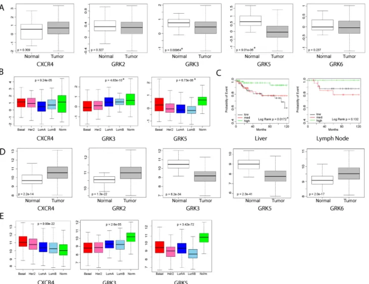
To determine whetherGRKexpression was different between the molecular breast cancer sub-types, genomic data from the TCGA database and publicly-available databases were analyzed as previously described by Harrell, et al [6]. Analysis of public human breast cancer microarray data shows that onlyGRK3andGRK5isotypes are expressed at statistically significant lower levels in tumor cells compared to normal breast tissue (Fig 1A). Also from this analysis,Fig 1B shows thatGRK3expression is lowest in the basal subtype (red box), whileGRK5was
decreased most in luminal subtypes (blue boxes). Although increased CXCR4 expression can indicate increased metastatic potential in some instances [30,31], differential expression of
CXCR4was not significant across these combined datasets (Fig 1A). When claudin-low and basal-type samples were grouped byGRK3expression level (Fig 1C), analysis of the human breast cancer data showed significant correlation of tumors expressing medium (black line) and low levels (red line) ofGRK3to liver metastasis and a similar trend toward lymph node metastasis, while tumors expressing high levels ofGRK3(green line) remained nearly metasta-sis-free. Similar results were seen in the analysis of the TCGA database (Fig 1D) where GRK3 and GRK5 again showed lower expression levels in tumor cells compared to normal breast tis-sue. Using this data, CXCR4 expression was shown to significantly increase in tumor cells com-pared to normal tissue as previously believed [30,31].Fig 1Eshows TCGA analysis comparing breast cancer subtypes and confirms that CXCR4 expression is highest and GRK3 is lowest in basal types, though these trends are seen in all the tumor subtypes to some degree. GRK5 expression is again shown to be downregulated the most in luminal B subtype, but could also be important in other types, including basal tumors. Taken together, there is potential clinical significance for decreasedGRK3expression in human basal breast cancer that could indicate metastatic potential.
3.2. GRK3 regulates CXCR4 function and the metastatic phenotype in
human TNBC cell lines
and DU4475 hadCXCR4:GRK3mRNA expression ratios of 7:1 and 6:1, respectively. Moder-ately invasive lines had ratios of 2:1 to 1:1 and the weakly invasive breast cancer lines had a
CXCR4:GRK3expression ratio less than 1 (Fig 2). Because the GPCR CXCR7 also binds to CXCL12 and has been shown to have a potential a role in tumor growth and metastasis [34], we also comparedCXCR7:GRK3expression ratios, but they did not correlate with invasive phe-notype (S4 Fig).
To further define the regulatory role of GRK3 in CXCL12/CXCR4-mediated function, the
CXCR4:GRK3ratios were experimentally altered by eitherGRK3overexpression or lentiviral transducedGRK3shRNA knockdown in the human cell lines 231 and MDA-MB-468, respectively. These two TNBC cell lines were chosen as comparators for being most different in intrinsicCXCR4:GRK3ratio (Fig 2) and invasive potential [32,33,35]. GRK3 was transiently overexpressed in the highly invasive MDA-MB-231 breast cancer cell line and
Fig 1. Genetic analysis of human breast cancer gene expression data associate GRK3 with tumor, basal subtype, and metastasis.Microarray datasets previously analyzed in Harrell, et al., were reanalyzed to (A) compareCXCR4andGRKexpression levels between normal breast tissue and tumor tissue, (B) determine the significance ofCXCR4,GRK3, andGRK5expression levels in human breast cancer subtypes, and (C) examine the association of
GRK3expression in metastasis to liver and lymph nodes. Microarray data in (C) was grouped based on relative level ofGRK3expression (low, medium, and high). (D) TCGA database was used to compareCXCR4andGRKexpression levels between normal breast tissue and tumor tissue, as in (A). (E) TCGA database was used to determine changes inCXCR4,GRK3, andGRK5expression levels in human breast cancer subtypes as in (B). Statistical significance determined by Students t-test (A,D), ANOVA (B,E), and Log-Rank test (C).
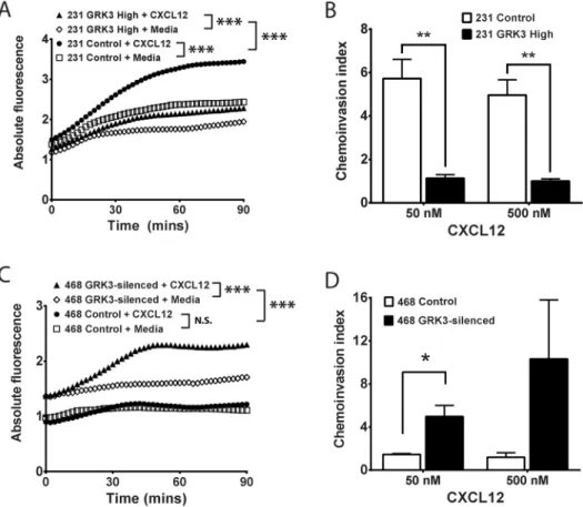
confirmed by Western Blot (S2 Fig). Though both control and GRK3-overexpressing MDA-MB-231 cells migrated significantly toward CXCL12, chemotaxis toward CXCL12 was significantly inhibited by GRK3 over expression compared to control transfected cells (Fig 3A). Given that metastasis is a multistep process that involves not only migration, but also invasion, we compared control and GRK3-overexpressing MDA-MB-231 invasion through matrix. GRK3-overexpressing MDA-MB-231 cells exhibited significantly less invasion compared to controls (Fig 3B).
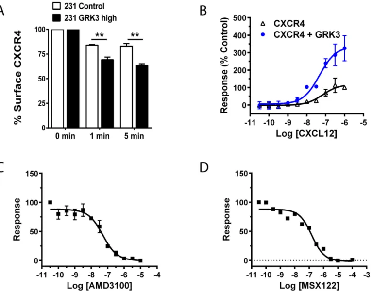
GRKs negatively regulates GPCR signaling by phosphorylation of terminal serine/threonine residues leading to receptor desensitization and internalization [11,16,17]. To confirm that overexpression of GRK3 was altering CXCL12/CXCR4 responses that culminated in increased internalization and removal of CXCR4 from the cell surface, MDA-MB-231 cells with overex-pressed GRK3 were compared to controls and had significantly more internalization of CXCR4 receptor in response to CXCL12 over time (Fig 4A).
Fig 2.CXCR4:GRK3ratio correlates with the invasiveness of human breast cancer lines.Human breast cancer lines were analyzed by quantitative real time PCR to determine the mRNA expression levels ofGRK3andCXCR4. Transcript copy number was determined using the standard curve method. Data shown are the average of two to four independent experiments. Error bars represent the SEM.
GPCR internalization following GRK phosphorylation is mediated by beta-arrestins [12, 36,37].
To show how GRK3 can influence CXCR4 internalization by recruitingβ-arrestin, we used the TANGO assay as described in the Materials and Methods.Fig 4Bshows the CXCL12 con-centration-dependent internalization of CXCR4 (black open triangles), which is significantly enhanced by the addition of GRK3 (blue closed circles). Known CXCR4 inhibitors AMD3100 (Fig 4C) and MSX-122 (Fig 4D) inhibit CXCL12-mediated arrestin recruitment in a concentra-tion-dependent manner. By comparison, the cancer-relevant receptor CXCR3, which has been shown to have functional interactions with CXCR4 [38–40], recruitsβ-arrestin in the TANGO assay when stimulated with its ligand CXCL11, but the response is not enhanced by GRK3 (S5A Fig). CXCL12 stimulation of CXCR3 does not recruitβ-arrestin, as expected (S5B Fig).
In contrast to MDA-MB-231, MDA-MB-468 are weakly-metastatic TNBC cells with a low
CXCR4:GRK3expression ratio (Fig 2), suggesting that GRK3 may be mitigating migration and
Fig 3. Alterations in GRK3 affect migratory responses of human breast cancer lines to CXCL12.GRK3 expression was altered by overexpression or shRNA silencing in human breast cancer lines. (A) Chemotaxis toward media or 50 nM CXCL12 of MDA-MB-231 transiently transfected with GRK3 or control plasmid was assessed by a real-time modified Transwell assay. Transfection efficiency was routinely 40%. Results shown are the mean of 5 independent experiments. (B) MDA-MB-231 invasion through Matrigel was analyzed after 24 hours and staining with Calcien-AM. The chemoinvasion index is defined as the ratio of relative
fluorescence of cells migrated toward CXCL12 over media control. Results shown are the mean of 3 independent experiments. (C) Chemotaxis toward media or 500nM CXCL12 of stably transduced MDA-MB-468 cells was assessed by a real-time modified Transwell assay. Results shown are the mean of 4
independent experiments. (D) MDA-MB-468 invasion through Matrigel was analyzed using a 96-well invasion assay. Results shown are the mean of 3 independent experiments. Error bars for all data represent the SEM. Statistical significance determined by analysis of covariance (ANCOVA) linear regression model (A and C) or by a two-tailed t-test (B and D):*p<0.05;**p<0.01;***p<0.001; N.S. not significant.
invasion through increased desensitization/internalization of CXCL12-activated CXCR4. To test this hypothesis, GRK3 was stably knocked down by lentiviral shRNA expression in MDA-MB-468 cells and confirmed by Western Blot (S2 Fig). Control MDA-MB-468 cells did not show statistically significant migration toward CXCL12. GRK3 knockdown in MDA-MB-468 cells significantly increasedin vitrochemotaxis (Fig 3C) and chemoinvasion (Fig 3D) toward CXCL12, thereby making their migratory phenotype more analogous to the aggressive MDA-MB-231 line. Taken together, the results indicate an important role for GRK3 in the
Fig 4. GRK3 regulates CXCL12-specific CXCR4 internalization andβ-arrestin recruitment.(A) Control cells (empty plasmid) and GRK3-overexpressed MDA-MB-231 cells were treated for the indicated times with 100 nM CXCL12 at 37°C. Surface expression of CXCR4 on the surface of cells was determined by flow cytometry. Data shown are the mean of three experiments normalized to the zero time point. Error bars represent SEM. Statistical analysis was performed using a two-tailed t-test.**p<0.01. (B) Using a TANGO arrestin-recruitment assay, HTLA cells were transfected with either CXCR4 alone or CXCR4 plus GRK3 as detailed in the Materials and Methods. Cells were plated in a 384 well plate and stimulated with CXCL12 at the indicated Molar concentrations. Luminescence was measured 24 hours post-stimulation. Error bars represent +/- SEM (n = 3). (C) TANGO results testing the CXCR4 antagonist AMD-3100. As in (B), HTLA cells were transfected with CXCR4 and GRK3 plasmids and stimulated with 10−7M CXCL12 (one concentration point above EC50) following pre-treatment with AMD3100 at the indicated Molar concentrations. (D) TANGO results testing the CXCR4 antagonist MSX-122.
HTLA cells were transfected with CXCR4 and GRK3 plasmids and stimulated with 10−7M CXCL12 following pre-treatment with MSX-122 at the indicated
Molar concentrations.
regulation of CXCL12/CXCR4-mediated functions and in the migratory phenotypes of human TNBC cell lines.
3.3. GRK3 gene silencing potentiates mammary tumor establishment
and metastasis
in vivo
To assess the role of GRK3 in metastasisin vivo, a syngeneic, immunocompetent mouse model of TNBC [21] was used. The mouse mammary tumor line 66cl4-luc is a weakly-metastatic, BALB/c tumor line expressing a luciferase construct that allows for tracking byin vivooptical imaging [24].GRK3mRNA and protein levels were silenced in the 66cl4-luc line by lentiviral shRNA transduction (S1 Fig) in an analogous fashion to silencing GRK3 in the non-invasive human MDA-MB-468 cells. Non-targeted control and GRK3-deficient 66cl4-luc cells were surgically implanted into the mammary fat pads of BALB/c mice and monitored by optical imaging for tumor growth and metastasis for six weeks, and organ-specific metastatic tumor burden was examined in detail by histopathology at experiment termination. Non-target con-trol 66cl4-luc cells established primary tumorsin vivo, but extended few metastases (Fig 5A andTable 1). In contrast, mice implanted with 66cl4-luc GRK3-deficient cells developed large primary tumors and distant metastases (Fig 5BandTable 1). Fluorescent intensity was ana-lyzed with Xenogen software and reported as average radiance (p/s/cm2/sr) to quantify primary and metastatic tumor burden over time. The analysis determined that primary tumor burden was not significantly different, albeit slightly increased on average, in GRK3-deficient tumors compared to controls throughout the 6-week period (Fig 5C). More notably, mice implanted with 66cl4-luc GRK3-deficient cells developed significantly more metastases at experiment ter-mination (Table 1andFig 5D). Histological data confirmed GRK3-deficient 66cl4 cells metas-tasized mostly to the lung, consistent with the model [28], and additionally to distant organ sites, such as liver, at a higher frequency (Table 1). Along with the optical imaging data, nec-ropsy of primary tumors showed that GRK3-deficient tumors displayed a different anatomy (Fig 5E), suggesting that these tumors may appear larger due to cell movement and resemble an invasive phenotype [41]. Thesein vivodata suggest that the metastasis of TNBC cells can be altered when the negative signaling regulator GRK3 is affected and that the sites of metastasis correlate with human data (Fig 1) as well as where CXCL12 is expressed.[7–9]
3.4. Silencing GRK3 expression does not alter cell proliferation, CXCL12
secretion, or anoikis
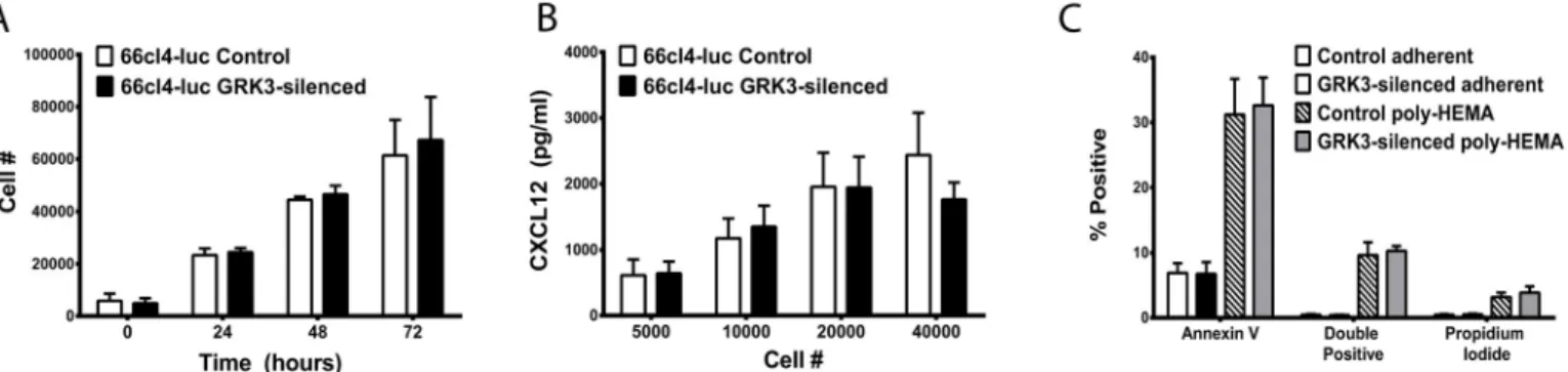
GRK3-deficient 66cl4-luc tumors appear larger in diameter than control tumors when visual-ized by optical imaging and necropsy (Fig 5), and studies by Woerner and colleagues have described an important role for GRK3 in regulating primary tumor growth in glioblastoma [14]. Since growth rates could impact metastatic potential in breast cancer, anin vitroassay was performed to assess intrinsic proliferation rates of GRK3-deficient and control 66cl4-luc cells. No significant difference was found in the proliferation of these 66cl4-luc cells inin
vitrocultures at baseline (Fig 6A) or in the presence of ligand CXCL12 or CXCR4-antagonist, AMD3100 (S6 Fig).
Autocrine stimulation from secreted CXCL12 has been shown to influence tumor behavior and phenotype [42], and CXCL12 expression in breast cancer cells has been correlated with decreased metastatic potential in humans [43]. To examine CXCL12 secretion from control and GRK3 deficient 66cl4-luc cells, CXCL12 in supernatants was measured by ELISA but was not found to be different between cell types (Fig 6B).
a process termed anoikis. Resistance to anoikis is a hallmark of metastatic progression in cancer [44]. Given that CXCR4 signaling is known to regulate anoikis in tumor progression [44,45], 66cl4-luc GRK3-deficient cells were compared to 66cl4-luc control cells for their apoptotic response to detachment; data showed that there was no intrinsic difference in anoikis between the different 66cl4-luc tumor typesin vitro(Fig 6C).
3.5. GRK3 deficiency increases migration of 66cl4-luc mammary tumor
cells to CXCL12
The analysis of human breast cancer cell lines in Figs2and3suggests that cells with higher
CXCR4:GRK3ratios are more metastatic because of increased migration and invasion toward CXCL12. Additionally, thein vivo66cl4-luc model shows increased distant metastasis of tumors to sites known to express high CXCL12 [7–9] after GRK3 silencing by lentiviral shRNA (Fig 5andTable 1). To determine whether GRK3-deficient 66cl4-luc metastasis could result from enhanced CXCL12/CXCR4-mediated migration, a kinetic chemotaxis assay was per-formed as previously described. Control 66cl4-luc cells did not migrate to CXCL12 over media
Fig 5. Increasing theCXCR4:GRK3ratio in 66cl4-luc mammary tumor cells increasesin vivometastasis.(A) Balb-c mice with 66cl4-luc control transduced cells visualized by optical imaging at 6 weeks post-implantation demonstrate only primary tumor growth in contrast to mice with 66cl4-luc GRK3 deficient cells (B), which show extensive and distant metastasis. Images shown inC. andD. are representative of two independent experiments.
Quantification of total (C) and metastatic (D) tumor in control versus GRK3-deficient tumors as imaged by luciferase activity. Data shown in (C) are n = 10 mice at weeks 0 and 2, n = 9 week 4, n = 7 week 6 (due to disease mortality). Data in (D) are n = 8 for Control, n = 7 for GRK3-deficient. (E) Necropsy of Balb-c mouse with Balb-control 66Balb-cl4-luBalb-c tumor Balb-cell implant showing small, enBalb-capsulated primary tumor (left panel). NeBalb-cropsy of animal with GRK3-silenBalb-ced 66Balb-cl4-luBalb-c tumor cell implant demonstrating larger, friable primary tumor and prominent neovascularization (right panel). All error bars represent SEM.
alone, while GRK3-deficient 66cl4-luc cells showed significantly enhanced directional migra-tion to CXCL12 (Fig 7A). Because 66cl4 cells are thought to perform poorly in standardin
vitroinvasion assays similar to those used for human cells inFig 3[28], a Cultrex 96-well tumor invasion plate was coated with a lower concentration basement membrane extract (0.2X BME) to assessin vitroinvasion. While detectable amounts of cells invaded the Matrigel, addi-tion of CXCL12 did not significantly increase the invasiveness of either control cells or GRK3 deficient 66cl4-luc cells over media alone (Fig 7B). Because breast cancer cells can have differ-ent behaviors and phenotypes in 3-dimensions versus 2-dimensions, 66cl4-luc cells were also tested in a 3D system of invasion (S7 Fig) with negative results similar to the 2D Transwell invasion assay.
4. Discussion
Breast cancer metastasis is known to be mediated in part by the CXCL12/CXCR4 signaling axis [7]. We now add G protein coupled receptor kinase 3 (GRK3) as an important regulator,
Table 1. GRK3-deficient 66cl4-luc mammary tumors disseminate distant metastasis.
Primary Tumor
Liver Metastasis
Lung Metastasis
Spleen Metastasis
Adrenal/Kidney Metastasis
Pancreas Metastasis
Skeletal Muscle Metastasis
GRK3 kd X X
GRK3 kd X X
GRK3 kd X X X X
GRK3 kd X
GRK3 kd X X X X X
GRK3 kd X X X
Control
Control X X
Control
Control X
Control
Control X X X
Control
doi:10.1371/journal.pone.0152856.t001
Fig 6. GRK3 deficient and control 66cl4-luc cells secrete soluble CXCL12, proliferate, and undergo apoptosis similarly.(A) Viable cell density was measured by colormetric assay (Cell Counting Kit-8, Dojindo) at indicated times and cell number estimated by standard curve (n = 3 + SEM). (B) Cells were plated in triplicate at the indicated number per well and incubated in culture conditions for 48 hours. CXCL12 levels in the supernatants were determined by sandwich ELISA and quantified by standard curve (n = 3; mean + SEM). (C) GRK3-deficient and control 66cl4-luc cells were incubated in adherent versus detached (poly-HEMA) conditions overnight and analyzed for apoptosis by flow cytometry for Annexin V and propidium iodide staining. Data is the mean of 4 independent experiments (error bars = SEM).
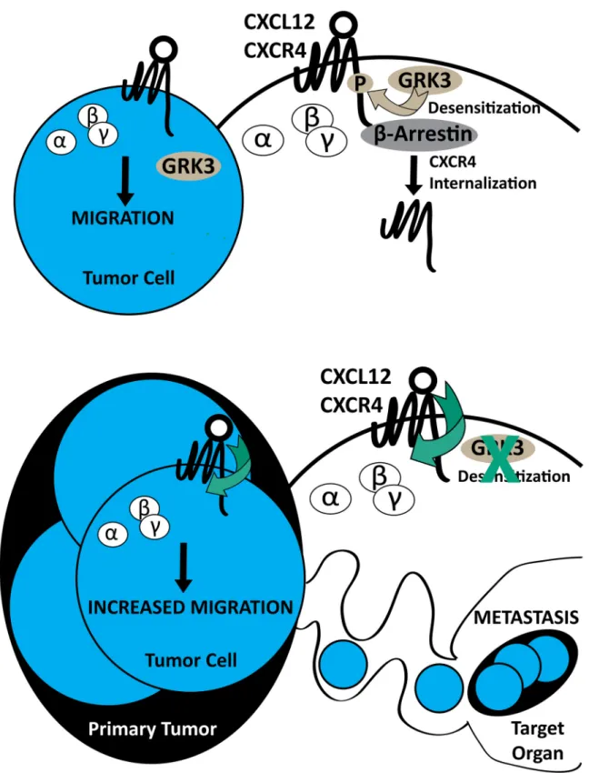
specifically with respect to invasion and metastasis, in human breast cancer tissue, TNBC cell lines, and in an immunocompetent mouse model. Previous work by others has suggested that quantitative levels of CXCR4 surface expression, as well as CXCL12 concentration and avail-ability, can affect the metastatic phenotype [9]. Recent evidence points to an even greater importance for CXCR4 expression in TNBC, which contributes to tumor size and metastatic potential as well as predicts poor prognosis in terms of overall and disease-free survival [46– 48]. However, further experiments have clarified that the magnitude of CXCR4 signaling, as perturbed by gene silencing, mutation, overexpression, or antagonism, is a key factor that affects increased migration, invasion, and metastasis [7–9,30,49]. In support of this argu-ment, Holland et al. showed that the absolute level of CXCR4 expressed on the surface of breast cancer cells alone did not accurately reflect or predict metastatic potential [10]. The data we present offers a mechanistic explanation for the role of CXCR4 signaling in breast cancer metastasis. The data show that relative GRK3 expression correlates with tumorgenicity, invasiveness, and metastatic potential in both human (Figs1,2and3) and mouse models (Fig 5andTable 1) and further, that manipulating GRK3 levels directly affects the chemotactic and metastatic potential of breast cancer cells (Figs3and7). Thus, we propose that high CXCR4 on the cell surface, combined with decreased intracellular regulatory GRK3, leads to impaired receptor internalization and more active CXCL12-mediated migration of the cancer cell (Fig 8).
We acknowledge that GRK3 in breast cancer is unlikely to be affecting CXCL12/CXCR4 interactions exclusivelyin vivoand could certainly be affecting additional chemokine receptors such as CXCR7 [50–52], CXCR3 [53,54], CCR7 [55–57], and others.[58] However, our data support a strong role for GRK3-mediated regulation of CXCL12 metastatic responses, arrestin-mediated recruitment, and CXCR4 receptor internalizationin vitro(Figs3,4and7). In con-trast, there is less compelling data for GRK3-mediated regulation of CXCR3 where we show that arrestin recruitment is independent (S5 Fig), and there was a lack of correlational expres-sion data supportingCXCR7:GRK3to the metastatic phenotype (S4 Fig). As genotyping has become an important tool to provide the prognosis and treatment of TNBC [59], the relative
Fig 7. GRK3-deficient 66cl4-luc mammary tumor cells have increased chemotaxis but not invasion.GRK3 was silenced by lentiviral shRNA in 66cl4-luc cells to examine the effect on migration. 100,000 cells were added per well to the top of a 96-well Fluoroblok Transwell plate, and 12.5nM (100ng/ ml) CXCL12 or media was in the lower chamber. Migration was monitored over 4 hours using a Fluoroskan Ascent FL plate reader. (A) Control transduced 66cl4-luc cells do not migrate toward CXCL12 (closed circle) over media control (open square) whereas GRK3 deficient 66cl4-luc cells display directional chemotaxis toward CXCL12 (closed triangle) over media control (open diamond). Statistics significance determined by analysis of covariance (ANCOVA) linear regression model:***p<0.001; N.S. not significant. (B) Control and GRK3 deficient 66cl4-luc cells were loaded into the upper chamber of Cultrex invasion plates in triplicate (seeMaterials and Methods). After addition of media alone or CXCL12 (50 or 100 nM) to the lower chamber, cells were incubated for 24 hours, stained with Calcein, and then counted for invasion into matrix (n = 3±SEM).
Fig 8. Summary model of GRK3 regulation of CXCR4-driven metastasis.Extracellular ligand stimulation of CXCL12 on G protein-coupled receptor (GPCR) CXCR4 elicits conformational changes of receptor, activation and dissociation of guanine nucleotide binding proteins, and downstream signaling for tumor cell migration (A). Negative regulation of surface receptor expression is mediated by G protein-coupled receptor kinase 3 (GRK3), which
gene expression of markers suchCXCR4andGRK3may provide additional prognostic infor-mation as it pertains specifically metastasis.
The metastatic potential of breast cancer cells is acutely sensitive to regulation of the CXCL12/CXCR4 signaling axis governed by receptor expression, desensitization, internaliza-tion, and recycling dynamics [9,18,19]. These properties of CXCR4 are directly influenced by phosphorylation by GRKs and subsequent downstream components, such asβ-arrestins [16, 17]. GRK3 has previously been shown to regulate tumor growth and proliferation in glioblas-toma (GBM). GRK3 expression is down-regulated by EGFR signaling in GBM compared to normal astrocytes, which leads to a corresponding increase in tumor growth.[14] Published data by others have also shown that down-regulation of GRK6 in medulloblastoma [15] and Lewis lung carcinoma [60] can similarly regulate tumor size, migration, and metastasis. In con-trast, our results correlate GRK3 with breast cancer metastatic potential via regulation of tumor cell migration and invasion (Figs3and7) without significant effect on tumor growth or detachment-induced death (Fig 6). These differences could potentially be explained by differ-ent cell types having altered GRK-specific phenotypes or redundancy based on tissue expres-sion and/or targeted GPCR-mediated regulation.
In vivobioluminescent imaging of 66cl4-luciferase tumors demonstrates an apparently larger, more diffuse tumor arising from GRK3-knockdown cells (Fig 5) despite similarity of cel-lular radiance quantification. One potential explanation is that the density of the tumor might be affected by the mobility of the individual GRK3-deficient tumor cells or by the recruitment of non-tumor cells into the tumor site. GRK3-deficient tumors also contained notable areas of necrotic tissue (data not shown). Since necrotic cells would no longer luminesce, it is also possi-ble that GRK3-deficient tumors did proliferate morein vivoin response to environmental cues not present in thein vitroassay, and that resultant tissue necrosis confounded optical quantifi-cation. The appearance of necrotic regions in primary tumors can also contribute to increased metastases [61,62]; however, this hypothesis was not directly tested in this study and remains a future endeavor.
High levels of CXCL12 known to exist in metastatic target tissues [7] raise the possibility that the functional outcome of altered GRK3 expression occurs distally from the primary tumor. Since 66cl4-luc cells are weakly invasive and this phenotype is unaltered byGRK3gene silencing (Fig 7B), we propose that the observation of increased metastases of GRK3-deficient 66cl4-luc cells is due to increased migration (Fig 7A), possiblywithinthe tumor microenviron-ment, leading to higher rates of intravasation that are controlled by other mechanisms. This interpretation is consistent with the larger, diffuse tumors imaged in mice implanted with 66cl4-luc GRK3-deficient cells (Fig 5). The sprawling, less dense tumor mass and increased chemotactic ability of the GRK3-deficient 66cl4-luc cells suggest that more motile cells within the primary tumor ultimately result in the increased distant metastases. However, our human cell line data do show an invasive phenotype, thus the findings using the 66cl4-luc cell line may be species and/or tumor specific. Future studies examining thein vivoandin vitromigratory, invasive, and metastatic potential of human derived breast cancer cells will be needed to see if these present results are generalizable to a broader array of malignant phenotypes.
growth by promoting angiogenesis [34]. The breast cancer cells used in the current study failed to demonstrate a correlation between CXCR7 levels and tumor cell migration or metastatic potential (S4 Fig). This may be due to low levels of CXCR7 expression in these cells. Further-more, current data implicate GRK2, not GRK3, as a potential regulator of arrestin-biased sig-naling through CXCR7, which fails to activate classic G protein pathways [64,66,67]. These authors acknowledge that GRK3 likely regulates other GPCRs that have relevance to cancer [14,68,69]; in agreement with their speculation, the data presented herein describe an impor-tant functional role for GRK3 in regulating CXCR4 that impacts metastatic breast cancer progression.
Most TNBC is associated with high CXCR4 expression, increased metastatic potential, and poor patient prognosis [47,48]. Our data emphasize the functional relationship of GRK3 as it pertains to CXCL12/CXCR4 migration in breast cancer and specific molecular subtypes. The analysis of human genetic data confirms potential involvement of GRK3 in tumor progression and metastasis (Fig 1). Across large datasets, decreasedGRK3expression correlated better with tumor (vs normal breast) than changes inCXCR4expression. In our resected mouse mammary tumors, theCXCR4:GRK3expression ratio was stable even after 6 weeks of tumor implantation and distant metastasis (S2 Fig), suggesting that theCXCR4:GRK3ratio is preserved and could be a useful prognostic indicator at extended times during the disease course. Others have proposed CXCR4 signaling and regulatory pathways as an attractive target for breast cancer treatment [8,10,48,70]. Considering the importance of GRK3 regulation of breast cancer migration, invasion, and metastasis described in this manuscript, understanding which tumors have more dysregulated signaling through CXCL12/CXCR4 could be used as a biomarker to predict which patients might respond better to CXCR4 antagonism or as an alternative, tar-geted therapy directed specifically at GRKs.
Supporting Information
S1 Fig. Altering theCXCR4:GRK3in 66cl4-luc mammary tumor cells byGRK3shRNA silencing.(A) 66cl4-luc murine mammary tumor cells were stably transduced with lentiviral GRK3 shRNA or non-target control plasmids and were analyzed by qRT-PCR to determine the mRNA expression levels ofGRK3andCXCR4after normalization to the IDUA housekeep-ing gene. Data is expressed as a ratio of CXCR4 to GRK3. 66cl4-luc Control n = 3, 66cl4-luc GRK3-deficient n = 4. (B) Representative GRK3 Western blot showing shRNA silenced GRK3-deficient 66cl4-luc cells compared to controls after immunoprecipitation. Blots were stripped and reprobed to confirm equal loading. Shown is actin blot of IP supernatant lanes. (C) Prior to implantation into Balb/c mice forin vivostudies, GRK3 is silenced approximately 50–60% in 66cl4-luc cells versus control as determined by qRT-PCR (n = 4). (D) GRK3 gene silencing was validated at 6 week experiment termination by qRT-PCT (n = 4). GRK3 mRNA expression was normalized to IDUA housekeeping gene and relative % expression determined byΔΔCTmethod. All error bars represent SEM.
(TIF)
GADPH in IP supernatants, bottom, confirm equal loading. (TIF)
S3 Fig. Surface receptor expression is not altered following stable GRK3 shRNA knockdown.
Human breast cancer line MDA-MB-468 (A, C) and murine breast cancer line 66cl4-luc (B, D) that have been stably transduced with GRK3 shRNA (red) or a control sequence (blue) and were stained for CXCR4 and CXCR3 and analyzed by flow cytometry. For human cells, antibod-ies used were: mouse anti-human CXCR4-PE (clone 12g5, isotype mouse 2a PE, Biolegend), anti-human CXCR3-APC (clone 49801, isotype mouse G1 APC, R&D Systems), and for mouse cells, rat anti-mouse CXCR4-PE (clone 2b11, isotype rat 2b PE, eBioscience) and rat anti-mouse CXCR3-APC (clone 220803, isotype rat 2a, R&D Systems). Negative controls were stained using equivalent amounts of isotype color controls. Shown are representative histograms of 3 independent experiments.
(TIF)
S4 Fig.CXCR7:GRK3relative copy number does not correlate with the metastatic potential of human breast cancer lines.Human breast cancer lines (in descending order moving from left to right of most highly metastatic to least metastatic) were analyzed by quantitative real time PCR to determine the mRNA expression levels ofGRK3andCXCR7. Transcript copy number was determined using the standard curve method. Data shown are the average of two to four independent experiments. Error bars represent the SEM.
(TIF)
S5 Fig. CXCR3 recruits arrestin independently of GRK3 in response to CXCL11 and does not recruit arrestin in response to CXCL12.(A) Using a TANGO arrestin-recruitment assay, HTLA cells were transfected with either CXCR3 alone or CXCR3 plus GRK3 as detailed in the Materials and Methods. Cells were plated in a 384 well plate and stimulated with CXCL11 at the indicated Molar concentrations. Luminescence was measured 24 hours post-stimulation. Error bars represent +/- SEM (n = 3). (B) HTLA cells were transfected with either CXCR3 alone or CXCR3 plus GRK3 as in (A). Cells were plated in a 384 well plate and stimulated with CXCL12 at the indicated Molar concentrations. Luminescence was measured 24 hours post-stimulation. Error bars represtent +/- SEM (n = 3).
(TIF)
S6 Fig. Addition of CXCL12 or AMD3100 does not alter the proliferation of 66cl4-luc cells over 72 hours.Viable cell density was measured by colormetric assay (Cell Counting Kit-8, Dojindo) at indicated times and cell number estimated by standard curve (n = 2 + SEM). 66cl4-luc non-target control (A) and GRK3-silenced (B) cell proliferation was tested with or without exogenous CXCL12 (100 ng/ml) added to the culture media. Since 66cl4-luc cells make large amounts of endogenous CXCL12 (Fig 6), 66cl4-luc non-target control (C) and GRK3-silenced (D) cell proliferation was also tested in presence or absence of CXCR4 antago-nist AMD3100 (5μg/ml).
(TIF)
is the mean of two independent experiments (error bars +/- SEM). (TIF)
S1 Methods. Supplemental Information.
(DOCX)
Acknowledgments
The authors wish to acknowledge the following funding sources in support of this research: North Carolina University Cancer Research Funds (UCRF) (TKT), K01 AI091863 (MJB), K08 AI070684 (TKT), R03AR059286 (TKT), NHLBI F31HL128029 (JMB), UNC Thurston Arthri-tis Research Center (TARC) Funds (TKT,MJB), NSF DMS-1106962 (YKT), and UNC Clinical Translation Science Award, Biostatistics Core TR000083-05 (YKT). The UNC Flow Cytometry Core Facility is supported in part by P30 CA016086 Cancer Center Core Support Grant to the UNC Lineberger Comprehensive Cancer Center. The authors also wish to acknowledge Dr. Bryan L. Roth for his technical expertise.
Author Contributions
Conceived and designed the experiments: TT MB DF JP NKD. Performed the experiments: MB DF MM DSS RT JP TT. Analyzed the data: MB DF MM DSS RT TT JS NKD JP RL YKT MFS. Contributed reagents/materials/analysis tools: GS TT JP JS JB RL MB DF MFS YKT. Wrote the paper: MB DF TT JB DSS RT MM.
References
1. Cleere DW. Triple-negative breast cancer: a clinical update. Community Oncology. 2010; 7(5). 2. Jemal A, Bray F, Center MM, Ferlay J, Ward E, Forman D. Global cancer statistics. CA: a cancer journal
for clinicians. 2011; 61(2):69–90. Epub 2011/02/08. doi:10.3322/caac.20107PMID:21296855. 3. Prat A, Parker JS, Karginova O, Fan C, Livasy C, Herschkowitz JI, et al. Phenotypic and molecular
characterization of the claudin-low intrinsic subtype of breast cancer. Breast cancer research: BCR. 2010; 12(5):R68. Epub 2010/09/04. doi:10.1186/bcr2635PMID:20813035; PubMed Central PMCID: PMC3096954.
4. Andre F, Zielinski CC. Optimal strategies for the treatment of metastatic triple-negative breast cancer with currently approved agents. Annals of oncology: official journal of the European Society for Medical Oncology / ESMO. 2012; 23 Suppl 6:vi46–51. Epub 2012/10/04. doi:10.1093/annonc/mds195PMID: 23012302.
5. Duffy MJ, McGowan PM, Crown J. Targeted therapy for triple-negative breast cancer: where are we? International journal of cancer Journal international du cancer. 2012; 131(11):2471–7. Epub 2012/05/ 15. doi:10.1002/ijc.27632PMID:22581656.
6. Harrell JC, Prat A, Parker JS, Fan C, He X, Carey L, et al. Genomic analysis identifies unique signa-tures predictive of brain, lung, and liver relapse. Breast cancer research and treatment. 2012; 132 (2):523–35. Epub 2011/06/15. doi:10.1007/s10549-011-1619-7PMID:21671017; PubMed Central PMCID: PMC3303043.
7. Muller A, Homey B, Soto H, Ge N, Catron D, Buchanan ME, et al. Involvement of chemokine receptors in breast cancer metastasis. Nature. 2001; 410(6824):50–6. Epub 2001/03/10.
8. Rhodes LV, Short SP, Neel NF, Salvo VA, Zhu Y, Elliott S, et al. Cytokine receptor CXCR4 mediates estrogen-independent tumorigenesis, metastasis, and resistance to endocrine therapy in human breast cancer. Cancer research. 2011; 71(2):603–13. Epub 2010/12/03. doi: 10.1158/0008-5472.CAN-10-3185PMID:21123450; PubMed Central PMCID: PMC3140407.
9. Smith MC, Luker KE, Garbow JR, Prior JL, Jackson E, Piwnica-Worms D, et al. CXCR4 regulates growth of both primary and metastatic breast cancer. Cancer research. 2004; 64(23):8604–12. Epub 2004/12/03. doi:10.1158/0008-5472.CAN-04-1844PMID:15574767.
11. Busillo JM, Armando S, Sengupta R, Meucci O, Bouvier M, Benovic JL. Site-specific phosphorylation of CXCR4 is dynamically regulated by multiple kinases and results in differential modulation of CXCR4 signaling. The Journal of biological chemistry. 2010; 285(10):7805–17. Epub 2010/01/06. doi:10.1074/ jbc.M109.091173PMID:20048153; PubMed Central PMCID: PMC2844224.
12. Lefkowitz RJ. G protein-coupled receptors. III. New roles for receptor kinases and beta-arrestins in receptor signaling and desensitization. The Journal of biological chemistry. 1998; 273(30):18677–80. Epub 1998/07/21. PMID:9668034.
13. King DW, Steinmetz R, Wagoner HA, Hannon TS, Chen LY, Eugster EA, et al. Differential expression of GRK isoforms in nonmalignant and malignant human granulosa cells. Endocrine. 2003; 22(2):135– 42. Epub 2003/12/11. doi:10.1385/ENDO:22:2:135PMID:14665717.
14. Woerner BM, Luo J, Brown KR, Jackson E, Dahiya SM, Mischel P, et al. Suppression of G-protein-cou-pled receptor kinase 3 expression is a feature of classical GBM that is required for maximal growth. Molecular cancer research: MCR. 2012; 10(1):156–66. Epub 2011/11/17. doi:10.1158/1541-7786. MCR-11-0411PMID:22086906; PubMed Central PMCID: PMC3262072.
15. Yuan L, Zhang H, Liu J, Rubin JB, Cho YJ, Shu HK, et al. Growth factor receptor-Src-mediated sup-pression of GRK6 dysregulates CXCR4 signaling and promotes medulloblastoma migration. Molecular cancer. 2013; 12:18. Epub 2013/03/19. doi:10.1186/1476-4598-12-18PMID:23497290; PubMed Cen-tral PMCID: PMC3599655.
16. Balabanian K, Levoye A, Klemm L, Lagane B, Hermine O, Harriague J, et al. Leukocyte analysis from WHIM syndrome patients reveals a pivotal role for GRK3 in CXCR4 signaling. The Journal of clinical investigation. 2008; 118(3):1074–84. Epub 2008/02/16. doi:10.1172/JCI33187PMID:18274673; PubMed Central PMCID: PMC2242619.
17. Tarrant TK, Billard MJ, Timoshchenko RG, McGinnis MW, Serafin DS, Foreman O, et al. G protein-cou-pled receptor kinase-3-deficient mice exhibit WHIM syndrome features and attenuated inflammatory responses. Journal of leukocyte biology. 2013. Epub 2013/08/13. doi:10.1189/jlb.0213097PMID: 23935208.
18. Lapteva N, Yang AG, Sanders DE, Strube RW, Chen SY. CXCR4 knockdown by small interfering RNA abrogates breast tumor growth in vivo. Cancer gene therapy. 2005; 12(1):84–9. Epub 2004/10/09. doi: 10.1038/sj.cgt.7700770PMID:15472715.
19. Yang P, Liang SX, Huang WH, Zhang HW, Li XL, Xie LH, et al. Aberrant expression of CXCR4 signifi-cantly contributes to metastasis and predicts poor clinical outcome in breast cancer. Current molecular medicine. 2014; 14(1):174–84. Epub 2013/11/22. PMID:24256053.
20. Finn RS, Dering J, Conklin D, Kalous O, Cohen DJ, Desai AJ, et al. PD 0332991, a selective cyclin D kinase 4/6 inhibitor, preferentially inhibits proliferation of luminal estrogen receptor-positive human breast cancer cell lines in vitro. Breast cancer research: BCR. 2009; 11(5):R77. Epub 2009/10/31. doi: 10.1186/bcr2419PMID:19874578; PubMed Central PMCID: PMC2790859.
21. Phoenix KN, Vumbaca F, Fox MM, Evans R, Claffey KP. Dietary energy availability affects primary and metastatic breast cancer and metformin efficacy. Breast cancer research and treatment. 2010; 123 (2):333–44. Epub 2010/03/06. doi:10.1007/s10549-009-0647-zPMID:20204498; PubMed Central PMCID: PMC2888909.
22. Benito M, Parker J, Du Q, Wu J, Xiang D, Perou CM, et al. Adjustment of systematic microarray data biases. Bioinformatics. 2004; 20(1):105–14. Epub 2003/12/25. PMID:14693816.
23. Parker JS, Mullins M, Cheang MC, Leung S, Voduc D, Vickery T, et al. Supervised risk predictor of breast cancer based on intrinsic subtypes. Journal of clinical oncology: official journal of the American Society of Clinical Oncology. 2009; 27(8):1160–7. Epub 2009/02/11. doi:10.1200/JCO.2008.18.1370 PMID:19204204; PubMed Central PMCID: PMC2667820.
24. Li J, Sahagian GG. Demonstration of tumor suppression by mannose 6-phosphate/insulin-like growth factor 2 receptor. Oncogene. 2004; 23(58):9359–68. Epub 2004/11/16. doi:10.1038/sj.onc.1208039 PMID:15543235.
25. Rutledge RG, Cote C. Mathematics of quantitative kinetic PCR and the application of standard curves. Nucleic acids research. 2003; 31(16):e93. Epub 2003/08/09. PMID:12907745; PubMed Central PMCID: PMC169985.
26. Barnea G, Strapps W, Herrada G, Berman Y, Ong J, Kloss B, et al. The genetic design of signaling cas-cades to record receptor activation. Proceedings of the National Academy of Sciences of the United States of America. 2008; 105(1):64–9. Epub 2008/01/01. doi:10.1073/pnas.0710487105PMID: 18165312; PubMed Central PMCID: PMC2224232.
28. Lou Y, Preobrazhenska O, auf dem Keller U, Sutcliffe M, Barclay L, McDonald PC, et al. Epithelial-mes-enchymal transition (EMT) is not sufficient for spontaneous murine breast cancer metastasis. Develop-mental dynamics: an official publication of the American Association of Anatomists. 2008; 237 (10):2755–68. Epub 2008/09/06. doi:10.1002/dvdy.21658PMID:18773493.
29. Diaz-Montero CM, McIntyre BW. Acquisition of anoikis resistance in human osteosarcoma cells does not alter sensitivity to chemotherapeutic agents. BMC cancer. 2005; 5:39. Epub 2005/04/15. doi:10. 1186/1471-2407-5-39PMID:15829011; PubMed Central PMCID: PMC1090563.
30. Li YM, Pan Y, Wei Y, Cheng X, Zhou BP, Tan M, et al. Upregulation of CXCR4 is essential for HER2-mediated tumor metastasis. Cancer cell. 2004; 6(5):459–69. Epub 2004/11/16. doi:10.1016/j.ccr.2004. 09.027PMID:15542430.
31. Schioppa T, Uranchimeg B, Saccani A, Biswas SK, Doni A, Rapisarda A, et al. Regulation of the che-mokine receptor CXCR4 by hypoxia. The Journal of experimental medicine. 2003; 198(9):1391–402. Epub 2003/11/05. doi:10.1084/jem.20030267PMID:14597738; PubMed Central PMCID:
PMC2194248.
32. Lacroix M, Leclercq G. Relevance of breast cancer cell lines as models for breast tumours: an update. Breast cancer research and treatment. 2004; 83(3):249–89. Epub 2004/02/06. doi:10.1023/B:BREA. 0000014042.54925.ccPMID:14758095.
33. Neve RM, Chin K, Fridlyand J, Yeh J, Baehner FL, Fevr T, et al. A collection of breast cancer cell lines for the study of functionally distinct cancer subtypes. Cancer cell. 2006; 10(6):515–27. Epub 2006/12/ 13. PubMed Central PMCID: PMC2730521.
34. Hernandez L, Magalhaes MA, Coniglio SJ, Condeelis JS, Segall JE. Opposing roles of CXCR4 and CXCR7 in breast cancer metastasis. Breast cancer research: BCR. 2011; 13(6):R128. Epub 2011/12/ 14. doi:10.1186/bcr3074PMID:22152016; PubMed Central PMCID: PMC3326570.
35. Prat A, Karginova O, Parker JS, Fan C, He X, Bixby L, et al. Characterization of cell lines derived from breast cancers and normal mammary tissues for the study of the intrinsic molecular subtypes. Breast cancer research and treatment. 2013; 142(2):237–55. Epub 2013/10/29. doi: 10.1007/s10549-013-2743-3PMID:24162158; PubMed Central PMCID: PMC3832776.
36. Ferguson SS, Downey WE 3rd, Colapietro AM, Barak LS, Menard L, Caron MG. Role of beta-arrestin in mediating agonist-promoted G protein-coupled receptor internalization. Science. 1996; 271
(5247):363–6. Epub 1996/01/19. PMID:8553074.
37. Zhang J, Ferguson SS, Barak LS, Menard L, Caron MG. Dynamin and beta-arrestin reveal distinct mechanisms for G protein-coupled receptor internalization. The Journal of biological chemistry. 1996; 271(31):18302–5. Epub 1996/08/02. PMID:8702465.
38. Costantini S, Raucci R, De Vero T, Castello G, Colonna G. Common structural interactions between the receptors CXCR3, CXCR4 and CXCR7 complexed with their natural ligands, CXCL11 and CXCL12, by a modeling approach. Cytokine. 2013; 64(1):316–21. Epub 2013/06/19. doi:10.1016/j. cyto.2013.05.024PMID:23773308.
39. Singh AK, Arya RK, Trivedi AK, Sanyal S, Baral R, Dormond O, et al. Chemokine receptor trio: CXCR3, CXCR4 and CXCR7 crosstalk via CXCL11 and CXCL12. Cytokine & growth factor reviews. 2013; 24 (1):41–9. Epub 2012/09/20. doi:10.1016/j.cytogfr.2012.08.007PMID:22989616.
40. Watts AO, van Lipzig MM, Jaeger WC, Seeber RM, van Zwam M, Vinet J, et al. Identification and profil-ing of CXCR3-CXCR4 chemokine receptor heteromer complexes. British journal of pharmacology. 2013; 168(7):1662–74. Epub 2012/11/23. doi:10.1111/bph.12064PMID:23170857; PubMed Central PMCID: PMC3605874.
41. McSherry EA, Donatello S, Hopkins AM, McDonnell S. Molecular basis of invasion in breast cancer. Cellular and molecular life sciences: CMLS. 2007; 64(24):3201–18. Epub 2007/10/25. doi:10.1007/ s00018-007-7388-0PMID:17957337.
42. Sauve K, Lepage J, Sanchez M, Heveker N, Tremblay A. Positive feedback activation of estrogen receptors by the CXCL12-CXCR4 pathway. Cancer research. 2009; 69(14):5793–800. Epub 2009/07/ 09. doi:10.1158/0008-5472.CAN-08-4924PMID:19584281.
43. Mirisola V, Zuccarino A, Bachmeier BE, Sormani MP, Falter J, Nerlich A, et al. CXCL12/SDF1 expres-sion by breast cancers is an independent prognostic marker of disease-free and overall survival. Eur J Cancer. 2009; 45(14):2579–87. Epub 2009/08/04. doi:10.1016/j.ejca.2009.06.026PMID:19646861. 44. Drury LJ, Wendt MK, Dwinell MB. CXCL12 chemokine expression and secretion regulates colorectal
carcinoma cell anoikis through Bim-mediated intrinsic apoptosis. PloS one. 2010; 5(9):e12895. Epub 2010/09/30. doi:10.1371/journal.pone.0012895PMID:20877573; PubMed Central PMCID: PMC2943927.
46. Chen HW, Du CW, Wei XL, Khoo US, Zhang GJ. Cytoplasmic CXCR4 high-expression exhibits distinct poor clinicopathological characteristics and predicts poor prognosis in triple-negative breast cancer. Current molecular medicine. 2013; 13(3):410–6. Epub 2013/01/22. PMID:23331013.
47. Chu QD, Panu L, Holm NT, Li BD, Johnson LW, Zhang S. High chemokine receptor CXCR4 level in tri-ple negative breast cancer specimens predicts poor clinical outcome. The Journal of surgical research. 2010; 159(2):689–95. Epub 2009/06/09. doi:10.1016/j.jss.2008.09.020PMID:19500800.
48. Gil M, Seshadri M, Komorowski MP, Abrams SI, Kozbor D. Targeting CXCL12/CXCR4 signaling with oncolytic virotherapy disrupts tumor vasculature and inhibits breast cancer metastases. Proceedings of the National Academy of Sciences of the United States of America. 2013; 110(14):E1291–300. Epub 2013/03/20. doi:10.1073/pnas.1220580110PMID:23509246; PubMed Central PMCID:
PMC3619300.
49. Ueda Y, Neel NF, Schutyser E, Raman D, Richmond A. Deletion of the COOH-terminal domain of CXC chemokine receptor 4 leads to the down-regulation of cell-to-cell contact, enhanced motility and prolifer-ation in breast carcinoma cells. Cancer research. 2006; 66(11):5665–75. Epub 2006/06/03. doi:10. 1158/0008-5472.CAN-05-3579PMID:16740704; PubMed Central PMCID: PMC2664111. 50. Salazar N, Munoz D, Kallifatidis G, Singh RK, Jorda M, Lokeshwar BL. The chemokine receptor
CXCR7 interacts with EGFR to promote breast cancer cell proliferation. Molecular cancer. 2014; 13:198. Epub 2014/08/30. doi:10.1186/1476-4598-13-198PMID:25168820; PubMed Central PMCID: PMC4167278.
51. Stacer AC, Fenner J, Cavnar SP, Xiao A, Zhao S, Chang SL, et al. Endothelial CXCR7 regulates breast cancer metastasis. Oncogene. 2015. Epub 2015/06/30. doi:10.1038/onc.2015.236PMID:26119946; PubMed Central PMCID: PMC4486335.
52. Wani N, Nasser MW, Ahirwar DK, Zhao H, Miao Z, Shilo K, et al. C-X-C motif chemokine 12/C-X-C che-mokine receptor type 7 signaling regulates breast cancer growth and metastasis by modulating the tumor microenvironment. Breast cancer research: BCR. 2014; 16(3):R54. Epub 2014/06/03. doi:10. 1186/bcr3665PMID:24886617; PubMed Central PMCID: PMC4076630.
53. Norton KA, Popel AS, Pandey NB. Heterogeneity of chemokine cell-surface receptor expression in tri-ple-negative breast cancer. American journal of cancer research. 2015; 5(4):1295–307. Epub 2015/06/ 24. PMID:26101698; PubMed Central PMCID: PMC4473311.
54. Zhu G, Yan HH, Pang Y, Jian J, Achyut BR, Liang X, et al. CXCR3 as a molecular target in breast can-cer metastasis: inhibition of tumor cell migration and promotion of host anti-tumor immunity. Oncotarget. 2015; 6(41):43408–19. Epub 2015/10/21.
55. Li F, Zou Z, Suo N, Zhang Z, Wan F, Zhong G, et al. CCL21/CCR7 axis activating chemotaxis accompa-nied with epithelial-mesenchymal transition in human breast carcinoma. Med Oncol. 2014; 31(9):180. Epub 2014/08/22. doi:10.1007/s12032-014-0180-8PMID:25142946.
56. Pang MF, Georgoudaki AM, Lambut L, Johansson J, Tabor V, Hagikura K, et al. TGF-beta1-induced EMT promotes targeted migration of breast cancer cells through the lymphatic system by the activation of CCR7/CCL21-mediated chemotaxis. Oncogene. 2016; 35(6):748–60. Epub 2015/05/12. doi:10. 1038/onc.2015.133PMID:25961925.
57. Tutunea-Fatan E, Majumder M, Xin X, Lala PK. The role of CCL21/CCR7 chemokine axis in breast can-cer-induced lymphangiogenesis. Molecular cancer. 2015; 14:35. Epub 2015/03/10. doi:10.1186/ s12943-015-0306-4PMID:25744065; PubMed Central PMCID: PMC4339430.
58. Mishan MA, Heirani-Tabasi A, Mokhberian N, Hassanzade M, Kalalian Moghaddam H, Bahrami AR, et al. Analysis of Chemokine Receptor Gene Expression in Esophageal Cancer Cells Compared with Breast Cancer with Insights into Metastasis. Iranian journal of public health. 2015; 44(10):1353–8. Epub 2015/11/18. PMID:26576348; PubMed Central PMCID: PMC4644580.
59. Stevens KN, Vachon CM, Couch FJ. Genetic susceptibility to triple-negative breast cancer. Cancer research. 2013; 73(7):2025–30. Epub 2013/03/29. doi:10.1158/0008-5472.CAN-12-1699PMID: 23536562; PubMed Central PMCID: PMC3654815.
60. Raghuwanshi SK, Smith N, Rivers EJ, Thomas AJ, Sutton N, Hu Y, et al. G protein-coupled receptor kinase 6 deficiency promotes angiogenesis, tumor progression, and metastasis. J Immunol. 2013; 190 (10):5329–36. Epub 2013/04/17. doi:10.4049/jimmunol.1202058PMID:23589623; PubMed Central PMCID: PMC3646980.
61. Bonfil RD, Bustuoabad OD, Ruggiero RA, Meiss RP, Pasqualini CD. Tumor necrosis can facilitate the appearance of metastases. Clinical & experimental metastasis. 1988; 6(2):121–9. Epub 1988/03/01. PMID:3345611.
63. Sun X, Cheng G, Hao M, Zheng J, Zhou X, Zhang J, et al. CXCL12 / CXCR4 / CXCR7 chemokine axis and cancer progression. Cancer metastasis reviews. 2010; 29(4):709–22. Epub 2010/09/15. doi:10. 1007/s10555-010-9256-xPMID:20839032; PubMed Central PMCID: PMC3175097.
64. Rajagopal S, Kim J, Ahn S, Craig S, Lam CM, Gerard NP, et al. Beta-arrestin- but not G protein-medi-ated signaling by the "decoy" receptor CXCR7. Proceedings of the National Academy of Sciences of the United States of America. 2010; 107(2):628–32. Epub 2009/12/19. doi:10.1073/pnas.0912852107 PMID:20018651; PubMed Central PMCID: PMC2818968.
65. Miao Z, Luker KE, Summers BC, Berahovich R, Bhojani MS, Rehemtulla A, et al. CXCR7 (RDC1) pro-motes breast and lung tumor growth in vivo and is expressed on tumor-associated vasculature. Pro-ceedings of the National Academy of Sciences of the United States of America. 2007; 104(40):15735– 40. Epub 2007/09/28. doi:10.1073/pnas.0610444104PMID:17898181; PubMed Central PMCID: PMC1994579.
66. Lipfert J, Odemis V, Engele J. Grk2 is an essential regulator of CXCR7 signalling in astrocytes. Cellular and molecular neurobiology. 2013; 33(1):111–8. Epub 2012/09/04. doi:10.1007/s10571-012-9876-5 PMID:22940879.
67. Luker KE, Gupta M, Steele JM, Foerster BR, Luker GD. Imaging ligand-dependent activation of CXCR7. Neoplasia. 2009; 11(10):1022–35. Epub 2009/10/02. PMID:19794961; PubMed Central PMCID: PMC2745668.
68. Desai AN, Salim S, Standifer KM, Eikenburg DC. Involvement of G protein-coupled receptor kinase (GRK) 3 and GRK2 in down-regulation of the alpha2B-adrenoceptor. The Journal of pharmacology and experimental therapeutics. 2006; 317(3):1027–35. Epub 2006/03/15. doi:10.1124/jpet.105.098996 PMID:16533872.
69. Salim S, Standifer KM, Eikenburg DC. Extracellular signal-regulated kinase 1/2-mediated transcrip-tional regulation of G-protein-coupled receptor kinase 3 expression in neuronal cells. The Journal of pharmacology and experimental therapeutics. 2007; 321(1):51–9. Epub 2007/01/27. doi:10.1124/jpet. 106.116921PMID:17255468.
70. Duda DG, Kozin SV, Kirkpatrick ND, Xu L, Fukumura D, Jain RK. CXCL12 (SDF1alpha)-CXCR4/ CXCR7 pathway inhibition: an emerging sensitizer for anticancer therapies? Clinical cancer research: an official journal of the American Association for Cancer Research. 2011; 17(8):2074–80. Epub 2011/ 02/26. doi:10.1158/1078-0432.CCR-10-2636PMID:21349998; PubMed Central PMCID: