ANALYSIS OF THE P53 REGULATOR MDM2 AND THE IDENTIFICATION OF THE NOVEL P53 TARGET GENE LRP1
Patrick Lee Leslie
A dissertation submitted to the faculty at the University of North Carolina at Chapel Hill in partial fulfillment of the requirements for the degree of Doctor of Philosophy in the
Curriculum in Genetics and Molecular Biology in the School of Medicine.
Chapel Hill 2016
ABSTRACT
Patrick Lee Leslie: Analysis of the p53 regulator MDM2 and identification of the p53 target gene LRP1
(Under the direction of Yanping Zhang)
The transcription factor p53 responds to many stresses and regulates many
different pathways. The earliest characterized functions of p53 include the induction of cell cycle arrest, apoptosis, and senescence; however, more recent studies have shown that p53 regulates other pathways, including lipid and glucose metabolism, DNA damage repair, and autophagy. While the activation of many of these pathways likely overlaps in many
homooligomerization and MDM2-‐MDMX heterooligomerization, my results show that MDM2 RING structural mutations that prevent MDM2 enzymatic function and MDMX binding retain the ability to homooligomerize. Interestingly, deletion of the regulatory central acidic domain of MDM2 inhibits the ability of MDM2 to homooligomerize but does not impede its ability to heterooligomerize with MDMX, suggesting that MDM2-‐MDM2 homooligomerization and MDM2-‐MDMX heterooligomerization occur through different mechanisms. In another study, I identified the gene low-‐density lipoprotein receptor related protein 1 (LRP1) as a novel p53 target gene. Further analysis revealed that LRP1 protein induction occurs in response to sub-‐lethal but not lethal p53-‐activating stresses.
Interestingly, although lethal p53-‐activating stress can induce LRP1 transcription, protein expression is impeded at the translational level. Collectively, these studies contribute to our knowledge of p53 regulation as well as the p53 regulome.
ACKNOWLEDGEMENTS
I could not have accomplished this dissertation without significant contributions from many other sources. I thank past and present members of the Zhang lab for technical expertise, helpful suggestions, and the cameraderie that made my time in graduate school worthwhile. In particular, Dr. Yong Liu served a major part in my growth as a scientist, and I am indebted for his assistance and advice on numerous aspects of my dissertation. I thank my mentor, Dr. Yanping Zhang, for allowing me the independence to explore new areas of research and to follow the data wherever it led, even if it was unfamiliar territory for the lab.
I also thank my dissertation committee members, Drs. Albert Baldwin, Adrienne Cox, Jay Brenman, and Michael Emanuele, for sharing their knowledge and advice on not only my research but also my professional development.
Finally, I thank my family, friends, and loved ones for providing support as well as an outlet through which I could put science aside every once in a while to relax and
recharge. My father, Barry Leslie, is the epitome of reliability and has always been there to support all of his children in whatever endeavors we decided to pursue. My mother, Rachel Leslie, who lost her battle with breast cancer in 2006, inspired my academic pursuit to contribute to the search for a better understanding (and hopefully better treatments) of one of the most deadly and ubiquitous diseases known to man-‐this dissertation is
the pursuit of perfection. My sister, Jennifer, and my brothers, Eric, Ed, Alan, and Tim, contributed to my competitive nature. The rest of my family has been wonderful as well, and I would like to present my appreciation and love to them all. My (future) extended family in North Carolina has been equally wonderful and supportive; I am very happy to have them all in my life. Finally, and most importantly, my soon-‐to-‐be wife, Carly, provided immeasurable support throughout my graduate school years, especially during late nights in the lab. She is my best friend, and I cannot wait to experience the rest of our lives together.
TABLE OF CONTENTS
LIST OF FIGURES ... xi
LIST OF ABBREVIATIONS ... xv
CHAPTER 1: INTRODUCTION ... 1
Discovery of p53 ... 3
Function of p53 ... 4
Regulation of p53 ... 7
p53 phosphorylation ... 9
p53 acetylation ... 11
p53 ubiquitination ... 12
p53 methylation ... 13
p53 differential gene activation ... 14
p53 threshold model ... 15
Differences in p53 response element ... 16
p53 post-‐translational modifications ... 17
p53 dynamic pattern ... 19
p53 co-‐regulation ... 20
p53 miRNA ... 21
What p53 programs are important for tumor suppression? ... 22
Conclusions related to p53 function and regulation ... 28
Discovery of MDM2 and MDMX ... 28
Function of MDM2 and MDMX ... 29
Background of MDM2 and MDMX ... 30
MDM2/MDMX oligomerization in vitro studies ... 34
Regulation of MDM2(/MDMX) oligomers ... 36
MDM2-‐MDMX heterooligomers are required in vivo ... 38
MDM2-‐MDMX heteroligomers are important, what about MDM2 homooligomerization? ... 43
Targeting the MDM2/MDMX oligomer ... 44
Expanding the tumor spectrum of MDM2/MDMX inhibitors ... 48
Concluding remarks on MDM2/MDMX ... 49
Dissertation questions and goals ... 50
How do different mutations in MDM2 affect its oligomerization capacity and its ability to degrade p53? ... 50
Identify novel p53 targets and determine how it fits into p53 regulation ... 50
Figures ... 52
CHAPTER 2: THE MDM2 ACIDIC DOMAIN AND RING DOMAIN PLAY DISTINCT ROLES IN MDM2-‐MDM2 HOMODIMERIZATION AND MDM2-‐MDMX HETERODIMERIZATION ... 58
Introduction ... 58
Results ... 60
MDM2 residue N447 is important for p53 degradation ... 60
RING-‐disruptive mutations inhibit MDM2 binding to MDMX
but not MDM2 ... 62
Deletion of extreme C-‐terminal residues prevents MDM2 interaction with MDM2 and MDMX ... 63
MDM2 acidic domain is necessary for interaction with MDM2 but not MDMX ... 64
Discussion ... 66
Experimental procedures ... 70
Figures ... 75
CHAPTER 3: LRP1 IS A NOVEL P53 TARGET GENE WHOSE PROTEIN PRODUCT IS INDUCED IN RESPONSE TO SUB-‐LETHAL STRESS ... 88
Introduction ... 88
Results ... 90
p53 induces LRP1 expression ... 90
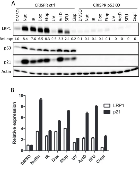
LRP1 is induced by several p53-‐activating stresses ... 92
Sub-‐lethal stresses induce LRP1 expression ... 93
Sub-‐lethal but not lethal doxorubicin induces LRP1 protein expression ... 95
LRP1 protein could be suppressed in a miRNA-‐dependent manner in response to lethal doxorubicin ... 98
LRP1 deletion confers a growth and survival advantage to established colon cancer cell line HCT116 in response to IR stress ... 99
Discussion ... 100
Experimental procedures ... 103
Figures ... 113
What could explain the difference in MDM2 homooligomerization
and MDM2-‐MDMX heterooligomerization? ... 143
Do MDM2-‐MDM2 homooligomers and MDM2-‐MDMX heterooligomers perform different functions in vivo? ... 145
How are p53-‐independent functions of MDM2 relative to MDM2-‐MDM2 homooligomerization and MDM2-‐MDMX heterooligomerization? ... 147
LRP1 is a novel p53 target gene that shows context-‐dependent protein induction involving a translation regulatory mechanism ... 149
Are miRNAs responsible for LRP1 dose-‐dependent expression? ... 151
Are lethal stress-‐induced translation/protein suppression mechanisms applicable to other p53 targets? ... 154
What are the intratumoral expression patterns of p53 target genes? ... 155
What accounts for stability change in response to high-‐dose doxorubicin? ... 160
What does p53-‐induced LRP1 accomplish for the cell? ... 161
Could LRP1 be a suitable drug target? ... 164
Conclusions ... 166
Figures ... 167
REFERENCES ... 175
LIST OF FIGURES
Figure 1-‐1. p53 contributes to the regulation of several genes and pathways ... 52
Figure 1-‐2. Diagram of p53 protein domains and modifications ... 53
Figure 1-‐3. MDM2 and MDMX share significant homology ... 54
Figure 1-‐4. Many different p53-‐activating stresses are channeled through MDM2 ... 55
Figure 1-‐5. MDM2 and MDMX form intramolecular interactions ... 56
Figure 1-‐6. The clinical efficacy of reactivation, inactivation, and stabilization of p53 through direct means or through MDM2/MDMX inhibition are being evaluated ... 57
Figure 2-‐1. Simulations of the different N447 mutations ... 75
Figure 2-‐2. MDM2 N447D but not N447A is able to degrade p53 ... 76
Figure 2-‐3. N447V and E mutants are unable to degrade p53 ... 77
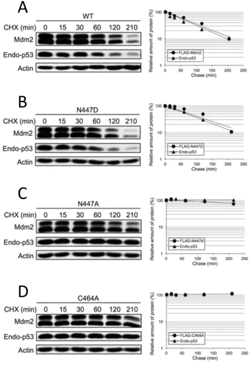
Figure 2-‐4. Half-‐life of p53 and MDM2 are extended in the presence of MDM2 N447A but not N447D ... 78
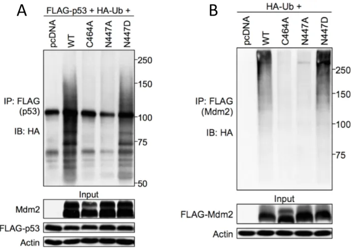
Figure 2-‐5. N447D but not N447A retains the ability to ubiquitinate itself and p53 ... 79
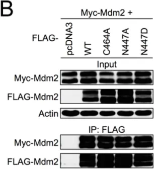
Figure 2-‐6. MDM2 heterooligomerization but not homooligomerization requires the proper structure of the RING domain ... 80
Figure 2-‐7. Deletion of the MDM2 RING domain or the extreme C-‐terminus inhibits p53 degradation ... 81
Figure 2-‐8. MDM2 RING or extreme C-‐terminal deletion prevents MDM2-‐ MDM2 homooligomerization and MDM2-‐MDMX heterooligomerization ... 82
Figure 2-‐9. AD deletion inhibits the ability of MDM2 to degrade p53 ... 83
Figure 2-‐10. AD deletion inhibits homooligomer but not heterooligomer formation in E3-‐competent MDM2 constructs ... 84
Figure 2-‐12. ARF affects the formation of MDM2-‐MDM2 homooligomers
and MDM2-‐MDMX heterooligomers ... 86
Figure 2-‐13. Summary of results ... 87
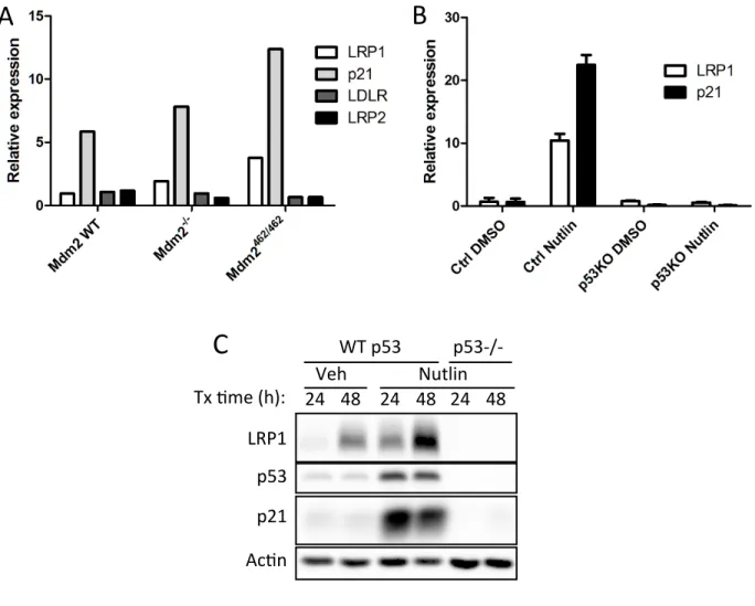
Figure 3-‐1. LRP1 transcript and protein are upregulated in response to nutlin-‐3a treatment ... 113
Figure 3-‐2. LRP1 expression is p53-‐dependent ... 114
Figure 3-‐3. LRP1 is a direct p53 transcription target ... 115
Figure 3-‐4. LRP1 is induced in response to canonical p53-‐activating stresses ... 116
Figure 3-‐5. U2OS cells show a similar trend in LRP1 expression ... 117
Figure 3-‐6. LRP1 induction is attenuated in the presence of the pleiotropic inhibitor caffeine ... 118
Figure 3-‐7. Sub-‐lethal doxorubicin can induce LRP1 expression ... 119
Figure 3-‐8. LRP1 protein is expressed in response to sub-‐lethal stresses ... 120
Figure 3-‐9. HCT116 cells show dose-‐dependent induction of LRP1 in response to etoposide ... 121
Figure 3-‐10. Caspase inhibition does not rescue LRP1 expression ... 122
Figure 3-‐11. LRP1 is a late-‐expressed gene that occurs in response to sub-‐lethal doses of doxorubicin ... 123
Figure 3-‐12. X-‐irradiation and UV irradiation produce distinct patterns of p53 induction and different ability to induce LRP1 ... 124
Figure 3-‐13. MCF7 cells show a similar pattern of p53 induction to HCT116 cells in response to doxorubicin ... 125
Figure 3-‐14. MCF cells show a similar pattern of p53 induction to HCT116 cells in response to IR and UV ... 126
Figure 3-‐15. LRP1 protein is induced gradually between 12 and 24 h after sub-‐lethal doxorubicin treatment ... 127
Figure 3-‐17. Collection of floating cells does not affect protein
expression levels ... 129 Figure 3-‐18. Doxorubicin dose-‐dependent decrease in LRP1 protein level does
not produce any detectable cleavage products ... 130 Figure 3-‐19. LRP1 protein also shows a dose-‐dependent decrease in response
to UV irradiation ... 131 Figure 3-‐20. LRP1 transcript expression is elevated in response to lethal
doxorubicin ... 132 Figure 3-‐21. LRP1 half-‐life is decreased upon treatment with lethal doxorubicin ... 133 Figure 3-‐22. Neither MG132 nor chloroquine can rescue LRP1 expression ... 134 Figure 3-‐23. De novo LRP1 translation is reduced in response to lethal
doxorubicin compared with sub-‐lethal doxorubicin ... 135 Figure 3-‐24. TargetScan results for LRP1 3’UTR ... 136 Figure 3-‐25. miR-‐103 and miR-‐107 show significantly higher induction in
response to lethal doxorubicin compared with sub-‐lethal doxorubicin ... 137 Figure 3-‐26. miR-‐103 and miR-‐107 are induced in a doxorubicin dose-‐
dependent manner ... 138 Figure 3-‐27. Primer design to confirm the presence of the miR-‐103/107
seed region in the LRP1 3’UTR in HCT116 cells ... 139 Figure 3-‐28. Predicted miR-‐103/107 binding site is present in HCT116 cells ... 140 Figure 3-‐29. HCT116 LRP1 knockout lines show a survival advantage ... 141 Figure 3-‐30. HCT116 LRP1 knockout lines show enhanced colony formation
ability compared with control cells ... 142 Figure 4-‐1. Small deletions of the MDM2 acidic domain may selectively
impair MDM2-‐MDM2 homooligomerization ... 167 Figure 4-‐2. A model based on the integration of the current study on p53
regulation of LRP1 and other studies investigating miRNA regulation ... 168 Figure 4-‐3. Intravenous drug delivery often delivers therapeutic doses of
Figure 4-‐4. Rapidly growing tumors display high levels of intratumoral
pressure ... 170 Figure 4-‐5. p53 WT tumors exposed to sub-‐optimal dosing regimens of some
drugs may employ p53 cell survival programs to increase their resistance ... 171 Figure 4-‐6. IR-‐dependent LRP1 induction does not show protein suppression
at high doses ... 172 Figure 4-‐7. LRP1 deletion does not affect p-‐H2AX resolution in cancer cells ... 173 Figure 4-‐8. TGF-‐beta signaling is not affected by LRP1 deletion in cancer cells ... 174
LIST OF ABBREVIATIONS 5FU: 5-‐fluorouracil
AD: acidic domain
APAF1: apoptotic peptidase activating factor 1 ApoE: apolipoprotein E
ARF: alternative reading frame ATM: ataxia telangiectasia mutated ATR: ATM-‐related
Aβ: amyloid-‐beta
BAX: Bcl-‐2-‐associated X protein Bcl-‐2: B-‐cell CLL/lymphoma 2
CDKN1A: cyclin-‐dependent kinase inhibitor 1A ChIP: chromatin IP
CK1/2: casein kinase 1/2 CQ: chloroquine
CRISPR: clustered regularly interspersed palindromic repeats DBD: DNA binding domain
DNA: deoxyribonucleic acid Dox: doxorubicin
DRAM: damage-‐regulated autophagy modulator FAO: fatty acid oxidation
GOF: gain-‐of-‐function
GPX1: glutathione peroxidase 1
HIPK2: homeodomain-‐interacting protein kinase 2 IP: immunoprecipitation
IR: ionizing radiation
LDL: low-‐density lipoprotein LDLR: LDL receptor
LRP1: LDLR-‐related protein 1 MCD: malonyl coA dehydrogenase MDM2: mouse double minute 2 MDMX: mouse double minute 4
MGMT: O-‐6-‐methylguanine DNA methyltransferase miRNA: microRNA
NES: nuclear exportation signal NLS: nuclear localization signal
NOXA: phorbol-‐12-‐myristate-‐13-‐acetate-‐induced protein 1 p53BD: p53 binding domain
p53ER: p53-‐estrogen receptor PCR: polymerase chain reaction PDGF: platelet-‐derived growth factor PIG3: p53-‐induced gene 3
PML: promyelocytic leukemia
PUMA: p53-‐upregulated modulator of apoptosis RE: response element
RING: really interesting new gene RISC: RNA-‐induced silencing complex RNA: ribonucleic acid
RP: ribosomal protein
RRM2B: ribonucleotide reductase subunit M2B SCO2: cytochrome C oxidase assembly protein SILAC: stable isotope labeling by amino acids in cells Sp1: specificity protein 1
SV40: simian virus 40
TGF-‐beta: transforming growth factor-‐beta
TIGAR: tp53-‐inducible glycolysis and apoptosis regulator tPA: tissue-‐type plasminogen activator
Ub: ubiquitin
ULK1: UNC51-‐like autophagy activating kinase UTR: untranslated region
UV: ultraviolet WT: wild-‐type
CHAPTER 1: INTRODUCTION1
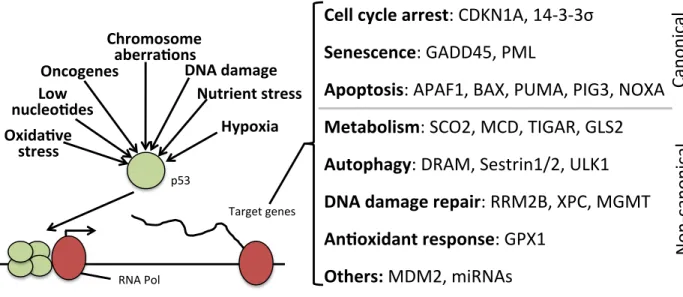
Known to many as the “guardian of the genome,” the transcription factor p53, which interestingly enough was originally characterized as an oncogene, has gradually gained widespread notoriety as a general stress response coordinator. Indeed, p53 can be
activated by several types of stress, including DNA damage, oncogene activation, ribosomal stress, hypoxia, nucleotide deficiency, oxidative stress, and chromosomal aberrations, among others (Figure 1-‐1). Likewise, p53 controls the expression of numerous genes involved in many different cellular processes corresponding to the nature of the activating stress (Figure 1-‐1). Early reports on p53 function discovered a prominent role for p53 in the induction of cell cycle arrest, apoptosis, and senescence, so-‐called canonical p53 functions. However, more recent studies have also shed light on various non-‐canonical functions of p53, including DNA damage repair, glucose and lipid metabolism, and
autophagy. These advancements in our knowledge of p53 have helped shape and change our perception of the role of p53 in the cellular and organismal context. Many novel p53 targets are being discovered as more and more high throughput screens are conducted. In Chapter 3, the identification and characterization of the novel p53 target gene low-‐density lipoprotein receptor-‐related 1 (LRP1) will be discussed.
In addition to understanding p53 function, another important research subject regarding p53 is how it is regulated. Commonly accepted as the most important negative regulator of p53, the gene product mouse double minute 2 (MDM2, sometimes referred to as H(uman)DM2) potently inhibits p53 function in two ways: MDM2 binds directly to the p53 activation domain thereby masking p53 function as a transcription factor, and MDM2 catalyzes the transfer of ubiquitin to p53 to mark p53 for proteasomal degradation. Studies on MDM2 have shown that it is as important in the context of p53 functions as p53 itself. Interestingly, as will be discussed in more detail below, the manipulation of MDM2 alone can produce phenotypes that are identical to the deletion or manipulation of p53,
suggesting that MDM2 is an important consideration for most, if not all, p53 studies. In Chapter 2, a mutational analysis of MDM2 will be described in detail. Some of the novel findings of this study include evidence that mutations in MDM2 that were previously thought to inhibit its ability to homooligomerize actually do not affect
homooligomerization. Importantly, I show evidence that homooligomer formation involves domains of MDM2 that are only beginning to be acknowledged for their role oligomer formation.
with a brief introduction describing the initial discovery and the surprisingly tortuous path towards the characterization of p53.
Discovery of p53
The initial discovery of p53 was reported by multiple labs that found that p53 interacts with the simian virus 40 (SV40) large T antigen. These labs, which included those of David Lane, Arnold Levine, Pierre May, Robert Carroll, and Alan Smith, found that upon immunoprecipitation of the SV40 large T antigen, a cellular protein of approximately 53 kDa in size could be co-‐precipitated.152, 163, 187, 212, 282 Further investigation into this 53-‐kDa protein, eventually named p53, found that not only could p53 be stabilized by SV40 large T antigen but p53 was generally stabilized in transformed tissues compared with normal tissues. These studies collectively showed that higher levels of p53 correlate with tumor formation and progression, which led to the initially false assumption that p53 functions as an oncoprotein. However, the characterization of p53 as an oncoprotein came under
scrutiny when completely opposite phenotypes were observed in cell lines transfected with cloned p53 constructs from different labs.174 These discrepancies were explained when sequencing of these clones revealed that wild-‐type p53 produced a cell suppression effect, whereas mutated versions of p53 acted as an oncogene and promoted cellular
normally tumor suppressive p53 could be transformed into a tumor-‐promoting factor through the mutation of single residues. Studies thereafter (by our own Dr. Dirk Dittmer studying in the Levine lab) confirmed that gain-‐of-‐function effects are associated with p53 mutations, which manifests in a pro-‐tumor phenotype.70
Function of p53
The function of p53 as tumor suppressor was further confirmed by studies analyzing the effects of p53 mutation. In patients who present with mutations in a single p53 allele, acquired either through germline transmission or through somatic mutation early in development, show a particularly high penetrance of tumor development of many types at a young age. This disorder, termed Li-‐Fraumeni syndrome, displays many of the characteristics observed upon deletion of p53 in mice, suggesting the autosomal dominant nature of the disease. Deletion of p53 in mice shows a similarly high rate of tumor
binding transcription factor composed of two dimers.198 Subsequent studies identified sequence-‐specific binding sites for p53 that are consistent with its symmetric oligomeric nature.80, 140 El-‐deiry et al. identified the p53 consensus binding site as consisting of two 5’-‐ RRRCWWGYYY-‐3’ half sites separated by 0-‐13 base pairs (R=purine, Y=pyrimidine,
W=adenine or thymine). The identification of the consensus p53 binding site was an
important discovery for the p53 field, as it allowed for the prediction of putative p53 target genes once genomic sequence data became available. Subsequent high-‐throughput analyses further refined the strongest p53 candidate response elements as consisting of the two decamer half sites separated by either zero or one base pair.32 Based on the identified p53 consensus binding sequence, multiple high-‐throughput studies have been conducted, identifying multiple verified and putative p53 response genes.32, 226, 281 Nonetheless, even before the development of high throughput techniques, shortly after the identification of the p53 consensus sequence, the first p53 target, p21 (also known as CDKN1A), was reported.81
Since the discovery of p21 as a p53-‐regulated gene, multiple other p53 target genes have been identified, including the apoptotic gene products PUMA, NOXA, BAX, AIP1, and APAF1, the autophagy gene products DRAM1, ULK1, and sestrin 1, the senescence gene products GADD45 and PML, the DNA damage repair gene products ribonucleotide
some of the mechanistic details remain obscure, many p53 target genes and pathways can have overlapping functions and can be activated in different combinations depending on context, which will be discussed below.
Another milestone in p53 research was achieved when the first successful crystal structure of p53 bound to its response element was reported. This crystal structure offered insight into which p53 residues interact directly with DNA.51 Details provided by the
crystal structure provided a physical explanation why common p53 mutations prevent p53 from binding to its response element.131 Interestingly, the vast majority of tumor-‐
associated p53 mutations are point mutations and not deletions, which can be explained by the selective pressure for clones that harbor the oncogenic GOF effects associated with p53 point mutations. Comparing mice deficient for p53 with mice harboring a single point mutation in p53 (R175H in human p53) revealed a relative survival advantage for the p53-‐ null mice, suggesting that p53 point mutations indeed promote cancer progression.164 In vitro experiments using cells derived from the p53 R175H mice revealed that these cells possessed several characteristics that are beneficial to rapid tumor growth, including increased transformation potential, increased clonogenicity, and more rapid proliferation, when compared with p53-‐null cells. Moreover, p53 point mutation-‐bearing mice display a different spectrum of tumors, compared with p53-‐null mice.235
been broadly classified into “structural mutants,” which induce the unfolding of p53, and “contact mutants,” which describe mutations that specifically affect that ability of p53 to bind DNA but do not significantly induce protein unfolding.51, 277 Although all p53
mutations likely unfold p53 to some extent, the unfolded nature of mutant p53 may play a role in its associated gain-‐of-‐function phenotypes. Interestingly, a recent study showed that several common p53 mutations reconfigure the folded p53 protein structure to allow binding to and redirection of the chromatin remodeling machinery, including the
methyltransferases MLL1 and MLL2 as well as the acetyltransferase MOZ. This results in the expression of a distinct set of genes that promotes tumor cell growth and metastasis.340 Other studies have also shown that mutant p53 could produce an opposite outcome
compared with WT p53 with respect to miRNA-‐mediated gene regulation.151 Because of the importance of p53 GOF mutations in cancers, this area of research remains popular and continues to evolve.
Regulation of p53
as rapidly proliferating cancer cells that lack the basic cell cycle regulation infrastructure are more prone than almost all normal cells to catastrophic outcomes from unrestrained proliferation in the presence of DNA damage or other defects in cellular structures (e.g., taxol-‐induced microtubule stabilization). Cancer treatment modalities, such as
radiotherapy and DNA damaging chemotherapies, exploit the rapid proliferative capacity of cancer cells and offer therapeutic value, as normal cells generally respond by inducing cell cycle arrest and/or DNA damage repair pathways through tumor suppressor genes such as p53. Interestingly, the absence of one or both alleles of p53 increases the sensitivity of mice to the development of tumors after a single dose of sub-‐lethal irradiation, which highlights the importance of p53 in preventing tumorigenic transformation.139 On the other hand, the absence or inhibition of p53 is sufficient to protect mice from otherwise lethal doses of irradiation.144, 322 In response to a lethal dose of irradiation, p53 WT mice show extensive apoptosis in many tissues and ultimately succumb to hematopoietic failure. These results suggest that p53 overactivation in normal tissue is just as important a consideration for the treatment of tumors as p53 underactivation in tumor tissue. To keep p53 inactive under normal conditions and active under stress, p53 is regulated at the transcriptional and translational levels;223 however, the main modality of p53 regulation occurs at the post-‐ translational level. The post-‐translational regulation of p53 is mediated predominantly in terms of p53 stability involving various post-‐translational marks (PTMs). p53 protein can be modified by phosphorylation, acetylation, ubiquitination, SUMOylation, methylation, and neddylation, among others. Some of the better characterized PTMs on p53 are described below and are shown in Figure 1-‐2.
p53 phosphorylation
Many p53 residues can be phosphorylated by various kinases under various conditions, contributing to the regulation of p53. Indeed, based on the rapid kinetics, p53 phosphorylation is thought to be one of the most important first steps for p53 stabilization in response to stress. One of the best characterized phosphorylation-‐inducing stresses is DNA damage, which results in p53 phosphorylation on several residues. Most prominent among these residues is serine 15, which is phosphorylated by ATM, ATR, and DNA-‐PK.26, 154, 209, 274 p53Ser15 phosphorylation results in p53 stabilization in part by inhibiting its ability to interact with its negative regulator MDM2.273 Interestingly, p53Ser15
phosphorylation also enhances the affinity of p53 for its response elements.192 Moreover, p53Ser15 phosphorylation behaves as a sort of gateway phosphorylation mark, whereby p53Ser15 phosphorylation precedes and acts as a prerequisite to the subsequent
phosphorylation of other p53 residues, including serines 6, 9, 20, and 46 as well as Thr18, by other kinases such as casein kinase 1, casein kinase 2, JNK, and HIPK2 (Figure 1-‐2).76, 262, 263 Importantly, mutation of p53Ser15 to alanine is sufficient to prevent the p53-‐dependent transactivation of target genes in response to DNA damage.192 p53Ser15 is often used as a DNA damage marker in response to various stresses.
some of which could be more important physiologically in the p53 response than even p53Ser15 phosphorylation. For example, mutation of one phosphorylation mark on MDM2 Ser395 (normally phosphorylated by ATM) to alanine prevents death in mice exposed to a lethal dose of ionizing radiation.92
p53 residue Ser46 is another well characterized phosphorylation site that is preferentially phosphorylated in response to apoptotic stress.229 Ser46 is phosphorylated by the kinase homeodomain-‐interacting protein kinase 2 (HIPK2) and has been used to differentiate between the apoptotic and cell cycle arrest programs of p53 (discussed more below).28, 29, 58, 69, 168 Similarly to the function of Ser15 phosphorylation as a gateway mark to additional phosphorylation marks, Ser46 phosphorylation facilitates the acetylation of Lys382,107 as p53 acetylation serves as another important mark for p53 regulation. p53 is also subject to dephosphorylation by various phosphatases. The most prominent p53 phosphatase is Wip1, which has been shown to dephosphorylate p53Ser15 but not p53Ser46.193 Interestingly, Wip1 is expressed in a p53-‐dependent manner, suggesting the existence of negative feedback loop involving Wip1 phosphatase.88 As one would
expect, p53 dephosphorylation of these residues by Wip1 has the opposite effect of p53 phosphorylation, namely inhibition of p53 transcriptional activity. Another p53
phosphatase that has been reported to inhibit p53 activity is protein phosphatase-‐1 (PP-‐1), which was shown to dephosphorylate Ser15 and Ser37.177 The protein phosphatase 2A (PP2A) subunit B56γ has also been reported as a p53 phosphatase that dephosphorylates residue Thr55.179 Interestingly, the authors report that B56γ-‐mediated dephosphorylation of Thr55 is necessary to stabilize p53 and induce apoptosis, suggesting that
p53 acetylation
As alluded to above, another important PTM on p53 is acetylation. The
acetyltransferases CBP/p300 (hereafter referred to as p300), PCAF, and Tip60 have been shown to directly acetylate various p53 Lys residues (p300: lysines 164, 370, 372, 373, 381, 382, and 386, PCAF: lysine 320, Tip60: lysine 120) thereby resulting in p53
activation.154, 188 Some of the acetylation sites in p53, especially near the C-‐terminus, overlap with ubiquitination sites within p53 that are crucial for its degradation. Therefore, it appears as though p53 acetylation functions in part by blocking ubiquitination and
proteasomal degradation. p53 lysine acetylation has also been shown to affect p53 function in other respects. In one study, mutant p53 that was unable to be acetylated on Lys120 showed increased binding affinity between p53 and the response elements for cell cycle arrest genes p21 and MDM2,294 suggesting that p53 acetylation may play a role in
differential gene transcription (discussed in detail below). Lys382 acetylation appears to play a similar role in specifying p53 transcriptional activity. Moreover, some p53
acetylation marks appear to interfere with or promote the acetylation or phosphorylation of other p53 residues, which could affect which genes are transcribed by p53 in response to a given stress;142 an example of this was provided in the preceding section whereby Ser46 phosphorylation enhances Lys382 acetylation.
activation of the various p53 programs.
p53 ubiquitination
A major avenue through which p53 is regulated on the post-‐translational level occurs through ubiquitin attachment. In fact, p53 degradation occurs primarily through the ubiquitin-‐proteasome pathway. The importance of ubiquitination on p53 degradation can be illustrated by MG132-‐mediated proteasome inhibition, which prolongs the half-‐life of p53 from less than 30 minutes to several hours.197 Ubiquitination generally occurs through a cascade of reactions involving the activation and transfer of individual ubiquitin (Ub) molecules to the substrate. Ub activation occurs when an E1 Ub-‐activating enzyme catalyzes the conjugation of an adenylate moiety to Ub. Once activated, an E2 Ub-‐
conjugating enzyme catalyzes the transfer of the activated Ub to itself thereby forming an E2-‐Ub intermediate. The final step of the Ub cascade is catalyzed by an E3 ligase, which binds to its specific substrate and the E2-‐Ub to facilitate the transfer and covalent attachment of Ub to the substrate. This reaction can occur in iterations to produce a polyubiquitin tail that can mark the targeted protein for degradation. E3 ligases constitute the largest and most diverse class of the ubiquitination cascade enzymes, which allows the ubiquitination system to target only a handful of proteins at a time. Interestingly, p53 can be ubiquitinated through multiple E3 ligases, including COP1,75 PirH2,171 and Topors,253 any of which can enhance p53 proteasomal degradation. However, the best characterized E3 ligase for p53 is MDM2 and its homolog MDMX. The ubiquitination mechanism of p53 remains unclear, as the extent of p53 ubiquitination could vary depending on the
remains unclear is how nuclear p53 is ubiquitinated and then transported to the cytoplasm where the proteasome resides. It is thought that a signal is required for p53 to be expelled to the cytoplasm, and studies have suggested that the signal is ubiquitin attachment.20, 96 One model proposes that p53 is monoubiquitinated in the nucleus by MDM2, which exposes the p53 nuclear export sequence (NES) and promotes p53 expulsion from the nucleus.160, 180 Interestingly, although MDM2 also possesses an NES, only the p53 NES and MDM2 ubiquitin ligase activity are necessary for p53 export. Once in the cytoplasm, p53 could be further polyubiquitinated and degraded by the proteasome.
Studies have also shown that once p53 becomes expelled from the nucleus (possibly through monoubiquitination), cytoplasmic p53 can exert transcription-‐independent
activity by directly interacting with proteins such as Bax, Bcl-‐2, and Bcl-‐xL.49, 50, 216 This leads to another unresolved question in the field, which is if p53 is monoubiquitinated and expelled into the cytoplasm, then what determines whether mono-‐Ub-‐p53 is immediately degraded or allowed to exert extra-‐transcriptional effects. Part of the answer appears in the fact that p53 can also be deubiquitinated through the action of the cytoplasmic
deubiquitinase USP10.335 Though the regulatory mechanism remains unclear, these results suggest that mono-‐Ub-‐p53 could be subject to deubiquitination under certain
circumstances after being expelled from the nucleus.
p53 methylation
Lys372 results in p53 stabilization and increased transcription of p53 target genes. Interestingly, p53 can also be methylated on Lys370 by Smyd2.114 In contrast to Lys372 methylation, Lys370 methylation was reported to suppress p53 activity. Moreover, methylation at Lys370 or Lys372 (by Set9) appeared to counter-‐regulate each other, suggesting that p53 methylation marks cross-‐talk with one another and produce different outcomes depending on the modified residues.
p53 differential gene activation
is the most beneficial outcome. On the other hand, because the generation of new cells requires significant investment of energy and resources, if DNA damage is resolvable, then the most beneficial outcome would be repair and adaptation. This dual function of p53 fits well with its role as a preserver of genomic fidelity and organismal fitness, as severe DNA damage can result in mutations that lead to transformation. An interesting question regarding the p53-‐regulated gene programs concerns the antagonistic effects of some programs towards others. One of the more obvious examples is that during the p53-‐ dependent induction of apoptosis, the simultaneous p53-‐dependent activation of DNA repair or antioxidant pathways could act counter to the intended apoptotic outcome. Indeed, p21 has long been known to exert anti-‐apoptotic effects, which suggests that suppressing p21 could be beneficial for a rapid apoptotic response.196, 316 Therefore, pleiotropic transcription factors such as p53 likely require a mechanism to selectively transcribe some genes but not others depending on the context. As one of the major mysteries that persists regarding p53 regulation, several models have been proposed to explain mechanistically how p53 decides which programs to activate, many of which have focused on differences in p53 itself (overall level, PTM marks, expression patterns, etc.). Notably, these proposed models are not necessarily mutually exclusive, and some of the models overlap considerably (e.g., PTMs on p53 affect the affinity between p53 and various response elements).
p53 threshold model
precise control of p53 levels in the absence of genotoxic stress showed a strong correlation between the level of p53 and the expression of both cell cycle arrest and apoptosis
genes.149 In this study, the authors provided additional evidence suggesting that a
physiological threshold dictates whether a cell responds to stress in an apoptotic manner. In one experiment, the authors showed that the activation of apoptosis requires a relatively long duration of high p53 levels, as low p53 induction for an extended duration failed to induce appreciable apoptosis. In another experiment, the authors showed that the theoretical p53 apoptotic threshold could be lowered by inhibiting anti-‐apoptotic Bcl-‐2 family member proteins. Interestingly, inhibition of Bcl-‐2 family proteins resulted in increased apoptosis in the presence of an otherwise sub-‐lethal level of p53. These results support a model whereby only high levels of p53 are able to induce apoptosis, whereas, while low levels of p53 can activate some apoptotic gene expression, the abundance of apoptotic gene expression does not surpass the threshold required for cell death. This model fits well with several studies that have shown that to trigger an apoptotic response, a certain threshold of pro-‐apoptotic gene expression (relative to anti-‐apoptotic gene expression) must occur.14, 286
Differences in p53 response elements
and apoptotic response elements in an attempt to identify a pattern. In one study
comparing the p53 REs of the cell cycle arrest gene p21 and the apoptotic gene TP53I3 (also called PIG3), the p53 RE for p21 was bound more rapidly than that of TP53I3 in response to DNA damage, suggesting that various p53 REs bind p53 at different rates and affinities.295 In another study, p53 was shown to bind preferentially to the REs for p21 and the
senescence target gene GADD45 in senescent cells. This binding pattern was not observed in cells treated with an acute dose of doxorubicin, which show increased accumulation of p53 at the promoters of apoptotic target genes.124 Using a biophysical approach, the Fersht group determined the in vitro dissociation constants for 20 known p53 response elements for the p53 tetramer and found that all of the low-‐affinity response elements corresponded to apoptotic genes, which could also be consistent with the p53 threshold model.321
Although these studies provide evidence that at least some of the p53 REs display different affinities for p53 binding, some of these studies have also hinted at the possibility that certain p53 PTMs could be required to enhance its binding to the low-‐affinity promoters.
p53 post-‐translational modifications
induction of apoptotic genes and an apoptotic outcome.229 The mutation of Ser-‐46 to Ala results in the attenuation of apoptosis induction by p53. p53 acetylation at residue Lys 382 occurs subsequent to Ser46 phosphorylation, and this mark is required for p53 apoptotic gene expression.107 Other PTMs include those induced by the E3 ubiquitin ligase E4F1, which monoubiquitinates p53 at Lys320 and results in the upregulation of the p53 cell cycle arrest program.166 Interestingly, Lys320 is not ubiquitinated by MDM2, suggesting an MDM2-‐independent mechanism. In addition to ubiquitination at Lys320, p53 can also be functionally acetylated at this residue. One study showed that p53 acetylation at Lys320 and Lys373 results in the expression of cell cycle arrest and apoptotic genes,
respectively.142 In this study, the authors show that acetylation of either residue prevents the other from being acetylated, implying antagonism between these two marks. Moreover, acetylation at Lys320 prevents the phosphorylation of N-‐terminal p53 residues thereby limiting p53 to high-‐affinity promoters such as those of cell cycle arrest gene p21. In contrast, acetylation of Lys373 promotes the phosphorylation of N-‐terminal p53 residues and allows p53 to bind to low-‐affinity promoters such as those of the apoptotic target genes. Another acetyl PTM mentioned above, acetylation of Lys120 also appears to dictate the affinity between p53 and cell cycle arrest and apoptosis gene response elements.294 Lys120 acetylation is necessary for the induction of p53 apoptotic genes, as the mutation of this residue inhibits the ability of p53 to upregulate apoptotic gene expression. Other PTMs could also play roles in the ability of p53 to bind to certain promoters, and this remains a popular area of p53 research.
p53 dynamic pattern
Most studies investigating p53 activation have analyzed lysates obtained from populations of cells. Using this method, the average p53 response across many cells can be determined; however, the effect of a given stress on the dynamic behavior of individual cells cannot be resolved. With advances in time-‐lapse microscopy and single cell tracking, the effects of stress on the p53 expression pattern in single cells could be determined. Analyzing single cells offers an opportunity unparalleled by other methods to determine how expression levels of certain genes change over time. In conjunction with fluorescently labeled p53 constructs, this method was used to precisely determine the dynamics of p53 expression in response to ionizing radiation (IR).159, 252 Interestingly, these studies showed that p53 expression in response to IR shows a pulsed pattern, whereas ultraviolet (UV) irradiation induced a sustained pattern of p53 activation. Interestingly, the pulsed patterns of p53 induction tend to result in non-‐lethal, reversible outcomes for the cell, whereas sustained activation of p53 results in cell death or senescence. The pulses of p53 in
response to IR show similar amplitudes but vary in frequency depending on the magnitude of the initial stress.159, 252 Importantly, the manipulation of the pulsed p53 induction
timing DNA damage to occur when p53 levels are highest, which predisposes the cell to a cell death outcome as opposed to a cell cycle arrest and survival outcome. Of note, the comparison in the Purvis study only analyzed the activation of p53 without addressing other potential differences related to the DNA damage stress itself. Because the authors compared the effects of two entirely different types of DNA damage (UV induces mainly thymidine dimers, whereas IR induces mainly double strand breaks) that activate different repair pathways, it is possible that this dichotomy of pulsed versus sustained p53
expression is unique to this stress comparison. Notably, the pulsed pattern of p53
induction has not been reported for other types of DNA damage. Moreover, our personal experience with p53 induction over time in response to a sub-‐lethal, brief (1 h) dose of the double strand break-‐inducing chemotherapeutic doxorubicin showed a pattern that most closely resembles the sustained pattern of UV-‐induced DNA damage. Constant doxorubicin treatment at the same concentration causes a similar albeit gradually increasing expression pattern for p53 (see Chapter 3). Thus, investigation and comparison of the p53 induction pattern in response to other types of stress may offer additional insight into the p53 dynamic pattern model of p53 induction and the nature of the pulsed pattern in response to IR.
p53 co-‐regulation model
through the concomitant binding of the promoter region by p53 and SMAD2/3.56 The nature of this co-‐regulatory mechanism appears to require individual binding sites for SMAD2/3 and p53, although these transcription factors are also able to bind directly to each other. Moreover, reports have suggested that the p53 and SMAD transcriptomes overlap substantially, particularly concerning cell cycle arrest genes,77 which is consistent with the roles of p53 and SMAD transcription factors in tumor suppression. The SMAD transcription factors have been implicated in cell cycle inhibition as well as cell cycle progression in a context-‐dependent manner, much like p53. In addition to the SMADs, p53 has been shown to bind directly to the transcription factor Sp1 (specificity protein 1).147, 148 p53-‐Sp1 co-‐regulation is required for the efficient upregulation of several p53 target genes, including p21 and PUMA.148 A recent high-‐throughput analysis further suggested that Sp1 co-‐regulation is a key determinant for p53-‐dependent apoptotic gene expression but not for cell cycle arrest gene expression.178 Collectively, these studies support a co-‐regulation mechanism that dictates whether p53 transactivates the expression of cell cycle arrest genes or apoptotic genes.
p53 miRNA model
Finally, another model that has recently been proposed regarding p53 differential gene regulation involves p53-‐dependent miRNA upregulation. miRNAs are small non-‐ coding RNA species approximately 22-‐23 nucleotides in length that bind to the 3’
genes are regulated by p53 as direct transcriptional targets.86 Interestingly, some p53-‐ regulated miRNAs have been reported to target known p53-‐regulated genes. In one such example, the p53-‐regulated miR-‐23a is expressed in response to p53-‐activating stresses. Upon upregulation, miR-‐23a can down-‐regulate the expression of the apoptotic p53 target gene APAF1. Through this mechanism, miR-‐23a expression can promote apoptotic
resistance in some cancer cells.43, 270, 333 Although these studies were performed in cancer cells, it is likely that p53-‐regulated miRNAs contribute to the regulation of other p53 target genes in normal cells as well. Of interest to my studies on p53 regulation of LRP1, the doses and stresses used in the studies showing p53-‐dependent miR-‐23a targeting of APAF1 were sub-‐lethal (~37 μM 5-‐fluorouracil in HCT116 cells), suggesting that miRNAs could play a role in the manipulation of p53 responses. These studies investigating p53-‐miRNA
feedback loops represent a relatively unknown aspect of p53 differential gene expression, which I address in part in Chapter 3 of this dissertation.
What p53 programs are important for tumor suppression?
At this point, one of the major issues that remains to be discussed is which p53 programs are most important for its tumor suppressor activity. Based on the earliest
variant (p53L25Q,W26S, so-‐called p5325,26), which shows deficiencies in activating cell cycle arrest and apoptosis, retain the ability to suppress KRAS-‐induced tumors.21 However, this mouse model retains the ability to induce senescence, which could account for the tumor suppressive effects of p5325,26. This possibility was addressed in a study by the Gu lab, where the authors analyze Tp533KR/3KR mice, which harbor three point mutations (K117R,
K161R, and K162R) that inhibit the acetylation of p53 and thus effectively abrogate the induction of p53-‐dependent cell cycle arrest, apoptosis, and senescence programs.181 In this paradigm-‐shifting study, the Tp533KR/3KR mouse remains resistant to spontaneous tumor development compared with Tp53-‐null mice, which develop early onset lymphomas. These results suggest that these canonical functions of p53, which have long been thought to be absolutely necessary for tumor suppression, are actually dispensable for p53-‐
mediated tumor prevention. In another mouse model that supports this conclusion, triple deletion of the p53 target genes p21, Puma, and Noxa, which are involved in cell cycle arrest, apoptosis, and senescence, reveals no difference in susceptibility to spontaneous tumor development.306 Collectively, these studies suggest that p53-‐dependent cell cycle arrest, apoptosis, and senescence are not necessary for effective tumor suppression, which in turn suggests that other non-‐canonical p53-‐regulated programs are sufficient for tumor suppression. Although p53 has been reported to exert extra-‐transcriptional activities on various cellular components (glucose-‐6-‐phosphate dehydrogenase130 and autophagy300), these functions of p53 also appear to be insufficient for tumor suppression.129 Therefore, p53 transcriptional activity appears to be key to its function in tumor suppression. Which pathways are most important for p53-‐mediated tumor suppression remain to be