MPLEx: A method for simultaneous pathogen inactivation and
extraction of samples for multi-omics profiling
Kristin E. Burnum-Johnson1,*, Jennifer E. Kyle1,*, Amie J. Eisfeld2, Cameron P. Casey1, Kelly G. Stratton1, Juan F. Gonzalez3, Fabien Habyarimana3, Nicholas M. Negretti4, Amy C. Sims5, Sadhana Chauhan6, Larissa B. Thackray7, Peter J. Halfmann2, Kevin B. Walters2, Young-Mo Kim1, Erika M. Zink1, Carrie D. Nicora1, Karl K. Weitz1, Bobbie-Jo M. Webb-Robertson8, Ernesto S. Nakayasu1, Brian Ahmer3, Michael E. Konkel4, Vladimir Motin6, Ralph S. Baric5, Michael S. Diamond7, Yoshihiro Kawaoka2, Katrina M. Waters1, Richard D. Smith1, and Thomas O. Metz1,‡
1Biological Sciences Division, Pacific Northwest National Laboratory, Richland, WA
2Infuenza Research Institute, Department of Pathobiological Sciences, School of Veterinary
Medicine, University of Wisconsin-Madison, Madison, WI
3Department of Microbial Infection and Immunity, Ohio State University, Columbus, OH
4 School of Molecular Biosciences, College of Veterinary Medicine, Washington State University,
Pullman, WA
5Department of Epidemiology, University of North Carolina at Chapel Hill, Chapel Hill, NC 6Department of Pathology, University of Texas Medical Branch, Galveston, TX
7Departments of Medicine, Molecular Microbiology, Pathology & Immunology, Washington
University School of Medicine, St. Louis, MO
8Computational and Statistical Analytics Division, Pacific Northwest National Laboratory,
Richland, WA
Abstract
The continued emergence and spread of infectious agents is of great concern, and systems biology approaches to infectious disease research can advance our understanding of host-pathogen relationships and facilitate the development of new therapies and vaccines. Molecular
characterization of infectious samples outside of appropriate biosafety containment can take place only subsequent to pathogen inactivation. Herein, we describe a modified Folch extraction using chloroform/methanol that facilitates the molecular characterization of infectious samples by enabling simultaneous pathogen inactivation and extraction of proteins, metabolites, and lipids for subsequent mass spectrometry-based multi-omics measurements. This single-sample metabolite, protein and lipid extraction (MPLEx) method resulted in complete inactivation of clinically important bacterial and viral pathogens with exposed lipid membranes, including Yersinia pestis, Salmonella Typhimurium, and Campylobacter jejuni in pure culture, and Yersinia pestis,
HHS Public Access
Author manuscript
Analyst
. Author manuscript; available in PMC 2018 January 26. Published in final edited form as:Analyst. 2017 January 26; 142(3): 442–448. doi:10.1039/c6an02486f.
A
uthor Man
uscr
ipt
A
uthor Man
uscr
ipt
A
uthor Man
uscr
ipt
A
uthor Man
uscr
Campylobacter jejuni, and West Nile, MERS-CoV, Ebola, and influenza H7N9 viruses in infection studies. In addition, >99% inactivation, which increased with solvent exposure time, was also observed for pathogens without exposed lipid membranes including community-associated methicillin-resistant Staphylococcus aureus, Clostridium difficile spores and vegetative cells, and adenovirus type 5. The overall pipeline of inactivation and subsequent proteomic, metabolomic, and lipidomic analyses was evaluated using a human epithelial lung cell line infected with wild-type and mutant influenza H7N9 viruses, thereby demonstrating that MPLEx yields biomaterial of sufficient quality for subsequent multi-omics analyses. Based on these experimental results, we believe that MPLEx will facilitate systems biology studies of infectious samples by enabling simultaneous pathogen inactivation and multi-omics measurements from a single specimen with high success for pathogens with exposed lipid membranes.
Infectious disease research is of global interest since the emergence and spread of infectious agents represent ongoing challenges due to population growth and associated increased livestock production to meet food demands, increased urbanization and land-use changes, and greater travel1–5. Emerging infectious diseases are often zoonotic and can be transmitted to humans from animals (e.g., Middle Eastern Respiratory Syndrome coronavirus (MERS-CoV); West Nile virus, influenza A viruses, and Ebola)3. As recently demonstrated by the 2014 Ebola virus infection diagnoses in the United States, the ease of world travel and increased global interdependence have added complexity to containing these infectious diseases6. Similarly, as new infectious diseases evolve and emerge, preexisting infectious diseases re-emerge with new genetic adaptations. For example, novel antigenically distinct subtypes of influenza A viruses that are not recognized by neutralizing antibodies and cause pandemic outbreaks7. Overuse of antimicrobial drugs and decreased compliance with vaccination policies has led to the development of resistant pathogens (e.g., drug-resistance Staphylococcus and Campylobacter)8, 9 and re-emergence of diseases that were previously under control (e.g., Pertussis and Measles)10, 11. In addition, uncontrolled neglected tropical diseases such as Dengue fever and West Nile encephalitis that are endemic in developing countries are now emerging in the United States12–14.
In response to these global threats, the National Institute of Allergy and Infectious Diseases (NIAID) Systems Biology for Infectious Disease Research Program supports research focusing on host-pathogen interactions that are characterized using combined multi-omics approaches and dataset integration to “develop and validate predictive models of infectious disease initiation, progression, and outcomes”15. Infectious disease research using a systems biology approach is imperative to understanding host-pathogen relationships and allows for development of new therapies and vaccines16–18. Specifically, proteomics, metabolomics, and lipidomics measurements can assist in unraveling host-pathogen relationships. Proteins are the major effectors of cellular pathways and represent the dynamic expression of information encoded within the genome during infection. Metabolites are intermediates and products of cellular pathways and represent the level at which most pharmaceuticals exert their effects19. In addition to having key functions in signaling pathways, energy storage and the structural integrity of cell membranes, lipids also function in host-pathogen interactions and immunomodulation since they act in first line recognition and host cell signaling during pathogen docking, invasion and intracellular trafficking20. However, proteomic,
A
uthor Man
uscr
ipt
A
uthor Man
uscr
ipt
A
uthor Man
uscr
ipt
A
uthor Man
uscr
metabolomic, and lipidomic characterizations of infectious biological specimens outside of appropriate biosafety level (BSL) containment laboratories can take place only subsequent to pathogen inactivation.
Non-enveloped viruses such as adenoviruses, noroviruses and bacterial spores are resistant to most disinfectants and require alternative methods for inactivation21, 22. Pathogens with exposed lipid membranes are more susceptible to disinfectants including detergents and other solvents23–25. Organic solvents render many pathogens non-infectious by solubilizing and disrupting their lipid membranes or envelopes26–28, and inactivation of pathogens by organic solvents has been leveraged for vaccine development29–32, transfusion fluids33–36, and sanitation37–39. In systems biology studies of pathogenic bacteria and viruses, it would be highly efficient to both inactivate pathogens and extract the molecular components needed for proteomic, metabolomic, and lipidomic analyses in a single step. Two of the most highly cited publications for lipid-based extraction methods, Folch et al.40 and Dyer et al.41, use high ratios of organic solvents to sample (e.g., 4:1), illustrating the broad utility of this technique in cell and tissue extraction. Recently, variations of the Folch technique have had broader utility in the extraction of metabolites and proteins42–45.
Here, we demonstrate that a modified Folch40 technique using chloroform/methanol for simultaneous protein, metabolite, and lipid extraction and subsequent mass spectrometry (MS)-based multi-omics analyses also results in concurrent pathogen inactivation. Specifically, we investigated if the metabolite, protein and lipid extraction (MPLEx) protocol46 could inactivate a diversity of infectious agents with exposed and embedded lipid bilayers including three Gram-negative bacteria (Yersinia pestis, Salmonella enterica subspecies enterica serovar Typhimurium, and Campylobacter jejuni), Gram-positive community-associated methicillin-resistant Staphylococcus aureus (MRSA) isolate USA300, Clostridium difficile strain 630 spores and vegetative cells, four RNA viruses (West Nile [WNV-New York 1999], MERS-CoV [icMERS], Ebola [Ebola-Zaire delta-VP30] and Influenza H7N9 [wild-type, A/Anhui/1/13 and mutant A/Anhui/103F-106M]). We also tested if MPLEx could inactivate adenovirus type 5, which does not contain an outer lipid bilayer.
MPLEx uses chloroform, methanol, and water (8:4:3) to induce a tri-phasic partitioning of the sample into metabolite, protein, and lipid fractions. Metabolites are located in the upper aqueous layer, whereas lipids are located in the lower organic layer after centrifugation, with a protein disc situated between the two phases (Fig. 1). In this study, MPLEx and subsequent assessment of pathogen inactivation was performed across multiple laboratories, where each laboratory has unique expertise and experience studying the pathogens in question. All studies presented in this manuscript were conducted in accordance with appropriate biosafety guidelines performed in BSL containment laboratories at the University of Wisconsin-Madison, Ohio State University, Washington State University, University of North Carolina at Chapel Hill, University of Texas Medical Branch, and Washington University School of Medicine, all of which are approved for such use by the Centers for Disease Control and Prevention and by the US Department of Agriculture.
A
uthor Man
uscr
ipt
A
uthor Man
uscr
ipt
A
uthor Man
uscr
ipt
A
uthor Man
uscr
The ability of MPLEx to inactivate pathogens was determined by comparing pathogen presence and abundance in either pre-treatment or positive controls vs. MPLEx-treated samples. S. Typhimurium, C. jejuni, MRSA, and C. difficile spore inactivation experiments (pre-treatment and MPLEx-treated) were replicated 20 times. Y. pestis and viral inactivation experiments were replicated 20 times for MPLEx treatment and a single sample was used for assessment of viral activity in the pre-treatment or positive control. We describe below the basic pathogen inactivation and simultaneous multi-omics extraction protocol. Because methods for culturing bacteria and viruses and for establishing infections vary depending on the model pathogen, we include those relevant experimental details in the description of each study in the Supplemental Material.
MPLEx inactivation protocol
1. In a biosafety cabinet (BSC), remove media from infected cells; immediately wash cells with a suitable buffer. The authors recommend a buffer that can be used to quench cell metabolism, such as a solution maintained at ~−40°C and consisting of 60% methanol and 0.85% ammonium bicarbonate in water47; remove rapid quenching solution
2. Add 150 µL of ice-cold 150 mM ammonium bicarbonate solution; scrape cells off of plates or wells for cells grown on agar or in well plate format. Collect cells and buffer into a 1.5 mL Sorenson microcentrifuge tube (or glass vials); if screw cap tops are required (e.g. for BSL-3+ level pathogens) use Fisher Screw Cap microcentrifuge tubes (Cat # 3468)
3. Add 600 µL of MPLEx solution (~-40°C chloroform/methanol [CHCl3/MeOH;
2:1, v/v and a 4-fold excess to sample volume])
4. Vortex for 10 s, leaving the samples on ice for 5 min, and vortex again for 10 s
5. Remove a 100 µl aliquot of mixed solution for inactivation assay
6. Centrifuge the remaining solution at 13,000 g for 10 min
7. Remove top phase to fresh tube, remove bottom phase to second tube leaving protein disc behind. Rinse the protein disc with 200 µL methanol and pellet at 9,000 g for 5 min. Decant the methanol solution and allow pellet to dry in BSC for 5 min
8. All tubes are then removed from BSC and BSL containment laboratories after appropriate disinfection and either stored at −80°C or dried in vacuo (for safe shipping of metabolite and lipid phases) and then stored at −80°C
Some of the bacterial pathogens and all of the viral pathogens were assessed in infection studies where infected host cells were treated with MPLEx and inactivation was evaluated via inoculation of the resulting cell lysates into fresh cells. For these infection studies, the MPLEx-treated cell lysates were diluted to retain host cell viability, as undiluted samples can induce toxicity. Some validations of the inactivation procedures were carried out with samples that had been dried in the speedvac, rather than the liquid/diluted extract prior to drying.
A
uthor Man
uscr
ipt
A
uthor Man
uscr
ipt
A
uthor Man
uscr
ipt
A
uthor Man
uscr
As depicted in Figure 1 and Figure 2, the MPLEx method for extraction of proteins, metabolites and lipids from a single sample for MS-based multi-omics profiling also simultaneously inactivates a number of clinically important bacterial and viral pathogens with exposed lipid membranes, including Y. pestis CO92, S. Typhimurium, and C. jejuni in pure culture and Y. pestis CO92, C. jejuni, WNV, MERS-CoV, Ebola and influenza viruses in infection studies. Near complete inactivation (>99%) was observed for pathogens without exposed lipid membranes including CA-MRSA, C. difficile 630 spores and vegetative cells after ≥ 20 min MPLEx exposure time, and Ad5 (see Supplemental Material and Table S1 for details).
In addition, to demonstrating that MPLEx yields biomaterial of sufficient quality for subsequent multi-omics analyses, Figure 3 highlights the reproducibility of proteomics, metabolomics and lipidomics data from human epithelial lung cells that were infected with either wild-type, A/Anhui/1/2013 (H7N9), or A/Anhui/1/2013 (H7N9) possessing two mutations in the NS1 gene (L103F and I106M) that reduce virus pathogenicity in mice (A. Eisfeld, S. Fan and Y. Kawaoka, unpublished results). To demonstrate the utility of the MPLEx protocol for both inactivating pathogens and for obtaining high quality metabolite, protein, and lipid fractions for respective omics analyses, we implemented the protocol in a systems biology study of influenza infection in a human lung epithelial cell line. The MPLEx protocol was implemented to extract proteins, metabolites and lipids from Calu-3 cells infected with wild-type (AH1) and mutant (FM) influenza viruses, or mock-infected controls for each of 6 timepoints; 0, 3, 7, 12, 18 and 24 h, and using 5 biological replicates per sample (treatment and timepoint). From this experiment, we quantified 23,688 peptides, 81 metabolites (50 were identified through matching of experimental mass spectra and retention indices to entries in the Agilent Fiehn Metabolomics Retention Time Locked Library)48, and 251 and 245 lipids from analyses in negative and positive electrospray mode analyses, respectively; therefore as previously reported46 the number of detected analytes with MPLEx provides comparable or more identifications compared to other methods. To assess reproducibility, we calculated the coefficient of variation (CV) of the 5 biological replicates within each of the datatypes (protein, metabolite, and lipid). For proteomics, metabolomics, and lipidomics, the maximum CV across features within each set of
biological replicates was <0.24 (proteomics), < 0.17 (metabolomics), <0.22 (lipidomics data collected under negative electrospray ionization (ESI)), and <0.19 (lipidomics data collected under positive ESI) (Table S2). Figure 3 shows viral peptides, ribosomal proteins,
metabolites and lipids significantly increasing in infected samples compared to matched mock controls due to infection duration across both viral strains (Table S2). Peptide, metabolite, and lipid statistics were performed by comparing data from matched virus and mock-infected controls using Analysis of Variance (ANOVA) with a Dunnett multiple test correction within time point and for qualitative changes via a G-test with a Bonferroni multiple test correction within time point49. Peptides, metabolites, and lipids with a p-value less than 0.05 were identified as significantly different (Table S2). The top of Figure 3 depicts the 123 viral peptides belonging to viral proteins; hemagglutinin (HA), matrix protein 1 (M1), neuraminidase (NA), nucleoprotein (NP), NS1, NS2, and RNA polymerase complex (PA, PB1, and PB2) significantly increased by G-test (only observed in infected samples) as early as 7 h with the number of significant peptides increasing from 7–24 h.
A
uthor Man
uscr
ipt
A
uthor Man
uscr
ipt
A
uthor Man
uscr
ipt
A
uthor Man
uscr
Statistical analysis of our proteomic data also found significant quantitative increases in the relative abundance of ribosomal subunits 40S and 60S with infection duration across both viral strains. During the influenza life cycle, single-stranded viral RNAs migrate to the nucleus where they are copied into mRNA by the viral RNA polymerase complex. Invading viruses do not harbor functional ribosomes in their virions; therefore, viral mRNAs hijack host cell ribosomes and use sophisticated mechanisms50 to enable selective translation of viral mRNAs. Free fatty acid metabolites myristic acid (14:0) and oleic acid (18:1) significantly increased at 18 h post infection across all 3 viral strains (Fig. 3). Ceramides (Cer) containing stearic acid (18:0) and tetracosanoic acid (24:0) significantly increased at 18 and 24 h in both AH1 and FM and at 12 h in AH1; likewise, Cer containing stearic acid (18:0) and nervonic acid (24:1) significantly increased at 18 h in both AH1 and FM and at 24 h in FM (Fig. 3). It has been previously reported that pathogenic avian influenza viruses, such as H7N9, decrease expression of lipid metabolism genes51. A NS1 mutation in the FM strain reduces overall pathogenicity; at the early 7 hour time point of infection the wild-type AH1 strain shows over a 30% increase in viral peptide compared to the FM mutant (Fig. 3). Correspondingly at this 7 h time point ribosome expression is only significantly increased in the AH1 virus and at the late 24 hour time point significant ribosomal expression is
increased by over 40% in the AH1 compared to FM.
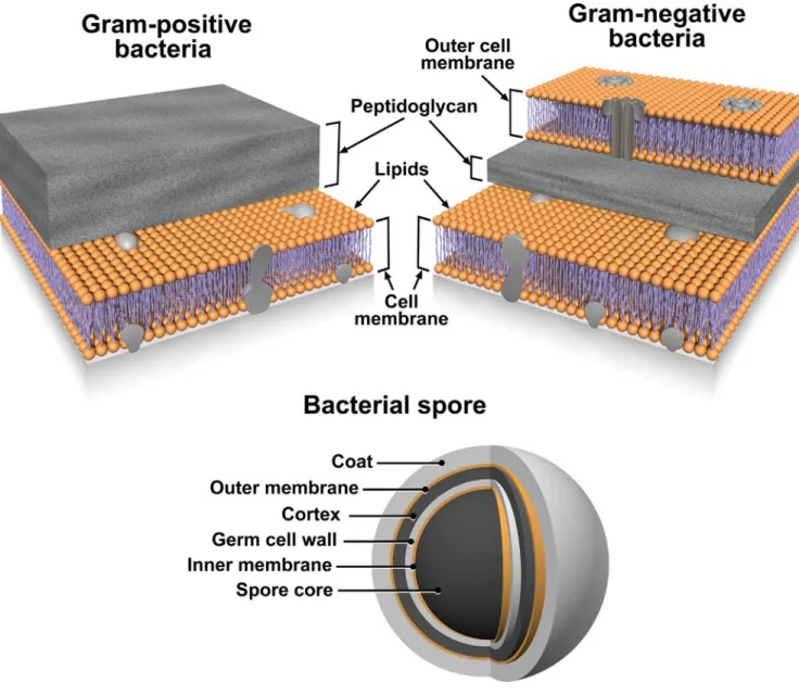
Based on these results we believe that MPLEx will benefit and facilitate systems biology studies of infectious samples by enabling both pathogen inactivation and multi-omics measurements from a single specimen with high success for pathogens with exposed lipid membranes. Figures 4 and 5 illustrate the bacterial and viral pathogens from this study that contain exposed and embedded lipid membranes; these membranes are denoted with orange color. Of the bacterial pathogens characterized in this study, Y. pestis CO92, S.
Typhimurium, and C. jejuni are Gram-negative bacteria, CA-MRSA is a Gram-positive bacterium, and C. difficile 630 is a spore forming Gram-positive bacterium (Fig. 4). The exposed walls for Gram-positive bacteria have a higher peptidoglycan (PG) and lower lipid content than Gram-negative bacteria. This bacterial classification is based on the Gram staining method, which differentiates bacteria by the chemical and physical properties of their cell walls; it detects PG, which is present in a thick layer in Gram-positive bacteria52. The inner membrane of dormant spores is composed of a bilayer of immobile phospholipids, which has similar phospholipid composition to growing bacteria but exhibits low
permeability to small molecules including water53, 54. This inner membrane surrounds the spore core which contains the genome. The basic spore architecture is conserved55–57; outside of the inner membrane is a layer of PG, known as the germ cell wall that is encircled in a thick layer of a modified form of PG, the cortex, which is essential for the acquisition and maintenance of heat resistance. The cortex is surrounded by an outer membrane derived from the mother cell that is essential for spore formation but confers no resistance
properties 58. A proteinaceous coat surrounds the outer membrane. In C. difficile 630 spores, the coat is further enclosed within a structure known as the exosporium. The partial
inactivation results presented for CA-MRSA (where only 2 out of 20 experimental replicates still retained a small level of bacterial activity) and C. difficile 630 spores reveals that MPLEx can dissolve protective PG layers encircling lipid bilayers and with long enough exposure times could lead to complete pathogen inactivation. The level of inactivation of C.
A
uthor Man
uscr
ipt
A
uthor Man
uscr
ipt
A
uthor Man
uscr
ipt
A
uthor Man
uscr
difficile 630 spores due to MPLEx treatment exposure time in min was 120 > 20 > 5 (Fig. 1), in addition in the two replicate experiments of CA-MRSA the experiment with the lower initial bacterial concentration (pre-treatment, 4.9 × 10⁸) did show complete inactivation due to MPLEx. WNV, MERS-CoV, Ebola, and Influenza H7N9 virus all contain lipid bi-layered envelopes (Fig. 5). WNV virions acquire their lipid envelope by budding into the
endoplasmic reticulum of the host cell; Coronaviruses such as MERS-CoV acquire their membranes in the ER-Golgi intermediate compartment (ERGIC) and use normal cellular processes to leave the cell; and Ebola and Influenza H7N9 virions acquire their lipid envelopes by budding from host plasma membranes often at lipid raft microdomains that are enriched in sphingomyelin and cholesterol59–61. Ad5 was the only virus that was not completely inactivated by MPLEx; Ad5 is a non-enveloped virus and therefore does not contain any lipid membranes (Fig. 5).
In conclusion, we show that MPLEx completely inactivates pathogens with exposed lipid membranes, such as enveloped viruses and Gram-negative bacteria, thereby facilitating multi-omics measurements from a single specimen in clinically important pathogen studies. Pathogens with internal or protected lipid membranes, such as Gram-positive bacteria and bacterial spores still show a significant decrease in activity (>99% reduction in the number of viable microorganisms after MPLEx treatment) related to both MPLEx solution exposure time and pre-treatment pathogen levels. Even Ad5, a virus without a lipid membrane, showed >99% decreased activity after MPLEx exposure, presumably since non-enveloped viruses can still be susceptible to organic solvent, in particular CHCl3, due to the
denaturation of proteins that are solvent sensitive28, 62. In summary, we believe our MPLEx method for concurrent pathogen inactivation and extraction of samples for multi-omics profiling will be broadly applicable to samples containing clinically important bacterial and viral pathogens with exposed lipid membranes since molecular characterization of infectious samples outside of appropriate biosafety containment can take place only subsequent to complete pathogen inactivation. We note that, for biosafety reasons, this protocol should be evaluated in each investigator’s laboratory for efficacy in pathogen inactivation, prior to implementation and particularly for pathogens that were not evaluated in our study.
Supplementary Material
Refer to Web version on PubMed Central for supplementary material.
Acknowledgments
This project was funded in part by the Systems Biology Program of the National Institute of Allergy and Infectious Diseases, National Institutes of Health, via grant U19AI106772. Portions of the work utilized capabilities developed under National Institute of General Medical Sciences grant GM103493. Multi-omics measurements were performed in the Environmental Molecular Science Laboratory, a U.S. DOE national scientific user facility at PNNL in Richland, WA. Battelle operates PNNL for the DOE under contract DE-AC05-76RLO01830. We would like to thank PNNL Graphic Designer Michael Perkins for assistance in preparing the figures.
REFERENCES
1. Heesterbeek H, Anderson RM, Andreasen V, Bansal S, De Angelis D, Dye C, Eames KTD, Edmunds WJ, Frost SDW, Funk S, Hollingsworth TD, House T, Isham V, Klepac P, Lessler J,
A
uthor Man
uscr
ipt
A
uthor Man
uscr
ipt
A
uthor Man
uscr
ipt
A
uthor Man
uscr
Lloyd-Smith JO, Metcalf CJE, Mollison D, Pellis L, Pulliam JRC, Roberts MG, Viboud C. I.N.I. I. Collaboration. Science. 2015; 347
2. Daszak P, Cunningham AA, Hyatt AD. Science. 2000; 287:443–449. [PubMed: 10642539] 3. Jones KE, Patel NG, Levy MA, Storeygard A, Balk D, Gittleman JL, Daszak P. Nature. 2008;
451:990–993. [PubMed: 18288193]
4. Lloyd-Smith JO, George D, Pepin KM, Pitzer VE, Pulliam JRC, Dobson AP, Hudson PJ, Grenfell BT. Science. 2009; 326:1362–1367. [PubMed: 19965751]
5. Keesing F, Belden LK, Daszak P, Dobson A, Harvell CD, Holt RD, Hudson P, Jolles A, Jones KE, Mitchell CE, Myers SS, Bogich T, Ostfeld RS. Nature. 2010; 468:647–652. [PubMed: 21124449] 6. Morens DM, Fauci AS. PLoS Pathogens. 2013; 9:e1003467. [PubMed: 23853589]
7. de Jong JC, Rimmelzwaan GF, Fouchier RAM, Osterhaus ADME. Journal of Infection. 2000; 40:218–228. [PubMed: 10908015]
8. Ruiz-Palacios GM. Clinical Infectious Diseases. 2007; 44:701–703. [PubMed: 17278063] 9. Grundmann H, Aires-De-Sousa M, Boyce J, Tiemersma E. Lancet. 2006; 368:874–885. [PubMed:
16950365]
10. Cherry JD. New England Journal of Medicine. 2012; 367:785–787. [PubMed: 22894554] 11. Omer SB, Salmon DA, Orenstein WA, deHart MP, Halsey N. New England Journal of Medicine.
2009; 360:1981–1988. [PubMed: 19420367]
12. Gubler DJ. Clinical Infectious Diseases. 2007; 45:1039–1046. [PubMed: 17879923]
13. Bouri N, Sell TK, Franco C, Adalja AA, Henderson DA, Hynes NA. Public Health Reports. 2012; 127:259–266. [PubMed: 22547856]
14. Hotez PJ, Bottazzi ME, Franco-Paredes C, Ault SK, Periago MR. PLoS Negl Trop Dis. 2008; 2:e300. [PubMed: 18820747]
15. Aderem A, Adkins JN, Ansong C, Galagan J, Kaiser S, Korth MJ, Law GL, McDermott JG, Proll SC, Rosenberger C, Schoolnik G, Katze MG. mBio. 2011; 2
16. Forst CV. Drug Discovery Today. 2006; 11:220–227. [PubMed: 16580599] 17. Zak DE, Tam VC, Aderem A. Annual Review of Immunology. 2014; 32:547–577.
18. Josset L, Tisoncik-Go J, Katze MG. Virus Research. 2013; 178:151–167. [PubMed: 23499671] 19. Fontana JM, Alexander E, Salvatore M. Translational Research. 2012; 159:430–453. [PubMed:
22633095]
20. Wenk MR. FEBS Letters. 2006; 580:5541–5551. [PubMed: 16859687]
21. Rutala WA, Weber DJ. Clinical Infectious Diseases. 2004; 39:702–709. [PubMed: 15356786] 22. Maillard JY. Journal of Hospital Infection. 2011; 77:204–209. [PubMed: 20850897]
23. Favero, M., Bond, W. Chemical disinfection of medical and surgical materials. In: Block, SS., editor. Disinfection, sterilization, and preservation. Philadelphia: Lippincott Williams & Wilkins; 2001.
24. McMurry LM, Oethinger M, Levy SB. Nature. 1998; 394:531–532. [PubMed: 9707111] 25. Feldman H, Wang S. Proc Soc Exp Biol Med. 1961; 106:736–738. [PubMed: 13698695] 26. Vidaver AK, Koski RK, Van Etten JL. Journal of Virology. 1973; 11:799–805. [PubMed:
16789137]
27. Olsen RH, Siak JS, Gray RH. Journal of Virology. 1974; 14:689–699. [PubMed: 4211861] 28. Feinstone SM, Mihalik KB, Kamimura T, Alter HJ, London WT, Purcell RH. Infection and
Immunity. 1983; 41:816–821. [PubMed: 6409813]
29. Fries LF, Waag DM, Williams JC. Infect Immun. 1993; 61:1251–1258. [PubMed: 8454328] 30. Parisius W, Macmorine HG. Applied Microbiology. 1969; 17:379–383. [PubMed: 5780397] 31. Evans RK, Nawrocki DK, Isopi LA, Williams DM, Casimiro DR, Chin S, Chen M, Zhu D-M,
Shiver JW, Volkin DB. Journal of Pharmaceutical Sciences. 2004; 93:2458–2475. [PubMed: 15349956]
32. Cham BE, Vickery K, Tohidi-Esfahani R, Cossart Y. Journal of Virological Methods. 2006; 137:160–163. [PubMed: 16808980]
34. Pelletier JPR, Transue S, Snyder EL. Best Practice & Research Clinical Haematology. 2006; 19:205–242. [PubMed: 16377551]
35. Dichtelmüller HO, Flechsig E, Sananes F, Kretschmar M, Dougherty CJ. Results in Immunology. 2012; 2:19–24. [PubMed: 24371563]
36. Hellstern P, Solheim BG. Transfusion Medicine and Hemotherapy. 2011; 38:65–70. [PubMed: 21779207]
37. Du W-X, Danyluk MD, Harris LJ. Journal of Food Science. 2010; 75:M7–M13. [PubMed: 20492187]
38. Macinga DR, Sattar SA, Jaykus L-A, Arbogast JW. Applied and Environmental Microbiology. 2008; 74:5047–5052. [PubMed: 18586970]
39. Turner RB, Fuls JL, Rodgers ND. Antimicrobial Agents and Chemotherapy. 2010; 54:1363–1364. [PubMed: 20047916]
40. Folch J, Lees M, Stanley GHS. Journal of Biological Chemistry. 1957; 226:497–509. [PubMed: 13428781]
41. Bligh EG, Dyer WJ. Canadian Journal of Biochemistry and Physiology. 1959; 37:911–917. [PubMed: 13671378]
42. Fay D, Bowman BU. J Virol. 1978; 27:432–435. [PubMed: 691117]
43. Belle JEL, Harris NG, Williams SR, Bhakoo KK. NMR in Biomedicine. 2002; 15:37–44. [PubMed: 11840551]
44. Ferraz TPL, Fiúza MC, dos Santos MLA, Pontes de Carvalho L, Soares NM. Journal of Biochemical and Biophysical Methods. 2004; 58:187–193. [PubMed: 15026205]
45. Jiang L, He L, Fountoulakis M. Journal of Chromatography A. 2004; 1023:317–320. [PubMed: 14753699]
46. Nakayasu ES, Nicora CD, Sims AC, Burnum-Johnson KE, Kim Y-M, Kyle JE, Matzke MM, Shukla AK, Chu RK, Schepmoes AA, Jacobs JM, Baric RS, Webb-Robertson B-J, Smith RD, Metz TO. mSystems. 2016; 1
47. Sellick CA, Hansen R, Maqsood AR, Dunn WB, Stephens GM, Goodacre R, Dickson AJ. Analytical Chemistry. 2009; 81:174–183. [PubMed: 19061395]
48. Kind T, Wohlgemuth G, Lee DY, Lu Y, Palazoglu M, Shahbaz S, Fiehn O. Analytical Chemistry. 2009; 81:10038–10048. [PubMed: 19928838]
49. Webb-Robertson B-JM, McCue LA, Waters KM, Matzke MM, Jacobs JM, Metz TO, Varnum SM, Pounds JG. Journal of Proteome Research. 2010; 9:5748–5756. [PubMed: 20831241]
50. Bushell M, Sarnow P. The Journal of Cell Biology. 2002; 158:395–399. [PubMed: 12163463] 51. Morrison J, Josset L, Tchitchek N, Chang J, Belser JA, Swayne DE, Pantin-Jackwood MJ, Tumpey
TM, Katze MG. Journal of Virology. 2014; 88:10556–10568. [PubMed: 24991006] 52. Bergey D, Holt J, Krieg N, Sneath P. 1994
53. Cowan AE, Olivastro EM, Koppel DE, Loshon CA, Setlow B, Setlow P. Proc Natl Acad Sci U S A. 2004; 101:7733–7738. [PubMed: 15126669]
54. Barra-Carrasco J, Olguin-Araneda V, Plaza-Garrido A, Miranda-Cardenas C, Cofre-Araneda G, Pizarro-Guajardo M, Sarker MR, Paredes-Sabja D. J Bacteriol. 2013; 195:3863–3875. [PubMed: 23794627]
55. Henriques AO, Moran CPJr. Annu Rev Microbiol. 2007; 61:555–588. [PubMed: 18035610] 56. McKenney PT, Driks A, Eichenberger P. Nat Rev Microbiol. 2013; 11:33–44. [PubMed:
23202530]
57. de Hoon MJ, Eichenberger P, Vitkup D. Curr Biol. 2010; 20:R735–R745. [PubMed: 20833318] 58. Paredes-Sabja D, Shen A, Sorg JA. Trends Microbiol. 2014; 22:406–416. [PubMed: 24814671] 59. Millet JK, Whittaker GR. Proc Natl Acad Sci U S A. 2014; 111:15214–15219. [PubMed:
25288733]
60. Adu-Gyamfi E, Johnson KA, Fraser ME, Scott JL, Soni SP, Jones KR, Digman MA, Gratton E, Tessier CR, Stahelin RV. J Virol. 2015
62. Ackermann, HW. Classification of bacteriophages, in The Bacteriophages. New York, NY: Oxford University Press; 2006.
A
uthor Man
uscr
ipt
A
uthor Man
uscr
ipt
A
uthor Man
uscr
ipt
A
uthor Man
uscr
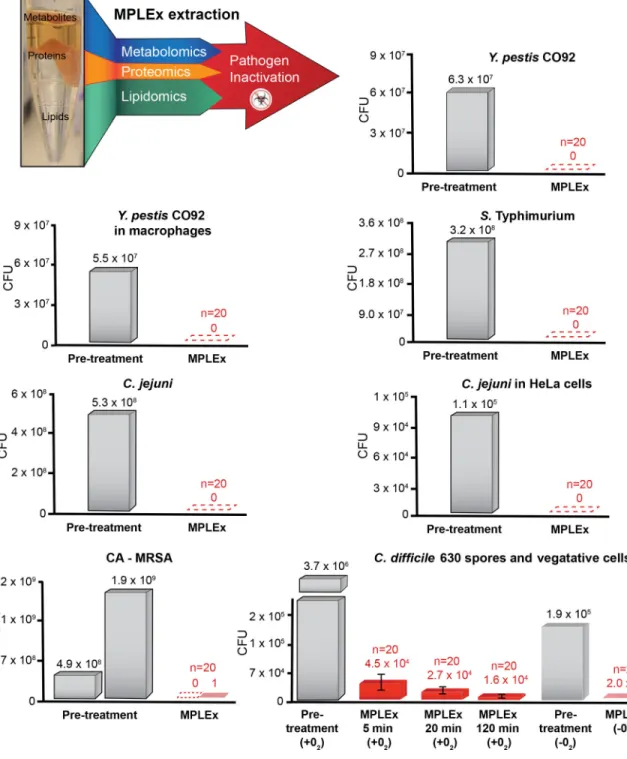
Figure 1. MPLEx Inactivation of Bacterial Pathogens
(Top, Left) Schematic of our MPLEx extraction method which uses chloroform, methanol, and water (8:4:3) to induce simultaneous pathogen inactivation and tri-phasic liquid partitioning into metabolite, protein, and lipid fractions. The bargraphs depict the level of pathogen inactivation after MPLEx extraction by quantifying the number of Colony Forming Units (CFU) in pre-treatment control samples (grey) and MPLEx-treated samples (red) in pure cultures of Yersinia pestis strain CO92 (Y. pestis CO92), Salmonella enterica subsp. enterica serovar Typhimurium (S. Typhimurium), Campylobacter jejuni (C. jejuni),
A
uthor Man
uscr
ipt
A
uthor Man
uscr
ipt
A
uthor Man
uscr
ipt
A
uthor Man
uscr
Methicillin-resistant Staphylococcus aureus USA300 (CA-MRSA), and Clostridium difficile strain 630 (C. difficile 630) spores and vegetative cells and in Y. Pestis CO92 and C. jejuni infection studies in macrophages and Hela cells, respectively.
A
uthor Man
uscr
ipt
A
uthor Man
uscr
ipt
A
uthor Man
uscr
ipt
A
uthor Man
uscr
Figure 2. MPLEx Inactivation of Viral Pathogens
The bargraphs depict the level of pathogen inactivation after MPLEx extraction by quantifying the number of Focus Forming Units (FFU) or Plaque Forming Units (PFU) in pre-treatment control samples (grey) and MPLEx-treated samples (red) in infection studies of West Nile (WNV, New York 1999), MERS-CoV (icMERS), Ebola (Ebola-Zaire delta-VP30) and Avian Influenza (H7N9).
A
uthor Man
uscr
ipt
A
uthor Man
uscr
ipt
A
uthor Man
uscr
ipt
A
uthor Man
uscr
Figure 3. Reliable multi-omic measurements resulting from MPLEx extraction of influenza infected Calu-3 cells
The heatmap depicts significant abundant changes between Calu-3 cells treated with wild-type (A/Anhui/1/13 [AH1]) and mutant (A/Anhui/103F-106M [FM]) Influenza H7N9 viral strains and time-matched (0, 3, 7, 12, 18 and 24 hours) mock-infected controls. Red color, significantly increase in virus vs. time matched mock (p-value < 0.05). The viral peptides labeled at the top of the heatmap include, hemagglutinin (HA), matrix protein 1 (M1), neuraminidase (NA), nucleoprotein (NP), non-structural (NS1), non-structural (NS2)
A
uthor Man
uscr
ipt
A
uthor Man
uscr
ipt
A
uthor Man
uscr
ipt
A
uthor Man
uscr
(corresponds to the two unlabeled viral peptides), and polymerase complex proteins (PA, PB1, and PB2); fatty acids, FA; Ceramide lipids, Cer.
A
uthor Man
uscr
ipt
A
uthor Man
uscr
ipt
A
uthor Man
uscr
ipt
A
uthor Man
uscr
Figure 4. The structural layers of Gram-positive and -negative Bacteria and Bacterial Spores
Lipid membranes are denoted with orange color.
A
uthor Man
uscr
ipt
A
uthor Man
uscr
ipt
A
uthor Man
uscr
ipt
A
uthor Man
uscr
Figure 5. The structure of lipid enveloped viruses; West Nile Virus (WNV), Middle East Respiratory Syndrome Coronavirus (MERS-CoV), Ebola, and Avian Influenza Virus
Lipid bilayers are denoted with orange color.