Epigenetic suppression of human telomerase (
hTERT
) is
mediated by the metastasis suppressor NME2 in a
G-quadruplex– dependent fashion
Received for publication, April 20, 2017, and in revised form, July 17, 2017 Published, Papers in Press, July 17, 2017, DOI 10.1074/jbc.M117.792077
Dhurjhoti Saha‡储1, Ankita Singh‡储1,2, Tabish Hussain‡, Vivek Srivastava‡3, Suman Sengupta‡, Anirban Kar‡4, Parashar Dhapola§储, Vishnu Dhople¶5, Ramesh Ummanni¶, and Shantanu Chowdhury‡§储6
From the‡Genomics and Molecular Medicine Unit,§G.N.R. Knowledge Centre for Genome Informatics, and储Academy of Scientific & Innovative Research (AcSIR), CSIR-Institute of Genomics and Integrative Biology, Council of Scientific and Industrial Research (CSIR), Mathura Road, New Delhi 110025, India and¶Centre for Chemical Biology, CSIR-Indian Institute of Chemical Technology, Hyderabad 500007, India
Edited by Joel Gottesfeld
Transcriptional activation of the human telomerase re-verse transcriptase (hTERT) gene, which remains repressed in adult somatic cells, is critical during tumorigenesis. Sev-eral transcription factors and the epigenetic state of the hTERTpromoter are known to be important for tight control ofhTERTin normal tissues, but the molecular mechanisms leading tohTERTreactivation in cancer are not well-under-stood. Surprisingly, here we found occupancy of the metasta-sis suppressor non-metastatic 2 (NME2) within thehTERT core promoter in HT1080 fibrosarcoma cells and HCT116 colon cancer cells and NME2-mediated transcriptional repression ofhTERTin these cells. We also report that loss of NME2 results in up-regulatedhTERTexpression. Mechanis-tically, additional results indicated that the RE1-silencing transcription factor (REST)–lysine-specific histone demeth-ylase 1 (LSD1) co-repressor complex associates with the hTERT promoter in an NME2-dependent way and that this assembly is required for maintaining repressive chromatin at thehTERTpromoter. Interestingly, a G-quadruplex motif at the hTERTpromoter was essential for occupancy of NME2 and the REST repressor complex on thehTERTpromoter. In light of this mechanistic insight, we studied the effects of G-quadruplex– binding ligands on hTERT expression and observed that several of these ligands repressed hTERT expression. Together, our results support a mechanism of
hTERT epigenetic control involving a G-quadruplex pro-moter motif, which potentially can be targeted by tailored small molecules.
Specialized DNA–protein assemblies called telomeres protect chromosome ends from being detected as DNA breaks
(1, 2). The ribonucleoprotein telomerase addsde novorepeats at the end of telomeres to maintain telomere length (3). Human telomerase comprises the catalytic reverse transcriptase (hTERT)7 and an RNA component (hTR) that provides the template for addition of telomeric repeats (4, 5). Lack of telom-erase results in shortening of telomeres because of the end rep-lication problem (6), and cells with critically short telomeres activate the DNA damage response, leading to cell cycle arrest or apoptosis (7, 8). This is the case in most normal somatic cells, which lack telomerase. Most cancer cells, however, have high levels of telomerase, and telomere length is maintained for initiation and survival of tumors (9). In normal cells, the limiting factor for telomerase activity is the level ofhTERT mRNA, which is under strong transcriptional control (10). In contrast, in about 85% of all cancers,hTERTexpression is reactivated (11), leading to malignant transformation and aggressive metastasis in many cases (12). The molecular mechanisms that underliehTERTreactivation from other-wise tight transcriptional control in normal somatic cells remain poorly understood.
In this context, the metastasis suppressor non-metastatic 2 (NME2; also known as nm23-H2) is of interest (13). Human nm23has several isoforms; of these, H1 (or NME1) and H2 are the most studied (14 –16). The role of NME2 in metastases suppression is well-described: overexpression of NME2 results in reduced metastasis of human oral squamous carcinoma, breast carcinoma, and murine melanoma cells (17–19), and the level ofNME2expression negatively correlates with advanced/
This work was supported by Wellcome Trust/Department of Biotechnology (DBT) India Alliance Grant 500127/Z/09/Z (to S. C.) and fellowships from the Wellcome Trust/DBT India Alliance (to D. S., S. S., V. S., and A. K.), Indian Council of Medical Research (to A. S.), and Council of Scientific and Indus-trial Research (to T. H.). The authors declare that they have no conflicts of interest with the contents of this article.
Author’s Choice—Final version free via Creative Commons CC-BY license. This article containssupplemental Figs. S1–S5 and Tables S1 and S2.
1Both authors made equal contributions to this work.
2Present address: National Institute of Cancer Prevention and Research,
Noida, Uttarpradesh 201301, India.
3Present address: Dept. of Biochemistry, School of Basic Sciences and
Research, Sharda University, Greater Noida, Uttar Pradesh 201306, India.
4Present address: Lineberger Comprehensive Cancer Center, University of
North Carolina, Chapel Hill, NC 27599-7295.
5Present address: Dept. of Functional Genomics, University of Greifswald,
Domstrasse 11, 17489 Greifswald, Germany.
6To whom correspondence should be addressed. E-mail: shantanuc@
igib.res.in.
7The abbreviations used are: hTERT, human telomerase reverse
transcrip-tase; REST, RE1-silencing transcription factor; LSD1, lysine-specific histone demethylase 1; NME2, non-metastatic 2; G4, G-quadruplex; MUT, mutated; co-IP, co-immunoprecipitation; TCP, tranylcypromine; PG4, potential G4; HDAC, histone deacetylase; MEM, modified Eagle’s medium.
Author’s Choice
metastatic stages in several tumor types (20). Notably, indepen-dent studies reported NME2-mediated transcription regula-tion of c-mycwhere association of NME2 to a G-rich sequence motif within the nuclease-hypersensitive element of the c-myc promoter was revealed (21). NME2 was also reported to regu-latePDGF-Aand vinculin transcriptionally, supporting its role as a regulatory factor (22, 23).
Herein we show that transcription of hTERT remains repressed in the presence of NME2, and loss of NME2 results in up-regulation ofhTERTexpression. NME2 binds to thehTERT core promoter, and the REST repressor complex associates with thehTERT promoter in an NME2-dependent manner. Results also revealed that the presence of an intact G-rich DNA secondary structure G-quadruplex (G4) motif in thehTERT core promoter was required for association of NME2 and the REST repressor complex at thehTERTpromoter. Notably, in the presence of NME2 and the REST repressor complex, epige-netic alterations restricted permissiveness of thehTERT pro-moter. Because altered NME2 has been detected in multiple cancer tissues (14, 17–19), it is of interest to understand the mechanisms underlying low NME2 and enhanced hTERT expression/activation.
Results
NME2 associates with the hTERT core promoter and transcriptionally represses hTERT
We noted a putative NME2-binding site on thehTERTcore promoter based on a previously reported motif from NME2 chromatin immunoprecipitation (ChIP)-sequencing experi-ments (24). Here we performed ChIP-PCR, with primers (span-ning from⫹40 to⫺230 bp with respect to thehTERT tran-scription start site) flanking the putative NME2-binding site, first in HT1080 fibrosarcoma cells and then in HCT116 colon cancer cells to confirm NME2 occupancy at the hTERT pro-moter (Fig. 1aandsupplemental Fig. S1a). To test the func-tional significance of the NME2 occupancy, endogenous hTERTexpression was checked in NME2-overexpressed or -si-lenced conditions in HT1080 and HCT116 cells. We found clear repression and an increase in hTERTexpression upon NME2 overexpression or silencing, respectively, and similar changes in hTERT protein levels (Fig. 1,bandc, and supple-mental Fig. S1b). The core promoter ofhTERTwas cloned into a luciferase reporter, and promoter activity was measured under NME2-altered conditions in HT1080 and HCT116 cells. NME2 expression andhTERTpromoter activity were found to be inversely correlated (Fig. 1,dande). Together, these data suggested NME2-mediated transcriptional re-pression ofhTERTin cancer cells.
This was further supported by fluorescence-activated cell sorting (FACS) and immunofluorescence analysis using stable NME2-overexpressing cells, which showed reduced expression ofhTERTrelative to vector-transformed cells (Fig. 1fand sup-plemental Fig. S1, c and d). Telomerase enzymatic activity was measured under NME2-overexpressed or -silenced conditions using a telomere repeat amplification protocol, and a reduc-tion or an increase in telomerase activity, respectively, was observed (supplemental Fig. S1e). Based on this, we asked
whether NME2 repressedhTERTin normal primary cells. In primary lung fibroblast MRC5 cells, we found repression or enhanced hTERT expression and promoter activity under NME2-overexpressed or -silenced conditions, respectively (Fig. 1,gandh).
To validate the occupancy of NME2 on thehTERTpromoter, an oligonucleotide pulldown assay was performed. A puta-tive 12-mer (⫺115 to ⫺127 bp from hTERT transcription start site) representing the NME2 DNA-binding motif (23) or its mutated/scrambled negative controls were used. Pull-down using anti-NME2 antibody showed specific binding to the unaltered (wild-type (WT)) relative to mutated (MUT) or scrambled motifs (Fig. 1i). Furthermore, a reporter plas-mid in which the mutant NME2 motif was inserted did not suppress promoter activity in the presence of NME2 in HT1080 and HCT116 cells (Fig. 1j). Together, these results suggested that NME2 binding was required for transcrip-tional repression ofhTERT.
Next, we checked the role, if any, of NME1, a close homolog of NME2, in transcriptional repression on hTERT. hTERT expression and protein levels remained unaltered when NME1 was specifically silenced (supplemental Fig. S1, f and g);hTERT promoter activity also was not affected upon NME1 silencing in HT1080 cells (supplemental Fig. S1h).
NME2 maintains repressive chromatin at the hTERT promoter by engaging the REST–LSD1 repressor complex
To understand the mechanism underlying NME2-mediated hTERTrepression, we asked whether other binding partners of NME2 were involved. To find possible nuclear interacting part-ners of NME2, we used LC-MS/MS after immunoprecipitation with anti-NME2 antibody from nuclear extract of HT1080 cells. Using a stringent analysis cutoff of 1% false discovery rate, 120 interacting partners of NME2 were detected (supplemental Fig. S2a and Table S1) that belonged to several important classes including chromatin remodeling (Gene Ontology anal-ysis;supplemental Fig. S2b). Of these, to specifically understand possible mechanisms underlying NME2-induced hTERT repression, we focused on chromatin modifiers HDAC1 and HDAC2. Interaction of NME2 with both HDAC1 and HDAC2 was further confirmed using co-immunoprecipitation (co-IP) experiments (Fig. 2a).
HDAC1 and HDAC2 are integral members of the REST repressor complex that engages the lysine-specific demethylase LSD-1 (25, 26). Therefore, we checked whether NME2 interacts with REST and LSD1. Co-IP with anti-NME2 antibody showed clear enrichment of REST and LSD1 in the anti-NME2-immu-noprecipitated fraction compared with its isotype control IgG (Fig. 2a). In addition, reverse co-IP with REST or anti-LSD1 confirmed NME2 interaction with REST–anti-LSD1 ( supple-mental Fig. S2c).
Next, we checked for REST and LSD1 occupancy on the hTERTpromoter and determined whether this was NME2-de-pendent. Both REST and LSD1 ChIPs showed occupancy on the hTERTpromoter where the ChIP signals were significantly reduced under NME2-silenced conditions (Fig. 2,bandc, and
(Fig. 2d). Moreover, sequential ChIP for NME2–REST and NME2–LSD1 further supported co-occupancy of NME2– REST–LSD1 complex on thehTERTpromoter in HT1080 and HCT116 cells (Fig. 2,eandf).
Next, we asked whether NME2-mediated engagement of REST–LSD1 affectedhTERTexpression. siRNA against REST attenuated NME2-induced repression ofhTERT expression, which was further derepressed using the LSD1 inhibitor tranyl-cypromine (TCP) (Fig. 2g). Silencing of all three, NME2, REST, and LSD1, led to a further increase inhTERTexpression (Fig. 2g). An increase in hTERTexpression and promoter activity was also evident upon inhibition of REST and LSD1 indepen-dently or together (supplemental Fig. S2, e and f). In addition,
histone methylation ChIP results showed an increase in histone activation marks (H3K4me2 and H3K4me) following silencing of REST (supplemental Fig. S2g). Taken together these results show role of NME2 in promoting repressive chromatin at the hTERT promoter through the REST–LSD1 complex to sup-presshTERTexpression.
Conversely, we expected permissive chromatin at thehTERT promoter upon silencing NME2. We checked histone activa-tion marks H3K9ac, H3K4Me2, and H3K4Me and found that all three marks were clearly enhanced in cells treated with siRNA against NME2 relative to control cells (si-control), whereas there was no significant change in total histone H3 occupancy (Fig. 2h).
Figure 1. NME2 occupies thehTERTpromoter and reduces its expression.a, NME2 ChIP showing occupancy of NME2 on thehTERTpromoter in HT1080 and HCT116 cell lines.ptprcgene serves as a negative control.bandc, real-time PCR (b) and Western blot analysis (c) forhTERTexpression (49, 50) upon NME2 overexpression and NME2 silencing in HT1080 and HCT116 cells. Thetop panelsshowhTERTexpression, and thelower panelsshow the expression of theGAPDH
loading control (c).Error barsrepresent S.E. (three biological replicates); * indicates apvalue⬍0.05; ** indicates apvalue⬍0.005.dande, luciferase reporter assay to measurehTERTpromoter activity upon NME2-overexpressed (d) and -silenced conditions (e).F-luc, firefly luciferase;R-luc,Renillaluciferase.Error bars
represent S.E. (three biological replicates); * indicates apvalue⬍0.05; ** indicates apvalue⬍0.005.f, flow cytometry analysis shows reduced expression of
hTERTin GFP-NME2 stably overexpressing cells in comparison with GFP vector (Vec) HT1080 cells.Error barsrepresent S.E. (three biological replicates); * indicates apvalue⬍0.05.gandh,hTERTexpression (g) and promoter activity (h) were measured in primary MRC5 normal lung fibroblasts in NME2-overex-pressed and -silenced conditions.Error barsrepresent S.E. (three biological replicates); * indicates apvalue⬍0.05.i, oligo pulldown assay for NME2 using biotin-labeled oligos with WT, MUT, and scrambled (Scr) NME2 motifs. Thelower panelrepresents the loading control.j, luciferase reporter assay forhTERT
NME2 mutants devoid of DNA-binding function are unable to repress hTERT transcription
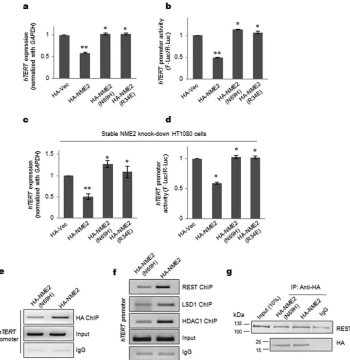
Mutants of NME2 that are unable to bind DNA (N69H and R34A) were reported earlier (27, 28). Using these mutants, we checked whether DNA binding by NME2 was required forhTERTrepression. Both the mutants, NME2(N69H) and NME2(R34A), did not show any repression ofhTERT expres-sion or promoter activity in HT1080 cells (Fig. 3,aandb). This was further confirmed using stable NME2 knockdown cells (supplemental Fig. S3a). As expected, on stable knockdown of NME2, telomerase expression and promoter activity increased significantly (supplemental Fig. S3, b and c).hTERTrepression and reduced promoter activity were observed on inducing HA-tagged NME2 (HA-NME2) but not when HA-NME2(N69H) or HA-NME2(R34A) were induced (Fig. 3,candd, and supple-mental Fig. S3d). Furthermore, using the mutant NME2(N69H), we checked whether occupancy of the REST–LDS1 complex was altered vis-à-vis wild-type NME2. First, using ChIP with HA antibody, we confirmed relatively reduced occupancy of the NME2(N69H) mutant compared with wild-type NME2 on the hTERT promoter (Fig. 3e). Next, using ChIP, we observed reduced occupancy of REST at thehTERTpromoter in the case of NME2(N69H) compared with NME2 (Fig. 3f) that was not due to any loss of REST interaction with the mutant NME2(N69H) as shown by co-IP experiments in HT1080 cells (Fig. 3g). Consistent with reduced REST occupancy, we further found both LSD1 and HDAC1 occupancy to be reduced at the hTERTpromoter in the case of NME2(N69H) relative to wild-type NME2 (Fig. 3f). Together, these studies confirmed DNA binding by NME2 to be a key factor in occupancy of the REST– LSD1 complex at thehTERTpromoter andhTERTrepression by NME2.
G-quadruplex motif at the hTERT promoter was required for NME2 and REST–LSD1 occupancy at the hTERT promoter
Potential G4 (PG4)-forming sequences were reported in the hTERTpromoter (29, 51). Interestingly, we observed that the NME2-binding motif (Fig. 4a) was within also within ahTERT promoter PG4-forming sequence, and because NME2 and pro-moter G4 interactions have been reported before (21, 30), we asked whether the G4 structure played any role in hTERT repression by NME2. Oligonucleotide representing the PG4-forming sequence adopted mixed parallel/antiparallel G4 structures in solution (Fig. 4b). For further work, we designed nucleotide substitutions such that key bases required for G4 stability were mutated, but the NME2-binding motif remained intact. This resulted in a disrupted G4 structure in solution (Fig. 4b). Next, a luciferase reporter construct was made by substi-tuting these bases (MUT-G4) using site-directed mutagenesis.
Upon expression of NME2, although promoter activity was repressed by⬃50% in the case of the G4-containing construct, for MUT-G4 the observed repression was relatively less in HT1080 and HCT116 cells (Fig. 4c).
To test NME2 interaction with the G4, we used oligonucle-otide pulldown with the G4 or the MUT-G4 sequence. On probing with anti-NME2 antibody, we found relatively reduced affinity of NME2 toward the MUT-G4 compared with G4 (Fig. 4d and supplemental Fig. S4). In addition, we checked and observed that a significantly reduced amount of REST was pulled down in the case of MUT-G4, supporting NME2–REST interaction (Fig. 4d). Furthermore, we found reduced intracel-lular occupancy of NME2 at thehTERTpromoter in the case of ectopically maintained MUT-G4 relative to G4 in ChIP with anti-NME2 antibody followed by PCR with plasmid-specific primers (Fig. 4e; also called transient ChIP (31)). This was also confirmed by ChIP using anti-REST or anti-LSD1 where reduced occupancy was observed for both REST and LSD1 on thehTERTpromoter in the case of MUT-G4 (Fig. 4f). Together, these results support roles of the G4 structural motif in NME2 binding and recruitment of the REST–LSD1 complex for hTERTrepression.
We next studied the effect of G4-binding ligands on telom-erase transcription. Because G4 ligands were noted to both acti-vate and repress gene transcription (32, 52), we screened 20 previously established intracellular G4-binding ligands (listed insupplemental Table S2). Of these, 11 ligands showed down-regulation (⬃50 – 85%) ofhTERTin HT1080 cells (Fig. 4gand
supplemental Table S2). Five of the 11 ligands also showed more than 50% reduction in hTERT promoter activity in reporter assays (Fig. 4h), and the ligand-induced effect on pro-moter activity was lost in the case of all five ligands when the G4 motif was mutated in the reporter construct. This further sup-ported a role of G4 –ligand interactions inside cells inhTERT repression (Fig. 4h).
Discussion
Multiple transcription factors including Sp1, c-Myc, p53, SP1, ETS, E2F, and AP1 have been reported to transcriptionally control hTERTexpression (10, 33, 34). However, how these function to control the epigenetic state of thehTERTpromoter is not precisely understood. For example, histone deacetylation repressed hTERT transcription, whereas treatment with the HDAC inhibitor trichostatin A resulted in activation ofhTERT (35), and a recent report suggested that LSD1, using its demeth-ylation activity, altered activating histone marks on thehTERT promoter to represshTERTtranscription (36). Although these studies indicated the participating regulatory factors,
mecha-Figure 2. NME2 interacts with REST complex and alters chromatin marks onhTERTto repress transcription.a, co-IP assay for validation of REST complex component interaction with NME2 as obtained from LC-MS/MS results. Co-IP for REST, LSD1, HDAC1, and HDAC2 was performed using nuclear extract from HT1080 cells.b– d, LSD1 (b), REST (c), and HDAC1/2 (d) ChIP in the NME2 transiently silenced condition showing reduced occupancy on thehTERTpromoter. The histogram represents the -fold change in LSD1, REST, and HDAC1/2 occupancy on thehTERTpromoter.Error barsrepresent S.E. (three biological replicates); * indicates apvalue⬍0.05.eandf, sequential chromatin immunoprecipitation for NME2–LSD1 (e) and NME2–REST (f) followed by PCR ofhTERTpromoter in HT1080 and HCT116 cell lines.g,hTERTmRNA expression was measured after silencing of REST and NME2, blocking of LSD1 using TCP, and combinations of both treatments.Error barsrepresent S.E. (three biological replicates); * indicates apvalue⬍0.05; ** indicates apvalue⬍0.005.h, ChIP for active histone methylation (H3K4me and H3K4me2) and acetylation (H3K9ac) marks and total histone3 (H3) onhTERTpromoter in NME2-silenced conditions in HT1080 cells.
nisms of how the repressors/histone modifiers were engaged on thehTERTpromoter have remained unclear.
Herein we show that NME2 transcriptionally represses hTERTthrough recruitment of the REST repressor complex, which includes REST, HDAC1, HDAC2, and LSD1. Using direct and sequential ChIP, we found that association of the REST repressor complex on thehTERTpromoter was
depen-dent on NME2. Moreover, co-immunoprecipitation experi-ments confirmed that NME2 interacted with members of the REST complex inside the nucleus. As a result, we observed that thehTERTpromoter is maintained in a repressive chromatin state. In other words, these data suggest that the loss of NME2 results in permissive chromatin at the hTERTpromoter and up-regulation ofhTERTexpression.
Figure 3. hTERT transcription repression is dependent on DNA binding by NME2.aandb,hTERTmRNA (a) and promoter activity (b) following expression of HA-tagged proteins HA-NME2, HA-NME2(N69H), and HA-NME2(R34A) (R34A and N69H are well-characterized NME2 mutants lacking DNA-binding activity) or the HA vector (Vec) control in HT1080 cells;GAPDHwas used as a control for mRNA expression.Error barsrepresent S.E. (three biological replicates); *,p⬍
Figure 4. Repression ofhTERTdepends on NME2 interaction withhTERTpromoter G-quadruplex.a, minimal promoter ofhTERTshowing the NME2-binding site (gray solid bar) and PG4-forming sequence (underlined).b, circular dichroism plot showing formation of the G4 motif in solution, whereas the sequence with mutations in key bases (MUT-G4) showed partial disruption of the G4 motif under similar conditions.TSS, transcription start site.c, luciferase reporter assay forhTERTpromoter activity upon transfection of either the wild-type G4 or the disrupted MUT-G4 plasmid along with HA vector or HA-NME2 in HT1080 and HCT116 cells.F-luc, firefly luciferase;R-luc,Renillaluciferase.Error barsrepresent S.E. (three biological replicates); * indicatesp⬍0.05.d, oligonu-cleotide pulldown assay with either the WT G4 or the disrupted MUT-G4 oligonuoligonu-cleotide in HT1080 nuclear lysate developed with the respective antibodies shows that both NME2 and REST have enhanced affinity for the G4 motif relative to MUT-G4.eandf, intracellular NME2 (and REST–LSD1) occupancy at the
Low NME2 has been associated with malignant transforma-tion in multiple cancers (14, 18). Independently, in oral, breast, and colon cancer, mechanisms of antimetastatic action of NME2 were suggested to be through induction of the epithelial phenotype (mesenchymal to epithelial transition) (37). Con-versely, increased telomerase due to telomerase reactivation was noted in progression of various cancers including acute leukemia, breast, prostate, lung, and melanoma (38). Recent studies further reveal that point mutations in thehTERT pro-moter leading to increased hTERTmRNA synthesis is key to hTERTreactivation in several cancers (10, 39). In this context, possible implications of low NME2 in up-regulation ofhTERT transcription through epigenetic alteration of thehTERT pro-moter could be of interest in understanding mechanisms of telomerase activation. Although results suggest engagement of the repressor complex in an NME2-dependent manner, further studies will be required to understand how other transcription factors at the hTERT promoter influence this mechanism. NME2 was also noted to inhibit telomerase catalytic activityin vitro(40). Together with findings reported here, this indicates a possible dual role of NME2 in control of telomerase in cancer cells.
Multiple lines of evidence support a role of G4 motifs in gene expression. Prominent among these are enrichment of poten-tial G4 forms within promoters across species (41– 43) and evi-dence supporting their role in gene expression (44) including their presencein vivoand identification of proteins that func-tion in associafunc-tion with G4s inside cells (45– 48). Interestingly, NME2 was reported to regulate transcription through interac-tion with a c-mycpromoter G4 motif (21). Together, these data prompted a closer look when we noted that thehTERT pro-moter harbors G4-forming sequences that were implicated in possiblehTERTexpression (29). Our results support a role of hTERTpromoter G4 motifs in hTERT repression. This encour-aged us to test G4 ligand-mediated repression of hTERT expression, which in turn may open potential ways of control-linghTERTactivation in cancer cells.
Experimental procedures
Cells and culture conditions
HT1080, HCT116, and MRC5 primary fibroblast cells were obtained from the American Type Cell Culture (ATCC) and maintained in modified Eagle’s medium (MEM), Dulbecco’s modified Eagle’s medium low glucose (DMEM) with high glu-cose, and MEM, respectively, and supplemented with 10% fetal bovine serum at 37 °C in 5% CO2.
ChIP
ChIP assays were performed as described (40) with the fol-lowing antibodies: rabbit anti-HA antibody (Abcam, ab9110), anti-rabbit IgG (Sigma), anti-REST antibody (Millipore, 17641), LSD1 (Abcam, ab17721), HDAC1 (Abcam, ab7028), HDAC2 (Abcam, ab51832), H3K4me (Abcam, ab8895), anti-H3K4me2 (Abcam, ab7766), anti-H3K4me3 (Abcam, ab8580), anti-histone3 (Abcam, ab1791), and anti-H3k9ac (Abcam, ab10812). For sequential ChIP, samples were treated as described for ChIP (40) before adding 3– 4g of the first anti-body and incubating overnight at 4 °C followed by 50l of
pro-tein G-Sepharose beads for 4 h at 4 °C on a rotator. Samples were then washed with washing buffers (low-salt buffers, high-salt buffers, and lithium chloride) and divided into two frac-tions. One part was processed further for ChIP-DNA elution, and 3– 4g of the second antibody was added to the second fraction and incubated again overnight at 4 °C followed by incu-bation with 50 l of protein G-Sepharose beads. Beads were washed (washing buffers as above), and the final DNA samples were obtained using a phenol, chloroform, and isoamyl alcohol precipitation method (as for basic ChIP protocol). The ChIP PCR primers used forhTERTpromoter were 5⬘ -CCAGGCCG-GGCTCCCAGTGGAT-3⬘(forward) and 5⬘ -GGCTTCCCAC-GTGCGCAGCAGGA-3⬘(reverse), and the amplicon length was 275 bp. For transient ChIP assays, cells were transfected with wild-typehTERTand mutanthTERTpromoter constructs (2g of plasmid/1⫻106cells). Transfected cells were harvested after 48 h, and ChIP was performed against REST, NME2, and LSD1 as described above. The eluted ChIP-DNA was checked for hTERTpromoter amplification by using primers derived partially from the vector construct and from thehTERT pro-moter sequence.
Real-time PCR
Total RNA was isolated using TRIzol威reagent (Invitrogen, Life Technologies) according to the manufacturer’s instruc-tions. RNA was quantified, and 2g of RNA was used for cDNA preparation using an Applied Biosciences kit. The relative tran-script expression level for genes was measured by quantitative real-time PCR using a SYBR Green-based method. Average -fold change was calculated by the difference in threshold cycles (Ct) between test and control samples.GAPDHgene was used as an internal control for normalizing the cDNA concentration of each sample. For TCP (concentration, 1M)-treated cells,
treatment was done after 24 h of transfection, and RNA was isolated 24 h post-treatment. G-quadruplex ligand (concentra-tion, 2M) treatment was done for 48 h.
siRNA transfection
SMARTpool ON-TARGET siRNAs against NME2, NME1, and REST were procured from GE Dharmacon and used according to the manufacturer’s protocol.
Luciferase assay
The minimal promoter region of hTERT harboring the NME2 motif and G-quadruplex sequences was cloned into pGL3-basic vector and transfected into HT1080 and HCT116 cells using Lipofectamine 2000 (Invitrogen). Plasmid (pGL4.73) containing a CMV promoter drivingRenillaluciferase was co-transfected as a transfection control for normalization. After 48 h, cells were harvested, and luciferase activities of cell lysate were measured using a Dual-Luciferase reporter assay kit (Pro-mega). For TCP (concentration, 1M)-treated cells, treatment
was done after 24 h of transfection, and luciferase activity was measured 24 h post-treatment. G-quadruplex ligand (concen-tration, 2M) treatment was done for 48 h.
Preparation of nuclear extracts
extract was isolated using a nuclear extraction kit (CelLytic, Sigma) according to the manufacturer’s protocol.
Co-IP
For immunoprecipitation experiments, 500 g of nuclear extract was incubated for 4 h at 4 °C with 4g of anti-NME2 antibody (Kamiya, KM1121 MC412). Immunoprecipitation was performed using a Catch and Release co-immunoprecipi-tation kit (Millipore) according to the manufacturer’s protocol.
Antibodies and Western blotting
For Western analysis, immunoprecipitated nuclear extracts were separated by 10% sodium dodecyl sulfate (SDS)-PAGE and transferred to polyvinylidene difluoride (PVDF) mem-branes (Immobilon FL, Millipore). The following primary antibodies were used for immunoblotting: anti-REST antibody (Millipore, 17– 641), HDAC1 (Abcam, ab7028), HDAC2 (Abcam, ab51832), LSD1 (Abcam, ab17721), anti-NME2 (Abcam, ab60602) (23, 24, 40), and anti-hTERT (Abcam, ab32020) (49, 50). The secondary antibodies used were anti-mouse and anti-rabbit alkaline phosphatase conjugates from Sigma. Western blots with antibodies against NME2, telomer-ase, REST, and LSD1 along with relevant molecular weight markers are shown insupplemental Fig. S5.
Immunofluorescence microscopy
Cells were grown on coverslips and at 100% confluence were fixed with 4% paraformaldehyde by incubating for 10 min at room temperature. Cells were permeabilized with 0.5% Tri-tonTMX-100 and treated with blocking solution (3% BSA in PBS) for 30 min at room temperature. After one PBS (1⫻) wash, cells were treated with anti-hTERT antibody (1:100) overnight at 4 °C. The next day cells were washed alternately with 1⫻PBS and PBS with Tween 20 three times and probed with Alexa Fluor威 594 for 1 h at room temperature. Cells were washed again alternately with PBS and PBS with Tween 20 three times and mounted with Prolong威Gold antifade reagent with DAPI. Images were taken as maximum intensity projections on a Leica TCS-SP8 confocal microscope.
Flow cytometry (FACS)
After trypsinization, both stable GFP-NME2-expressing and corresponding GFP vector-transformed cells were fixed in 4% paraformaldehyde for 10 min. After washing in PBS, cells were permeabilized in 0.5% Triton X-100 in PBS for 10 min. After blocking in 3% BSA in PBS for 1 h at 4 °C followed by washing in PBS, cells were incubated with primary anti-hTERT antibody (Abcam, ab94523) (1:100) at 4 °C overnight. After washing in PBS, cells were incubated with Alexa Fluor 594-conjugated sec-ondary antibody for 2 h at 4 °C. As a negative control, cells were fixed and incubated with secondary antibody only without incubation with primary antibody. Finally, after washing in PBS, cells were resuspended in PBS for acquisition by flow cytometry (BD Biosciences FACSAria III) using the Cy3 chan-nel (for Alexa Fluor 594) to detecthTERTexpression.
Oligonucleotide pulldown assay
For pulldown assays, 2g of biotinylated NME2 motif, motif mutant M1, wild-type G4, and MUT-G4 and 300g of nuclear
extract (prepared from HT1080 cells) were incubated for 1 h at room temperature in binding buffer. Then the whole complex was incubated with 60l of streptavidin-agarose (Invitrogen) for 4 h at 4 °C. In all experiments, the beads were washed three times with washing buffer (50 mMTris-HCl (pH 7.5) and 150
mMNaCl), and the bound proteins were eluted by boiling in 5⫻
SDS sample buffer (20 mMTris-HCl (pH 6.8), 10% glycerol, 4%
SDS, 100 mMdithiothreitol, 4 mMEDTA, and 0.025%
Coomas-sie Brilliant Blue R-250) and subjected to Western blot analysis using anti-REST antibody (Abcam) and anti-NME2 antibody (Kamiya).
Separation of immunoprecipitate and preparation of peptide mixtures
Protein identification using mass spectrometry was per-formed as follows. First, the immunoprecipitate was resolved by 12.5% SDS-PAGE followed by visualization with colloidal Coo-massie Brilliant Blue. The bands that appeared specifically in the immunoprecipitate were excised manually using sterile blades. Further procedures including distaining and digestion with trypsin followed by extraction of peptides were performed manually. In brief, gel pieces were distained in 50 mM
ammo-nium bicarbonate and acetonitrile (50%, v/v) and dehydrated with 95% (v/v) acetonitrile. The dehydrated gel pieces were soaked with a sufficient volume of trypsin solution (20 ng/l trypsin in 25 mMammonium bicarbonate) and incubated at
37 °C overnight. Upon digestion, the trypsinized solution was pooled with a further peptide extraction from gel pieces using 50% (v/v) acetonitrile containing 0.1% (v/v) acetic acid and incubating for 30 min at 37 °C with constant shaking. The pep-tide extract collected as supernatant was pooled, vacuum-dried to about 10l, and purified using a micro ZipTip (Millipore). The purified mixture was vacuum-dried, and peptides were reconstituted in 2% acetonitrile containing 0.1% (v/v) acetic acid and further analyzed by LC-MS/MS.
Identification of proteins using LC-MS/MS
acquisi-tion method was set to isolate up to 20 of the most intense ions depending upon signal intensity for fragmentation in the linear ion trap using collision-induced dissociation. Target ions already selected for MS/MS were dynamically excluded for 60 s. The general conditions of the mass spectrometer for data col-lection were 1.6 –1.7-kV electrospray voltage and ion secol-lection threshold of 2000 counts for MS/MS. For identification of pro-teins from MS data, an automated database search was per-formed using Proteome Discoverer 1.3.0.339 (Thermo Scien-tific) with the Sequest algorithm. A human UniProt FASTA database and a decoy database of 1% false discovery rate were used for identification. Two possible missed cleavages for tryp-sin enzyme specificity with a mass tolerance of 10 ppm (parent ion) and 0.8 Da (fragment ion) and methionine oxidation dynamics were considered for the database search. The identity of the proteins was confirmed based on high-confidence pep-tide identification containing at least two peppep-tides per protein (with rank 1 peptides in proteins; XCorr score,ⱖ2.45 or 2.85 for doubly and triply charged peptides, respectively).
Site-directed mutagenesis
A QuikChange site-directed mutagenesis kit (Agilent tech-nologies) was used to generate various mutants ofNME2gene. Mutagenesis reactions were performed according to the man-ufacturer’s instructions with the wild-type c-DNA cloned in pRSET-A used as a template. The positive clones were screened for the mutation by sequencing.
Author contributions—D. S. and S. C. designed the experiments. T. H. and D. S. performed immunofluorescence experiments. D. S. and A. S. performed ChIP-seq. D. S., P. D., and A. K. performed ChIP-seq validation. A. S. performed the biophysical experiments. S. S. and T. H. performed flow cytometry experiments. T. H. and D. S. performed ligand screening. V. D., R. U., and D. S. performed proteomics experiments. V. S. performed the telomere repeat ampli-fication protocol. D. S., T. H., and S. C. contributed to data compila-tion, images, and manuscript writing. S. C. conceived the idea, con-ceptualized the study, wrote and edited the manuscript, and is responsible for correspondence.
Acknowledgment—We thank Jean-Francois Riou (Muséum National d’Histoire Naturelle) for providing the library of G4-specific ligands as a gift. We thank S. Neidle (University College London), S. Balasubra-manian (University of Cambridge), the M. P. Teulade-Fichou labora-tory (Institut Curie), and Patrick Mailliet (Riou laboralabora-tory) for G4 ligands used in this study.
Note added in proof—An incorrect gel section was inadvertently used in the REST ChIP panel in Fig. 3fof the version of this article that was published as a Paper in Press on July 17, 2017. This error has now been corrected and does not affect the results or conclusions of this work.
References
1. Longhese, M. P. (2008) DNA damage response at functional and dysfunc-tional telomeres.Genes Dev.22,125–140
2. O’Sullivan, R. J., and Karlseder, J. (2010) Telomeres: protecting chromo-somes against genome instability.Nat. Rev. Mol. Cell Biol.11,171–181 3. Greider, C. W., and Blackburn, E. H. (1989) A telomeric sequence in the
RNA of Tetrahymena telomerase required for telomere repeat synthesis.
Nature337,331–337
4. Greider, C. W., and Blackburn, E. H. (1985) Identification of a specific telomere terminal transferase activity in Tetrahymena extracts.Cell.43,
405– 413
5. Sandin, S., and Rhodes, D. (2014) Telomerase structure. Curr. Opin. Struct. Biol.25,104 –110
6. Greider, C. W. (1996) Telomere length regulation.Annu. Rev. Biochem.
65,337–365
7. Deng, Y., Chan, S. S., and Chang, S. (2008) Telomere dysfunction and tumour suppression: the senescence connection. Nat. Rev. Cancer8,
450 – 458
8. Hayashi, M. T., Cesare, A. J., Fitzpatrick, J. A., Lazzerini-Denchi, E., and Karlseder, J. (2012) A telomere-dependent DNA damage checkpoint in-duced by prolonged mitotic arrest.Nat. Struct. Mol. Biol.19,387–394 9. Shay, J. W., and Wright, W. E. (2010) Telomeres and telomerase in normal
and cancer stem cells.FEBS Lett.584,3819 –3825
10. Akincilar, S. C., Unal, B., and Tergaonkar, V. (2016) Reactivation of telom-erase in cancer.Cell. Mol. Life Sci.73,1659 –1670
11. Shay, J. W., and Wright, W. E. (2005) Senescence and immortalization: role of telomeres and telomerase.Carcinogenesis.26,867– 874 12. Stewart, S. A., Hahn, W. C., O’Connor, B. F., Banner, E. N., Lundberg, A. S.,
Modha, P., Mizuno, H., Brooks, M. W., Fleming, M., Zimonjic, D. B., Popescu, N. C., and Weinberg, R. A. (2002) Telomerase contributes to tumorigenesis by a telomere length-independent mechanism.Proc. Natl. Acad. Sci. U.S.A.99,12606 –12611
13. Steeg, P. S., Bevilacqua, G., Kopper, L., Thorgeirsson, U. P., Talmadge, J. E., Liotta, L. A., and Sobel, M. E. (1988) Evidence for a novel gene associated with low tumor metastatic potential.J. Natl. Cancer Inst.80,200 –204 14. Thakur, R. K., Yadav, V. K., Kumar, P., and Chowdhury, S. (2011)
Mech-anisms of non-metastatic 2 (NME2)-mediated control of metastasis across tumor types. Naunyn Schmiedebergs Arch. Pharmacol. 384,
397– 406
15. Steeg, P. S., Zollo, M., and Wieland, T. (2011) A critical evaluation of biochemical activities reported for the nucleoside diphosphate kinase/ Nm23/Awd family proteins: opportunities and missteps in understanding their biological functions.Naunyn Schmiedebergs Arch. Pharmacol.384,
331–339
16. Galasso, A, and Zollo, M. (2009) The Nm23-H1-h-Prune complex in cel-lular physiology: a ‘tip of the iceberg’ protein network perspective.Mol. Cell. Biochem.329,149 –159
17. Hartman, J., Ström, A., Gustafsson, J.-A. (2009) Estrogen receptorin breast cancer— diagnostic and therapeutic implications. Steroids 74,
635– 641
18. Hartsough, M. T., and Steeg, P. S. (2000) Nm23/nucleoside diphosphate kinase in human cancers.J. Bioenerg. Biomembr.32,301–308
19. Miyazaki, H., Fukuda, M., Ishijima, Y., Takagi, Y., Iimura, T., Negishi, A., Hirayama, R., Ishikawa, N., Amagasa, T., and Kimura, N. (1999) Overex-pression of nm23-H2/NDP kinase B in a human oral squamous cell car-cinoma cell line results in reduced metastasis, differentiated phenotype in the metastatic site, and growth factor-independent proliferative activity in culture.Clin. Cancer Res.5,4301– 4307
20. Herak Bosnar, M., Bago, R., Konjevoda, P., and Pavelic´, J. (2008) Gene expression profiling of Nm23-H2 overexpressing CAL 27 cells using DNA microarray.Neoplasma55,447– 454
21. Thakur, R. K., Kumar, P., Halder, K., Verma, A., Kar, A., Parent, J.-L., Basundra, R., Kumar, A., and Chowdhury, S. (2009) Metastases suppressor NM23-H2 interaction with G-quadruplex DNA within c-MYC promoter nuclease hypersensitive element induces c-MYC expression.Nucleic Ac-ids Res.37,172–183
22. Ma, D., Xing, Z., Liu, B., Pedigo, N. G., Zimmer, S. G., Bai, Z., Postel, E. H., and Kaetzel, D. M. (2002) NM23-H1 and NM23-H2 repress transcrip-tional activities of nuclease-hypersensitive elements in the platelet-de-rived growth factor-A promoter.J. Biol. Chem.277,1560 –1567 23. Thakur, R. K., Yadav, V. K., Kumar, A., Singh, A., Pal, K., Hoeppner, L.,
24. Yadav, V. K., Thakur, R. K., Eckloff, B., Baral, A., Singh, A., Halder, R., Kumar, A., Alam, M. P., Kundu, T. K., Pandita, R., Pandita, T. K., Wieben, E. D., and Chowdhury, S. (2014) Promoter-proximal transcription factor binding is transcriptionally active when coupled with nucleosome reposi-tioning in immediate vicinity.Nucleic Acids Res.42,9602–9611 25. Roizman, B. (2011) The checkpoints of viral gene expression in productive
and latent infection: the role of the HDAC/CoREST/LSD1/REST repres-sor complex.J. Virol.85,7474 –7482
26. Bithell, A. (2011) REST: transcriptional and epigenetic regulator. Epig-enomics3,47–58
27. Postel, E. H., Weiss, V. H., Beneken, J., and Kirtane, A. (1996) Mutational analysis of NM23-H2/NDP kinase identifies the structural domains criti-cal to recognition of a c-myc regulatory element.Proc. Natl. Acad. Sci. U.S.A.93,6892– 6897
28. Postel, E. H., Abramczyk, B. A., Gursky, S. K., and Xu, Y. (2002) Structure-based mutational and functional analysis identify human NM23-H2 as a multifunctional enzyme.Biochemistry41,6330 – 6337
29. Palumbo, S. L., Ebbinghaus, S. W., and Hurley, L. H. (2009) Formation of a unique end-to-end stacked pair of G-quadruplexes in the hTERT core promoter with implications for inhibition of telomerase by G-quadruplex-interactive ligands.J. Am. Chem. Soc.131,10878 –10891
30. Shan, C., Yan, J.-W., Wang, Y.-Q., Che, T., Huang, Z.-L., Chen, A.-C., Yao, P. F., Tan, J. H., Li, D., Ou, T. M., Gu, L. Q., and Huang, Z. S. (2017) Design, synthesis and evaluation of isaindigotone derivatives to downregulate c-myc transcription via disrupting the interaction of NM23-H2 with G-quadruplex.J. Med. Chem.60,1292–1308
31. Lavrrar, J. L., and Farnham, P. J. (2004) The use of transient chromatin immunoprecipitation assays to test models for E2F1-specific transcrip-tional activation.J. Biol. Chem.279,46343– 46349
32. Rhodes, D., and Lipps, H. J. (2015) G-quadruplexes and their regulatory roles in biology.Nucleic Acids Res.43,8627– 8637
33. Kyo, S., Takakura, M., Fujiwara, T., and Inoue, M. (2008) Understanding and exploiting hTERT promoter regulation for diagnosis and treatment of human cancers.Cancer Sci.99,1528 –1538
34. Ramlee, M. K., Wang, J., Toh, W. X., and Li, S. (2016) Transcription reg-ulation of the human telomerase reverse transcriptase (hTERT) gene.
Genes7,E50
35. Cong, Y. S., and Bacchetti, S. (2000) Histone deacetylation is involved in the transcriptional repression of hTERT in normal human cells.J. Biol. Chem.275,35665–35668
36. Zhu, Q., Liu, C., Ge, Z., Fang, X., Zhang, X., Strååt, K., Björkholm, M., and Xu, D. (2008) Lysine-specific demethylase 1 (LSD1) is required for the transcriptional repression of the telomerase reverse transcriptase (hTERT) gene.PLoS One3,e1446
37. Yadav, V. K., Kumar, A., Mann, A., Aggarwal, S., Kumar, M., Roy, S. D., Pore, S. K., Banerjee, R., Mahesh Kumar, J., Thakur, R. K., and Chowdhury, S. (2014) Engineered reversal of drug resistance in cancer cells—metasta-ses suppressor factors as change agents.Nucleic Acids Res.42,764 –773 38. Artandi, S. E., and DePinho, R. A. (2010) Telomeres and telomerase in
cancer.Carcinogenesis31,9 –18
39. Li, Y., Cheng, H. S., Chng, W. J., and Tergaonkar, V. (2016) Activation of mutant TERT promoter by RAS-ERK signaling is a key step in malignant progression of BRAF-mutant human melanomas.Proc. Natl. Acad. Sci. U.S.A.113,14402–14407
40. Kar, A., Saha, D., Purohit, G., Singh, A., Kumar, P., Yadav, V. K., Kumar, P., Thakur, R. K., and Chowdhury, S. (2012) Metastases suppressor NME2 associates with telomere ends and telomerase and reduces telomerase activity within cells.Nucleic Acids Res.40,2554 –2565
41. Rawal, P., Kummarasetti, V. B., Ravindran, J., Kumar, N., Halder, K., Sharma, R., Mukerji, M., Das, S. K., and Chowdhury, S. (2006) Genome-wide prediction of G4 DNA as regulatory motifs: role in Escherichia coli global regulation.Genome Res.16,644 – 655
42. Verma, A., Halder, K., Halder, R., Yadav, V. K., Rawal, P., Thakur, R. K., Mohd, F., Sharma, A., and Chowdhury, S.(2008) Genome-wide computa-tional and expression analyses reveal G-quadruplex DNA motifs as con-served cis-regulatory elements in human and related species.J. Med. Chem.51,5641–5649
43. Huppert, J. L., and Balasubramanian, S. (2007) G-quadruplexes in promot-ers throughout the human genome.Nucleic Acids Res.35,406 – 413 44. Verma, A., Yadav, V. K., Basundra, R., Kumar, A., and Chowdhury, S.
(2009) Evidence of genome-wide G4 DNA-mediated gene expression in human cancer cells.Nucleic Acids Res.37,4194 – 4204
45. Gray, L. T., Vallur, A. C., Eddy, J., and Maizels, N. (2014) G quadruplexes are genomewide targets of transcriptional helicases XPB and XPD.Nat. Chem. Biol.10,313–318
46. Biffi, G., Di Antonio, M., Tannahill, D., and Balasubramanian, S. (2014) Visualization and selective chemical targeting of RNA G-quadruplex structures in the cytoplasm of human cells.Nat. Chem.6,75– 80 47. Lipps, H. J., and Rhodes, D. (2009) G-quadruplex structures:in vivo
evi-dence and function.Trends Cell Biol.19,414 – 422
48. Schaffitzel, C., Berger, I., Postberg, J., Hanes, J., Lipps, H. J., and Plückthun, A. (2001)In vitrogenerated antibodies specific for telomeric guanine-quadruplex DNA react withStylonychia lemnaemacronuclei.Proc. Natl. Acad. Sci. U.S.A.98,8572– 8577
49. Xi, L., Schmidt, J. C., Zaug, A. J., Ascarrunz, D. R., and Cech, T. R. (2015) A novel two-step genome editing strategy with CRISPR-Cas9 provides new insights into telomerase action and TERT gene expression.Genome Biol.
16,231
50. Schmidt, J. C., Dalby, A. B., and Cech, T. R. (2014) Identification of human TERT elements necessary for telomerase recruitment to telomeres.eLife
3,e03563
51. Yu, Z., Gaerig, V., Cui, Y., Kang, H., Gokhale, V., Zhao, Y., Hurley, L. H., and Mao, H. (2012) Tertiary DNA structure in the single-stranded hTERT promoter fragment unfolds and refolds by parallel pathways via coopera-tive or sequential events.J. Am. Chem. Soc.134,5157–5164