Boosting protein stability with the
computational design of
b
-sheet surfaces
Doo Nam Kim,
1Timothy M. Jacobs,
2and Brian Kuhlman
1,3*
1
Department of Biochemistry and Biophysics, University of North Carolina at Chapel Hill, Chapel Hill, North Carolina 2
Program in Bioinformatics and Computational Biology, University of North Carolina at Chapel Hill, Chapel Hill, North Carolina 3
Lineberger Comprehensive Cancer Center, University of North Carolina at Chapel Hill, Chapel Hill, North Carolina
Received 20 October 2015; Revised 18 December 2015; Accepted 21 December 2015 DOI: 10.1002/pro.2869
Published online 23 December 2015 proteinscience.org
Abstract:b-sheets often have one face packed against the core of the protein and the other facing solvent. Mutational studies have indicated that the solvent-facing residues can contribute signifi-cantly to protein stability, and that the preferred amino acid at each sequence position is depend-ent on the precise structure of the protein backbone and the iddepend-entity of the neighboring amino acids. This suggests that the most advantageous methods for designingb-sheet surfaces will be approaches that take into account the multiple energetic factors at play including side chain rotamer preferences, van der Waals forces, electrostatics, and desolvation effects. Here, we show that the protein design software Rosetta, which models these energetic factors, can be used to dramatically increase protein stability by optimizing interactions on the surfaces of smallb-sheet proteins. Two design variants of theb-sandwich protein from tenascin were made with 7 and 14 mutations respectively on itsb-sheet surfaces. These changes raised the thermal midpoint for unfolding from 458C to 648C and 748C. Additionally, we tested an empirical approach based on increasing the number of potential salt bridges on the surfaces of theb-sheets. This was not a robust strategy for increasing stability, as three of the four variants tested were unfolded.
Keywords: protein stability; computational protein design; Rosetta molecular modeling program; b-sheets; electrostatic interactions; charge zipper proteins
Introduction
Approximately one quarter of all known protein structures are comprised almost exclusively of b -strands and connecting loops.1 These proteins often adopt b-sandwich or b-barrel folds in which it is common for one face of a b-sheet to point towards the hydrophobic core of the protein while the other face points towards solvent. As would be expected, the core facing residues play a critical role in deter-mining protein stability as they form tight van der
Waals and hydrogen bonding interactions with other residues in the protein. However, the solvent-facing residues can also play a strong role in dictating pro-tein stability, as they frequently form specific inter-actions with residues from neighboring b-strands as well as nearby residues on the same b-strand. For this reason, there has been considerable effort aimed at understanding the sequence and structure fea-tures that contribute tob-sheet stability.2–5
Mutagenesis studies and statistical analyses of naturally occurring b-sheets have shown that some amino acids have a greater intrinsic propensity to adopt b-strands. The b-branched amino acids (Ile, Val, and Thr) and aromatic residues are over-represented in b-strands, while the charged amino acids (Arg, Lys, Glu, and Asp) and turn residues (Gly and Pro) are underrepresented. Similar studies have also examined the preferences for various amino acids to be placed near each other on adjacent Additional Supporting Information may be found in the online
version of this article.
Conflict of interest: The authors have no conflict of interest with this work.
Grant sponsor: NIH; Grant number: GM073960.
b-strands.6,7 Two of most favored pairings are
aro-matic pairs and the formation of salt bridges using aspartate or glutamate paired with arginine or lysine. These preferences have been used widely to design and stabilize model b-hairpins and b-sheets constructed from synthetic peptides,8,9 but there
have been relatively few studies that have focused on using these principles for the large-scale redesign ofb-sheets that are incorporated in folded proteins.
An important feature of b-sheets in well-folded proteins is that they are fairly rigid, and each resi-due in the sheet has a unique set of phi and psi angles as well as a unique set of neighbors, each with distinct geometries that dictate which direction side chains will be projected. An important conse-quence of this variability is that although there are general preferences for particular amino acids and amino acid pairs to stabilizeb-sheets, the preferred amino acid at a specific residue position depends strongly on the precise structure surrounding that residue.7 This complexity and diversity suggests
that the most advantageous methods for designing
b-sheets will be approaches that take into account the multiple factors that contribute to stability including: side chain rotamer preferences, van der Waals interactions, hydrogen bonding, desolvation effects, and electrostatics.10
Over the last 20 years, methods for computa-tional protein design have emerged as a powerful approach for optimizing sequences based on multi-component energy functions. These protocols have been used to stabilize proteins, design new protein structures and interactions, and more recently cre-ate large macromolecular assemblies.11–13 In these
studies, b-sheet surfaces have been designed in the context of larger goals, but there have been few studies that have specifically probed how effective these approaches are at designing b-sheet surfaces. For instance, is it possible to dramatically stabilize naturally occurring proteins by just redesigning their b-sheet surfaces? Mayo and coworkers opti-mized an energy function for the design of b-sheet surfaces and tested the protocol on the redesign of
b-sheets from two proteins, in one case there was a modest decrease in protein stability and in the other case the melting temperature increased by 88C.14 In
this study, we used the molecular modeling program Rosetta to redesign b-sheet surfaces of the fibronec-tin type III domain of the protein tenascin (TNfn3).
TNfn3 forms a Greek key fold with three b -strands in one sheet and four b-strands in a second sheet. It has been studied extensively as a model system for protein folding and stability,15,16and
pre-vious studies have demonstrated that its stability can be improved via mutation. In most cases, the stabilizing mutations have been located in the pro-tein core, or the redesigns included a mixture of mutations from various regions of the protein.17–19
Unlike the Mayo study, we did not employ an energy function and modeling protocol specifically created for b-sheet surfaces, but rather used the all-atom energy function in Rosetta, which has been parameterized with a diverse set of sequence design and structure prediction tests.20,21The primary
com-ponents of the energy function are a damped Lennard-Jones potential that models dispersion forces and steric repulsion, an implicit solvation model that penalizes the burial of polar groups, an orientation-dependent hydrogen bonding term that has been parameterized to be used with damped Coulomb electrostatics, and knowledge-based terms that score dihedral preferences and the intrinsic preferences of the amino acids to be in alternative secondary structures. The Coulomb electrostatics term is a more recent addition to the Rosetta force field that has been benchmarked computationally,21
but few experimental tests have been performed with it.
approach was inspired by previous studies that dem-onstrated that arrays of salt bridges could be used to favor the formation of heterodimeric over homodi-meric coiled-coils.22 Charge repulsion between
like-charged groups disfavored homodimers while charge attraction favored the heterodimers. A significant challenge in the design ofb-sheet proteins is how to specify which b-strands will pair with each other. This is especially problematic for tertiary folds in which strands distant in primary sequence are paired in the final folded structure. Kinetically, it is more straightforward for strands close in primary sequence to pair, and many structure prediction algorithms suffer from predicting too many local contacts when performingab initiostructure predic-tion on b-sheet proteins.23 TNfn3 is an excellent
example of a protein with a topology that is difficult for design and prediction and contains b-strand
contacts distant in primary sequence; it includes strand pairing between the third and sixth b -strands as well as the second and fifth b-strands. Interestingly, we observed that through mutation it is possible to place charged residues on TNfn3 in such a way that every b-strand has the opposite charge of the b-strands that are paired with it, and that b-strands that are close in primary sequence, but are not paired in the final structure, end up with the same charge (Fig. 1). We reasoned that this arrangement of charges should favor the folding and stability of the protein by creating favorable electro-static interactions in the folded state, while simulta-neously disfavoring kinetically accessible misfolded states.
across from glutamates or aspartates in b-sheets, while there is an energetic penalty for placing like-charged amino acids near each other.6,24 However,
charged residues also have lower intrinsic preferen-ces for adopting b-strands.7,25,26 This suggests that
although charge patterning may stabilize the desired pair interactions, the new charged residues may also disfavorb-strand formation.
Results
To test the Rosetta design protocol and energy func-tion onb-sheet surfaces we designed and character-ized two variants of TNfn3. In the exhaustive simulation, all surface positions on both b-sheets of TNfn3 were allowed to vary. This included 18 posi-tions on the four-stranded sheet and 10 posiposi-tions on the three-stranded sheet (Fig. 2). All amino acids except for cysteine and proline were allowed at each position. Interestingly, Rosetta only mutated 5 dues on the four-stranded sheet and mutated 8 resi-dues on the three-stranded sheet (Fig. 3). All but one residue on strands 3 and 4, which are in the four-stranded sheet, were kept as the wild-type amino acid. We refer to this design as RE, for Rosetta exhaustive. The total calculated energy for RE is 2195 REUs (Rosetta Energy Units, negative values are more favorable) relative to 2180 REUs for the wild type protein. The hydrogen bond score
is more favorable for RE compared to the WT pro-tein (214 vs. 210 REUs), as well the electrostatics term (267 vs.262; Table I). New interactions pre-dicted to occur in RE include hydrogen bonds between T66 and E68, E68 and R70, and D11 with R18.
We also performed a design run in which only 8 residues were allowed to vary on the 4-stranded sheet and 5 residues on the 3-stranded sheet (Fig. 2). These residues were picked to emphasize the for-mation of new pair contacts between strands. Resi-dues 44, 36, 68, and 86 were all varied and form a line across the 4-stranded b-sheet, similarly with residues 48, 32, 72, and 82. This design simulation produced a sequence with 3 mutations on the 4-stranded sheet and 4 mutations on the 3-4-stranded sheet. We refer to this design as RS, for Rosetta sparse. The total calculated energy for RS was2190 REU. As with the exhaustive design, there were improved hydrogen bonding and electrostatics ener-gies compared to the wild type sequence with scores of267 and211 REUs respectively. New interactions included a hydrogen bond between E32 and R72, and a tight valine-valine interaction formed between V18 and V57.
variants we varied most of the residues that were varied in the RE (exhaustive) simulation. In one of these cases, we started the charge patterning with the first b-strand forced to be negatively charged, while in the second case we started with the firstb -strand positively charged. We refer to these designs as NE, for negative exhaustive, and PE, positive exhaustive. In PE, the first, fourth, fifth and sixth
b-strands are positively charged, while the other strands are negatively charged. The reverse is true for NE. To pick which charged residues were placed at each residue position, we performed a constrained design simulation with Rosetta where residues on the positive strands were constrained to lysine or arginine, and residues on the negative strands were constrained to be aspartate or glutamate. The final PE and NE designs have 22 and 19 mutations, respectively, and were predicted to include 18 and 14 surface salt bridges, respectively. Interestingly, the total score for the PE design is more favorable than the score for the NE design,2192 versus2188 REUs. One contribution to this difference is that the NE design results in a higher net charge for the pro-tein (214) compared with PE (25) and wild type (29; Table II).
In addition to the charge patterned exhaustive designs, we also created a PS (positive sparse) and a NS (negative sparse) design. These simulations used the same charge patterning rules that were used for
the PE and NE designs. The PS design has 9 muta-tions relative to wild type and the NS design has 12 mutations.
All six of the designs (RE, RS, PE, PS, NE, and NS) along with the wild type protein were expressed
in E. coli and purified with metal affinity
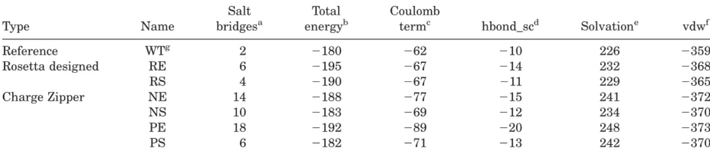
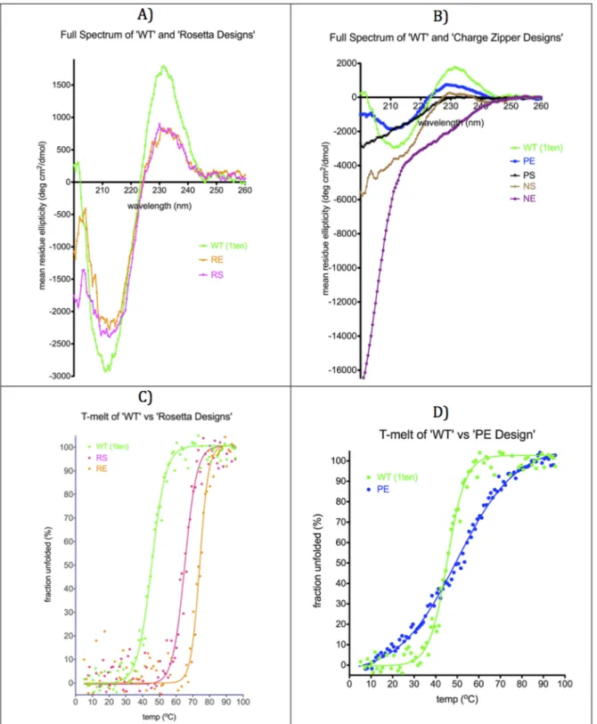
chroma-tography followed by gel filtration. Circular dichro-ism was used to determine if the proteins were folded. At low concentrations of salt, RE, RS, and PE all exhibited a CD spectrum consistent with a folded b-protein, while PS, NE, and NS have CD spectra indicative of random coil (Fig. 4). The ther-mal stabilities of the folded proteins were measured by monitoring the CD signal at 220 nm as a function of temperature. Both of the Rosetta designed sequences were dramatically stabilized relative to the WT protein with thermal unfolding tempera-tures of 74.18C (RE) and 64.18C (RS) compared with 45.48C for the wild type protein. Like the WT pro-tein, the designs also refolded when returning to room temperature (Supporting Information Fig. S5). These experiments were performed with 0M NaCl. At a concentration of 1 M sodium chloride, the designs were also more stable than the wild type protein, 58.28C (WT), 82.28C (RE), and 77.78C (RS). Similar increases in stability were observed for RE and RS in chemical denaturation experiments with guanidine hydrochloride (Table II, Supporting Infor-mation Fig. S6).
Table I. Computed Stabilities for Proteins
Type Name
Salt bridgesa
Total energyb
Coulomb
termc hbond_scd Solvatione vdwf
Reference WTg 2 2180 262 210 226 2359
Rosetta designed RE 6 2195 267 214 232 2368
RS 4 2190 267 211 229 2365
Charge Zipper NE 14 2188 277 215 241 2372
NS 10 2183 269 212 234 2370
PE 18 2192 289 220 248 2373
PS 6 2182 271 213 242 2370
aNumber of salt bridges on theb-sheet surfaces. Explanation of a salt bridge is in Supporting Information materials. bTotal energy for the protein as computed with Rosetta (unit is REU).27
cCoulombic electrostatic potential with a distance-dependent dielectric (unit is REU).21 dSidechain-sidechain hydrogen bond energy (unit is REU).
eLazaridis-Karplus solvation energy (unit is REU).
fvan der waals (5 “Lennard-Jones attractive between atoms in different residues”1“Lennard-Jones repulsive between
atoms in different residues”; unit is REU).
gFibronectin type III domain from tenascin (PDB code: 1ten).
Table II. Experimentally Measured Stabilities for Proteins
Type Name
Net
charge Mutations
Tm
(0MNaCl)
Tm
(1MNaCl)
m
(Kcal/mol * M)
DGu (0MGdnHCl) (Kcal/mol)
Reference WT 29 0 45.48C 58.28C 2.42 3.82
Rosetta designs RE 25 14 74.18C 82.28C 2.73 6.86
RS 27 7 64.18C 77.78C 2.56 5.27
Charge Zipper NE 214 19 Not folded Not folded Not folded Not folded
NS 212 12 Not folded Not folded Not folded Not folded
PE 25 22 49.98C 47.68C N/A N/A
Of the charge zipper designs, only PE is folded at low concentrations of salt and has a thermal unfolding temperature that is 58C greater than the wild type protein. Interestingly however, PE is not stabilized by salt like the wild type protein, and at 1M NaCl has a thermal stability that is 118C lower than the wild type protein. Intrigued by the dra-matic changes in stability with changes in salt
Discussion
Our results demonstrate that protein stability can be dramatically increased by redesigning only the solvent exposed face of small b-sheet proteins. Since the Rosetta design protocol aims to optimize several energetic features, including van der Waals contacts, intrinsic secondary structure preferences and elec-trostatic interactions, it is not straightforward to assign the increase in stability to any single feature. However, it is interesting that like WT TNfn3 both of the Rosetta designs, RE and RS, are stabilized by high salt concentrations. This suggests that the sta-bility of these variants is not entirely dependent on the formation of salt-bridges between oppositely charged amino acids, as these interactions are pre-dicted to become weaker at higher salt concentra-tions. Consistent with this conclusion, explicitly placing oppositely charged amino acids on the sur-face was not a simple recipe for boosting the stabil-ity of TNfn3. Three of the four charge zipper designs failed to fold at low salt concentrations. The charge zipper design that does fold, PE, is unlike the other TNfn3 variants, in that it is destabilized by high salt concentrations. This suggests that the redesign did have the intended effect of making protein sta-bility more dependent on surface electrostatic inter-actions. In contrast to our results with b-sheets, surface salt bridges have been shown to have a more dominant role in stabilizing helical proteins.22,28,29
This is likely to be in part because the charged amino acids, Arg, Lys, Glu, and Asp have a higher intrinsic propensity to be in helices compared with
b-strands.26
One of our goals in testing charge patterning on TNfn3 was our hope that it would provide a way to
dictate, whichb-strands would pair with each other, and in particular destabilize pairing between strands that are close in primary sequence but are not intended to be paired. We thought that this would be a simple approach to incorporate in the de novo design of b-sandwich proteins, a problem that is still unsolved. The results suggest that charge patterning does not provide a simple solution, and indicate that the correct strand pairing will need to be specified by the many different structural fea-tures that go into determiningb-sheet stability.
It is striking that in the design simulation where all residues on the surfaces of the b-sheets were allowed to vary, Rosetta only mutated 14 out of 28 residues. This is despite the fact that the design simulation starts from a completely random sequence, and uses a stochastic sampling protocol to find a low energy sequence. This suggests that most native residues on the b-sheet surfaces of TNfn3 are already optimized for stability, and highlights the fact that every residue in a b-sheet is in a unique environment, where the most favorable residue depends on the precise positioning of neighboring backbone atoms.30
Materials and Methods
Computational design and analysis of proteins
(backbone refinement).31 The script used to perform
these simulations is provided in the Supporting Information. Residues not allowed to change their amino acid identities were allowed to adopt different rotamers (“NATAA”); 1,000–10,000 independent sim-ulations were performed for each set of design parameters (80–800 cpu hours spent, number of design trajectories did not affect greatly the final design selection), and the lowest energy sequence for each set was selected for experimental characterization.
Protein expression and purification
All proteins were expressed using a 6-Histidine tagged PQE-80L vector in the BL21* strain of E. coli. Isopropyl b-D21-thiogalactopyranoside (IPTG) was used at 0.40.8 OD600 to induce and the
pro-teins were expressed overnight at 188C. Cell pellets were sonicated, and after additional centrifugation, supernatant was applied to a Ni-NTA column (GE healthcare). The purified solutions were further purified by size exclusion chromatography (GE healthcare HiLoad 16/60 Superdex 75 pg or HiLoad 16/600 Superdex 200 pg).
Circular dichroism
Secondary structure identification and melting tem-perature measurement were performed using circu-lar dichroism with JASCO J-815 CD spectrometer. All measurements were done with 20 lM protein concentration. All mean residue ellipticity values shown in this article are CD values of protein sam-ple after extracting CD values of buffer only. Data Integration Time (D.I.T) for ellipticity measurements was increased to 8 s from 4 s especially when high concentration of NaCl was used as buffers. When high concentration of NaCl was used as buffers, analysis of full spectrum of the ellipticity was not meaningful when wavelength is less than 205 nm. Nonlinear regression (sigmoidal dose-response) was used to fit all melting temperatures by Prism soft-ware ver. 5.0a.32Similar thermal unfolding tempera-tures were obtained by fitting the data to the Gibbs Helmholtz equation with nonlinear regression by Mathematica 10.33
Fluorescence
All chemical denaturations were evaluated by meas-uring fluorescence emission spectra (310–400 nm) with a Fluoromax 3 spectrofluorometer. Similar as in Gilbrethet al.,17we plotted fluorescence intensity vs. [GdnHCl] at wavelength 365 nm after excited at 295 nm. All measurements were performed with 5 lM protein concentration at 20 mM sodium phos-phate pH 7.0 except PS where the measurement was done in 20 mM sodium phosphate pH 7.0 and 100 mMNaCl.
Acknowledgment
The authors thank Dr. Bryan Der, Dr. Ryan Hallet, Dr. Hayretin Yumerefendi, and Dr. Joseph Harrison for guidance in cloning and protein purification, and Dr. Ashutosh Tripathy for helpful discussions.
References
1. Gerstein M (1998) How representative are the known structures of the proteins in a complete genome? A comprehensive structural census. Fold Des 3:497–512. 2. Lassila K, Datta D, Mayo S (2002) Evaluation of the
energetic contribution of an ionic network to beta-sheet stability. Protein Sci 11:688–690.
3. Der BS, Kluwe C, Miklos AE, Jacak R, Lyskov S, Gray JJ, Georgiou G, Ellington AD, Kuhlman B (2013) Alter-native computational protocols for supercharging pro-tein surfaces for reversible unfolding and retention of stability. PLoS One 8:e64363.
4. Lawrence MS, Phillips KJ, Liu DR (2007) Supercharg-ing proteins can impart unusual resilience. J Am Chem Soc 129:10110–10112.
5. Miklos AE, Kluwe C, Der BS, Pai S, Sircar A, Hughes RA, Berrondo M, Xu J, Codrea V, Buckley PE, Calm AM, Welsh HS, Warner CR, Zacharko MA, Carney JP, Gray JJ, Georgiou G, Kuhlman B, Ellington AD (2012) Structure-based design of supercharged, highly ther-moresistant antibodies. Chem Biol 19:449–455. 6. Smith CK, Regan L (1995) Guidelines for protein
design: the energetics of beta sheet side chain interac-tions. Science 270:980–982.
7. Minor D, Kim P (1994) Context is a major determinant ofb-sheet propensity. Nature 371:264–267.
8. Riemen AJ, Waters ML (2009) Design of highly stabi-lized beta-hairpin peptides through cation-p interac-tions of lysine and N-methyllysine with an aromatic pocket. Biochemistry 48:1525–1531.
9. Kiehna SE, Waters ML (2003) Sequence dependence of
b-hairpin structure: comparison of a salt bridge and an aromatic interaction. Protein Sci 12:2657–2667. 10. Street AG, Mayo SL (1999) Intrinsic beta-sheet
propen-sities result from van der Waals interactions between side chains and the local backbone. Proc Natl Acad Sci U S A 96:9074–9076.
11. Hu X, Wang H, Ke H, Kuhlman B (2008) Computer-based redesign of a beta sandwich protein suggests that extensive negative design is not required for de novo beta sheet design. Structure 16:1799–1805. 12. Kuhlman B, Dantas G, Ireton GC, Varani G, Stoddard
BL, Baker D (2003) Design of a novel globular protein fold with atomic-level accuracy. Science 302:1364–1368. 13. Gonen S, DiMaio F, Gonen T, Baker D (2015) Design of ordered two-dimensional arrays mediated by noncova-lent protein-protein interfaces. Science 348:1365–1368. 14. Street AG, Datta D, Gordon DB, Mayo SL (2000)
Designing protein beta-sheet surfaces by Z-score opti-mization. Phys Rev Lett 84:5010–5013.
15. Lappalainen I, Hurley MG, Clarke J (2008) Plasticity within the obligatory folding nucleus of an immunoglobulin-like domain. J Mol Biol 375:547–559. 16. Ng SP, Billings KS, Randles LG, Clarke J (2008)
Manipulating the stability of fibronectin type III domains by protein engineering. Nanotechnology 19: 384023.
18. Jacobs SA, Diem MD, Luo J, Teplyakov A, Obmolova G, Malia T, Gilliland GL, Neil KTO (2012.) Design of novel FN3 domains with high stability by a consensus sequence approach. 25:107–117. [JOURNAL]
19. Strickler SS, Gribenko AV, Gribenko AV, Keiffer TR, Tomlinson J, Reihle T, Loladze VV, Makhatadze GI (2006) Protein stability and surface electrostatics: a charged relationship. Biochemistry 45:2761–2766. 20. Leaver-Fay A, O’Meara MJ, Tyka M, Jacak R, Song Y,
Kellogg EH, Thompson J, Davis IW, Pache Ra, Lyskov S, Gray JJ, Kortemme T, Richardson JS, Havranek JJ, Snoeyink J, Baker D, Kuhlman B (2013) Scientific benchmarks for guiding macromolecular energy func-tion improvement. Methods Enzymol 523:109–143. 21. O’Meara MJ, Leaver-Fay A, Tyka MD, Stein A,
Houlihan K, DiMaio F, Bradley P, Kortemme T, Baker D, Snoeyink J, Kuhlman B (2015) Combined covalent-electrostatic model of hydrogen bonding improves structure prediction with Rosetta. J Chem Theory Comput 11:609–622.
22. O’Shea EK, Lumb KJ, Kim PS (1993) Peptide “Velcro”: design of a heterodimeric coiled coil. Curr Biol 3:658– 667.
23. Ruczinski I, Kooperberg C, Bonneau R, Baker D (2002) Distributions of beta sheets in proteins with applica-tion to structure predicapplica-tion. Proteins Struct Funct Genet 48:85–97.
24. Zhu H, Braun W (1999) Sequence specificity, statistical potentials, and three-dimensional structure prediction with self-correcting distance geometry calculations of beta-sheet formation in proteins. Protein Sci 8:326– 342.
25. Minor DL, Kim PS (1994) Measurement of the beta-sheet-forming propensities of amino acids. Nature 367: 660–663.
26. Fujiwara K, Toda H, Ikeguchi M (2012) Dependence of alpha-helical and beta-sheet amino acid propensities on the overall protein fold type. BMC Struct Biol 12:18. 27. Tyka MD, Keedy DA, Andre I, Dimaio F, Song Y,
Richardson DC, Richardson JS, Baker D (2011) Alter-nate states of proteins revealed by detailed energy landscape mapping. J Mol Biol 405:607–618.
28. Walther TH, Gottselig C, Grage SL, Wolf M, Vargiu AV, Klein MJ, Vollmer S, Prock S, Hartmann M, Afonin S, Stockwald E, Heinzmann H, Nolandt OV, Wenzel W, Ruggerone P, Ulrich AS (2013) Folding and self-assembly of the TatA translocation pore based on a charge zipper mechanism. Cell 152:316–326.
29. Spek EJ, Bui aH, Lu M, Kallenbach NR (1998) Surface salt bridges stabilize the GCN4 leucine zipper. Protein Sci 7:2431–2437.
30. Kuhlman B, Baker D (2000) Native protein sequences are close to optimal for their structures. Proc Natl Acad Sci U S A 97:10383–10388.
31. Leaver-Fay A, Tyka M, Lewis SM, Lange OF, Thompson J, Jacak R, Kaufman K, Renfrew PD, Smith CA, Sheffler W, Davis IW, Cooper S, Treuille A, Mandell DJ, Richter F, Ban YE, Fleishman SJ, Corn JE, Kim DE, Lyskov S, Berrondo M, Mentzer S, Popovic´ Z, Havranek JJ, Karanicolas J, Das R, Meiler J, Kortemme T, Gray JJ, Kuhlman B, Baker D, Bradley P (2011) ROSETTA 3: An object-oriented soft-ware suite for the simulation and design of macromole-cules. Methods Enzymol 487:545–574.
32. Prism Version 5.0a for Mac OS X (2008) GraphPad Software, San Diego, CA, USA. Available at: http:// graphpad.com/scientific-software/prism.