Journal of Global Pharma Technology
Available Online at:
www.jgpt.co.inRESEARCH ARTICLE
Effect of Isolate Catechin GMB4 in Expression of GRP78 and
Tunel Assay in Rat Cataract Model
Nugraha Wahyu Cahyana
1*,Edi Widjajanto
2,Umi Kalsum
3,Seskoati
Prayitnaningsi
41.Doctoral Program of Medical Science, Faculty of Medicine, Brawijaya University, Malang,
East Java, Indonesia.
2.Department of Clinical Pathology, Faculty of Medicine, Brawijaya University, Malang, East
Java, Indonesia.
3.Doctoral Program of Pharmacology, Faculty of Medicine, Brawijaya University, Malang, East
Java, Indonesia.
4.Department of Ophthalmology, Faculty of Medicine, Brawijaya University, Malang, East
Java, Indonesia.
*Corresponding Author: Nugraha Wahyu Cahyana
Abstract
This research is to explore glucose-regulated protein 78 (GRP78) expression and Tunel assay, signs of stress of endoplasmic reticulum (ER) and apoptosis in rat cataract model. Cataract was formed by inducing a subcutaneous injection of 19 µmol/kg sodium selenite to ten-day-old Wistar rats. The neonatal rats were divided into five groups randomly (n=5 in each group): a control group, and four cataract-induction groups, treated with either0, 50, 100, 200 mg/kg catechin Isolate From GMB4 Clone Green Tea. We performed slit-lamp biomicroscopic analysis,
immunohistochemistry for GRP78 and Tunel.Both eyes of all rats in Group 1 did not exhibit cataract
formation. In Group 2, one out of five (20%) developed grade 3 cataracts and the remaining four out of
five (80%) developed grade 4 cataracts. The grade of cataract formation decreased in groups 3, 4 and
5. The diff erence in exhibited cataract in the lens of all rats between Group 2 and any eyes of
groups 3 or 4 and 5 were significant (P = 0.022, 0,001, 0,001). The mean amount of Tunel cells
and GRP78 of group II rats were significantly (P < 0.01) higher than the levels in Group I, Group III, Group IV and Group V. Stress of Endoplasmic reticulum and apoptosis in the lens that increased following cataract formation in rats was suppressed by catechin Isolate From GMB4 Clone Green Tea.
Keywords: Cataract, Endoplasmic reticulum stress, Apoptosis, Catechin.
Introduction
A cataract is defined as any opacification of the lens that causes symptoms, including decreased visual acuity, decreased color perception, decreased contrast sensitivity, and glare disability, which eventually result in blindness. 1 According to the World Health Organization, 25 million people are affected by cataracts, which is also the leading cause of blindness worldwide. 2.3 Cataract is a major health problem and the major cause of blindness throughout the world caused by opacity that develops in the crystalline lens of the eye, [1-3].
One of the most common types of cataracts is related to age. The accumulation of unfolded proteins in the ER lumen causes Endoplasmic reticulum (ER) stress [6, 7]. ER stress has been proven as a self-protection mechanism of cells; it can restore the homeostasis within the ER. However, ER stress that isn’t treated and ongoing will eventually cause apoptosis namely ER stress-mediated cell death [8].
Recently, ER stress-mediated apoptosis pathway has been proven to play a crucial role in the degenerative pathophysiology of neurodegenerative disorders [9-13] retinitis pigments, and another eye disease [14-17]. Glucose-regulated protein 78 (GRP78) are the key markers of ER stress. The mechanisms during ER stress: GRP78 is involved in the unfolding protein reaction and the protection mechanism during ER stress.
The expression of GRP78 is an indication of ER stress, while apoptosis is detected by Tunel assay. The aim of this study is to investigate the expression of GRP78 and Tunel Assay in the Cataract model, the involvement of ER stress and its association with the apoptosis of lens epithelium cells, in rat cataract model.
Green tea, which contains polyphenol flavonoid compounds called catechins as its main component, is widely consumed throughout Asia[18]. One hundred grams of green tea contained 12 to 14% isolated catechins. The result of HPLC analysis showed that EGCG and ECG were main components from catechins isolation of green tea GMB-4 clone [19]. Catechins are a class of catechin compound group consisting of epicatechin (EC), epigallocatechin (EGC), epicatechin gallate (ECG), epigallocatechin gallate (EGCG), catechin, gallocatechin, catechin gallate, and gallocatechin gallate.
EGCG is predominant catechin which has content from 48% up to 55% in total polyphenols of green tea leaves [20]. Neither in-vivo nor in-vitro research on catechins isolated from green tea GMB4 clone in relation to cataract has ever been performed. As such, this study was needed to understand the effects of catechins isolated from GMB4 clone on the expression of GRP78 and Tunel Assay in the rat cataract model.
Materials and Methods
This study was performed in Biosains Laboratory of Brawijaya University. Twenty-five Wistar-albino rat pups were housed with their mother in special wire-bottom cages and in standard conditions (12-hour daylight-dark cycle, ventilated, constant room temperature). It has been considered that solid-bottom cages are more adequate for the housing of the rat pups. The rat pups were divided into five groups (four experimental and one control), each consisting of five pups. Group 1 received only a subcutaneous saline injection and was the control group. In Group 2, sodium-selenite (19 nmol/g body weight, Sigma Chem.
Co., St Louis, USA) was injected subcutaneously on a postpartum Day 10. In Group 3, subcutaneous sodium-selenite (19 nmol/g body weight) was injected on a postpartum Day 10 and injection of isolate catechin intraperitoneally (50 mg/kg body weight), starting one day before sodium-selenite injection (on postpartum Day 9) and was continued for 5 days (till postpartum Day 13). The procedures performed on Group 3 rats were also performed on Group 4 and Group 5, the diff erence being the dosage of isolate catechin. Group 4 had used 100 mg/kg body weight of isolate catechin and Group 5 was 200 mg/kg body weight.
Table 1: Treatment groups studied GROUPS
(N = 5)
INJECTIONS (DAY 9,10,11,12, 13)
INJECTION (DAY 10)
GROUP 1 Saline Saline
GROUP 2 Saline Na2SeO3(19 μmol/kg BW)
GROUP 3 Catechin 50 mg/KgBW Na2SeO3(19 μmol/kgBW) + Catechin 50 mg/kg BW
GROUP 4 Catechin 100 mg/KgBW Na2SeO3(19 μmol/kgBB) + Catechin 100mg/kgBW
GROUP 5 Catechin 200 mg/KgBW Na2SeO3(19 μmol/kgBB) + Catechin 200mg/kgBW
On a postpartum day 17, all rats were anesthetized with intraperitoneal ketamine injection (80 mg/kg BW) and
two hours. All lenses were evaluated and were morphologically staged for cataract development and grading was performed
by slit-lamp biomicroscopy on a scale of 0 to 4 as follows in Table 2 [26]:
Table 2: Grade of Lenticular Opacification GRADE
GRADE 0 Normal transparent lens
GRADE 1 The lens with a subcapsular opacity
GRADE 2 Was a nuclear cataract
GRADE 3 Was a strong nuclear cataract with opacity in the perinuclear area
GRADE 4 Was a mature dense opacity involving the entire lens
Lens photos ×25 magnifications were taken using a camera attached to slit-lamp (Topcon, Tokyo, Japan) (Figure 1). The lens was then taken immediately after euthanasia, the eyes were enucleated. Frozen lens samples were weighed and homogenized in ice-cold phosphate-buffered saline solution (0.01 mol/L and pH 7.4). Homogenization procedures were carried out using Bullet Blend tissue Homogenizer (Next Advanced Inc, Averill Park, NY, USA), according to the manufacturer’s instructions at 4 ℃.
These homogenates were centrifuged at 10 000 g for 30min at 4 ℃, and supernatants were obtained. Supernatants were used for the measurement of the levels of GRP78 and Tunel assay using a GRP78 and Tunel assay kit (ImmuchromGmbH, Hessen, Germangradey). Data are presented as mean ± standard deviation and differences between groups were analyzed using one-way ANOVA with
SPSS 17.0 Statistical Package. The post-doc test was used in the ANOVA was significant. P< 0.01 was considered statistically significant. This study was approved by the Institutional Review/ Ethics Board of Brawijaya University [ref: 1114-KEP-UB, dated 24th April 2019]. All methods were performed in accordance with guidelines and regulations.
Results
The mean GRP78 levels lenses (13, 33 ± 2,875) of Group II rats were significantly (P <0,001) higher than the levels in Group I lens I (4,833 ± 2,639), Group III (10,833 ± 2,926), Group IV (9,667 ± 1,751), and Group V (6,333 ± 1,329) (Figure 2). A significant difference was also observed in the GRP78 level in the lens (P <0.001) between group III and group I. The GRP78 level in the lens decreased and the level of lens opacity increased in group II. Furthermore, the GRP78 level decreased gradually and the opacity level decreased in accordance with the administration of catechin doses (groups III, IV, and V).
0 2 4 6 8 10 12 14 16 18
Group 1 Group 2 Group 3 Group 4 Group 5
M
ea
n
o
f
G
RP
7
8
E
x
press
io
n
Treatment Groups
Figure 2: Expression of GRP78 in Each Treatment Group. Results are expressed as the mean ± SD. *P<0, 01 vs Group 1, #P<0, 01 vs. Group 2
GRP78 levels in the lenses from the Na2SeO3 group were found to be significantly (p <0.01) higher than those in the control group and the catechin group.
Treatment with catechins in the catechin + Na2SO3 group (Figure 2) significantly (p <0.01) decreased GRP78 levels.
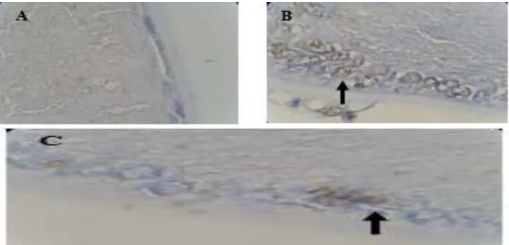
Figure 3: Effect of catechin on DNA fragmentation in the lens epithelium induced by cataract. Photomicrographs of terminal deoxynucleotidyl transferase-mediated dUTP nick end labeling (TUNEL) positive cells in the lens epithelium. (A) control group, (B) cataract-induction group, (C) cataract-induction and 200 mg/kg BW catechin group
0 5 10 15 20
Group 1 Group 2 Group 3 Group 4 Group 5
M
e
an
o
f
Tu
n
e
l e
xp
re
ss
io
n
Treatmant Groups *#
Figure 4: Expression of Tunel in Each Treatment Group. Results are expressed as the mean ± SD. *P<0, 01 vs Group 1, #P<0, 01 vs. Group 2
The mean number of Tunel cells in Group II mice (18 ± 1,264) was significantly (P <0.001) higher than the number of Tunel cells in Group I (4.3 ± 2.136), Group III (14 ± 2.562), Group IV (10 ± 2,136), and Group V (7 ± 2,097) (Figure. 4). A significant difference was also observed in the number of Tunel
doses (groups III, IV, and V).
Discussion
Radiation, galactose, streptozocin, and selenite are used in most experimental cataract models to cause cataracts [21-23]. Selenite cataract is similar in many respects to human cataracts. In 1978, Ostadalova et al. first used selenite, which is one of the most widely used pharmacological agents in experimental cataract studies, in such a sample [23].
The basic mechanism of selenite in the formation of cataracts is that it acts as an oxidant, thereby causing damage to the lens [23-27]. It also causes lipid peroxidation in the crystalline lens, induces hydrogen peroxide, and lowers GSH in the lens [27]. Effects of sodium selenite towards retinal cells have been researched and resulted in increased expression of GRP78 that shows reticulum endoplasmic stress in the retina but research on the effect on lens epithelium has not yet been conducted.
We found that GRP78, a stress protein on ER and an important marker for the protection mechanism of ER stress, was elevated after Cataract. Stresses like oxygen deficiency, low glucose, and low Ca2+ can lead to disruption of protein metabolism in cells; then the accumulation of unfolded protein response (UPR) elevates GRP78 expression to keep the ER homeostasis improving protein synthesis and transport[29, 30].
Recent studies revealed that UPR had a significant role in neuronal degenerative disorders, such as Alzheimer's disease, but few reported the role of UPR in cataracts. The existence of a protective mechanism against apoptosis during ER stress after Cataract provides us a window of opportunity for recovery of visual function before the occurrence of irreversible widespread damage.
How to promote the protection mechanism of ER stress and attenuate injury mechanism after the cataract is a potential interest of study in the future. Cataract formation is one of the most common causes of irreversible visual loss associated with aging, thus much interest is being laid on recognition of a drug that will help to prevent or treat cataractogenesis. The present investigations were undertaken to determine the efficacy of
isolated catechin from green tea GMB 4 to prevent the progression of cataract on in vivo animal models. A cataract is a protein deterioration disorder characterized by irreversible modification and accumulation of lens proteins. Once the cataract is formed it cannot be reverted back; thus the study is focusing on the prevention of lens opacity and it lacks a positive control.
Selenite-induced cataract model (a single subcutaneous injection of sodium selenite at a dose of 19 μmol/kg BB is the universally accepted animal model for studying oxidative stress induced experimental cataract as it shows almost all the events associated in human age-related cataracts such as membrane damage, calcium accumulation, endoplasmic reticulum stress, lenticular apoptosis, and proteolysis of lens proteins [17].
It is essential to check whether the anti cataractogenic potential of Catechin is by the prevention of endoplasmic reticulum stress or lenticular apoptosis. Several studies have shown that the presence of significant amounts of vitamins, carotenoids, caffeine, acetyl-L-carnitine, ebselen, quercetin, flavonoids, phenyl ester of caffeic acid and curcumin exerts inhibitory effects on cataracts [30-35].
Nevertheless, no agent can completely block or delay lens opacification. Recent studies have shown that the green tea catechins have antioxidant, anti-inflammatory, antiangiogenic and antibacterial effects [36-40]. Such catechins bind reactive oxygen and nitrogen species and exert indirect antioxidant effects by stimulating the synthesis of endogenous antioxidant enzymes, such as superoxide dismutase, glutathione reductase, glutathione-S-reductase, catalase, and quinone reductase. Because of those results, green tea will inhibit lipid peroxidation and DNA mutation. Green tea has high levels of catechin and shows more strongly antioxidant activity than vitamins C and E [38-42].
Reported that catechin prevents H2O2-induced oxidative stress in the lens epithelial cells. Chen et al [46].Reported that eye drops with catechin exhibit potent protective effects on ultraviolet B radiation-induced corneal oxidative damage in mice; the effects are likely due to increased defense system, antioxidant activity, lipid peroxidation inhibition, and protein oxidation inhibition. Recent studies have demonstrated that catechin also protects human γB-crystallin from UV-induced damage and cultured human lens epithelial cells from hyperglycemia-induced damage [47, 49].
Catechin prevents tryptophan oxidation in cataractous human lens γ-crystallin in the presence of H2O2 [47].Heo et al. [50-59].Showed that catechin increased cell count and cell viability after the UV irradiation of cultured human lens epithelial cells, indicating that catechin can protect lens epithelium against UV damage.
No studies have thus far been conducted on the preventive effects of catechin in experimental cataract models. This research shows that after sodium selenite is given, lens opacity will occur and followed by an increase in GRP 78 (Figure 1) and cells with positive Tunel (Figure 3). Then the opacity level decrease followed by a decrease in both GRP78 (Figure 2) and cells with positive Tunel (Figure 4) in the group given catechin
50 mg/kg BW (Group 3), 100mg / kg BW (Group 4) and 200mg / KgBW (Group 5). This can be interpreted that sodium selenite causes lens opacity due to endoplasmic reticulum stress and apoptosis in the lens epithelium. Furthermore, catechins are given so as to reduce endoplasmic reticulum stress and apoptosis by decreasing GRP78 and positive Tunel cells and decreasing turbidity from the lens.
This can be interpreted that sodium selenite causes lens opacity due to stress endoplasmic reticulum and apoptosis in the lens epithelium. Furthermore, catechins are given so as to reduce endoplasmic reticulum stress and apoptosis by decreasing GRP78 and positive Tunel cells and decreasing opacity from the lens.
Conclusion
The present research demonstrated that catechin significantly inhibits the development of cataracts by inhibiting reticulum endoplasmic stress and apoptosis.
Acknowledgments
The authors are grateful for Hidayat Sujuti, MD, M.Sc., Ph, D., SpM for his constructive feedback on our manuscript.
References
1. Sullivan PM, Luff AJ, Aylward GW (1997) Results of primary retinal reattachment surgery: a prospective audit. Eye, 11: 869-871.
2. Yang CH, Lin HY, Huang JS, Ho TC, Lin CP, Chen MS et al (2004) Visual outcome in primary macula-off rhegmatogenous retinal detachment treated with scleral buckling. J. Formos Med. Assoc., 103: 212-217.
3. Chang PY, Yang CM, Yang CH, Huang JS, Ho TC, Lin CP et al (2005) Clinical characteristics and surgical outcomes of pediatric rhegmatogenous retinal detachment in Taiwan. Am J. Ophthalmol., 139: 1067-1072.
4. Cook B, Lewis GP, Fisher SK, Adler R (1995) Apoptotic photoreceptor degeneration in experimental retinal detachment. Invest Ophthalmol. Vis. Sci., 36: 990-996.
5. Chang CJ, Lai WW, Edward DP, Tso MO (1995) Apoptotic photoreceptor cell death after traumatic retinal detachment in humans. Arch. Ophthalmol., 113: 880-886.
6. Harding HP, Zhang Y, Ron D (1999) Protein translation and folding are coupled by an endoplasmic-reticulum-resident kinase. Nature, 397: 271-274. 7. Patil CK, Li H, Walter P (2004) Gcn4p
and novel upstream activating sequences regulate targets of the unfolded protein response. PLoS Biol., 2: E246.
8. Momoi T (2004) Caspases involved in ER stress-mediated cell death. J. Chem. Neuroanat, 28: 101-105.
9. Lindholm D, Wootz H, Korhonen L
(2006) ER stress and
Differ., 13: 385-392.
10. Sekine Y, Takeda K, Ichijo H (2006) The ASK1-MAP kinase signaling in ER stress and neurodegenerative diseases. Curr. Mol. Med., 6: 87-97. 11. Yoshida H (2007) ER stress and
diseases. FEBS J., 274: 630-658.
12. Katayama T, Imaizumi K, Manabe T, Hitomi J, Kudo T, Tohyama M (2004) Induction of neuronal death by ER stress in Alzheimer’s disease. J. Chem. Neuroanat., 28: 67-78.
13. Szegezdi E, Duffy A, O’Mahoney ME, Logue SE, Mylotte LA, O0brien T et al (2006) ER stress contributes to ischemia- induced cardiomyocyte apoptosis. Biochem Biophys Res Commun., 349: 1406-1411.
14. Tam BM, Moritz OL (2007) Dark rearing rescues P23H rhodopsin- induced retinal degeneration in a transgenic Xenopus laevis model of retinitis pigmentosa: a chromophore-dependent mechanism characterized by production of N-terminally truncated mutant rhodopsin. J. Neurosci., 27: 9043-9053.
15. Ryoo HD, Domingos PM, Kang MJ, Steller H (2007) Unfolded protein response in a Drosophila model for retinal degeneration. EMBO J., 26: 242-252.
16. Roybal CN, Marmorstein LY, Vander Jagt DL, Abcouwer SF (2005) Aberrant accumulation of fibulin-3 in the endoplasmic reticulum leads to activation of the unfolded protein response and VEGF expression. Invest Ophthalmol Vis Sci., 46: 3973-3979. 17. Abcouwer SF, Marjon PL, Loper RK,
Vander Jagt DL (2002) Response of VEGF expression to amino acid deprivation and inducers of endoplasmic reticulum stress. Invest Ophthalmol Vis Sci., 43: 2791-2798. 18. Kao, Hiipakka, Liao (2000) Modulation of
obesity by a green tea catechin. Am J Clin Nutr., 72(5):1232-4.
19. Susanti, Ciptati, Aulanniam, Rudijanto Qualitative analysis of catechins from green tea GMB-4 clone using HPLC and LC-MS/MS. 2015. Asian Pacific Journal of Tropical Biomedicine 5(12). DOI: 10.1016/j.apjtb.2015.09.013
20. Velayutham, Babu, Liu (2008) Green tea catechins and cardiovascular health: an update. Curr Med. Chem., 15(18):1840-50. 21. Hiraoka T, Clark JI (1995) Inhibition of
lens opacification during the early stages of cataract formation. Invest Ophthalmol Vis Sci., 36(12): 2550-2555.
22. Chitkara DK (1999) Cataract formation mechanisms. In Yanoff M, Duker JS (Editors). Ophthalmology. Mosby, St Louis, MO, 481-488.
23. Ostadalova I, Babicky A, Obenberger J (1978) Cataract induced by administration of a single dose of sodium selenite to suckling rats. Experientia, 34(2):222-223. 24. Dherani M, Murthy GV, Gupta SK, Young
IS, Maraini G, Camparini M, Price GM, John N, Chakravarthy U, Fletcher AE (2008) Blood levels of vitamin C, carotenoids and retinol are inversely associated with cataract in a North Indian population. Invest Ophthalmol Vis Sci., 49(8): 3328-3335.
25. Christen WG, Liu S, Glynn RJ (2008) Dietary carotenoids, vitamins C and E, and risk of cataract in women: a prospective study. Arch Ophthalmol., 126(1):102-109.
26. Aydemir O, Güler M, Kaya M (2012) Protective effects of ebselen on selenite-induced experimental cataract in rats. J. Cataract Refract Surg., 38(12):2160-2166. 27. Manikandan R, Thiagarajan R, Beulaja S,
Sudhandiran G, Arumugam M (2010) Effect of curcumin on selenite-induced cataractogenesis in Wistar rat pups. Curr Eye Res, 35(2):122-129.
28. Hayashi T, Saito A, Okuno S, Ferrand-Drake M, Chan PH (2003) Induction of GRP78 by ischemic preconditioning reduces endoplasmic reticulum stress and prevents delayed neuronal cell death. J. Cereb. Blood Flow Metab, 23: 949-961.
29. Lee AS (2005) The ER chaperone and signaling regulator GRP78/ BiP as a monitor of endoplasmic reticulum stress. Methods, 35: 373-381.
30. Prokofyeva E, Wegener A, Zrenner E (2013) Cataract prevalence and prevention in Europe: a literature review. Acta Ophthalmol., 91(5):395-405.
Saxena R (2009) Advances in pharmacological strategies for the prevention of cataract development. Indian J. Ophthalmol., 57(3):175-183. 32. Dherani M, Murthy GV, Gupta SK, Young
IS, Maraini G, Camparini M, Price GM, John N, Chakravarthy U, Fletcher AE (2008) Blood levels of vitamin C, carotenoids and retinol are inversely associated with cataract in a North Indian population. Invest Ophthalmol Vis Sci., 49(8): 3328-3335.
33. Christen WG, Liu S, Glynn RJ (2008) Dietary carotenoids, vitamins C and E, and risk of cataract in women: a prospective study. Arch Ophthalmol., 126(1):102-109.
34. Aydemir O, Güler M, Kaya M (2012) Protective effects of ebselen on selenite-induced experimental cataract in rats. J. Cataract Refract Surg., 38(12):2160-2166. 35. Manikandan R, Thiagarajan R, Beulaja S,
Sudhandiran G, Arumugam M (2010) Effect of curcumin on selenite-induced cataractogenesis in Wistar rat pups. Curr Eye Res, 35(2):122-129.
36. Koo MW, Cho CH (2004) Pharmacological effects of green tea on the gastrointestinal system. Eur. J. Pharmacol., 500(1-3):177-185.
37. Singh AK, Seth P, Anthony P, Husain MM, Madhavan S, Mukhtar H, Maheshwari RK(2002) Green tea constituent epigallocatechin-3-gallate inhibits angiogenic differentiation of human endothelial cells. Arch Biochem Biophys, 401(1):29-37.
38. Yang CS, Chung JY, Yang G, Chhabra SK, Lee MJ (2000) Tea and tea polyphenols in cancer prevention. J. Nutr., 130(2S):472S-478S.
39. Miura Y, Chiba T, Tomita I, Koizumi H, Miura S, Umegaki K, Hara Y, Ikeda M, Tomita T (2001) Tea catechins prevent the development of atherosclerosis in apoprotein E-deficient mice. J. Nutr., 131(1): 27-31.
40. Murase T, Nagasawa A, Suzuki J, Hase T, Tokimitsu I (2002) Beneficial effects of tea catechins on diet-induced obesity: Stimulation of lipid catabolism in the liver. Int. J. Obes. Relat Metab Disord., 26(11):1459-1464.
41. Crespy V, Williamson G (2004) A review of the health effects of green tea catechins in in vivo animal models. J. Nutr., 134(12):3431S-3440S.
42. Wu LY, Juan CC, Ho LT, Hsu YP, Hwang LS (2004) Effect of green tea supplementation on insulin sensitivity in Sprague-Dawley rats. J. Agric. Food Chem., 52(3):643-648.
43. Cavet ME, Harrington KL, Vollmer TR, Ward KW, Zhang JZ (2011) Anti-inflammatory and anti-oxidative effects of the green tea polyphenol epigallocatechin gallate in human corneal epithelial cells. Mol. Vis., 17: 533-542.
44. Emoto Y, Yoshizawa K, Kinoshita Y, Yuri T, Yuki M, Sayama K, Shikata N, Tsubura A (2014) Green tea extract suppresses N-methyl-Nnitrosourea-induced
photoreceptor apoptosis in Sprague-Dawley rats. Graefes Arch Clin Exp. Ophthalmol., 252(9):1377-1384.
45. Cia D, Vergnaud-Gauduchon J, Jacquemot N, Doly M (2014) Epigallocatechingallate (EGCG) prevents H2O2-induced oxidative stress in primary rat retinal pigment epithelial cells. Curr Eye Res., 39(9):944-952.
46. Chen MH, Tsai CF, Hsu YW, Lu FJ (2014) Epigallocatechin gallate eye drops protect against ultraviolet B-induced corneal oxidative damage in mice. Mol. Vis., 20:153-162.
47. Chaudhury S, Bag S, Bose M, Das AK, Ghosh AK, Dasgupta S (2016) Protection of human γB-crystallin from UV-induced damage by epigallocatechin gallate: spectroscopic and docking studies. Mol. Biosyst., 12(9):2901-2909.
48. Chaudhury S, Ghosh I, Saha G, Dasgupta S (2015) EGCG prevents tryptophan oxidation of cataractous ocular lens human γ-crystallin in presence of H2O2. Int. J. Biol. Macromol., 77: 287-292.
49. Ye P, Lin K, Li Z, Liu J, Yao K, Xu W (2013) Epigallocatechin gallate regulates expression of apoptotic genes and protects cultured human lens epithelial cells under hyperglycemia. Mol Biol. (Mosk) 47(2):251-257.
22(3):183-186.
51. Shimazawa M, Ito Y, Inokuchi Y, Hara H (2007) Involvement of double-stranded RNA-dependent protein kinase in ER stress-induced retinal neuron damage. Invest Ophthalmol Vis Sci., 48: 3729-3736.
52. Shimazawa M, Inokuchi Y, Ito Y, Murata H, Aihara M, Miura M et al (2007) Involvement of ER stress in retinal cell death. Mol. Vis., 13: 578-587.
53. Ciccaglione AR, Marcantonio C, Tritarelli E, Equestre M, Vendittelli F, Costantino A et al (2007) Activation of the ER stress gene gadd153 by hepatitis C virus sensitizes cells to oxidant injury. Virus Res., 126: 128-138.
54. Li J, Holbrook NJ (2004) Elevated gadd153/chop expression and enhanced c-Jun N-terminal protein kinase activation sensitizes aged cells to ER stress. Exp. Gerontol., 39: 735-744.
55. Nakazawa T, Matsubara A, Noda K,
Hisatomi T, She H, Skondra D et al (2006) Characterization of cytokine responses to retinal detachment in rats. Mol. Vis., 12: 867-878.
56. Arroyo JG, Yang L, Bula D, Chen DF (2005) Photoreceptor apoptosis in human retinal detachment. Am J. Ophthalmol., 139: 605-610.
57. Yang L, Bula D, Arroyo JG, Chen DF (2004) Preventing retinal detachment-associated photoreceptor cell loss in Bax- deficient mice. Invest Ophthalmol. Vis Sci., 45: 648-654.
58. Zacks DN, Hanninen V, Pantcheva M, Ezra E, Grosskreutz C, Miller JW (2003) Caspase activation in an experimental model of retinal detachment. Invest Ophthalmol. Vis Sci., 44: 1262-1267.