Original Research Article
Multi detector computed tomography evaluation in chronic obstructive
pulmonary disease and correlation with severity of disease
Ajay Kumar
1*, Ankita Rohira
1, Ashish Vijay
1, Abhay Sharma
2INTRODUCTION
Chronic obstructive pulmonary disease (COPD) has been the 4th leading cause of the death and is projected to rank 5th in 2020 as a worldwide burden of disease according to the study published in the American Journal of Respiratory and Critical Care Medicine.1 It is defined in
the functional terms as the slowly progressive disorder characterized by the airflow limitation which does not change markedly over several months. The limitation of airflow is associated with the inflammatory response of
lungs to various 2 noxious particles or gases.2 The
diagnosis of COPD depends on pulmonary function tests (PFTs) and spirometry. A ratio of the forced expiratory volume in one second and the percentage forced vital capacity (FEV1/FVC%) of less than 70% predicted in a patient with a post-bronchodila to rFEV1 of less than 80% of predicted value is diagnostic for COPD.3
It is well-known that lung has the strong compensatory ability, and injury to more than 30.0% of the lung tissues is typically essential to result in the abnormal lung
1Department of Radiodiagnosis, Index Medical College Hospital and Research centre Indore, Madhya Pradesh, India 2Department of Medicine, Index Medical College Hospital and Research centre Indore, Madhya Pradesh, India
Received: 17 May 2019
Revised: 05 July 2019
Accepted: 09 July 2019
*Correspondence:
Dr. Ajay Kumar,
E-mail: [email protected]
Copyright: © the author(s), publisher and licensee Medip Academy. This is an open-access article distributed under the terms of the Creative Commons Attribution Non-Commercial License, which permits unrestricted non-commercial use, distribution, and reproduction in any medium, provided the original work is properly cited.
ABSTRACT
Background: Multi Detector computed tomography (MDCT) may effectively characterize and quantify the extent of emphysema and the air trapping related to the small airway’s disease. Here we highlight the computed-tomography findings of Chronic Obstructive Pulmonary Disease (COPD) and correlation with the Spirometrics values.
Methods: The study group included the total of 100 adult patients of either sex with a clinical suspicion of COPD and those who undergone MDCT of thorax. Lung function of the patients with the COPD stages mild to very severe was evaluated by both the MDCT and Spirometrics Pulmonary Function Tests (PFTs). The scanning was done at maximum end inspiration and maximum end expiration.
Results: There was a preponderance of male patients with highly significant correlation between values of mean lung density and low attenuation values (p<0.000I). MDCT correlated well with those obtained from spirometric Pulmonary Function Tests in the patients with COPD and that the correlation at expiration was superior to that at inspiration.
Conclusions: The study concludes that Multi-detector computed tomography is the invaluable tool in defining and quantifying COPD and the characterization of emphysematous changes.
Keywords: Airway obstruction, Chronic Obstructive Pulmonary Disease, Emphysema, Imaging, Multi detector Computed Tomography, Pulmonary Function Tests, Spirometry
function. By the time abnormal lung function is diagnosed, irreparable damage has likely occurred. Dual phase MDCT with the routine radiation dose has also been used for diagnosis of COPD with the favorable results.4 While COPD is the convenient umbrella term, its
scheduled use obscures fact that morphologic manifestations of this group of the obstructive diseases vary extensively, a fact that is readily obvious to clinical radiologist. Multi-detector computed tomography scanners provide noninvasive methods to study the lung pathology in COPD. Quantitative procedures based on CT are accessible to quantify emphysema and airway dimensions and the consistent body of literature recommends that CT represents the major tool in the clinical setting helping to precisely detect location, severity of disease and quantify the extent, as well as the small airway disease (SAD).5 Thus, the purpose of the
study was to determine usefulness of MDCT performed in both inspiration & expiration to quantitatively assess pulmonary function in the COPD patients.
METHODS
This prospective study was carried out on 100male-female patients of COPD of all age meeting the inclusion criteria, who were visited to department of radio-diagnosis under the index medical college hospital and research centre Indore an informed consent was taken from all patients or his attendant before the patient was subjected for evaluation.
Inclusion Criteria
Patients showing clinical signs and Symptoms consistent with the diagnosis of COPD and those who undergone MDCT of thorax. Patients who were willing to participate in the study and have given written consent were included.
Exclusion Criteria
• Any co-existent lung pathology or lung malignancy • Patients who were not willing to participate in the
study and did not give written consent were excluded.
Procedure methodology
There were 111COPD patients were enrolled in this study, 7 patients were refused to participate in the study, and 4 were not fit in the inclusion criteria. Finally, 100 patients were taken. Detailed history and physical examination was done and recorded on the predesigned proforma that was prepared in English and local language which was used during an interview from each patient. Patient's personal history, physical examination findings like name, age, sex, demographic profile, Height, Weight, BMI, blood pressure and all relevant clinical and radiological examination was done and recorded. Pulmonary Function tests: Pulmonary function tests were
performed by an Easy One spirometer with the patients in a seated position. The Spirometric data were collected on the same day when CT scan was acquired.
Table 1: Gold criteria for severity of airflow obstruction in COPD.
Stage Spirometric findings
Mild FEV1/FVC<0.70 FEV1>80.0% predicted
Moderate FEV1/FVC<0.70
50.0%≤ FEV1<80.0% predicted
Severe FEV1/FVC<0.70
30.0%≤ FEV1<50.0% predicted
Very Severe
FEV1/FVC<0.70
FEV1<30.0% predicted or FEV1 <50.0% predicted plus chronic respiratory failure
Radiological examinations
Plain radiography
Standard posteroanterior chest radiographs of the patients were obtained in all patients. Radiographs were evaluated to detect the presence of emphysema.
Computed Tomography
Computed tomography of the thorax was performed for all patients on multi-detector high resolution computed tomography (SIEMENS SOMATOM Definition AS 64) in the helical mode without the intravascular contrast material.
Confounders
“Present demonstration of metabolic factors” was defined in an affirmative response to question “Have you, at any time in your life, suffered from chest pain, expectoration, and cough with sputum?” and was it was within the previous year. “At what age did these changes were noticed?”
Statistical Analysis
Data was analyzed using Statistical Package for Social Sciences, version 23 (SPSS Inc., Chicago, IL). Results for continuous variables are presented as mean±standard deviation, whereas results for categorical variables are presented as number (percentage). The level P <0.05 was considered as the cutoff value for significance
RESULTS
COPD patients was found to be 59.81±7.83 years
(Table 2).
Table 2: Demographic profile of the studied patients.
Demographic details No. of patients
(n=100) %
Age (years)
41-50 14 14.0
51-60 31 31.0
61-70 37 37.0
>70 18 18.0
Gender Male 63 63.0
Female 37 37.0
Smoking (packs per year)
0 11 11.0
20-25 41 41.0
25-30 44 44.0
>30 4 4.0
The classification of COPD severity based on gold criteria and 53.0% were having mild COPD followed by moderate COPD (26.0%), severe COPD was in 13.0% while very severe was only in 8.0% patients. (Table 3).
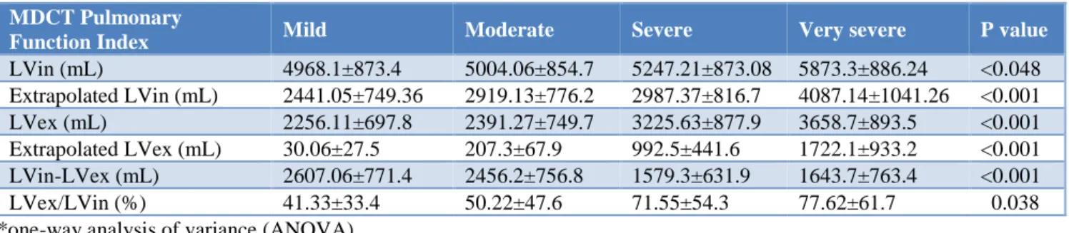
Significant differences among the five COPD groups were also found in the difference between LVin and LVex (p <0.0001) and the lvex/lvin % (p <0.0001).
Table 3: Spirometric classification of COPD severity based on GOLD criteria, 2008; among the
studied patients.
Severity of
COPD Standard Value
No. of patients %
Mild
FEV1/FVC <0.70 FEV1 ≥ 80% predicted
53 53.0
Moderate
FEV1/FVC < 0.70 50% ≤ FEV1 <80% predicted
26 26.0
Severe
FEV1/FVC < 0.70 30% ≤ FEV1 < 50% predicted
13 13.0
Very Severe
FEV1 < 30% predicted or FEV1 < 50% predicted plus chronic respiratory failure
8 8.0
As with the spirometric PFT indexes, most MDCT pulmonary function indexes, especially during full expiration, were significantly different in patients with stage severe and very severe COPD as compared with those with lower stages (Table 4).
Table 4: MDCT Pulmonary Function Indexes by Chronic Obstructive Pulmonary Disease (COPD) Stage.
MDCT Pulmonary
Function Index Mild Moderate Severe Very severe P value
LVin (mL) 4968.1±873.4 5004.06±854.7 5247.21±873.08 5873.3±886.24 <0.048 Extrapolated LVin (mL) 2441.05±749.36 2919.13±776.2 2987.37±816.7 4087.14±1041.26 <0.001 LVex (mL) 2256.11±697.8 2391.27±749.7 3225.63±877.9 3658.7±893.5 <0.001 Extrapolated LVex (mL) 30.06±27.5 207.3±67.9 992.5±441.6 1722.1±933.2 <0.001 LVin-LVex (mL) 2607.06±771.4 2456.2±756.8 1579.3±631.9 1643.7±763.4 <0.001
LVex/LVin (%) 41.33±33.4 50.22±47.6 71.55±54.3 77.62±61.7 0.038
*one-way analysis of variance (ANOVA)
Table 5: Correlation between Spirometric Pulmonary Function Test (PFT) Indexes and MDCT Pulmonary Function Indexes in Patients with Chronic Obstructive Pulmonary Disease (COPD).
MDCT pulmonary function index FVC (% predicted FEV1 (% Predicted) FEV1/FVC%
LVin (mL) -0.289a -0.011 -0.529a
LVin (mL) -0.643a -0.409a -0.755a
LVin – LVex (mL) 0.379a 0.453a 0.247
LVex/LVin (%) -0.587a -0.479a -0.603a
Extrapolated LVin (mL) -0.443a -0.163 -0.643a
Extrapolated LVex (mL) -0.739a -0.497c -0.837a
FVC = forced vital capacity, FEV1 = forced expiratory volume in 1 second, FVC% = percentage forced vital capacity, LVin = inspiratory lung volume, LVex = expiratory lung volume. ap <0.001, bp <0.05. cp <0.01.
The correlations between PFT indexes and MDCT pulmonary function indexes are depicted in Table 5. A strong correlation was obtained between the extrapolated LVex and COPD stage (r =0.802, p<0.001) and between the extrapolated LVex and FEV1/FVC% (r=-0.837, p<0.001). Strong negative correlations were found
-0.643, p < 0.001). Other correlations either were weak to moderate or were not significant.
DISCUSSION
Chronic obstructive pulmonary disease (COPD) is a major cause of chronic morbidity and is the 12th leading cause of disability in the world.6 It is defined in
functional terms as a slowly progressive disorder characterized by airflow limitation that does not changemarkedly over several months. The limitation of airflow is associated with an inflammatory response of the lungs to various 2 noxious particles or gases.1
The primary parameters of diagnostic assessment with spirometry are Forced Expiratory Volume in the 1st second (FEV1) and Forced Vital Capacity (FVC). Reductions in FEV1, FVC and the ratio of FEV1 to FVC are hallmarks of airway obstruction. The criterion for a diagnosis of COPD is an FEV1/FVC ratio of less than 70% and a post bronchodilator FEV1 less than 80% of the predicted value 3 confirms airflow limitation.7
We have used the cross-sectional design which is a type of observational study, Zaporozhan et al,and Chen H et al, also performed the cross-sectional study to detect the role of MDCT in COPD patients.8,9 Other than the above
mentioned studies mostly were the case reports in which one or two patients were analyzed. We haven’t gone for case and control study because it was not feasible for us to take a CT from a healthier person without any concrete reason for taking it.
COPD was defined by history of dyspnea, chronic cough or sputum production and history of exposure to risk factors and a ratio of FEV1 (forced expiratory volume in 1st second) to FVC (forced vital capacity) <0.7 measured 20 minutes after administration of broncodilator (salbutamol). Patients were defined as clinically stable if they had no hospital admission, respiratory infection or exacerbation in prior three months. Patients with bronchial asthma (defined as an increase in FEV1 by 200 mL or 15% above the baseline value after administration of a bronchodilator), history of myocardial infarction within the preceding four months, unstable angina, congestive heart failure, thyroid disease, liver disease, renal disease, gastrointestinal or other hemorrhage, blood transfusion within the last three months and any malignancy were excluded from the study. In the present study the mean age of the studied COPD patients was found to be 59.81±7.83 years which was comparable to the studies done by Marzieh Nojomi et al, Nienke Nakkenet al, Shaheena Parveen et al, Shah Mohammad Abbas Waseem et al, which implies that the majority of patients suffer from Chronic obstructive pulmonary disease in 5th and 6th decade of their lives.10-13 In the
present study the distribution of patients on the basis of their gender shows that the majority of patients were males (63.0%) followed by females (37.0%) and this result was comparable to several studies like Marzieh
Nojomi et al, Shaheena Parveen et al, Shah Mohammad Abbas Waseem et al, Naser Ahmed et al, and as mentioned in the above table who also found the prevalence of male was greater than females in COPD disease.12-14
Shaheena Parveen et al, reported Smokers constituted 60% of the studied patients; Smokers constituted 95% of patients in the study by Matthias John.12,15 More than
50% of smoker in our study were consuming 11-20 pack years of smoking which is lower than the smoking burden reported in the studies by C. Coteet al.16 This could be
explained on the basis of lower smoking habits of our society.
In the present study COPD severity based on gold criteria and 53.0% were having mild COPD followed by moderate COPD (26.0%), severe COPD was in 13.0% while very severe was only in 8.0% patients. Shah Mohammad Abbas Waseem et al, reported similar result as in present study with 55.4% mild and 34.2 % moderate while 10.4% severe or very severe.13 A. Robalo Nunes et
al, also reported the comparable result.17 This implies that
mild and moderate COPD was in more prevalence than severe and very severe.
Association between MDCT Pulmonary Function Indexes with Chronic Obstructive Pulmonary Disease (COPD) Stage was found to be statistically significant (p<0.05) for the stages of COPD and lung volume and the similar results were depicted by Chen H et al, Kauczor et al, measured lung attenuation at the paired high resolution CT done at full inspiration and full expiration and corelated values with results of PFT and obtained that inspiratory mean lung density and expiratory attenuation increase were able to differentiate the patients with the obstructive and restrictive ventilatory impairment from the healthy subjects and that scans obtained at the full expiration provided the best results.9,18
Zaporozhan et al, used 3D high resolution CT data found at inspiration and expiration for quantitative evaluation of the emphysema and reported that the emphysema volumes measured from the expiratory scans were more consistent with the PFT results.8
The limitation of the study was, the sample size was small. Authors didn’t compare the standard dose MDCT and low dose MDCT in same patients because of the concern about the increased radiation exposure. Also, there were no controls with COPD who under-went standard dose MDCT and low dose MDCT.
The strength of the present study was that we have used the standard protocol for diagnosis of the chronic obstructive pulmonary disease (COPD).
CONCLUSION
COPD in the individual patients. Pulmonary function
tests are inexpensive and initial diagnostic tool to detect airflow limitation. Computed Tomography accurately depicts even minute changes in underlying lung parenchyma and can help quantify the severity of disease. The study concludes that Multi-detector computed tomography is the invaluable tool in defining and quantifying COPD and characterization of the emphysematous changes.
Funding: No funding sources Conflict of interest: None declared
Ethical approval: The study was approved by the Institutional Ethics Committee
REFERENCES
1. Pauwels RA, Buist AS, Calverley PM, Jenkins CR, Hurd SS. Global strategy for the diagnosis, management, and prevention of chronic obstructive pulmonary disease: NHLBI/WHO Global Initiative for Chronic Obstructive Lung Disease (GOLD) Workshop summary. Am J Resp Crit Care Med. 2001;163(5):1256-76.
2. Pauwels R, Anthonisen N, Bailey WC, Barnes P, Buist S, Calverley P, et al. Global strategy for the diagnosis, management, and prevention of COPD. National Heart, Lung and Blood institute and World Health Organization Global Initiative of COPD (GOLD) Executive summary; 2004 Update.
3. Gómez FP, Rodriguez-Roisin R. Global Initiative for Chronic Obstructive Lung Disease (GOLD) guidelines for chronic obstructive pulmonary disease. Curr Opin Pulm Med. 2002;8(2):81-6. 4. Chen H, Chen RC, Guan YB, Li W, Liu Q, Zeng
QS. Correlation of pulmonary function indexes determined by low-dose MDCT with spirometric pulmonary function tests in patients with chronic obstructive pulmonary disease. Am J Roentgenol. 2014;202(4):711-8.
5. Fernandes L, Fernandes Y, Mesquita AM. Quantitative computed tomography imaging in chronic obstructive pulmonary disease. Lung India. 2016;33(6):646.
6. American Thoracic Society. Standards for the diagnosis and care of patients with chronic obstructive pulmonary disease. Am J Respir Crit Care Med. 1995;152 (5pt2):S77-S83.
7. Yadav KV, Sajith S, Srinuvasan S, Chidambaram R. Role of multi-detector computed tomography (MDCT) in evaluation of chronic obstructive pulmonary disease (COPD) and spirometry correlation. Ind J Res. 2018;7:152-4.
8. Zaporozhan J, Ley S, Eberhardt R, Weinheimer O, Iliyushenko S, Herth F, et al. Paired
inspiratory/expiratory volumetric thin-slice CT scan for emphysema analysis: comparison of different quantitative evaluations and pulmonary function test. Chest. 2005;128(5):3212-20.
9. Chen H, Chen RC, Guan YB, Li W, Liu Q, Zeng QS. Correlation of pulmonary function indexes determined by low-dose MDCT with spirometric pulmonary function tests in patients with chronic obstructive pulmonary disease. Am J Roentgenol. 2014;202(4):711-8.
10. Nojomi M, Afshar AE, Saberi M. Prevalence of anemia in patients with chronic obstructive pulmonary disease. Pak J Med Sci. 2011;27(5):1046-50.
11. Nakken N, Janssen DJ, van den Bogaart EH, Muris JW, Vercoulen JH, Custers FL, et al. Knowledge gaps in patients with COPD and their proxies. BMC Pulmonary Med. 2017;17(1):136.
12. Parveen S, Rangreze I, Ahmad SN, Mufti SA, Khan SS. Prevalence of anemia in patients with COPD and its potential impact on morbidity of COPD patients. Int J Clinic Med. 2014;5(08):452.
13. Waseem SMA, Srivastava VK, Bano R, Singh S, Dhunagana H. Anemia as co-morbidity in COPD: comparative study of oxidant anti-oxidant imbalance in anemic and non-anemic COPD patients. Int J Contemp Med Res. 2017;4(6):1223-7. 14. Ahmed N, Parvin R, Azad MA. Anemia in Patients with Chronic Obstructive Pulmonary Disease in a Tertiary Care Hospital in Bangladesh. J Enam Med Coll. 2014;4(3):151-5.
15. John M, Hoernig S, Doehner W, Okonko D, Witt C, Danker S. Anemia and inflammation in COPD. Chest. 2005;127:825-9.
16. Cote C, Zilberberg MD, Mody SH, Dordelly LJ, Celli B. Haemoglobin level and its clinical impact in a cohort of patients with COPD. Euro Respiratory J. 2007;29(5):923-9.
17. Nunes AR, Tátá M. The impact of anaemia and iron deficiency in chronic obstructive pulmonary disease: A clinical overview. Revista Portuguesa de Pneumologia (English Edition). 2017;23(3):146-55. 18. Kauczor HU, Hast J, Heussel C, Schlegel J,
Mildenberger P, Thelen M. CT attenuation of paired HRCT scans obtained at full inspiratory/expiratory position: comparison with pulmonary function tests. Euro Radiol. 2002;12(11):2757-63.