Dr N. Radha MD (RD) et al JMSCR Volume 07 Issue 11 November 2019 Page 460
Role of Computed tomography in Evaluation of Cerebrovascular Accidents
Authors
Dr N. Radha MD (RD)*, Dr Y. Trinadh, Dr Sravani
Department of Radio-Diagnosis, Andhra Medical College, Visakhapatnam, India *Corresponding Author
Dr N. Radha MD (RD)
Abstract
Background: Stroke is the third leading cause of death throughout the world. The prolonged morbidity and extended hospitalization required by these patients makes the disease one of the most devastating in medicine. An acute stroke is defined as a focal or global deficit of brain function lasting for more than 24 hours which had occurred within 2 weeks of the patient's presentation and which was considered on admission to have vascular cause. Computed tomography has greatly facilitated the diagnosis and management of stroke and added significantly to our understanding of pathophysiological brain alterations. The purpose of the present study is to document the presence or absence of haemorrhage or infarcts, to determine the size, location and reasonably assessing the territory to blood vessels involved and to detect the incidence of negative cases of clinically suspected stroke.
Materials & Methods: 300 cases of clinically suspected CVA, which was submitted for CT evaluation from October 2016 to September 2018 were included in this study.
Results: Out of the 300 patients clinically suspected of CVA submitted for CT scan study of the brain. 190 patients i.e., 63.33% had infarcts, 75 patients i.e., 25% had haemorrhage, 4 patients i.e., 1.33%had C.V.T., 15 patients i.e., 5% had S.A.H, 5 patients i.e., 1.66% had tumours, 11 patients i.e., 3.66% had normal scans. Infarcts formed the major group of the CVA cases i.e., 63.33%. Involving most commonly the R.MCA territory in 50 patients i.e., 26.31 %. Haemorrhage formed the second major group of the CVA cases i.e., 25%. Involving most commonly the L.MCA territory in 20 patients i.e., 26.66%.
Conclusion: CT scanning is a "Gold Standard" technique for the diagnosis of acute stroke and management of stroke depends upon "accurate diagnosis" and should be ideally done in all cases.
Introduction
Cerebrovascular accident or stroke is defined as an acute loss of focal and at times global (applied to patients in deep coma and those with subarachnoid haemorrhage) cerebral function, the symptoms lasting more than 24 hours or leading to death with no apparent cause other than that of vascular origin (WHO)1.
Cerebrovascular accidents are one of the leading causes of death after heart disease and cancer in
the developed countries and one of the leading causes of death in India. The exact prevalence rate of this disease in the Indian population is not known, although it accounts for about one percent of admissions to general hospital. The incidence rate and death rate from stroke increases dramatically with age. About 15 to 30% of patients die with each episode of cerebral infarction and 16 to 80% with cerebral haemorrhage. Those who survive are usually left
http://jmscr.igmpublication.org/home/ ISSN (e)-2347-176x ISSN (p) 2455-0450
Dr N. Radha MD (RD) et al JMSCR Volume 07 Issue 11 November 2019 Page 461 with permanent disability. Thus, stroke becomes a
great medical and social problem. Accurate and early diagnosis may improve the morbidity and mortality rates in the future as newer and more effective therapies are currently being instituted2. The advent of CT in early 1970s greatly facilitated the diagnosis and management of stroke and added significantly to our understanding of pathophysiological brain alterations in case of humans. With CT it is now possible for the first time to non-invasively and reliably diagnose and distinguish between stroke due to cerebral infarction and stroke due to haemorrhage. In addition, other brain lesions, at times, may clinically present as stroke like syndromes such as primary or metastatic brain tumour or subdural hematoma that can usually be clearly differentiated by CT examination. However, it is a relatively new and scarcelyavailable facility in a yet developing country like India. Its use is further restricted bypatient's economic status.
There are several reasons for performing Brain CT of patients with cerebrovascular accidents3.
1) To establish the diagnosis.
2) To identify types of stroke amenable by surgery.
3) To exclude intracranial hemorrhage. 4) To diagnose spontaneous subarachnoid
haemorrhage.
Thanks to the high spatial and density resolution capability of a CT, it is one of the most accurate methods available for identifying and localizing an infarction within the brain. Ischemic infarction, haemorrhagic infarction and intracerebral hematoma are usually differentiated. CT also permits identification of the acute and chronic sequence that may develop after a sequence of infarction. These include, in acute phase, brain swelling and conversion of a bland into haemorrhagic infarct and in chronic phase, cystic parenchymal change, cortical atrophy and focal ventricular dilatation2.
Despite many improvements in MR technology, CT is still the method of choice for most of the patients being evaluated for cerebrovascular
accidents. CT is a good diagnostic instrument even in early phase of acute ischemic stroke. In combination with new helical CT technique (CT angiography) all-important decisions regarding early therapeutics can be answered.
In CT evaluation of stroke, additional and frequent valuable information may be gained when CT scans are performed both before and after contrast administration. Contrast administration aids in identifying other types of brain lesions that maypresent clinically as stroke and permits detection of up to 13% of infarcts, which are evasive on non-contrast scans. Although the underlying nature of the vascular pathology causing an infarction is not directly revealed on CT, frequently distinguishing patho-physiological alternations will be evident on contrast enhanced CT which in combination with alteration seen on non-contrast CT will suggest the correct diagnosis between two major causes of infarction, embolism and primary vaso-occlusive diseases. In this differentiation follow up, non-contrast CT are frequently valuable during the last 2-3 weeks as distinctive differences in the temporal evolution of these two conditions may be revealed. The difference between the two has important therapeutic implications2.
Clinical approach to stroke has undergone many changes in the past few years. CT scan has become an essential part of the assessment and has given a more objective basis to management and use of the IV contrast material. After non-contrast CT and the availability of follow up studies in many instances significantly aids in the determination of the correct vascular aetiology of the stroke, as does correlation of CT changes with patient's age, sex, history and neurological deficit.
Materials and Methods
Dr N. Radha MD (RD) et al JMSCR Volume 07 Issue 11 November 2019 Page 462 in patients admitted in our hospital as per the
proforma.
Inclusion Criteria
All patients with clinical diagnosis of acute stroke admitted in the King George hospital, Visakhapatnam were eligible for the study.
Exclusion Criteria
Patients with neurological defects due to obvious cause other than vascular, such as hypoglycaemia, diabetic keto acidosis were excluded in this study.
Results
300 cases admitted to King George hospital, Visakhapatnam with the clinical diagnosis of acute stroke were taken up for the study. The present study was done between October 2016 to September 2018.
Out of 300 patients clinically suspected of CVA submitted for CT scan study of brain,
190 patients had infarction 75 patients had hemorrhage 15 patients had SAH
11 Patients had normal scans 5 patients had tumorous pathology
4 patients had cerebral venous thrombosis
Age- In our study the age of the patient varied from second decade to eighth decade. The youngest patient was 21 years old and oldest was 86 years. It was observed that both infarction and hemorrhage were most common in the age group between 60-69 yrs.
Table 1: Infarcts in the Age groups
Sex- Among the 300 cases included in the study, 200 patients were males, i.e., 66.66% and 100 patients were females (33.33%).
Infarction in males 67% Infarction in females 33% Male: Female ratio: 1.05:0.5 Hemorrhage in males: 66.66% Hemorrhage in Females: 33.3% Male: Female ratio: 2.6:1.3
Comparatively in our study cerebral hemorrhage incidence in men was more.
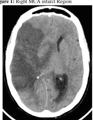
Figure 1: Right MCA infarct Region
Dr N. Radha MD (RD) et al JMSCR Volume 07 Issue 11 November 2019 Page 463
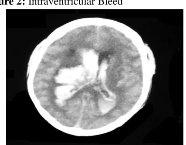
Figure 2: Intraventricular Bleed
Risk Factors
Hypertension: Among the risk factors, past history of hypertension was given importance. 90 patients i.e., 30% had history of pre-existing hypertension, however many patients admitted that they were not tested for hypertension before the onset of stroke. 50 patients i.e., 55.55% with hypertension showed cerebral hemorrhage 30 patients i.e., 33.33% with hypertension showed Infarct.
Diabetes Mellitus: In our study: 24% of the patients had a history of Diabetes mellitus. Many patients were not tested previously for evidence of diabetes before the onset of stroke. Out of 300 patients 120 patients had diabetes. 66.6% of the diabetic patients had cerebral infarction i.e., in 80 patients. 33.3% of the diabetic patients had cerebral hemorrhage i.e., 40 patients. HEART DISEASE In our study history revealed the existence of heart disease in 20% of the patients i.e., in 60 cases. Out of 60 cases, 29 cases had cardiac disease, which was previously detected, 26 cases had ECG changes after stroke attack and 5 cases had silent cardiac chest pain.
Figure 3: Right PCA Infarct
Involvement of Vascular Territory
Cerebral Infarction: Out of 300 cases of CT evaluation of CVA, 190 cases of infarcts were diagnosed that accounts for 63.33%.
50 patients had infarct in right MCA territory accounting for 26.31%.
40 patients had infarct in left MCA territory accounting for 21.05%
20 Patients had infarct in right PCA territory accounting for 10.52%
10 patients had infarct in left PCA territory accounting for 5.26%
5 patients had infarct in left ACA territory accounting for 2.63%
5 Patients had infarct in right ACA territory accounting for 2.63%
15 patients had infarct in right MCA and PCA territory accounting for 7.89%
10 patients had infarct in left MCA and PCA territory accounting for 5.26%
20 patients had infarct in both MCA territories accounting for 10.52%
5 patients had infarct in vertebro-basilar artery territory except PCA branch accounting for 2.63%
10 patients had lacunar infarcts accounting for 5.26%
Intracerebral Hemorrhage: In our study of 300 cases of clinically suspected CVA, 75 cases were turned out to be intracerebral hemorrhage, which accounts for 25%.
Out of 75 cases of intracerebral hemorrhage. 20 Patients had intracerebral hemorrhage
in left MCA territory accounting 26.66% 15 Patients had intracerebral hemorrhage
in right MCA territory accounting for 20% 5 Patients had intracerebral hemorrhage in
right PCA territory accounting for 6.66% 5 patients had intracerebral hemorrhage in
left PCA territory accounting for 6.6% 6 patients had intracerebral hemorrhage in
right ACA territory accounting for 8.8% 2 patients had intracerebral hemorrhage in
Dr N. Radha MD (RD) et al JMSCR Volume 07 Issue 11 November 2019 Page 464 5 patients had intracerebral hemorrhage in
right MCA and PCA territories accounting for 6.66%
6 patients had intracerebral hemorrhage in left MCA & PCA territories accounting for 8.0%
4 Patients had intracerebral hemorrhage in both MCA territories accounting for 5.33%
2 patients had haemorrhagic infarction accounting for 2.66%.
5 patients had Intra Cerebral Hemorrhage in vertebro-basilar artery territory accounting for 6.66%
In our study left MCA territory was the most commonly affected site.
Table 3: Incidence of ICH
Sub Arachnoid Hemorrhage as a Percentage of all Strokes - In my study of 300 cases of CVA, 15 cases had subarachnoid hemorrhage i.e., accounting for 5% These studies of primary subarachnoid hemorrhage include SAH due to aneurysmal rupture, rupture of A-V malformations and SAH of unknown cause.
Cerebral Venous Thrombosis- In our study we had 4 cases of CVT out of 300 cases and the percentage calculation was 1.33% Interestingly all of our CVT patients were females and all the cases were subjected to CT study in the post-partum period.
Clinically Suspected CVA but Normal on CT scan of Brain- Out of 300 cases of clinically suspected CVA subjected to CT Study, 11 cases
turned out to be normal accounting for 3.66%. These cases are taken as negative cases. There are technical problems to detect infarction but certainly the hemorrhage is ruled out in all cases.
Tumors- In our study stroke mimics — tumours detected in 5 cases out of 300 cases of suspected CVA, which accounts for 1.66% of the total study. Signs and symptomatology of tumour were mimicking the signs and symptoms of stroke, hence Neurophysicians suspected these cases as stroke, which turned out to be of tumorous pathology on computed tomography scanning.
Discussion
This study was directed to evaluate the role of CT scan in patients presenting with acute cerebro-vascular accident in differentiating between haemorrhage, infarct and other causes of stroke. Before the advent of CT scan and in places where CT scan is not yet available, physicians were mainly dependent on the history, physical findings and the Allen's method of scoring to differentiate between haemorrhage and infarct using this scoring system. Allen studied 174 cases of acute stroke and was able to make an accurate diagnosis in 90% of cases4.
However, the scoring system had certain limitations as it is dependent on the history given by the relatives of patients and sometimes, they are not able to give a clear description of signs and symptoms which correlated with the scoring system. 100% accuracy in distinguishing haemorrhage from ischemic stroke based on clinical findings was not possible.
Previous studies have reported the usefulness of CT scan in patients suffering from stroke by its ability to differentiate between haemorrhage and infarct and other causes of stroke and thus aiding in the clinical management. Oxford shire Community Stroke project that assesses 325 consecutive patients of acute stroke highlighting the role of usefulness of CT scan3.
Dr N. Radha MD (RD) et al JMSCR Volume 07 Issue 11 November 2019 Page 465 findings has proved to be of prognostic value in
the evaluation of these patients. The use of CT coupled with early acute phase therapy of stroke such as thrombolytic therapy has shown to improveoutcome in the acute stroke patients. Cerebral CT is a mainstay in emergency diagnostic work up of acute stroke patients and conveys important information within a few hours after the ictus. Hans Peter Harring et al found that in a recent series of patients with MCA territory infarctions, the incidence of positive findings was 68% in cerebral CT scans performed within 2 hours of stroke onset increasing to 89% within 3 hours, thus emphasizing the great value of emergency cerebral CT scanning in acute stroke management, which is superior to MRI5.
In the present study 300 patients of stroke were analysed and of them 190 patients had infarct i.e., 63.33%, 75 patients had haemorrhage i.e., 25%, 4 patients had CVT i.e., 1.33% and 15 patients had Sub Arachnoid haemorrhage i.e., 5%, 5 patients had tumoursi.e., 1.66% and 11 patients had normal scan i.e., 2.2%.
In studies done from India Mehta JK and Jacob reported an incidence of 60% infarcts and 30% haemorrhage, 8% subarachnoid haemorrhage in a case study of 50 patients. Ghosh SK and Row Chowdhary in a study of 30 patients with stroke had reported an incidence of infarct in 33.3% of cases and intracerebral haemorrhage in 60% of cases. In this study that had a different experience in the severity, the incidence of haemorrhage is seen to be higher than that of infarction.
In studies done by Rochester, Minnesota, (1945-1984) 7.2% patients had sub arachnoid haemorrhage. Carlisle, England 7.0% patients had sub arachnoid haemorrhage. Auckland, New Zealand 6.8% patients had sub arachnoid haemorrhage. Perth, Australia 4.5% patients had sub arachnoid haemorrhage.
In my study of 300 cases of CVA, 15 cases had subarachnoid haemorrhage i.e., accounting for 5%.These studies of primary subarachnoid haemorrhage include SAH due to aneurysmal rupture, rupture of A-V malformations and SAH
of unknown cause. Out of 300 cases of clinically suspected CVA subjected to CT study, 11 cases turned out to be normal accounting for 3.66%. These cases are taken as negative cases. There are technical problems to detect infarction but certainly the haemorrhage is ruled out in all cases. Moha, Briton (40) reported 3 patients with mass lesions (one subdural hematoma, one hydrocephalus and one metastasis) from 197 patients who had presented with acute stroke. In the Oxfordshire community stroke project five non-stroke lesions were detected by CT (2 gliomas, one metastasis, and 2 subdural hematomas) among 325 patients who were clinically diagnosed as having a definite stroke. In the present study of 300 patients, 5 cases of gliomas, 4 cases of CVT and 15 cases of sub arachnoid haemorrhage was detected and the patients presented with an acute stroke like picture3.
Conclusion
The following conclusions were made
1) CT scanning is the "Gold standard" technique for diagnosis of acute stroke as the rational management of stroke depends on "Accurate diagnosis" and should be ideally being done in all cases.
2) The results and factors obtained from our study correlates well with studies done in different parts of the world
Since risk factors such as hypertension, diabetes, cardiac disease and previous episodes of stroke play major role in the evolution of cerebrovascular accidents, it is suggested that
1) Such patients should be investigated carefully.
2) Sudden onset of neurological deficit or unexplained headache should further be investigated for the possibility of CVA. 3) If treatment is given early some of the
Dr N. Radha MD (RD) et al JMSCR Volume 07 Issue 11 November 2019 Page 466
Summary
300 patients who were clinically suspected of stroke were subjected to computed tomographic study.
Among these 300 patients 63.33% of patients had infarcts, 25% patients had intracerebral haemorrhage, 1.33% patients had cerebral venous thrombosis, 5% patients had subarachnoid haemorrhage, 1.66% of patients had tumorous pathology and 3.66% had normal scans.
It was observed that both infarction and intracerebral haemorrhage were most common in the age group between 60-69 years.
➢ Men were affected commonly in stroke cases. ➢ Risk factors like hypertension, Diabetes mellitus and cardiac disease plays major role in the evolution of stroke.
Out of 90 patients who had history of pre-existing hypertension, 55% of them showed cerebral haemorrhage and 33.33% of patients showed infarction. Out of 120 diabetic patients, 73% of patients had cerebral infarction and 27% of patients had cerebral haemorrhage. 20% of our patients had pre-existing heart disease.
Commonest territory affected was right middle cerebral artery territory in cases of cerebral infarction, which accounts for 26.31%. However, most of the large infarcts were noted involving more than one arterial territory.
In cases of intracerebral hemorrhage putamen and external capsule were commonly affected i.e., in 46.66% of cases.
Interestingly all of our cerebral venous thrombosis patients were females and all cases were subjected to computed tomographic scanning in the post-partum period.
11 cases (3.66%) were turned out to be normal on brain scan though there were technical problems to detect infarction, certainly the hemorrhage is ruled out in all cases.
5 case (1.66%) show tumours mimicking stroke symptomatology. In spite of our sincere effort most of the patients did not turned up for follow-up scans. Intraventricular extension was noted in 30 cases accounting for 40% which ofthem had bad prognosis.
References
1. Hatano S. Experience from a multicenter register, Bull. WHO.1976; 54: 541-553. 2. 2. Osborn AG. Stroke , Chapter 11, In :
Osborn AG, ed. Diagnostic neuroradiology, St. Louis: Mosby, 1994; 330-385.
3. Sandercock P, Molyneux A, Warlow C. Value of CT in patients with stroke: Oxfordshire Community Stroke Project, BMJ. 1985; 290: 193-197.
4. Allen CMC. Clinical diagnosis of the acute stroke syndrome, Quarterly Journal of Medicine. 1983; New Series II (208): 515-523.