1357
J. Exp. Med. The Rockefeller University Press • 0022-1007/97/10/1357/08 $2.00 Volume 186, Number 8, October 20, 1997 1357–1364Neutrophil Emigration in the Skin, Lungs, and Peritoneum:
Different Requirements for CD11/CD18 Revealed by
CD18-deficient Mice
By Joseph P. Mizgerd,
*
Hiroshi Kubo,
*
Gregory J. Kutkoski,
*
Sabrina D. Bhagwan,
*Karin Scharffetter-Kochanek,
‡Arthur L. Beaudet,
‡and Claire M. Doerschuk
*From the *Physiology Program, Harvard School of Public Health, Boston, Massachusetts 02115; and ‡Department of Molecular and Human Genetics, Baylor College of Medicine, Houston, Texas 77030
Summary
To determine the role of CD11/CD18 complexes in neutrophil emigration, inflammation was
induced in the skin, lungs, or peritoneum of mutant mice deficient in CD18 (CD18
2/2mu-tants). Peripheral blood of CD18
2/2mutants contained 11-fold more neutrophils than did
blood of wild-type (WT) mice. During irritant dermatitis induced by topical application of
croton oil, the number of emigrated neutrophils in histological sections of dermis was 98% less
in CD18
2/2mutants than in WT mice. During Streptococcus pneumoniae pneumonia, neutrophil
emigration in CD18
2/2mutants was not reduced. These data are consistent with expectations
based on studies using blocking antibodies to inhibit CD11/CD18 complexes, and on
observa-tions of humans lacking CD11/CD18 complexes. The number of emigrated neutrophils in
lung sections during Escherichia coli pneumonia, or in peritoneal lavage fluid after 4 h of S.
pneu-moniae peritonitis, was not reduced in CD18
2/2mutants, but rather was greater than the WT
values (240
6
30 and 220
6
30% WT, respectively). Also, there was no inhibition of
neutro-phil emigration during sterile peritonitis induced by intraperitoneal injection of thioglycollate
(90
6
20% WT). These data contrast with expectations. Whereas CD11/CD18 complexes are
essential to the dermal emigration of neutrophils during acute dermatitis, CD18
2/2mutant
mice demonstrate surprising alternative pathways for neutrophil emigration during pneumonia
or peritonitis.
A
cute emigration of neutrophils requires CD11/CD18
complexes under most circumstances (for review see
ref-erence 1). Antibodies against CD11/CD18 inhibit
neutro-phil emigration during acute inflammation in animals (2, 3).
Leukocytes from human patients with leukocyte adhesion
deficiency type 1 (LAD-1),
1a disease arising from
muta-tions in the gene for CD18, lack CD11/CD18 complexes.
In LAD-1 patients, neutrophil emigration is not observed
using Rebuck skin windows or skin chambers (4–7), and
infected peritoneal, laryngeal, esophageal, periodontal,
gin-gival, pharyngeal–glottic, dermal, or umbilical tissues of
LAD-1 patients are devoid of emigrated neutrophils (4, 5, 8).
In the lung, neutrophil emigration occurs via CD11/
CD18-dependent, but also via CD11/CD18-independent
pathways. Antibodies against CD11 or CD18 inhibit
neu-trophil emigration in response to Escherichia coli, E. coli LPS,
Pseudomonas aeruginosa, phorbol ester, IgG immune
com-plexes, or IL-1 (3, 9–12), but these antibodies do not
in-hibit pulmonary emigration in response to Streptococcus
pneumoniae, hydrochloric acid, or C5a complement
frag-ments (3, 10). In contrast to the other tissues examined, the
lungs from an autopsied LAD-1 patient contained
emi-grated neutrophils (8).
Mice deficient in CD18 (CD18
2/2mutants) have now
been derived (Scharffetter-Kochanek, K., submitted for
pub-lication). Neutrophils from these mice do not express
CD11/CD18 adhesion complexes. CD18
2/2mutants display
a phenotype resembling that seen in humans with LAD-1,
including neutrophilia, peripheral lymphadenopathy,
sple-nomegaly, and skin lesions. In this paper, CD18
2/2mutant
mice were used to determine the roles of CD11/CD18
complexes in neutrophil emigration during inflammation
in the skin, lungs, or peritoneum.
Methods
Animals. The CD18 gene was targeted for disruption in em-bryonic murine stem cells using a previously published targeting construct (13), blastocysts containing mutant stem cells were
1Abbreviations used in this paper: LAD-1, leukocyte adhesion deficiency
transferred to murine foster mothers, and a homozygous mouse line was established by selective breeding, as previously described (14–17). Mouse genotypes were confirmed by Southern blotting. Wild-type mice were from the same genetic background (mixed 129/Sv and C57BL/6). Mice were studied at 8–18 wk of age. All experiments received institutional approval.
CD11/CD18 Expression. Expression of CD11/CD18 on circu-lating neutrophils was examined by flow cytometry. Mice were killed by a lethal overdose of halothane. Blood was collected from the inferior vena cava, erythrocytes were hypotonically lysed, and leukocytes were stained with saturating concentrations of the fol-lowing rat monoclonal antibodies from PharMingen (San Diego, CA): M17/4 (anti–murine CD11a), M1/70 (anti–murine CD11b), or H129.19 (anti–murine CD4, used as a control anti-body, nonbinding for neutrophils). Antibodies against CD11a and CD4 were directly conjugated to FITC. Antibodies against CD11b were biotinylated, and CD11b-labeled cells were second-arily labeled by streptavidin conjugated to FITC (PharMingen). Cells were fixed with 1% paraformaldehyde, and then green fluo-rescence of 5,000 cells in the neutrophil population (identified and gated using scatter profiles) was measured using an Ortho Cytofluorograf 50HTM flow cytometer equipped with a Cicero
interface system (Cytomation, Fort Collins, CO).
Expression of CD11/CD18 adhesion complexes by leukocytes in the lungs was examined by immunohistochemistry. After an overdose of halothane, lungs were removed, inflated with a 1:1 mixture of 0.9% NaCl and Tissue-Tek OCT compound embed-ding medium (Miles Labs., Inc., Elkhart, IN), and snap-frozen in liquid N2. Adhesion molecules were identified in 6-mm sections
by antibodies M17/4 (PharMingen) against mouse CD11a, and M1/70 (PharMingen), against mouse CD11b. Antibody control sections were treated with nonspecific rat IgG. After treatment with goat anti–rat secondary antibodies, sites of antibody labeling were stained red using an alkaline phosphatase–based detection system (Kirkegaard and Perry Laboratories, Inc., Gaithersburg, MD). All slides were counterstained with hemotoxylin (Fisher Scientific Co., Fairlawn, NJ).
Dermatitis. Irritant dermatitis was induced by topical applica-tion of croton oil. Mice were anesthetized by methoxyflurane in-halation, and each side of one ear was treated with 10 ml of 2% croton oil (Sigma Chemical Co., St. Louis, MO) in 4:1 acetone/ olive oil. After 6 h, mice were killed by an overdose of halothane inhalation. Ear widths were measured using spring-loaded cali-pers. Peripheral blood was collected from the inferior vena cava. Blood leukocytes were counted with a hemacytometer after erythrocyte lysis, and leukocyte differentials were counted in blood smears stained with LeukoStat (Fisher Scientific Co., Pitts-burgh, PA). Each ear was removed, fixed in 10% formalin, em-bedded in paraffin, sectioned, and stained with hematoxylin and eosin for examination by light microscopy.
Morphometric analysis was used to quantify neutrophil emi-gration using a drawing tube to reflect a grid onto microscopically viewed histologic sections. For each ear, the volume densities of emigrated neutrophils within four 110-mm-wide cross-sections, separated by 1.5-mm intervals, were assessed by point counting (18). A total of 547–1,677 points/ear were counted. Each point was assessed as falling on (a) epidermis, dermis, or cartilage, and (b) an emigrated neutrophil or not an emigrated neutrophil. The volume fraction of emigrated neutrophils in each ear was stan-dardized to the cartilage volume of that ear, a value not expected to change during acute (6-h) dermatitis, by dividing the neutro-phil volume fraction by the cartilage volume fraction.
Cutaneous edema was quantified as the percentage of increase
in ear width in the right (croton oil–treated) compared to the left (untreated) ear for each mouse. The width of each ear was mea-sured five times with spring-loaded calipers. Edema (expressed as percentage swelling) was calculated as 100 times the difference in ear widths divided by the width of the untreated ear.
Pneumonia. Pneumonias were induced by intratracheal instil-lation of bacteria, as previously described (15, 19). Mice were anesthetized by intramuscular injection of ketamine hydrochlo-ride (100 mg/kg) and acepromazine maleate (5 mg/kg). The tra-cheas were surgically exposed, and 2.3 ml/g body wt of S.
pneu-moniae (5 3 109 CFU/ml) or E. coli (107 CFU/ml) were instilled
intratracheally. All bacterial suspensions contained 5% colloidal carbon to mark the deposition of the instillate. Radiotracers for measurement of edema formation (see below) were injected into the tail vein 15 min before intratracheal instillation (125I-albumin)
and 2 min before euthanasia (51Cr-RBC). After 6 h of infection,
mice were killed by overdose of halothane. The peritoneal and thoracic cavities were rapidly opened, the heart vessels were tied off to prevent pulmonary blood loss, peripheral blood samples were drawn from the inferior vena cava, and the lungs were re-moved and fixed via intratracheal instillation of 6% glutaralde-hyde under 22 cm H2O pressure. Circulating leukocytes were
counted as above.
Pulmonary neutrophils were quantified by morphometry in histological sections (18). Carbon black–containing lung regions were embedded in paraffin, and 5–7 mm thick sections were cut and stained with hematoxylin and eosin. A counting grid (10 3 10, covering 70,000 mm2 of the magnified field) was reflected
onto the field of view using a drawing tube. Randomly selected fields of pneumonic peripheral lung that were largely free of non-capillary blood vessels and bronchioles or larger airways were ex-amined. A total of three grids (300 points) were counted for each lung, and every point was classified as landing on (a) air space or tissue and (b) neutrophil or not a neutrophil. The quantities of neutrophils in air space or tissue were expressed as volume % of the respective compartment (alveolar air space or septal tissue). The volume % of the alveolated region of lung occupied by air space and by tissue did not differ among infected and uninfected WT and CD182/2 mutant mice (data not shown). The ratio of emigrated neutrophils (% alveolated region) to septal neutrophils (% alveolated region) was calculated for pneumonic regions from each mouse.
Pulmonary edema was measured before dissection of lungs for morphometry, as previously described (15, 19). In brief, 125
I-albu-min and 51Cr-RBC activities of blood and plasma samples and of
excised fixed lungs were measured to characterize the pulmonary plasma and blood volumes. The pulmonary intravascular plasma content, calculated from the hematocrit and the lung blood vol-ume, was subtracted from the total lung plasma content to yield extravascular plasma volume, and extravascular plasma volume per lung was expressed as ml pulmonary edema (15, 19).
Peritonitis. Peritonitis was induced and characterized as previ-ously described (15, 16). After anesthetizing mice by intramuscu-lar injection of ketamine hydrochloride (100 mg/kg) and acepro-mazine maleate (5 mg/kg), iodinated albumin (0.3 mCi/mouse) was injected into the tail vein. S. pneumoniae (109 CFU/mouse)
LeukoStat (Fisher Scientific Co.). The 125I-albumin recovered by
lavage was expressed as the percentage of 125I-albumin injected
and reflects plasma leakage into the peritoneal cavity (15, 16). Bacterial clearance was inferred from the loss of viable bacteria re-covered in peritoneal lavage, and CFU rere-covered 4 or 24 h after instillation were expressed as a percentage of the original number of CFU injected. Sterile peritonitis was induced in separate groups of mice by intraperitoneal injection of 1 ml of sterile-fil-tered 4% thioglycollate in PBS, and mice were processed as above.
Statistics. Groups consisted of four or five mice. Data were presented as mean 6 SEM. Data from different groups (WT ver-sus CD182/2 or infected versus uninfected) were compared by t
test. Because circulating blood cell counts in CD182/2 mutant mice were not normally distributed and were highly variable, these data were compared by Mann-Whitney U test and ex-pressed as medians in addition to mean 6 SEM values. Differ-ences were considered statistically significant when p ,0.05.
Results
Expression of CD11/CD18 Adhesion Complexes.
Circu-lating neutrophils from WT mice expressed both CD11a
and CD11b on their surface, as measured by flow
cytome-try (Fig. 1). Neutrophils from CD18
2/2mutants, however,
did not express CD11a or CD11b (Fig. 1). Similarly,
leu-kocytes from peripheral lymph nodes or spleens of WT
mice expressed CD11a and/or CD11b, whereas lymph
node and spleen cells from CD18
2/2mutant mice did not
express CD11a or CD11b, as measured by flow cytometry
(data not shown).
Expression of CD11/CD18 by leukocytes marginated
within lung capillaries was examined by
immunohis-tochemistry. Lungs from WT mice contained leukocytes
positive for CD11a or CD11b, but no leukocytes in
CD18
2/2lungs were positive for CD11a or CD11b. The
genetic mutation of CD18 resulted in the complete loss of
immunologically recognizable CD11a and CD11b in
leu-kocytes of CD18
2/2mutant mice.
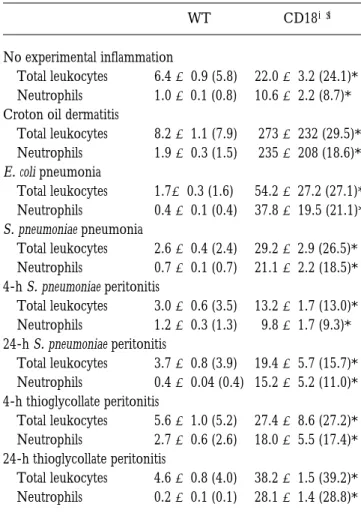
Leukocytosis and Splenomegaly.
All CD18
2/2mice with
or without experimentally induced inflammations had
ele-vated circulating leukocyte and neutrophil counts when
compared with WT (Table 1).
CD18
2/2mice exhibited splenomegaly (spleen weights
of 171.7
6
11.2 and 62.3
6
5.3 mg in CD18
2/2and WT
mice with bacterial peritonitis, respectively; p
,
0.05).
Nei-ther liver weights (1.02
6
0.04 and 0.94
6
0.04 g in
CD18
2/2and WT mice, respectively) nor body weights
(18.9
6
0.6 and 19.2
6
0.8 g in CD18
2/2and WT mice,
respectively) differed between WT and mutant mice with
bacterial peritonitis.
Neutrophil Emigration During Dermatitis.
Croton oil
ap-plication induced significant neutrophil emigration in WT
mice (Fig. 2). In contrast, there was no increase in
emi-grated neutrophils in the dermis of CD18
2/2mutant mice
after 6 h of croton oil dermatitis (Fig. 2).
Dermal Edema.
After 6 h, croton oil induced edema
ac-cumulation in both WT and CD18
2/2ears (Table 2).
There was, however, significantly less edema accumulation
in the CD18
2/2ears than in WT ears (Table 2).
Neutrophil Emigration During Pneumonia.
More neutrophils
Figure 1. Expression ofCD11a–CD18 and CD11b– CD18 by peripheral blood neu-trophils from WT and CD182/2 mutant mice. Blood was with-drawn from the inferior vena cava of mice after 6 h of croton oil dermatitis. Shown are repre-sentative linear-scale fluores-cence histograms from WT (1/ 1) or CD18-mutant (2/2) neutrophils labeled with mono-clonal antibodies H129.19 against murine CD4 as a nonbinding con-trol (A), M17/4 against murine CD11a (B), or M1/70 against murine CD11b (C).
Table 1. Circulating Leukocytes in WT and CD182/2 Mice
WT CD182/2
No experimental inflammation
Total leukocytes 6.4 6 0.9 (5.8) 22.0 6 3.2 (24.1)*
Neutrophils 1.0 6 0.1 (0.8) 10.6 6 2.2 (8.7)*
Croton oil dermatitis
Total leukocytes 8.2 6 1.1 (7.9) 273 6 232 (29.5)*
Neutrophils 1.9 6 0.3 (1.5) 235 6 208 (18.6)*
E. coli pneumonia
Total leukocytes 1.76 0.3 (1.6) 54.2 6 27.2 (27.1)*
Neutrophils 0.4 6 0.1 (0.4) 37.8 6 19.5 (21.1)*
S. pneumoniae pneumonia
Total leukocytes 2.6 6 0.4 (2.4) 29.2 6 2.9 (26.5)*
Neutrophils 0.7 6 0.1 (0.7) 21.1 6 2.2 (18.5)*
4-h S. pneumoniae peritonitis
Total leukocytes 3.0 6 0.6 (3.5) 13.2 6 1.7 (13.0)*
Neutrophils 1.2 6 0.3 (1.3) 9.8 6 1.7 (9.3)*
24-h S. pneumoniae peritonitis
Total leukocytes 3.7 6 0.8 (3.9) 19.4 6 5.7 (15.7)*
Neutrophils 0.4 6 0.04 (0.4) 15.2 6 5.2 (11.0)*
4-h thioglycollate peritonitis
Total leukocytes 5.6 6 1.0 (5.2) 27.4 6 8.6 (27.2)*
Neutrophils 2.7 6 0.6 (2.6) 18.0 6 5.5 (17.4)*
24-h thioglycollate peritonitis
Total leukocytes 4.6 6 0.8 (4.0) 38.2 6 1.5 (39.2)*
Neutrophils 0.2 6 0.1 (0.1) 28.1 6 1.4 (28.8)*
Blood was drawn from the inferior vena cava. Values represent mean 6 SEM 3 106 cells/ml. Median values are shown in parentheses.
were present in the alveolar septae of uninfected CD18
2/2mutants than in those of uninfected WT mice (Fig. 3).
There were almost no neutrophils in the alveolar air spaces
of WT and CD18
2/2mutant mice (Fig. 4).
During E. coli and S. pneumoniae pneumonias, the
vol-ume fraction of septal tissue occupied by neutrophils
in-creased in both WT and CD18
2/2mutant mice (Fig. 3).
CD18
2/2mutants had more neutrophils in alveolar septae
for each type of pneumonia than did WT mice (Fig. 3).
Neutrophils emigrated into the alveolar air spaces during
E. coli and S. pneumoniae pneumonias for both WT and
CD18
2/2mutant mice (Fig. 4). During either pneumonia,
CD18
2/2mutants had more neutrophils in their air spaces
than did WT mice (Fig. 4). The two types of bacteria
in-duced a similar degree of neutrophil emigration in WT
mice, but fewer neutrophils (P
,
0.05) emigrated during E.
coli than during S. pneumoniae pneumonia in CD18
2/2mu-tant mice (Fig. 4).
The ratio of alveolar air space neutrophils to septal
neu-trophils in pneumonic regions is an index of the ability of
neutrophils to extravasate. In WT mice, the ratio of air
space to septal neutrophils was 1.0
6
0.2 and 1.1
6
0.1
during E. coli and S. pneumoniae pneumonias, respectively.
In CD18
2/2mutants, this ratio was 1.7
6
0.2 and 2.3
6
0.3 for E. coli and S. pneumoniae, respectively, significantly
greater than WT for each type of pneumonia (P
,
0.05).
Pulmonary Edema.
Pulmonary edema during E. coli
pneumonia did not differ between WT and CD18
2/2mice
(Table 2). Edema accumulation was significantly greater in
CD18
2/2mutants than in WT mice during S. pneumoniae
pneumonia (Table 2).
Figure 2. Dermal neutrophil emigration in WT and CD182/2 mice 6 hafter topical application of 2% croton oil. Emigrated neutrophils were quantitated morphometrically and expressed as mean 6 SEM standard-ized volume fractions for WT (closed bars) or CD182/2 (open bars) mice.
*Significant differences from WT; ‡significant differences from control
(untreated) ears (P ,0.05).
Figure 3. Neutrophils in the alveolar septae of uninfected WT or CD182/2 mice and 6 h after intratracheal instillation of E. coli or S. pneu-moniae. Septal neutrophils were quantitated morphometrically and expressed as the mean 6 SEM volume percent of septal tissue occupied by neutro-phils for WT (closed bars) or CD182/2 (open bars) mice. *Significant
differ-ences from WT; ‡significant differences from uninfected mice (P ,0.05).
Figure 4. Neutrophils in the alveolar air spaces of uninfected WT or CD182/2 mice and 6 h after intratracheal instillation of E. coli or S. pneu-moniae. Emigrated neutrophils were quantitated morphometrically and expressed as the mean 6 SEM volume percent of alveolar air space occu-pied by neutrophils for WT (closed bars) or CD182/2 (open bars) mice.
*Significant differences from WT; ‡significant differences from uninfected
mice (P ,0.05).
Table 2. Edema Accumulation in WT and CD182/2 Mice
WT CD182/2
Dermatitis
(percent increase in ear width)
Croton oil, 6 h 41.8 6 3.4 17.8 6 8.7*
Pneumonia (ml/lung)
E. coli 6 h 78.1 6 21.5 96.6 6 15.3
S. pneumoniae 6 h 54.9 6 12.1 129.2 6 24.0*
S. pneumoniae peritonitis
(%125I-albumin recovered)
4 h 2.7 6 0.7 1.4 6 0.3
24 h 1.2 6 0.1 2.6 6 0.4*
Thioglycollate peritonitis (%125I-albumin recovered)
4 h 4.0 6 1.1 2.5 6 0.5
24 h 1.6 6 0.2 5.8 6 1.2*
Edema accumulation was quantified during dermatitis, pneumonia, or peritonitis, as described in the text. Values represent mean 6 SEM.
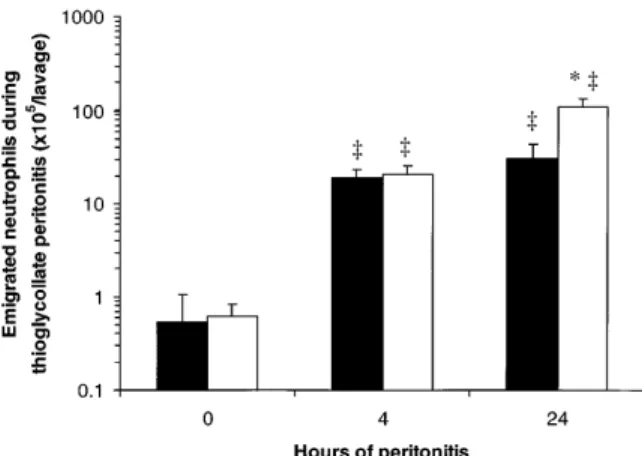
Neutrophil Emigration During Peritonitis.
There were few
neutrophils in the uninfected peritoneal cavities of WT and
CD18
2/2mice (Fig. 5). After intraperitoneal injection of
S. pneumoniae, neutrophil accumulation was apparent 4 h
later, and was greatly increased after 24 h of peritonitis in
both WT and CD18
2/2mutant mice (Fig. 5). More
neu-trophils were recovered in the peritoneal lavage fluids of
CD18
2/2mutants than of WT mice after 4 or 24 h of
peri-tonitis (220
6
30 and 500
6
50% WT, respectively; Fig.
5). After intraperitoneal injection of thioglycollate, there
was no significant difference between CD18
2/2mutants
and WT mice in the number of neutrophils recovered by
peritoneal lavage after 4 h (110
6
30% WT; Fig. 6). By 24 h,
more neutrophils were recovered from CD18
2/2mutants
than from WT mice (360
6
80% WT; Fig. 6).
Peritoneal Edema.
Peritoneal edema did not significantly
differ between mutant and WT mice after 4 h of bacterial
or thioglycollate peritonitis (Table 2). After 24 h of either
peritonitis, there was more peritoneal edema in CD18
2/2mutants than in WT mice (Table 2).
Bacterial Clearance During Peritonitis.
Bacterial clearance
did not significantly differ between WT and CD18
2/2mu-tant mice. After 4 h, 24
6
10% of injected CFU were
re-covered in the peritoneal lavage from WT mice, and 45
6
9% of injected CFU from CD18
2/2mice. After 24 h,
,
0.1%
of injected CFU were recovered from either WT or
CD18
2/2mice.
Discussion
Neutrophil emigration was reduced and edema
accumu-lation compromised in CD18
2/2mutant mice with croton
oil dermatitis. In contrast, neither neutrophil emigration
nor edema accumulation were compromised in CD18
2/2mutants during S. pneumoniae or thioglycollate peritonitis
or during E. coli or S. pneumoniae pneumonia. These data
confirm that CD11/CD18 complexes are essential to
neu-trophil emigration under at least some circumstances, but
they definitively demonstrate that
CD11/CD18-indepen-dent pathways for neutrophil emigration can be used
dur-ing acute inflammation in the peritoneum and the lung.
These results may be compared and contrasted with (a)
studies using blocking antibodies to inhibit CD11/CD18
interactions with ligands, (b) observations with human
LAD-1 patients, (c) studies with CD18 hypomorphic
mu-tant mice with decreased but not eliminated expression of
CD18, and (d) mutant mice deficient in CD11a or in
CD11b.
Antibodies against CD18 inhibit cutaneous emigration
of neutrophils in response to diverse stimuli (2), and in this
study cutaneous emigration of neutrophils was inhibited
in CD18
2/2mutant mice during croton oil dermatitis.
Whereas antibodies against CD18 inhibit acute peritoneal
emigration of neutrophils during S. pneumoniae or
thiogly-collate peritonitis (3, 20) and acute pulmonary emigration
of neutrophils during E. coli pneumonia (3), however,
neu-trophil emigration was not inhibited during any of these
inflammatory reactions in mice completely deficient in
CD11/CD18 adhesion complexes due to a CD18 null
mu-tation. This discrepancy suggests that the murine regulation
of adhesion pathways differs during lifelong deficiency of
CD11/CD18 compared with acute inhibition of CD11/
CD18 by antibodies.
Tissues from human LAD-1 patients with a lifelong
defi-ciency of CD11/CD18 complexes are generally lacking in
neutrophils, even when infected by bacteria or fungi.
Biop-sies from diseased periodontal, gingival, pharyngeal–glottic,
dermal, or umbilical tissues from LAD-1 patients show a
lack of neutrophils in infected or inflamed sites (4, 5).
Au-topsy of a LAD-1 patient revealed infection without
neu-trophil emigration in the peritoneum, larynx, and
esopha-gus (8). The lungs of the autopsied patient were also
Figure 5. Peritoneal neutrophil emigration in WT and CD182/2 mice0, 4, or 24 h after intraperitoneal injection of S. pneumoniae. Emigrated neutrophils were quantitated in peritoneal lavage fluids and expressed as mean 6 SEM neutrophils/lavage for WT (closed bars) or CD182/2 (open bars) mice. *Significant differences from WT; ‡significant differences from
0-h (uninfected) mice (P ,0.05).
Figure 6. Peritoneal neutrophil emigration in WT and CD182/2 mice
0, 4, or 24 h after intraperitoneal injection of thioglycollate. Emigrated neutrophils were quantitated in peritoneal lavage fluids and expressed as mean 6 SEM neutrophils/lavage for WT (closed bars) or CD182/2 (open bars) mice. The data from 0 h of peritonitis (no thioglycollate) are the same as shown in Fig. 5. *Significant differences from WT; ‡significant
infected, but in contrast to the other sites examined,
emi-grated neutrophils were observed (8). Thus, humans can
recruit neutrophils via CD11/CD18-independent
path-ways under at least some circumstances. During acute
in-flammations in this study, the CD18
2/2mutant mice
re-cruited neutrophils into the lungs and the peritoneum, but
not into the skin.
Mutant mice that express 2–16% of WT levels of CD11/
CD18 on their leukocyte surfaces (CD18 hypomorphs)
have been previously characterized (13). The numbers of
neutrophils recovered by peritoneal lavage 4 h after
intra-peritoneal injection of thioglycollate were not significantly
different in CD18 hypomorphs when compared with WT
mice (13). Thus, either the remaining CD11/CD18
medi-ates neutrophil emigration in the hypomorphs, or
alterna-tive pathways for neutrophil emigration are used in the
hy-pomorphs as in the CD18
2/2mutants.
Mutant mice lacking CD11a–CD18 secondary to a
CD11a mutation have been generated, and their
neutro-phils are compromised in their ability to emigrate during
thioglycollate peritonitis (21). In contrast, CD18
2/2mu-tants lack CD11a–CD18 as well as the other members of
the CD11/CD18 family, yet neutrophil emigration is not
compromised in these mice during peritonitis. Thus, the
CD18
2/2mutants, but not the CD11a–CD18 mutants,
in-duce CD11/CD18-independent neutrophil emigration
dur-ing thioglycollate peritonitis. Mutant mice deficient in CD11b
do not demonstrate reduced neutrophil emigration during
thioglycollate peritonitis (22, 23). CD11a–CD18 function,
however, remains critical to neutrophil emigration in
CD11b-deficient mice as indicated by CD11a-blocking antibodies
(23). It is unclear why mutant mice with single deficiencies
in CD11a–CD18 or CD11b–CD18 require CD11a–CD18 for
neutrophil emigration under circumstances in which CD18
2/2mutants do not demonstrate impaired emigration. CD18
2/2mutants are deficient in CD11a–CD18, CD11b–CD18,
CD11c–CD18, and CD11d–CD18, whereas the CD11a
2/2and CD11b
2/2mutant mice each express three of the four
adhesion complexes. Furthermore, the CD18
2/2mutants
have extremely high circulating neutrophil counts, which
has not been reported for the CD11a
2/2or CD11b
2/2mu-tants. These factors may contribute to the differing
recruit-ment of CD11a-independent pathways for the emigration
of neutrophils in the peritoneal cavities of these mutant
mice.
To our knowledge, this is the first evidence that a
CD11/CD18-independent pathway can be used by
neu-trophils in nonpulmonary tissues during the first hours of
acute inflammation. CD11/CD18-independent
neutro-phil emigration in the peritoneum has been observed over
prolonged (12–24-h) periods of inflammation (20, 24).
Macrophages recovered after 72 h of protease
peptone-induced peritonitis can elicit CD11/CD18-independent
neutrophil emigration when transferred to naïve peritoneal
cavities before eliciting acute (4 h) streptococcal peritonitis
(25). CD11/CD18-independent neutrophil emigration also
occurs in the joints during chronic inflammation (26). In
CD18
2/2mutants, the peritoneal emigration of neutrophils
occurred in the absence of CD11/CD18 after only 4 h of
peritonitis.
These data suggest that neutrophils can use different
ad-hesion molecule pathways in emigrating from the systemic
vasculature in the skin (during croton oil dermatitis) and in
the peritoneum (during S. pneumoniae or thioglycollate
peritonitis). These tissues may regulate the emigration of
neutrophils differently. Whereas neutrophil emigration into
the skin was quantified in situ by morphometric analysis of
histologic sections, neutrophil emigration into the
perito-neum was quantified in lavage fluid. Studying the roles of
adhesion molecules in neutrophil emigration by
examina-tion of lavage fluid during peritonitis is complicated by the
fact that leukocytes use adhesion molecules, including
CD11/CD18 and ICAM-1, to adhere to the peritoneal
mesothelium (27, 28). Thus, leukocytes may be less likely
to adhere to the mesothelium and may be more likely to be
lavaged in situations in which CD11/CD18-ICAM-1
in-teractions are disrupted. Despite these confounding factors
in interpreting lavage data, it remains clear that neutrophils
emigrated during peritonitis in CD18
2/2mutant mice.
Neutrophil emigration was observed in situ during both E.
coli and S. pneumoniae pneumonias in CD18
2/2mutant
mice as well.
The observation of increased emigration of neutrophils
in the lungs and peritoneal cavities of CD18
2/2mutants
when compared with WT mice likely resulted from the
in-creased numbers of circulating neutrophils in CD18
2/2mutant mice, although other explanations such as decreased
resolution of inflammation (22) are also possible. Animals
rendered neutrophilic by administration of G-CSF
demon-strate increased neutrophil numbers in bronchoalveolar
lav-age fluid during pneumonia induced by S. pneumoniae,
Klebsiella pneumoniae, or E. coli (29–31), and increased
neu-trophil numbers in peritoneal lavage fluid during peritonitis
induced by Listeria monocytogenes, fecal flora, or E. coli (32–
34). The number of circulating neutrophils in CD18
2/2mutants with pneumonia or peritonitis was increased 8–fold
or more when compared with WT mice. Thus, the
in-creased neutrophil emigration observed in CD18
2/2mu-tants with pneumonia or peritonitis likely resulted from
their peripheral neutrophilia.
In conclusion, CD18
2/2mutant mice displayed the
ex-pected phenotype of reduced acute neutrophil emigration
when studied during croton oil dermatitis. CD18
2/2The authors thank Ervin Melulini for preparation of histological slides, and Amy Imrich for assistance with flow cytometry.
This work was supported by United States Public Health Service grants HL 48160, HL 52466, and AI 32177. Karin Scharffetter-Kochanek was supported by a Heisenberg grant from the Deutsche Forschungsge-meinschaft.
Address correspondence to Claire M. Doerschuk, Physiology Program, Harvard School of Public Health, Building I Room 305, 665 Huntington Ave., Boston, MA 02115. Phone: 1706; FAX: 617-432-3468; E-mail: [email protected]
Received for publication 6 June 1997 and in revised form 7 August 1997.
References
1. Carlos, T.M., and J.M. Harlan. 1994. Leukocyte-endothelial adhesion molecules. Blood. 84:2068–2101.
2. Arfors, K., C. Lundberg, L. Lindbom, K. Lundberg, P.G. Be-atty, and J.M. Harlan. 1987. A monoclonal antibody to the membrane glycoprotein complex CD18 inhibits polymor-phonuclear leukocyte accumulation and plasma leakage in vivo. Blood. 69:338–340.
3. Doerschuk, C.M., R.K. Winn, H.O. Coxson, and J.M. Har-lan. 1990. CD18-dependent and -independent mechanisms of neutrophil adherence in the pulmonary and systemic mi-crovasculature of rabbits. J. Immunol. 114:2327–2333. 4. Bowen, T.J., H.D. Ochs, L.C. Altman, T.H. Price, D.E. Van
Epps, D.L. Brautigan, R.E. Rosin, W.D. Perkins, B.M. Ba-bior, S.J. Klebanoff, and R.J. Wedgwood. 1982. Severe re-current bacterial infections associated with defective adher-ence and chemotaxis in two patients with neutrophils deficient in a cell-associated glycoprotein. J. Pediatr. 101:932–940. 5. Anderson, D.C., F.C. Schmalsteig, M.J. Finegold, B.J. Hughes,
R. Rothlein, L.J. Miller, S. Kohl, M.F. Tosi, R.L. Jacobs, T.C. Waldrop, et al. 1985. The severe and moderate pheno-types of heritable Mac-1, LFA-1 deficiency: their quantitative definition and relation to leukocyte dysfunction and clinical features. J. Infect. Dis. 152:668–689.
6. Buescher, E.S., T. Gaither, J. Nath, and J.I. Gallin. 1985. Ab-normal adherence-related functions of neutrophils, mono-cytes, and Epstein-Barr virus-transformed B cells in a patient with C3bi receptor deficiency. Blood. 65:1382–1390. 7. Weisman, S.J., R.L. Berkow, G. Plautz, M. Torres, W.A.
McGuire, T.D. Coates, R.A. Haak, A. Floyd, R. Jersild, and R.L. Baehner. 1985. Glycoprotein-180 deficiency: genetics and abnormal neutrophil activation. Blood. 65:696–704. 8. Hawkins, H.K., S.C. Heffelfinger, and D.C. Anderson. 1992.
Leukocyte adhesion deficiency: clinical and postmortem ob-servations. Pediatr. Pathol. 12:119–130.
9. Mulligan, M.S., G.P. Wilson, R.F. Todd, C.W. Smith, D.C. Anderson, J. Varani, T.B. Issekutz, M. Miyasaka, T. Tama-tani, M. Miyasaka, et al. 1993. Role of b1, b2 integrins, and ICAM-1 in lung injury after deposition of IgG and IgA im-mune complexes. J. Immunol. 150:2407–2417.
10. Hellewell, P.G., S.K. Young, P.M. Henson, and G.S. Worthen. 1994. Disparate roles of the b2-integrin CD18 in
the local accumulation of neutrophils in pulmonary and cuta-neous inflammation in the rabbit. Am. J. Respir. Cell Mol.
Biol. 10:391–398.
11. Kumasaka, T., N.A. Doyle, W.M. Quinlan, L. Graham, and C.M. Doerschuk. 1996. Role of CD11/CD18 in neutrophil emigration during acute and recurrent Pseudomonas
aerugi-nosa-induced pneumonia in rabbits. Am. J. Pathol. 148:1297–
1305.
12. Qin, L., W.M. Quinlan, N.A. Doyle, L. Graham, J.E. Sligh, F. Takei, A.L. Beaudet, and C.M. Doerschuk. 1996. The roles of CD11/CD18 and ICAM-1 in acute Pseudomonas
aeruginosa-induced pneumonia in mice. J. Immunol. 157:
5016–5021.
13. Wilson, R.W., C.M. Ballantyne, C.W. Smith, C. Montgom-ery, A. Bradley, W.E. O’Brien, and A.L. Beaudet. 1993. Gene targeting yields a CD18-mutant mouse for study of in-flammation. J. Immunol. 151:1571–1578.
14. Sligh, J.E., Jr., C.M. Ballantyne, S.S. Rich, H.K. Hawkins, C.W. Smith, A. Bradley, and A.L. Beaudet. 1993. Inflamma-tory and immune responses are impaired in mice deficient in intercellular adhesion molecule 1. Proc. Nat. Acad. Sci. USA. 90:8529–8533.
15. Bullard, D.C., L. Qin, I. Lorenzo, W.M. Quinlin, N.A. Doyle, R. Bosse, D. Vestweber, C.M. Doerschuk, and A.L. Beaudet. 1995. P-selectin/ICAM-1 double mutant mice: acute emigration of neutrophils into the peritoneum is com-pletely absent but is normal into pulmonary alveoli. J. Clin.
Invest. 95:1782–1788.
16. Bullard, D.C., E.J. Kunkel, H. Kubo, M.J. Hicks, I. Lorenzo, N.A. Doyle, C.M. Doerschuk, K. Ley, and A.L. Beaudet. 1996. Infections and deficiency of leukocyte rolling and re-cruitment in E-/P-selectin mutant mice. J. Exp. Med. 183: 2329–2336.
17. Scharffetter-Kochanek, K., K. Norman, D.C. Bullard, I. Lorenzo, N.P. van Nood, S. Rich, W. Smith, and A.L. Beaudet. 1996. Generation and characterization of a CD18 complete knock-out mouse—new insights in the function of the CD18 molecule? J. Investig. Dermatol. 106:809.
18. Weibel, E.R. 1990. Morphometry: stereological theory and practical methods. In Models of Lung Disease: microscopy and Structural Methods, Volume 47. J. Gil, editor. Marcel Dekker, Inc., New York. 199–252.
19. Mizgerd, J.P., B.B. Meek, G.J. Kutkoski, D.C. Bullard, A.L. Beaudet, and C.M. Doerschuk. 1996. Selectins and neutro-phil traffic: margination and Streptococcus pneumoniae-induced emigration in murine lungs. J. Exp. Med. 184:639–645. 20. Conlan, J.W., and R.J. North. 1994. Listeria monocytogenes,
but not Salmonella typhimurium, elicits a CD18-independent mechanism of neutrophil extravasation into the murine peri-toneal cavity. Infect. Immunol. 62:2702–2706.
show normal CTL responses to virus, but fail to reject immu-nogenic tumor. J. Exp. Med. 183:1415–1426.
22. Coxon, A., P. Rieu, F.J. Barkalow, S. Askari, A.H. Sharpe, U.H. von Andrian, M.A. Arnaout, and T.N. Mayadas. 1996. A novel role for the b2 integrin CD11b/CD18 in neutrophil apoptosis: a homeostatic mechanism in inflammation.
Immu-nity. 5:653–666.
23. Lu, H., C.W. Smith, J. Perrard, D. Bullard, L. Tang, S.B. Shappell, M.L. Entman, A.L. Beaudet, and C.M. Ballantyne. 1997. LFA-1 is sufficient in mediating neutrophil emigration in Mac-1 deficient mice. J. Clin. Invest. 99:1340–1350. 24. Winn, R.K., and J.M. Harlan. 1993. CD18-independent
neutrophil and mononuclear leukocyte emigration into the peritoneum of rabbits. J. Clin. Invest. 92:1168–1173. 25. Mileski, W., J. Harlan, C. Rice, and R. Winn. 1990.
Strep-tococcus pneumoniae-stimulated macrophages induce neu-trophils to emigrate by a CD18-independent mechanism of adherence. Circ. Shock 31:259–267.
26. Issekutz, T.B., M. Miyasaka, and A.C. Issekutz. 1996. Rat blood neutrophils express very late antigen 4 and it mediates migration to arthritic joint and dermal inflammation. J. Exp.
Med. 183:2175–2184.
27. Andreoli, S.P., C. Mallett, K. Williams, J.A. McAteer, R. Rothlein, and C.M. Doerschuk. 1994. Mechanisms of poly-morphonuclear leukocyte mediated peritoneal mesothelial cell injury. Kidney Int. 46:1100–1109.
28. Liberek, T., N. Topley, W. Luttman, and J.D. Williams. 1996. Adherence of neutrophils to human peritoneal
me-sothelial cells- role of intercellular adhesion molecule-1. J.
Am. Soc. Nephrol. 7:208–217.
29. Nelson, S., W. Summer, G. Bagby, C. Nakamura, L. Stew-art, G. Lipscomb, and J. Andresen. 1991. Granulocyte col-ony-stimulating factor enhances pulmonary host defenses in normal and ethanol-treated rats. J. Infect. Dis. 164:901–906. 30. Lister, P.D., M.J. Gentry, and L.C. Preheim. 1993.
Granulo-cyte colony-stimulating factor protects control rats but not ethanol-fed rats from fatal pneumococcal pneumonia. J.
In-fect. Dis. 168:922–926.
31. Freeman, B.D., R. Correa, W. Karzai, C. Natanson, M. Patterson, S. Banks, Y. Fitz, R.L. Danner, L. Wilson, and P.Q. Eichacker. 1996. Controlled trials of rG-CSF and CD11b-directed MAb during hyperoxia and E. coli pneumo-nia in rats. J. Appl. Physiol. 80:2066–2076.
32. Serushago, B.A., Y. Yoshikai, T. Handa, M. Mitsuyama, K. Muramori, and K. Nomoto. 1992. Effect of recombinant hu-man granulocyte colony-stimulating factor (rhG-CSF) on murine resistance against Listeria monocytogenes. Immunology. 75:475–480.
33. Lundblad, R., M.Y. Wang, G. Kvalheim, E. Lingaas, and K. Giercksky. 1995. Granulocyte colony-stimulating factor im-proves myelopoiesis and host defense in fulminant intra-abdomi-nal sepsis in rats. Shock. 4:68–73.
34. Dunne, J.R., B.J. Dunkin, S. Nelson, and J.C. White. 1996. Effects of granulocyte colony stimulating factor in a non-neutropenic rodent model of Escherichia coli peritonitis. J.