REVIEW
Nanotechnology: a promising method
for oral cancer detection and diagnosis
Xiao‑Jie Chen
1, Xue‑Qiong Zhang
2*, Qi Liu
3, Jing Zhang
1,4and Gang Zhou
1,4*Abstract
Oral cancer is a common and aggressive cancer with high morbidity, mortality, and recurrence rate globally. Early detection is of utmost importance for cancer prevention and disease management. Currently, tissue biopsy remains the gold standard for oral cancer diagnosis, but it is invasive, which may cause patient discomfort. The application of traditional noninvasive methods‑such as vital staining, exfoliative cytology, and molecular imaging‑is limited by insuf‑ ficient sensitivity and specificity. Thus, there is an urgent need for exploring noninvasive, highly sensitive, and specific diagnostic techniques. Nano detection systems are known as new emerging noninvasive strategies that bring the detection sensitivity of biomarkers to nano‑scale. Moreover, compared to current imaging contrast agents, nano‑ particles are more biocompatible, easier to synthesize, and able to target specific surface molecules. Nanoparticles generate localized surface plasmon resonances at near‑infrared wavelengths, providing higher image contrast and resolution. Therefore, using nano‑based techniques can help clinicians to detect and better monitor diseases during different phases of oral malignancy. Here, we review the progress of nanotechnology‑based methods in oral cancer detection and diagnosis.
Keywords: Oral cancer, Nanotechnology, Molecular imaging, Biomarker detection
© The Author(s) 2018. This article is distributed under the terms of the Creative Commons Attribution 4.0 International License (http://creat iveco mmons .org/licen ses/by/4.0/), which permits unrestricted use, distribution, and reproduction in any medium, provided you give appropriate credit to the original author(s) and the source, provide a link to the Creative Commons license, and indicate if changes were made. The Creative Commons Public Domain Dedication waiver (http://creat iveco mmons .org/ publi cdoma in/zero/1.0/) applies to the data made available in this article, unless otherwise stated.
Background
Cancer is a critical public health problem worldwide that has brought great burden to society. In 2016, an esti-mated 1,685,210 new cases and 595,690 cancer deaths
occurred in the United States alone [1]. Oral cancer is
the sixth most common cancer globally and has a 5-year
survival rate of around 50% [2]. According to US cancer
statistics, approximately 31,910 new cases of oral cancer
and 6490 oral cancer deaths occurred in 2016 [3]. Oral
cancer is an aggressive cancer that mainly affects oral epithelial cells, may develop metastasis, and even results
in death [4]. The major type of malignancy is oral
squa-mous cell carcinomas (OSCC), which accounts for more
than 90% of all oral cancers [5]. These tumors may invade
the mucosa of the tongue, buccal, floor of mouth, alveo-lar and the hard palate, and the tongue is reported to be
the most common subsite, with poor prognosis [1, 6].
Oral carcinogenesis is often due to long-term exposure to various potential risk factors, which may lead to
accu-mulation of multiple genetic mutations [4]. Several major
risk factors for oral cancer, including smoking, alcohol consumption, and human papillomavirus infection, with
smoking acting as the leading cause of cancer death [3,
7]. Besides, habitual use of the areca nut is another risk
factor that closely associated with oral cancer, especially
in Indian subcontinent [8].
The formation of oral cancer is a multifactorial and
multistep process [6]. Oral leukoplakia, oral
erythropla-kia, oral lichen planus, oral submucous fibrosis, actinic keratosis, and discoid lupus erythematosus are common oral potentially malignant disorders (OPMD) that are known to have the potential for malignant
transforma-tion [8, 9]. Thus, early detection of OPMD and oral
can-cer is critical for the prognosis of diseases [5]. To date,
scalpel biopsy and histopathological examinations are still the standard diagnostic procedures applied to ascer-tain the oral potentially malignant and malignant lesions
[17, 18]. However, the biopsy procedure is often invasive,
Open Access
which may cause patients anxiety and discomfort [10]. The selection of resection margins depends largely on the histopathological assessments, and the results can be affected by the quality of the specimens and pathologists’
subjective judgments [11, 12]. In addition, the
assess-ments are unable to detect small numbers of genetically abnormal cells at the margins, thus leaving the risk of
recurrence [13, 14].
In the past few decades, a variety of pain-free diag-nostic strategies have been developed. Non-invasive visual tools such as toluidine blue (TB) staining, auto-fluorescence (VELscope) and chemiluminescence (Vizi-Lite) have been used solely or in combination as adjuvant
tests to detect potentially malignant lesions [15–19]. In
oral epithelial dysplasia cases, the sensitivity and speci-ficity of TB, VELscope and ViziLite are reported to be 84.1% and 15.3, 77.3 and 27.8, 56.8 and 65.8%,
respec-tively [15]. Exfoliated cells, serum, and saliva are the most
commonly used non-invasive samples for oral cancer detection since they are easily accessible, convenient, and
cost-effective [11, 20]. For oral cancer diagnosis, the
sen-sitivity and specificity of exfoliative cytology is reported
to be 93.5 and 50.6%, respectively [21]. The biomarker
with high sensitivity and specificity in serum is com-bined detection of Cyclin D1 and epidermal growth fac-tor recepfac-tor (EGFR), while the reliable marker in saliva is
CD44 [22, 23]. Imaging techniques are used as diagnostic
adjuncts to the histopathological assessments since they
are noninvasive and done in real-time [24]. Radiographic
imaging modalities-including magnetic resonance imag-ing (MRI), computed tomography (CT), cone beam computed tomography (CBCT), and positron emission tomography (PET)-are commonly used for clinical
estab-lishment of oral cancer stages and treatment plans [24,
25]. Raman spectroscopy, elastic scattering spectroscopy,
diffuse reflectance spectroscopy, narrow-band imaging, and confocal reflectance microscopy are common opti-cal diagnostic methods that distinguish malignant lesions from normal oral mucosa by reflecting changes within
tissues through returned optical signals [11, 26–32].
However, these noninvasive methods still have some
limitations [12]. The visual tools are highly subjective and
depend on the expertise of the investigators [16–18]. The
main deficiency of exfoliative cytology technology, which is based on the quantitative cytomorphometry and DNA aneuploidy, is the low detection specificity, resulting from
the collection of disaggregated cells [12, 33, 34].
Moreo-ver, the sensitivity for traditional detection methods is limited as the biomarkers with low concentrations in the
tissue samples or body fluids may not be detected [35].
Although the imaging methods have provided real-time cancer cell morphology, their sensitivity for detecting
small, earlier intraepithelial lesions are insufficient [36].
Thus, novel detection methods need to be explored to bring clinical benefits, including (1) accurately predicting the malignant risk of OPMDs, (2) specifically detecting oral cancer based on molecular targeting, (3) providing ultrasensitive detection strategies at nano-scale, (4) mak-ing real-time suggestions for the extent of surgical resec-tion margins, and (5) monitoring oral cancer prognosis in a convenient way after treatment.
According to the US National Nanotechnology
Initia-tive, nanotechnology refers to the manipulation of matter
with the length scale of 1–100 nm in at least one
dimen-sion [37, 38]. In the past few decades, nanotechnologies
have been applied in various fields, especially in the
med-ical field [39]. One of the most hotly researched subfield
of nanotechnology is nanomedicine, which increases
the possibility of specific targeted cancer therapy [40].
Moreover, nanotechnology is also a useful tool for can-cer detection, and monitoring the disease as it
metasta-sizes [41–44]. To date, nanotechnology has been applied
in the detection and diagnosis of various cancers, such as cervical cancer, lung cancer, breast cancer, gastric cancer,
nasopharyngeal cancer, and oral cancer [45–52]. As far as
we know, the application of nano-based detection meth-ods for oral cancer has not been systematically reviewed. In this review, we highlighted the various nanotechnolo-gies that have been developed for oral cancer detection and diagnosis. The application of nanotechnology for in vitro and in vivo bioimaging of oral cancer was shown
in Fig. 1.
Nanotechnology‑based detection and diagnostic methods
Nano‑based molecular imaging
Magnetic resonance imaging
Magnetic resonance imaging (MRI) is reported to be suitable for the assessment of the primary tumor and bone invasion, as well as the outlining of the actual tumor
borders during surgery [25, 53]. Commonly used positive
MRI contrast agents-Gd3+ complexed with
diethyltri-amine-pentaacetic acid (Gd-DTPA) or tetra azacyclodo-decane-1,4,7,10-tetraacetic acid (Gd-DOTA)-can shorten
tissue longitudinal relaxation times (T1) [54]. However,
the contrast agents distribute throughout the entire body after being intravenously injected, but do not specifically accumulate in tumors. In addition, the blood circula-tion life time for Gd-DTPA or Gd-DOTA is very short,
approximately only 1–1.5 h [55]. The contrast agents
usually consist of superparamagnetic nanoparticles with
coating layers [56].
With the advancement in nanotechnology, various types of nanoparticles have been applied as specific MRI
contrast agents for cancer screening [54]. Nano-contrast
markers and prolonged blood circulation half-life,
exhib-iting better MRI contrast properties [57]. The most
com-monly studied superparamagnetic iron oxide (SPIO) and ultrasmall superparamagnetic iron oxide (USPIOs) nan-oparticles, which can shorten T2 and T2*, have already been used as negative contrast agents for detecting liver
and spleen diseases [58].
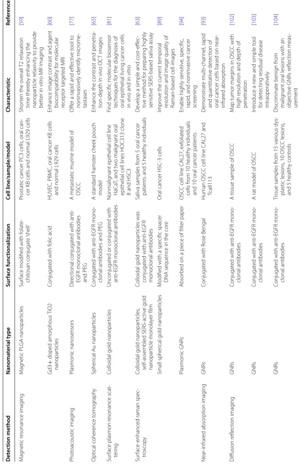
Nano-contrast agents have also been studied in oral cancers. For example, Asifkhan et al. combined the folate
preconjugated chitosan and magnetic poly (lactide-co
-glycolide) (PLGA) nanoparticles to create an MRI
con-trast agent (Fig. 2) [59]. The overall T2 relaxation time
was shortened, and the nanoparticle relaxivity was
enhanced thereby providing better imaging contrast [59].
Meanwhile, the folate receptor positive KB oral cancer cells showed increased nanoparticle uptake and caused
significant enhancement in cytotoxicity [59]. This nano
agent not only provided high contrast cancer imaging but also simultaneously provided cancer therapy. Another novel magnetic nano-contrast agent was developed based
on Gd3+ doped amorphous TiO2 and was suitable for T1
weighted MRI [60]. The size of this agent was reported
to be about 25 nm, which is much smaller than SPIO
(50 nm) [58]. The potential of inducing hemolysis,
plate-let aggregation, and plasma coagulation was studied, and
no adverse reaction was reported [60]. As a consequent,
the folic acid conjugated nanoparticles were specifically aggregated on the surface of folate receptor positive oral
cancer KB cells, leaving normal L929 cells unstained [60].
Notably, this nano-contrast agent showed enhanced lon-gitudinal relaxivity, magnetic resonance, and excellent biocompatibility for MRI.
Optical coherence tomography
Optical coherence tomography (OCT) is a direct simu-lation of ultrasound. It produces cross-sectional archi-tectural images of subsurface tissues, such as epithelial layers and basement membranes, using infrared light with a penetration depth of about 2 mm, and is suitable for early oral cancer detection and oral dysplasia
moni-toring [61]. The resolution of OCT is reported to be
around 10 μm which is higher than that of other non-invasive diagnostic techniques, such as CT, MRI, and
ultrasound [50, 62]. Although OCT is a non-invasive and
real-time clinical diagnostic method for cell and stromal morphology imaging, the contrast remains insufficient,
especially between neoplastic and normal tissues [63].
Gold nanoparticles are promising OCT contrast agents. They are biocompatible, easy to synthesize, and can provide localized surface plasmon resonances at near-infrared wavelengths that avoid predominant
absorption in tissues [64]. For example, the EGFR
mon-oclonal antibodies conjugated Au nanoparticles with a diameter of 71 nm have been applied to enhance the con-trast of OCT images of oral dysplasia in a hamster model
[65]. Meanwhile, microneedles and ultrasound were
uti-lized to overcome the obstacle for Au NP delivery. This multimodal delivery was demonstrated to be effective in improving OCT penetration depth and resulted in an approximately 150% increased contrast level in oral
car-cinogenesis [65].
Photoacoustic imaging
Photoacoustic imaging is a new emerging optical diagnostic technology. By using a short laser pulse, it
Fig. 2 Representation of the magnetic core–shell hybrid nanoparticles for receptor targeted MRI (Reprinted with permission from [59]. Copyright
generates ultrasound transients from tissues, thereby causing transient thermoelastic expansions after
opti-cal absorption [66–68]. These photoacoustic waves
are being then transformed into photoacoustic images according to their arrival times after collected by an
ultrasound transducer [69, 70]. The ultrasound
pro-vides high spatial resolution for structural phenotyping and is a useful tool for assessing lymph nodes
follow-ing a radical surgery [71, 72]. Consequently, the optical
contrast can be significantly improved while
maintain-ing the high spatial resolution of ultrasound [73].
Com-pared to conventional optical imaging, photoacoustic
imaging has improved imaging depth, about 6 cm [69].
Though various exogenous contrast agents-such as methylene blue, ICG, and GNs-have been used to enhance the photoacoustic imaging contrast, the gold nanoparticles are considered a more attractive contrast
agent due to their ability to conjugate biomolecules and their production of stronger photoacoustic imaging
signals [67, 69, 74]. To date, photoacoustic imaging has
demonstrated great potential in brain, breast, and
pros-tate cancer diagnosis [67, 73, 75, 76].
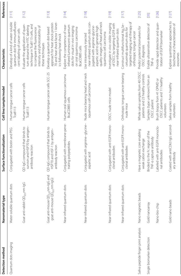
Luke et al. introduced ultrasound-guided spectro-scopic photoacoustic imaging technology for detecting lymph node micrometastases in a metastatic murine
model of OSCC (Fig. 3) [77]. Using anti-EGFR antibody
conjugated molecularly activated plasmonic nanosen-sors (MAPS), the study showed that the MAPS shifted their absorption spectrum to the near-infrared region
[77]. In addition, large ultrasound-guided spectroscopic
photoacoustic signals appeared in micrometastases as small as 50 mm within 30 min after MAPS injection
[77]. These findings offer an alternate to sentinel lymph
node biopsy analysis of oral cancer resection.
Fig. 3 Representation of the photoacoustic imaging using anti‑EGFR antibody conjugated molecularly activated MAPS. a A schematic of the
Surface plasmon resonance scattering
Surface plasmon waves are formed by collective
oscil-lation of conduction electrons in noble metals [78].
Recently, gold nanoparticles have been commonly applied for surface plasmon resonance scattering since they can resonantly scatter visible and near-infrared light
due to their surface plasmon oscillation [78]. In
addi-tion, they are easy to prepare, readily bioconjugated, and have low cytotoxicity, making them suitable for
biomo-lecular labeling and targeting [79]. It is reported that the
conjugated nanoparticles tended to aggregate together, inducing a greatly enhanced surface plasmon resonance
scattering compared to unconjugated nanoparticles [80].
El-Sayed et al. recorded surface plasmon resonance scattering images and surface plasmon resonance
absorption spectra after cell incubation [81].
Light-scat-tering images showed that the EGFR conjugated nano-particles bind specifically to the surface of the cancer cells with high concentration, while the binding to
non-cancerous cells was nonspecific and random [81]. Micro
absorption spectra showed that the absorption maxi-mum for conjugated nanoparticles was 545 nm, without aggregation tendency, while unconjugated colloidal gold nanoparticles accumulated inside cells and aggregated
with an absorption maximum around 552 nm [81]. As a
result, the anti-EGFR antibody conjugated nanoparticles showed 600% greater affinity to malignant oral epithelial cell lines HOC 313 clone 8 and HSC 3 than to the
non-malignant cell line HaCaT [81]. In addition, the surface
plasmon resonance property of gold nanoparticles was shown to have the ability to increase Raman scattering
in saliva samples of oral cancer patients [63, 78]. High
optical signals were produced by enhanced surface plas-mon resonance when the gold nanoparticles gathered around the target cancerous cells, due to their
conjuga-tion with anti-EGFR [63]. The sensitivity was observed to
be around 70% of the current technique, which needs to
be further improved [63].
Surface‑enhanced Raman spectroscopy
Raman spectroscopy is a vibrational spectroscopic tech-nique based on inelastic interactions between light and
matter [82]. The normal, premalignant, or malignant
lesions are distinguished by inelastic scattering of light, which can be a laser in the visible, near-infrared, or
near-ultraviolet range [83]. The signals in normal
tis-sues are homogeneous but heterogeneous in malignant cells, reflecting the changes in chemical
characteriza-tion and molecular structure of the lesions [84]. Raman
spectroscopy is a near-field effect and has a low penetra-tion depth. Its clinical applicapenetra-tion has been limited by the weak Raman signal intensity and the slow speed of
spec-trum acquisitions [78, 83].
Recently, nanoparticles have been applied as exog-enous contrast agents, in order to acquire Raman signal
with high speed and resolution [85–87]. After directly
adsorbed on the nanoparticle surface, the molecules emit an amplified Raman scattering intensity, known as
sur-face-enhanced Raman scattering (SERS) [83, 88]. A study
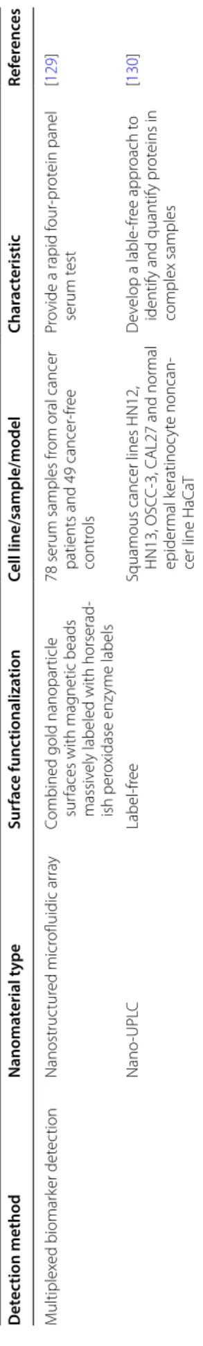
introduced small, spherical, near-infrared region sensi-tive and SERS acsensi-tive gold nanoparticles with highly nar-row intra-nanogap structures for single oral cancer cell
HSC-3 imaging (Fig. 4) [89]. The gold nanoparticles can
selectively target intracellular organelles and were specif-ically distributed in cytoplasm, mitochondria, and nuclei. Finally, high speed Raman imaging was achieved within
30 s with a high resolution of 50 × 50 pixels [89].
Nanospheres, nanorods, nanocubes, nanobranches, and nanobipyramids are different shapes of gold
nano-particles [90, 91]. Gold nanorods (GNRs) have received
much attention for molecular imaging because of their advantage of higher index sensitivity over spherical and cubic gold nanoparticles, which means minor changes in the surrounding environment of GNRs can result in sig-nificant longitudinal surface plasmon resonance (LSPR)
peak wavelength variation [90, 92]. Since the index
sensitivities and longitudinal plasmon wavelengths of nanorods increase with aspect ratios, the use of nanorods with large aspect ratios can provide near-infrared region plasmon wavelengths and high index sensitivity for
opti-cal techniques [90, 91].
Wang et al. conjugated GNRs with rose bengal (RB), a specific probe for oral cancer cell target, and
moni-tored optical absorption in the near-infrared region [93].
The RB molecules have the ability to bind with the pro-tein or nucleic acid of cancer cell lysate, whereafter the RB-GNR probes aggregated, inducing red-shift in the
near-infrared absorption wavelength [93]. This RB-GNR
platform provided a specific and quantitative method for oral cancer cell lysate analysis with a detection sensitivity
of 2000 cells/ml [93]. Liu et al. described a paper-based
SERS technology in combination with exfoliative cytol-ogy for screening of exfoliated cells from oral cancer
patients and healthy individuals [94]. Cells were placed
on a plasmonic paper with GNRs adsorbed on it, and spectra were acquired afterward. Sensitivity and speci-ficity were both 100% for distinguishing exfoliated cells
from normal and cancer tissues, based on the I1600/1440
and I1440/1340 peak ratios of the spectra values [94]. This
paper-based SERS platform has overcome the drawbacks of traditional exfoliative cytology, such as low sensitivity
and subjective cytologic interpretation [94].
Diffusion reflection imaging
while the rest undergoes multiple elastic scattering and
gets diffusely reflected [95]. The reflected light is greatly
affected by cytologic and morphologic changes during epithelial tissue cancerization, including nuclear size, col-lagen content, extracellular matrix structure, epithelial
thickness, and blood flow variation [28, 96]. It is reported
that recording diffuse reflectance images can help to determine surgical margins and is a useful tool to
differ-entiate normal mucosa, OPMD, and oral cancer [96–98].
In oral cancer, 14.3% of tumor margins after surgical
excision were identified to have residual carcinoma [99].
Accurate determination of tumor margins is critical for
complete surgical resection of residual diseases in oral
cancer and may reduce the high rate of recurrence [100].
The accuracy of routine microscopic examination after frozen sections is limited by the 30.7–47.3% shrinkage
of the frozen tissues [101]. Meanwhile, for the
paraffin-embedded tissue section, results are only available after the operation, making the intraoperative
identifica-tion challenging [101]. Thus, efforts should be made to
achieve a real-time and high sensitive way for more com-plete tumor resections.
Ankri et al. conjugated GNRs to monoclonal antibod-ies against EGFR and evaluated the margins of human
Fig. 4 Graphical representation of the SERS active gold nanoparticles for oral cancer cell HSC‑3 imaging. a synthetic scheme of Raman dye
(44DP)‑coded Au‑NNPs using four different kinds of DNA‑AuNPs as core particles. b the solution color and HR‑TEM image of 44DP‑coded Au‑NNPs.
OSCC specimens by diffusion reflection imaging [102]. Air scanning electron microscopy was used to visualize the nanorods in tissues, showing the GNRs-EGFR spread a distance of 1 mm between the tumor and the healthy regions. Diffusion reflection imaging was then performed in a resolution of 1 mm, suggesting that the tumor edge is in the region of 4–5 mm, which is consistent with the commonly used cutoff of 5 mm for a close margin
[100]. This study group has also tested diffusion
reflec-tion imaging of GNRs-EGFR on a mice OSCC model
induced by 4-nitroquinoline-N-oxide [103]. GNRs
specif-ically attached to areas histologspecif-ically identified as OSCC, with high reflectance at 780 nm over 17 intensity units. The overall specificity and sensitivity was 97 and 87%,
respectively [103]. Moreover, the reflectance spectrum
at 780 nm was found to be moderate in areas of carci-noma in situ, but absent in normal epithelium. The opti-cal properties showed significant changes-more than 80% of the invasive cancer and more than 30% of carcinoma
in situ [103]. The group has also found that this
modal-ity is suitable for discriminating benign from malignant oral lesions since the reflectance intensity increased as
the dysplastic changes increased [104]. Thus, the group
has demonstrated that diffusion reflection imaging is a promising technique for the screening of malignant oral lesions and detecting residual disease during operation.
Quantum dots imaging
Quantum dots are nanometer-sized semiconductor crys-tals that luminesce through quantum confinement effects
[105, 106]. Quantum dots have several advantages that
could overcome the limitations of conventional fluores-cent dyes, such as size-tunable emission, wide excita-tion spectra, strong luminescence and excellent stability
against photobleaching [106–108]. In addition,
chang-ing the size and composition of quantum dots allows for obtaining a wide range of spectrum, from ultraviolet to
the near infrared [109, 110].
Currently, quantum dots have been applied in the molecular and cell imaging of OSCC both in vitro and in vivo. It has been demonstrated that quantum dots have high fluorescence intensity, low nonspecific binding, and good stability against photobleaching for the in vitro imaging of human oral cancer cells Tca8113, SCC-25 and
BcaCD885 [111–114]. Most of the quantum dots used for
in vivo imaging were linked to molecules with the ability
to target cancer cells [115]. Recently, it was reported that
the near-infrared quantum dots with an emission wave-lengths range of 700–900 nm have strong tissue
penetra-tion and are not harmful in vivo [114, 115]. Meanwhile,
quantum dots with emission wavelengths between 400 and 600 nm are able to avoid the interference of tissue autofluorescence, making them suitable for bioimaging
[116, 117]. Studies have proven that quantum dots with
an emission wavelength of 800 nm conjugated with EGFR monoclonal antibodies or arginine–glycine–aspartic acid sequence can generate high quality images of OSCC
(Fig. 5) [117–119]. The technique also offers great
poten-tial in personalized therapy for OSCC [117–119].
Nano‑based ultrasensitive biomarker detection
Currently, plenty of novel proteomic, genomic, and tran-scriptomic biomarkers are being researched. Exploration of tumor molecular biomarkers-such as tumor necrosis factor-alpha (TNF-α), vascular endothelial growth fac-tor (VEGF), EGFR, and interleukin 6 (IL 6)-holds great
promise for early cancer detection and diagnosis [22,
120, 121]. Routine measurement methods-including
enzyme-linked immunosorbent assay (ELISA), immu-nohistochemistry, Western Blot, and polymerase chain reaction-still bear a limited detection sensitivity ranging
from pM to fM (10−12 to 10−15 M) concentration
lev-els [22, 23, 35]. The application of nanotechnology may
enhance the detection sensitivity for biomarkers with low
concentrations in the tissue samples or body fluids [122,
123].
The saliva peptide finger print technique is a useful tool for salivary proteomics analysis and can predict potential
biomarkers valuable for cancer diagnosis [124]. A study
utilized matrix-assisted laser-desorption ionization-time-of-flight mass spectrometry (MALDI-TOF–MS) for ana-lyzing the expression spectrum of salivary peptides in 40
OSCC patients and 23 normal controls [125].
Nanomate-rial-based magnetic beads were used for selective enrich-ment of low-molecular-mass peptides. It is noteworthy that 50 proteins expression levels were significantly dif-ferent between OSCC patients and healthy controls. As a result, the mass peaks of 1285.6 and 1432.2 Da, which were both identified as histatin-3, were correlated with OSCC progression. This study introduced a novel high-throughput, non-invasive strategy for valuable oral
cancer biomarkers screening [125]. The specific
advan-tages of magnetic beads constructed on nanomaterial over other types of separation beads have not yet been illustrated.
A nano-based single biomarker detection method has also been utilized for oral cancer detection. A study detected TNF-α by gold protein chip method using a total internal reflection fluorescence
micros-copy (TIRFM) [35]. A 4 × 5 nanoarray incorporating
500 nm diameter gold spots was achieved on 10 mm square glass substrates. The TNF-α detection sensitiv-ity was reported to be at the attomolar (aM)
concen-tration level (× 10−18), enabling ultra-sensitive oral
cancer detection [35]. However, this method could
study described the analysis of oral cancer bio marker EGFR with exfoliative cytology specimens of 41 OPMD or OSCC patients and 11 healthy volunteers, using
a nano-bio-chip sensor technique [126]. A total of 51
measurement parameters were collected, and biochem-ical and morphologic changes were further analyzed. The EGFR expression level-along with nuclear area, nuclear diameter, and nuclear-to-cytoplasmic ratio-was significantly altered in oral lesions with diagnosed
squamous cell carcinoma or dysplasia [126]. Using
ultra-sensitive atomic force microscopy (AFM) and field emission scanning electron microscopy (FESEM) with high resolution (~ 1 nm), another study exhibited the substructure of single human saliva exosomes and interpreted the nanoscale structures of exosomes under varying forces, revealing reversible mechanical
defor-mation [127]. Further, cell-type specific marker CD63
was detected by using 10 nm gold beads on individual exosomes. The nanoscale biomechanical, morpho-logical, and surface biomolecular properties of saliva exosomes are found to be critical for the oral cancer
diagnosis [127]. Although these two systems have made
it possible for the quantitative analysis of cellular bio-markers, the systems described above can only be used for single biomarker analysis.
It is well-known that single oral cancer biomarkers
cannot provide reliable diagnoses [128]. Multiplexed
biomarker detection can minimize false positives and
negatives arising from single biomarker analysis [128].
A multiplexed biomarker detection approach meas-ured a four-protein panel of biomarkers using an
ultra-sensitive electrochemical microfluidic array [129]. The
microfluidic device contained an array of nanostructured sensors, and plenty of magnetic beads were labeled. The four-protein panel-including interleukin-6, interleu-kin-8, VEGF, and VEGF-C-was analyzed in 78 oral can-cer patient serum samples and 49 controls, and showed a clinical diagnostic sensitivity and specificity for 89
and 98%, respectively [129]. The study provided a
low-cost, easily fabricated method for accurate clinical oral cancer diagnosis. Another study analyzed proteins bio-markers in conditioned media of oral squamous cell lines HN12, HN13, OSCC-3 and CAL27 by utilizing a nano ultra-performance liquid chromatography
(nano-UPLC) ion-mobility mass spectrometry [130]. A total of
approximately 952 proteins-including known cancer bio-marker proteins IL-6, IL-8, VEGF-A, and VEGF-C were identified. This nano-UPLC-Q-TOF assay provided a high-throughput approach to quantify proteins and com-pare protein expression levels across different samples,
Fig. 5 Schematic illustration of surface modification, bioconjugation, and theranostic application of Ag2Se QDs coupled with cetuximab (Reprinted
Table
1
Summar
y of nanot
echnolo
gy based metho
ds f or or al c anc er det ec tion and diagnosis Det ec tion method Nanoma terial t ype Sur fac e func tionaliza tion Cell line/sample/model Char ac teristic Ref er enc es M ag netic r esonance imag ing M ag netic PL GA nanopar ticles Sur
face modified with f
olat e‑ chit osan conjugat e ‘ shell ’ Pr
ostatic cancer PC3 cells
, oral can
‑
cer KB cells and nor
mal L929 cells
Shor
ten the o
verall
T2 r
elaxation
time ther
eb
y enhancing the
nanopar ticle r elaxivit y t o pr ovide bett er in vitr
o MR imag
ing [ 59 ] G d3 + doped amor phous TiO2 nanopar ticles Conjugat
ed with f
olic acid
HUVEC, PBMC, oral cancer KB cells and nor
mal L929 cells
Enhance image contrast and agent biocompatibilit
y f or molecular recept or tar get ed MRI [ 60 ] Phot oacoustic imag ing Plasmonic nanosensors Dir ec tional conjugat
ed with anti
‑
EGFR monoclonal antibodies and PEG
A metastatic mur
ine model of
OSC
C
O
ffer a rapid and eff
ec tiv e t ool t o nonin vasiv
ely identify micr
ome ‑ tastases [ 77 ] Optical coher ence t omog raph y Spher ical A u nanopar ticles Conjugat
ed with anti
‑EGFR mono
‑
clonal antibodies and PEG
A standar
d hamst
er cheek pouch
model
Enhance the contrast and penetra
‑
tion depth in
viv o OC T images [ 65 ] Sur
face plasmon r
esonance scat
‑
ter
ing
Colloidal gold nanopar
ticles
Unconjugat
ed or conjugat
ed with
anti
‑EGFR monoclonal antibodies
Nonmalig
nant epithelial cell line
HaC
aT
, and t
w
o malig
nant oral
epithelial cell lines HOC313 clone 8 and HSC3 Find specific molecular biosensor techniques f
or the diag
nosis of
oral epithelial living cancer cells in viv
o and in
vitr o [ 81 ] Sur face
‑enhanced raman spec
‑
tr
oscop
y
Colloidal gold nanopar
ticles , self ‑assembled SERS ‑ac tiv e gold nanopar ticle monola yer film
Colloidal gold nanopar
ticles was
conjugat
ed with anti
‑EGFR
monoclonal antibodies
Saliva samples fr
om 5 oral cancer
patients and 5 health
y individuals
D
ev
elop a simple and cost
‑eff
ec
‑
tiv
e method f
or pr
epar
ing highly
sensitiv
e SERS
‑based saliva assa
y
[
63
]
Small spher
ical gold nanopar
ticles
M
odified with a specific spacer DNA sequence in the cor
e
Oral cancer HSC
‑3 cells
Impr
ov
e the cur
rent t
emporal
resolution and image qualit
y of
Raman
‑based cell images
[
89
]
Plasmonic GNRs
Absor
bed on a piece of filt
er paper
OSC
C cell line CAL27, exf
oliat
ed
cells fr
om 10 health
y individuals
and 10 oral cancer patients
Enable highly sensitiv
e, specific
,
rapid
, and nonin
vasiv e cancer scr eening [ 94 ] Near ‑infrar ed absor ption imag ing GNRs Conjugat
ed with R
ose Bengal
Human OSC
C cell line CAL27 and
Tca8113 D emonstrat e multi ‑channel , rapid and quantitativ e det ec tion of
oral cancer cells based on near
‑ infrar ed absor ption [ 93 ] Diffusion r eflec tion imag ing GNRs Conjugat
ed with anti
‑EGFR mono
‑
clonal antibodies
A tissue sample of OSC
C
M
ap tumor mar
gins in OSC
C with
high r
esolution and depth of
penetration [ 102 ] GNRs Conjugat
ed with anti
‑EGFR mono
‑
clonal antibodies
A rat model of OSC
C
Intr
oduce a ne
w and simple t
ool for det ec ting r esidual disease intraoperativ ely [ 103 ] GNRs Conjugat
ed with anti
‑EGFR mono
‑
clonal antibodies
Tissue samples fr
om 15 var
ious dys
‑
plastic lesions
, 10 OSC
C lesions
,
and 5 health
y contr ols Discr iminat e benig n fr om malig
nant oral lesions with an
objec
tiv
e GNRs r
Table 1 (c on tinued) Det ec tion method Nanoma terial t ype Sur fac e func tionaliza tion Cell line/sample/model Char ac teristic Ref er enc es
Quantum dots imag
ing
W
at
er
‑soluble quantum dots
Conjugat
ed with biotin and PEG
Human t
ongue cancer cells
Tca8113
D
ev
elop of a k
ind of wat
er
‑soluble
quantum dot f
or immunofluor
es
‑
cent labeling of cancer cells
[ 111 ] G oat anti ‑rabbit QD 655 nm ‑IgG QD
‑IgG compound that binds t
o
sur
vivin and HSP70 b
y antigen–
antibody r
eac
tion
Human t
ongue cancer cells
Tca8113
Evaluat
e the application of quan
‑
tum dotsand the FIT
C labeling
technique in
Tca8113 cells
, and
to compar
e the fluor
escence
int
ensit
y and phot
ostabilit y of these t echniques [ 112 ] G oat anti ‑mouse QD 525 nm ‑IgG and goat anti ‑mouse QD 655 nm ‑IgG) QD
‑IgG compound that binds t
o
HSP70 and HSF
‑1 b y antigen– antibody r eac tion Human t
ongue cancer cells SC
C
‑25
D
ev
elop a quantum dot
‑based
appr
oach f
or heat shock pr
ot
ein
70 and heat shock fac
tor 1 k
inet
‑
ics f
ollo
wing heat shock
[
113
]
Near
‑infrar
ed quantum dots
Conjugat
ed with membrane
‑pene
‑
trating polypeptides
Human oral squamous car
cinoma
BcaCD885 cells
Explor
e the compet
ence of near
‑
infrar
ed luminescent quantum
dots f
or visual in
viv
o imag
ing
on oral squamous car
cinoma BcaCD885 cells [ 114 ] Near ‑infrar
ed quantum dots
Conjugat
ed with ar
ginine –gly cine – aspar tic acid
Nude mice bear
ing head and neck
squamous cell car
cinoma Use intra venously injec ted near ‑infrar
ed quantum dots con
‑
jugat
ed with ar
ginine
‑gly
cine
‑
aspar
tic acid t
o generat
e high
qualit
y images of head and neck
squamous cell car
cinoma [ 118 ] Near ‑infrar
ed quantum dots
Conjugat
ed with anti
‑EGFR mono
‑
clonal antibodies
OSC
C nude mice model
In
vestigat
e in
viv
o visible imag
ing
of OSC
C b
y tar
geting EGFR with
near
‑infrar
ed quantum dots
[
119
]
Near
‑infrar
ed quantum dots
Conjugat
ed with anti
‑EGFR mono ‑ clonal antibodies Or thot opic t ongue cancer ‑bear ing nude mice Construc t multifunc tional A g2 Se –
cetuximab quantum dots f
or
tar
get
ed imag
ing and therap
y of or thot opic t ongue cancer [ 117 ]
Saliva peptide finger pr
int analysis
Nano mag
netic beads
Ha
ve a mag
netic cor
e enabling
w
eak cation ex
change
Whole saliva samples fr
om 40 OSC
C
patients and 23 health
y contr ols Pr edic t pot ential biomar kers f or OSC C diag nosis [ 125 ] Single biomar ker det ec tion G old nanoar ra y Binded t
o the F
c r
eg
ion of the
TNF ‑α captur e antibody Samples (t ype unk no wn) fr om an OSC C patient Enable ultrasensitiv e det ec tion of TNF ‑α [ 35 ] Nano ‑bio ‑chip
Labeled with anti
‑EGFR monoclo
‑
nal antibodies
Brush biopsy fr
om 41 OP
MD or
OSC
C patients and 11 health
y
volunt
eers
Pr
ovide rapid det
ec
tion and quan
‑
titation of EGFR biomar
ker
[
126
]
G
old nano beads
Coat
ed with antiCD63 IgG second
‑
ar
y antibody
Saliva samples fr
Table
1
(c
on
tinued)
Det
ec
tion method
Nanoma
terial t
ype
Sur
fac
e func
tionaliza
tion
Cell line/sample/model
Char
ac
teristic
Ref
er
enc
es
M
ultiplex
ed biomar
ker det
ec
tion
Nanostruc
tur
ed micr
ofluidic ar
ra
y
Combined gold nanopar
ticle
sur
faces with mag
netic beads
massiv
ely labeled with horserad
‑
ish per
oxidase enz
yme labels
78 serum samples fr
om oral cancer
patients and 49 cancer
‑fr
ee
contr
ols
Pr
ovide a rapid f
our
‑pr
ot
ein panel
serum t
est
[
129
]
Nano
‑UPL
C
Label
‑fr
ee
Squamous cancer lines HN12, HN13, OSC
C
‑3, CAL27 and nor
mal
epider
mal k
eratinoc
yt
e noncan
‑
cer line HaC
aT
D
ev
elop a lable
‑fr
ee appr
oach t
o
identify and quantify pr
ot
eins in
complex samples
[
130
]
PLG
A
poly lac
tide
-c
o-gly
colide
,
O
SCC
or
al squamous c
ell car
cinoma,
TNF
-α
tumor necr
osis fac
tor
-alpha,
EGFR
epider
mal g
ro
wth fac
tor r
ec
ept
or
,
PEG
poly
eth
ylene gly
col
,
UPL
C
ultr
a-per
for
manc
e liquid chr
oma
tog
raph
y,
GNRs
gold nanor
ods
,
HUVEC
human pr
imar
y endothelial c
ells
,
PBM
C
per
ipher
al blood mononuclear c
ells
,
OCT
optical c
oher
enc
e t
omog
raph
without the need for stable isotope labeling. The identifi-cation of peptides was unlimited with the fragmentation
technique [130].
Conclusion and perspective
Ranking as one of the top 10 cancers worldwide, oral can-cer has a poor prognosis and a high recurrence rate, and the time and accuracy of diagnosis directly affects disease
outcomes [131]. In the past few decades,
nanotechnol-ogy has brought new techniques to cancer diagnosis [36,
38, 132, 133]. The performance parameters of
nanopar-ticles-such as biocompatibility, function-specific size and shape, blood circulation half-life, and targeting to specific cell surface molecules-can be controlled by modulating their fabrication materials, methods or surface chemistry, making nanoparticles a promising diagnostic material
[79]. The present review article has critically introduced
nano-based detection strategies for oral cancers, and
summarized various kinds of nanomaterials, sample
types, and the characteristic of each technique in Table 1.
The pros and cons of each nanotechnology for bioimag-ing and molecular detection of oral cancer were shown in
Fig. 6. In the oral cavity, the use of nanoparticles has not
only achieved noninvasive real-time diagnosis with high sensitivity and specificity but also assisted with accurately identifying surgical margins, indicating the potential to reduce the reliance on tissue biopsy and histopathologi-cal assessment in many cases.
Nano-based contrast agents for MRI, OCT and pho-toacoustic imaging have lower toxicity, prolonged blood circulation half-life, and the ability to target unique cell surface molecules. Compared to routine contrast agents, nano agents exhibit better image contrast properties and improved penetration depth. In optical imaging, nanoparticles enable sufficient signals and sub-cellular spatial resolution. They can generate surface plasmon resonance at near-infrared wavelengths, gathered around
the targeted cell surface, and the optical resonance prop-erties of nanorods can be regulated over a broad range by adjusting their sizes and shapes. Quantum dots with size-tunable emission, wide excitation spectrum, high intensity of luminescence, and excellent photochemical stability have overcome the disadvantages of traditional fluorescence markers. As for cancer biomarker detection, nano-based materials-such as nano beads, gold nanoar-ray, and nano-bio-chips-offer high throughput screen-ing for potential biomarkers and have brought the level of detection sensitivity to the nanoscale. Therefore, the small and earlier intraepithelial lesions missed by com-mon techniques can potentially be detected by nanotech-nologies, making oral diseases more readily cured.
Nano-based diagnostic methods act as a promising tool to provide real-time, convenient, and cost-effec-tive diagnosis for oral cancer detection and diagnosis. They can provide molecular targeted imaging, analyze biomarkers at nano-scale, enable intraoperative identi-fication of surgical resection margins, and monitor oral cancer prognosis after treatment. Although these tech-nologies have been studied in ex vivo studies of tissue and saliva samples and in vivo studies in animal models, further efforts should be employed before these strate-gies can be successfully applied in clinical diagnosis.
Abbreviations
OPMD: oral potentially malignant disorders; OSCC: oral squamous cell carcino‑ mas; TB: toluidine blue; EGFR: epidermal growth factor receptor; MRI: magnetic resonance imaging; CT: computed tomography; CBCT: cone beam computed tomography; PET: positron emission tomography; Gd‑DTPA: Gd3+ complexed with diethyltriamine‑pentaacetic acid; Gd‑DOTA: tetra azacyclododecane‑ 1,4,7,10‑tetraacetic acid; SPIO: superparamagnetic iron oxide; USPIO: ultrasmall superparamagnetic iron oxide; MAPS: molecularly activated plasmonic nanosensors; SERS: surface‑enhanced Raman scattering; GNRs: gold nanorods; LSPR: longitudinal surface plasmon resonance; RB: rose bengal; TNF‑α: tumor necrosis factor‑alpha; VEGF: vascular endothelial growth factor; IL 6: interleukin 6; ELISA: en‑zyme‑linked immunosorbent assay; MALDI‑TOF–MS: matrix‑ assisted laser‑desorption ionization‑time‑of‑flight mass spectrometry; TIRFM: total internal reflection fluorescence microscopy; AFM: atomic force micros‑ copy; FESEM: field emission scanning electron microscopy; PEG: polyethylene glycol; UPLC: ultra‑performance liquid chromatography; PLGA: poly lactide‑co‑ glycolide; HUVEC: human primary endothelial cells; PBMC: peripheral blood mononuclear cells.
Authors’ contributions
XJC designed the review and drafted the manuscript. XQZ, QL, JZ and GZ helped with the manuscript editing. GZ reviewed the manuscript drafts. All authors read and approved the final manuscript.
Author details
1 The State Key Laboratory Breeding Base of Basic Science of Stomatology (Hubei‑MOST) and Key Laboratory of Oral Biomedicine Ministry of Education, School and Hospital of Stomatology, Wuhan University, Wuhan 430079, Peo‑ ple’s Republic of China. 2 School of Chemistry, Chemical Engineering and Life Sciences, Wuhan University of Technology, Wuhan 430070, People’s Republic of China. 3 Division of Pharmacoengineering and Molecular Pharmaceutics and Center for Nanotechnology in Drug Delivery, Eshelman School of Phar‑ macy, University of North Carolina at Chapel Hill, Chapel Hill, NC 27599, USA. 4 Department of Oral Medicine, School and Hospital of Stomatology, Wuhan University, Wuhan 430079, People’s Republic of China.
Acknowledgements
This work was supported by Grants from National Natural Science Foundation of China (No. 81771080, No. 81371147) to Professor Zhou Gang.
Competing interests
The authors declare that they have no competing interests.
Availability of data and materials
Not applicable.
Consent for publication
Not applicable.
Ethics approval and consent to participate
Not applicable.
Funding
Not applicable.
Publisher’s Note
Springer Nature remains neutral with regard to jurisdictional claims in pub‑ lished maps and institutional affiliations.
Received: 6 March 2018 Accepted: 1 June 2018
References
1. Siegel RL, Miller KD, Jemal A. Cancer statistics, 2016. CA Cancer J Clin. 2016;66:7–30.
2. Calixto G, Bernegossi J, Fonseca‑Santos B, Chorilli M. Nanotechnology‑ based drug delivery systems for treatment of oral cancer: a review. Int J Nanomed. 2014;9:3719–35.
3. Torre LA, Bray F, Siegel RL, Ferlay J, Lortet‑Tieulent J, Jemal A. Global cancer statistics, 2012. CA Cancer J Clin. 2015;65:87–108.
4. Tanaka T, Ishigamori R. Understanding carcinogenesis for fighting oral cancer. J Oncol. 2011;2011:603740.
5. Warnakulasuriya S. Global epidemiology of oral and oropharyngeal cancer. Oral Oncol. 2009;45:309–16.
6. Ng JH, Iyer NG, Tan MH, Edgren G. Changing epidemiology of oral squamous cell carcinoma of the tongue: a global study. Head Neck. 2017;39:297–304.
7. Jemal A, Bray F, Center MM, Ferlay J, Ward E, Forman D. Global cancer statistics. CA Cancer J Clin. 2011;61:69–90.
8. Arakeri G, Patil SG, Aljabab AS, Lin KC, Merkx MAW, Gao S, et al. Oral submucous fibrosis: an update on pathophysiology of malignant transformation. J Oral Pathol Med. 2017;46:413–7.
9. Khan Z, Khan S, Christianson L, Rehman S, Ekwunife O, Samkange‑ Zeeb F. Smokeless tobacco and oral potentially malignant disorders in south asia: a systematic review and meta‑analysis. Nicotine Tob Res. 2017;20:12–21.
10. Benergossi J, Calixto G, Fonseca‑Santos B, Aida KL, de Cassia Negrini T, Duque C, et al. Highlights in peptide nanoparticle carriers intended to oral diseases. Curr Top Med Chem. 2015;15:345–55.
11. Liu D, Zhao X, Zeng X, Dan H, Chen Q. Non‑invasive techniques for detection and diagnosis of oral potentially malignant disorders. Tohoku J Exp Med. 2016;238:165–77.
12. Mercadante V, Paderni C, Campisi G. Novel non‑invasive adjunctive techniques for early oral cancer diagnosis and oral lesions examina‑ tion. Curr Pharm Des. 2012;18:5442–51.
13. Wikner J, Grobe A, Pantel K, Riethdorf S. Squamous cell carcinoma of the oral cavity and circulating tumour cells. World J Clin Oncol. 2014;5:114–24.
14. Brennan JA, Mao L, Hruban RH, Boyle JO, Eby YJ, Koch WM, et al. Molecular assessment of histopathological staging in squamous‑cell carcinoma of the head and neck. N Engl J Med. 1995;332:429–35. 15. Awan KH, Morgan PR, Warnakulasuriya S. Assessing the accuracy of
tools for oral potentially malignant disorders—a clinicopathological evaluation. Clin Oral Investig. 2015;19:2267–72.
16. Chainani‑Wu N, Madden E, Cox D, Sroussi H, Epstein J, Silverman S Jr. Toluidine blue aids in detection of dysplasia and carcinoma in suspi‑ cious oral lesions. Oral Dis. 2015;21:879–85.
17. Balasubramaniam AM, Sriraman R, Sindhuja P, Mohideen K, Parameswar RA, Muhamed Haris KT. Autofluorescence based diagnostic techniques for oral cancer. J Pharm Bioallied Sci. 2015;7:S374–7.
18. Kammerer PW, Rahimi‑Nedjat RK, Ziebart T, Bemsch A, Walter C, Al‑ Nawas B, et al. A chemiluminescent light system in combination with toluidine blue to assess suspicious oral lesions‑clinical evaluation and review of the literature. Clin Oral Investig. 2015;19:459–66.
19. Giovannacci I, Vescovi P, Manfredi M, Meleti M. Non‑invasive visual tools for diagnosis of oral cancer and dysplasia: a systematic review. Med Oral Patol Oral Cir Bucal. 2016;21:e305–15.
20. Ye X, Zhang J, Tan Y, Chen G, Zhou G. Meta‑analysis of two computer‑ assisted screening methods for diagnosing oral precancer and cancer. Oral Oncol. 2015;51:966–75.
21. Sekine J, Nakatani E, Hideshima K, Iwahashi T, Sasaki H. Diagnostic accu‑ racy of oral cancer cytology in a pilot study. Diagn Pathol. 2017;12:27. 22. Fernandez‑Olavarria A, Mosquera‑Perez R, Diaz‑Sanchez RM, Serrera‑
Figallo MA, Gutierrez‑Perez JL, Torres‑Lagares D. The role of serum biomarkers in the diagnosis and prognosis of oral cancer: a systematic review. J Clin Exp Dent. 2016;8:e184–93.
23. Sannam Khan R, Khurshid Z, Akhbar S, Faraz Moin S. Advances of salivary proteomics in oral squamous cell carcinoma (OSCC) detection: an update. Proteomes. 2016;4:E41.
24. Keshavarzi M, Darijani M, Momeni F, Moradi P, Ebrahimnejad H, Masoudifar A, et al. Molecular imaging and oral cancer diagnosis and therapy. J Cell Biochem. 2017;118:3055–60.
25. Sarrion Perez MG, Bagan JV, Jimenez Y, Margaix M, Marzal C. Utility of imaging techniques in the diagnosis of oral cancer. J Craniomaxillofac Surg. 2015;43:1880–94.
26. Mian SA, Yorucu C, Ullah MS, Rehman IU, Colley HE. Raman spectros‑ copy can discriminate between normal, dysplastic and cancerous oral mucosa: a tissue‑engineering approach. J Tissue Eng Regen Med. 2016;11:3253–62.
27. Green B, Cobb AR, Brennan PA, Hopper C. Optical diagnostic techniques for use in lesions of the head and neck: review of the latest develop‑ ments. Br J Oral Maxillofac Surg. 2014;52:675–80.
28. Stephen MM, Jayanthi JL, Unni NG, Kolady PE, Beena VT, Jeemon P, et al. Diagnostic accuracy of diffuse reflectance imaging for early detection of pre‑malignant and malignant changes in the oral cavity: a feasibility study. BMC Cancer. 2013;13:278.
29. Piazza C, Del Bon F, Paderno A, Grazioli P, Perotti P, Barbieri D, et al. The diagnostic value of narrow band imaging in different oral and oropharyngeal subsites. Eur Arch Otorhinolaryngol. 2016;273:3347–53. 30. Lucchese A, Gentile E, Romano A, Maio C, Laino L, Serpico R. The poten‑
tial role of in vivo reflectance confocal microscopy for evaluating oral cavity lesions: a systematic review. J Oral Pathol Med. 2016;45:723–9. 31. Gentile E, Maio C, Romano A, Laino L, Lucchese A. The potential role of
in vivo optical coherence tomography for evaluating oral soft tissue: a systematic review. J Oral Pathol Med. 2017;46:864–76.
32. Sokolov K, Aaron J, Hsu B, Nida D, Gillenwater A, Follen M, et al. Optical systems for in vivo molecular imaging of cancer. Technol Cancer Res Treat. 2003;2:491–504.
33. Omar E. Future imaging alternatives: the clinical non‑invasive modali‑ ties in diagnosis of oral squamous cell carcinoma (OSCC). Open Dent J. 2015;9:311–8.
34. Liu Y, Li Y, Fu Y, Liu T, Liu X, Zhang X, et al. Quantitative prediction of oral cancer risk in patients with oral leukoplakia. Oncotarget. 2017;118(10):3055–60.
35. Lee K, Lee S, Yu H, Kang SH. Ultra‑sensitive detection of tumor necrosis factor‑alpha on gold nano‑patterned protein chip formed via E‑beam nanolithography by total internal reflection fluorescence microscopy. J Nanosci Nanotechnol. 2010;10:3228–31.
36. Sharma P, Brown S, Walter G, Santra S, Moudgil B. Nanoparticles for bioimaging. Adv Colloid Interface Sci. 2006;123–126:471–85. 37. Ogle OE, Byles N. Nanotechnology in dentistry today. West Indian Med
J. 2014;63:344–8.
38. Jaishree V, Gupta PD. Nanotechnology: a revolution in cancer diagnosis. Indian J Clin Biochem. 2012;27:214–20.
39. Ho D, Wang CH, Chow EK. Nanodiamonds: the intersection of nano‑ technology, drug development, and personalized medicine. Sci Adv. 2015;1:e1500439.
40. Wang ZQ, Liu K, Huo ZJ, Li XC, Wang M, Liu P, et al. A cell‑targeted chemotherapeutic nanomedicine strategy for oral squamous cell carcinoma therapy. J Nanobiotechnol. 2015;13:63.
41. Gharat SA, Momin M, Bhavsar C. Oral squamous cell carcinoma: current treatment strategies and nanotechnology‑based approaches for pre‑ vention and therapy. Crit Rev Ther Drug Carrier Syst. 2016;33:363–400. 42. Bao C, Conde J, Curtin J, Artzi N, Tian F, Cui D. Bioresponsive antisense DNA gold nanobeacons as a hybrid in vivo theranostics platform for the inhibition of cancer cells and metastasis. Sci Rep. 2015;5:12297. 43. Han Y, An Y, Jia G, Wang X, He C, Ding Y, et al. Theranostic micelles
based on upconversion nanoparticles for dual‑modality imaging and photodynamic therapy in hepatocellular carcinoma. Nanoscale. 2018;10:6511–23.
44. Halo TL, McMahon KM, Angeloni NL, Xu Y, Wang W, Chinen AB, et al. NanoFlares for the detection, isolation, and culture of live tumor cells from human blood. Proc Natl Acad Sci USA. 2014;111:17104–9. 45. Zdobnova TA, Lebedenko EN, Deyev Scapital Em C. Quantum dots for
molecular diagnostics of tumors. Acta Nat. 2011;3:29–47.
46. Jaiswal JK, Mattoussi H, Mauro JM, Simon SM. Long‑term multiple color imaging of live cells using quantum dot bioconjugates. Nat Biotechnol. 2003;21:47–51.
47. Lee MH, Lee DH, Jung SW, Lee KN, Park YS, Seong WK. Measurements of serum C‑reactive protein levels in patients with gastric cancer and quantification using silicon nanowire arrays. Nanomedicine. 2010;6:78–83.
48. Adarsh N, Ramya AN, Maiti KK, Ramaiah D. Unveiling NIR aza–boron– dipyrromethene (BODIPY) dyes as Raman probes: surface‑enhanced raman scattering (SERS)‑guided selective detection and imaging of human cancer cells. Chemistry. 2017;23:14286–91.
49. Gonda K, Watanabe M, Tada H, Miyashita M, Takahashi‑Aoyama Y, Kamei T, et al. Quantitative diagnostic imaging of cancer tissues by using phosphor‑integrated dots with ultra‑high brightness. Sci Rep. 2017;7:7509.
50. Pande P, Shrestha S, Park J, Gimenez‑Conti I, Brandon J, Applegate BE, et al. Automated analysis of multimodal fluorescence lifetime imaging and optical coherence tomography data for the diagnosis of oral cancer in the hamster cheek pouch model. Biomed Opt Express. 2016;7:2000–15.
51. Jackson AW, Chandrasekharan P, Ramasamy B, Goggi J, Chuang KH, He T, et al. Octreotide functionalized nano‑contrast agent for targeted magnetic resonance imaging. Biomacromol. 2016;17:3902–10. 52. Kwon OS, Song HS, Conde J, Kim HI, Artzi N, Kim JH. Dual‑color emissive
upconversion nanocapsules for differential cancer bioimaging in vivo. ACS Nano. 2016;10:1512–21.
53. Hinni ML, Zarka MA, Hoxworth JM. Margin mapping in transoral surgery for head and neck cancer. Laryngoscope. 2013;123:1190–8.
54. Cheng W, Ping Y, Zhang Y, Chuang KH, Liu Y. Magnetic resonance imaging (MRI) contrast agents for tumor diagnosis. J Healthc Eng. 2013;4:23–45.
55. Bennett KM, Jo J, Cabral H, Bakalova R, Aoki I. MR imaging techniques for nano‑pathophysiology and theranostics. Adv Drug Deliv Rev. 2014;74:75–94.
56. Villaraza AJ, Bumb A, Brechbiel MW. Macromolecules, dendrimers, and nanomaterials in magnetic resonance imaging: the interplay between size, function, and pharmacokinetics. Chem Rev. 2010;110:2921–59. 57. Aryal S, Key J, Stigliano C, Landis MD, Lee DY, Decuzzi P. Positron
emitting magnetic nanoconstructs for PET/MR imaging. Small. 2014;10:2688–96.
58. Brigger I, Dubernet C, Couvreur P. Nanoparticles in cancer therapy and diagnosis. Adv Drug Deliv Rev. 2002;54:631–51.
59. Shanavas A, Sasidharan S, Bahadur D, Srivastava R. Magnetic core‑shell hybrid nanoparticles for receptor targeted anti‑cancer therapy and magnetic resonance imaging. J Colloid Interface Sci. 2017;486:112–20. 60. Chandran P, Sasidharan A, Ashokan A, Menon D, Nair S, Koyakutty
with enhanced longitudinal relaxivity for targeted cancer imaging. Nanoscale. 2011;3:4150–61.
61. Green B, Tsiroyannis C, Brennan PA. Optical diagnostic systems for assessing head and neck lesions. Oral Dis. 2016;22:180–4. 62. Troutman TS, Barton JK, Romanowski M. Optical coherence
tomography with plasmon resonant nanorods of gold. Opt Lett. 2007;32:1438–40.
63. Kah JC, Kho KW, Lee CG, James C, Sheppard R, Shen ZX, et al. Early diagnosis of oral cancer based on the surface plasmon resonance of gold nanoparticles. Int J Nanomed. 2007;2:785–98.
64. Oldenburg AL, Hansen MN, Zweifel DA, Wei A, Boppart SA. Plasmon‑ resonant gold nanorods as low backscattering albedo contrast agents for optical coherence tomography. Opt Express. 2006;14:6724–38. 65. Kim CS, Wilder‑Smith P, Ahn YC, Liaw LH, Chen Z, Kwon YJ. Enhanced
detection of early‑stage oral cancer in vivo by optical coherence tomography using multimodal delivery of gold nanoparticles. J Biomed Opt. 2009;14:034008.
66. Bayer CL, Wlodarczyk BJ, Finnell RH, Emelianov SY. Ultrasound‑guided spectral photoacoustic imaging of hemoglobin oxygenation during development. Biomed Opt Express. 2017;8:757–63.
67. Zhang M, Kim HS, Jin T, Yi A, Moon WK. Ultrasound‑guided photoacous‑ tic imaging for the selective detection of EGFR‑expressing breast can‑ cer and lymph node metastases. Biomed Opt Express. 2016;7:1920–31. 68. Xu C, Chen F, Valdovinos HF, Jiang D, Goel S, Yu B, et al. Bacteria‑like
mesoporous silica‑coated gold nanorods for positron emission tomography and photoacoustic imaging‑guided chemo‑photothermal combined therapy. Biomaterials. 2018;165:56–65.
69. Jiang Y, Pu K. Advanced photoacoustic imaging applications of near‑ infrared absorbing organic nanoparticles. Small. 2017;13:1700710. 70. Bao C, Conde J, Pan F, Li C, Zhang C, Tian F, et al. Gold nanoprisms as
a hybrid in vivo cancer theranostic platform for in situ photoacous‑ tic imaging, angiography, and localized hyperthermia. Nano Res. 2016;9:1043–56.
71. Palasz P, Adamski L, Gorska‑Chrzastek M, Starzynska A, Studniarek M. Contemporary diagnostic imaging of oral squamous cell carcinoma—a review of literature. Pol J Radiol. 2017;82:193–202.
72. Bui NQ, Cho SW, Moorthy MS, Park SM, Piao Z, Nam SY, et al. In vivo photoacoustic monitoring using 700‑nm region Raman source for targeting Prussian blue nanoparticles in mouse tumor model. Sci Rep. 2018;8:2000.
73. Weber J, Beard PC, Bohndiek SE. Contrast agents for molecular photoa‑ coustic imaging. Nat Methods. 2016;13:639–50.
74. Liang S, Li C, Zhang C, Chen Y, Xu L, Bao C, et al. CD44v6 monoclonal antibody‑conjugated gold nanostars for targeted photoacoustic imag‑ ing and plasmonic photothermal therapy of gastric cancer stem‑like cells. Theranostics. 2015;5:970–84.
75. Fan Q, Cheng K, Yang Z, Zhang R, Yang M, Hu X, et al. Perylene–diimide‑ based nanoparticles as highly efficient photoacoustic agents for deep brain tumor imaging in living mice. Adv Mater. 2015;27:843–7. 76. Horiguchi A, Shinchi M, Nakamura A, Wada T, Ito K, Asano T, et al. Pilot
study of prostate cancer angiogenesis imaging using a photoacoustic imaging system. Urology. 2017;108:212–9.
77. Luke GP, Myers JN, Emelianov SY, Sokolov KV. Sentinel lymph node biopsy revisited: ultrasound‑guided photoacoustic detection of micro‑ metastases using molecularly targeted plasmonic nanosensors. Cancer Res. 2014;74:5397–408.
78. Hou C, Galvan DD, Meng G, Yu Q. Long‑range surface plasmon resonance and surface‑enhanced Raman scattering on X‑shaped gold plasmonic nanohole arrays. Phys Chem Chem Phys. 2017;19:24126–34. 79. Lee SH, Lee JB, Bae MS, Balikov DA, Hwang A, Boire TC, et al. Current
progress in nanotechnology applications for diagnosis and treatment of kidney diseases. Adv Healthc Mater. 2015;4:2037–45.
80. Sokolov K, Follen M, Aaron J, Pavlova I, Malpica A, Lotan R, et al. Real‑ time vital optical imaging of precancer using anti‑epidermal growth factor receptor antibodies conjugated to gold nanoparticles. Cancer Res. 2003;63:1999–2004.
81. El‑Sayed IH, Huang X, El‑Sayed MA. Surface plasmon resonance scatter‑ ing and absorption of anti‑EGFR antibody conjugated gold nano‑ particles in cancer diagnostics: applications in oral cancer. Nano Lett. 2005;5:829–34.
82. Yan B, Li B, Wen Z, Luo X, Xue L, Li L. Label‑free blood serum detection by using surface‑enhanced Raman spectroscopy and support vector machine for the preoperative diagnosis of parotid gland tumors. BMC Cancer. 2015;15:650.
83. Harmsen S, Wall MA, Huang RM, Kircher MF. Cancer imaging using surface‑enhanced resonance Raman scattering nanoparticles. Nat Protoc. 2017;12:1400–14.
84. Guze K, Pawluk HC, Short M, Zeng H, Lorch J, Norris C, et al. Pilot study: Raman spectroscopy in differentiating premalignant and malignant oral lesions from normal mucosa and benign lesions in humans. Head Neck. 2015;37:511–7.
85. Galloway TA, Cabo‑Fernandez L, Aldous IM, Braga F, Hardwick LJ. Shell isolated nanoparticles for enhanced Raman spectroscopy studies in lithium‑oxygen cells. Faraday Discuss. 2017;205:469–90.
86. Wang YW, Reder NP, Kang S, Glaser AK, Yang Q, Wall MA, et al. Raman‑encoded molecular imaging with topically applied SERS nanoparticles for intraoperative guidance of lumpectomy. Cancer Res. 2017;77:4506–16.
87. Conde J, Bao C, Cui D, Baptista PV, Tian F. Antibody‑drug gold nano‑ antennas with Raman spectroscopic fingerprints for in vivo tumour theranostics. J Control Release. 2014;183:87–93.
88. Liu R, Zhao J, Han G, Zhao T, Zhang R, Liu B, et al. Click‑functionalized SERS nanoprobes with improved labeling efficiency and capability for cancer cell imaging. ACS Appl Mater Interfaces. 2017;9:38222–9. 89. Kang JW, So PTC, Dasari RR, Lim DK. High resolution live cell Raman
imaging using subcellular organelle‑targeting SERS‑sensitive gold nanoparticles with highly narrow intra‑nanogap. Nano Lett. 2015;15:1766–72.
90. Chen H, Kou X, Yang Z, Ni W, Wang J. Shape‑ and size‑dependent refrac‑ tive index sensitivity of gold nanoparticles. Langmuir. 2008;24:5233–7. 91. El‑Sayed MA. Some interesting properties of metals confined in
time and nanometer space of different shapes. Acc Chem Res. 2001;34:257–64.
92. Tian F, Conde J, Bao C, Chen Y, Curtin J, Cui D. Gold nanostars for effi‑ cient in vitro and in vivo real‑time SERS detection and drug delivery via plasmonic‑tunable Raman/FTIR imaging. Biomaterials. 2016;106:87–97. 93. Wang JH, Wang B, Liu Q, Li Q, Huang H, Song L, et al. Bimodal optical
diagnostics of oral cancer based on rose bengal conjugated gold nanorod platform. Biomaterials. 2013;34:4274–83.
94. Liu Q, Wang J, Wang B, Li Z, Huang H, Li C, et al. Paper‑based plasmonic platform for sensitive, noninvasive, and rapid cancer screening. Biosens Bioelectron. 2014;54:128–34.
95. Chen C, Florian K, Rajesh K, Max R, Christian K, Florian S, et al. Recover‑ ing the superficial microvascular pattern via diffuse reflection imaging: phantom validation. Biomed Eng Online. 2015;14:87.
96. Jayanthi JL, Nisha GU, Manju S, Philip EK, Jeemon P, Baiju KV, et al. Dif‑ fuse reflectance spectroscopy: diagnostic accuracy of a non‑invasive screening technique for early detection of malignant changes in the oral cavity. BMJ Open. 2011;1:e000071.
97. Lalla Y, Matias M, Farah CS. Oral mucosal disease in an Australian urban Indigenous community using autofluorescence imaging and reflec‑ tance spectroscopy. Aust Dent J. 2015;60:216–24.
98. Miller DM, Jokerst NM. Flexible silicon sensors for diffuse reflectance spectroscopy of tissue. Biomed Opt Express. 2017;8:1512–24. 99. Varvares MA, Poti S, Kenyon B, Christopher K, Walker RJ. Surgical mar‑
gins and primary site resection in achieving local control in oral cancer resections. Laryngoscope. 2015;125:2298–307.
100. Tasche KK, Buchakjian MR, Pagedar NA, Sperry SM. Definition of “Close Margin” in oral cancer surgery and association of margin distance with local recurrence rate. JAMA Otolaryngol Head Neck Surg. 2017;143:1166–72.
101. Abbas SA, Ikram M, Tariq MU, Raheem A, Saeed J. Accuracy of frozen sections in oral cancer resections, an experience of a tertiary care hospi‑ tal. J Pak Med Assoc. 2017;67:806–9.
102. Ankri R, Ashkenazy A, Milstein Y, Brami Y, Olshinka A, Goldenberg‑Cohen N, et al. Gold nanorods based air scanning electron microscopy and diffusion reflection imaging for mapping tumor margins in squamous cell carcinoma. ACS Nano. 2016;10:2349–56.

![Fig. 2 Representation of the magnetic core–shell hybrid nanoparticles for receptor targeted MRI (Reprinted with permission from [59]](https://thumb-us.123doks.com/thumbv2/123dok_us/8344684.2217359/4.892.88.807.756.1047/representation-magnetic-hybrid-nanoparticles-receptor-targeted-reprinted-permission.webp)