R E S E A R C H
Open Access
Obstructive sleep apnoea is associated with
dynamic intra-thoracic central airway
collapse: results of a 10-year multi-centre
retrospective analysis
Thomas David Crowhurst
1,2,3*, Petar Tcherveniakov
2, Brett Lorraine
2, Jonathan Frederick Polasek
1,2,
Phan Tien Nguyen
1,2and Aeneas Yeo
1,2Abstract
Background:Disproportionate bulging of the pars membranosa into the central airway on expiration, now termed
‘excessive dynamic airway collapse’(EDAC), has poorly characterised significance. Despite physiological arguments supporting an association between obstructive sleep apnoea (OSA) and dynamic intra-thoracic central airway collapse, this is unproven.
Methods:An audit was conducted of patients having had both a dynamic CT chest and a diagnostic
polysomnogram between 2009 and 2018 and within 2 years of one another. Dynamic airway collapse was quantified by the presence of EDAC (≥75% collapse) at any of five sites plus a composite index of mean cross-sectional area (CSA) change. Linear regression models were used to assess associations between pre-defined variables.
Results:A total of 59 patients was included. Median apnoea-hypopnoea index (AHI) was 13 events/hour (IQR: 5–31
events/hour | range 0–153 events/hour). EDAC was not identified. Median composite dynamic airway collapse index was 18% (IQR: 9–30% | range: 0–63%). Unadjusted and adjusted linear regression analyses demonstrated statistically significant associations between degree of dynamic intra-thoracic central airway collapse and multiple OSA indices. In the adjusted model, every 10% worsening in dynamic airway collapse was associated with an increase in apnoea-hypopnoea index (AHI) of 5 events/hour (95% confidence interval: 0.9–10 events/hour |p= 0.019). No association was found between dynamic airway collapse and symptoms or healthcare utilisation.
Conclusions:This study supports an association between dynamic intra-thoracic central airway collapse and OSA,
despite the failure to identify EDAC, which was likely due to retrospectively audited CT scans not being performed to research standards.
Keywords:Obstructive sleep apnoea, Tracheobronchomalacia, Excessive dynamic airway collapse
© The Author(s). 2020Open AccessThis article is licensed under a Creative Commons Attribution 4.0 International License, which permits use, sharing, adaptation, distribution and reproduction in any medium or format, as long as you give appropriate credit to the original author(s) and the source, provide a link to the Creative Commons licence, and indicate if changes were made. The images or other third party material in this article are included in the article's Creative Commons licence, unless indicated otherwise in a credit line to the material. If material is not included in the article's Creative Commons licence and your intended use is not permitted by statutory regulation or exceeds the permitted use, you will need to obtain permission directly from the copyright holder. To view a copy of this licence, visithttp://creativecommons.org/licenses/by/4.0/.
* Correspondence:[email protected]
The research was presented in abstract form at the Australasian Sleep Association conference in Sydney in October 2019.
1Discipline of Medicine, University of Adelaide, Adelaide, SA 5000, Australia 2Central Adelaide Local Health Network, 1 Port Road, Adelaide, SA 5000,
Australia
Introduction
Excessive central airway collapse (ECAC) is caused ei-ther by structural deficiency of airway cartilage (tra-cheobronchomalacia) or by disproportionate bulging of the pars membranosa into the airway lumen (ex-cessive dynamic airway collapse | ‘EDAC’) with pres-ervation of the semicircular configuration of the cartilaginous anterolateral central airway. Although the phenomenon of EDAC has been long recognised, inconsistent terminology and definitions of severity have previously impeded progress. A classification sys-tem has been proposed and the threshold for abnor-mal collapse in healthy volunteers has been more rigorously defined, being a≥75% reduction in cross-sectional airway area (CSA) on spirometry-controlled dynamic CT chest (Murgu & Colt, 2007; Boiselle et al., 2009; O’Donnell et al., 2012; Litmanovich et al.,
2010). Nonetheless understanding of EDAC remains incomplete and, importantly, the association of this condition with other comorbidities and respiratory symptoms remains uncertain (Murg & Colt, 2013; Matus et al., 2016).
Pathophysiological principles suggest an association should exist between EDAC and obstructive sleep ap-noea (OSA). Repetitive tension generated by inspira-tory efforts against an occluded upper airway in sleep could promote the development of EDAC. Chronic airway inflammation from associated micro-aspiration could also contribute to reduced elasticity. Only three studies have examined links between the two condi-tions, with mixed results (Ehtisham et al., 2015; Kola-kowski et al., 2018; Good et al., 2018). These studies have important limitations including the conceptual-isation and analysis of dynamic intra-thoracic central airway behaviour as a categorical phenomenon, with collapse of the pars membranosa being either ‘normal’ or ‘excessive’ but with no gradation in between; this arbitrarily simplifies the inherently continuous nature of dynamic intra-thoracic central airway collapse, overvalues the strength of the research that has de-fined the threshold for pathological collapse, and limits nuance in what remains an exploratory area of investigation. Potential associations between OSA and degree of dynamic intra-thoracic central airway col-lapse clearly warrant further study.
We hypothesised that EDAC (as a categorical variable) would be associated with OSA. Additionally, noting the limitations of earlier studies, we hypothesised that de-gree of dynamic intra-thoracic central airway collapse (as a continuous variable) would be associated with OSA. Separately, we hypothesised that both EDAC and degree of dynamic intra-thoracic central airway collapse would be associated with increased symptom burden and / or healthcare utilisation.
Materials and methods
Study design
This multi-centre retrospective audit was approved by the local research ethics committee (Central Adelaide Local Health Network Research Ethics Committee ap-proval number 137). Inclusion criteria were (a) dynamic CT chest for any indication between 1 January 2009 and 31 December 2018 at any public hospital in the state, (b) diagnostic polysomnography (level 1 or 2) within 2 years of the dynamic CT chest and (c) available records for audit.
We excluded patients with known tracheobronchoma-lacia or risk factors for this separate form of ECAC (be-ing current or previous tracheostomy, compressive lesion, relapsing polychronditis and cystic fibrosis).
Diagnostic investigations
Dynamic CT chest
The retrospective nature of this audit, its chronological duration and the inclusion of multiple sites renders it impossible to describe the technical specifics of each scanner. Dynamic CT chest involved acquisition of im-ages at full inspiration and full expiration. Patients were coached to perform breathing manoeuvres by radiogra-phers, however spirometry-control and respiratory physiologist supervision were not employed.
CTs were reviewed in Picture Archiving Communica-tions System / Radiology Information System (Care-stream Health Systems, Version 11.4.1102, Rochester, New York). Fine slice axial inspiratory images were reviewed in lung windows (window: 1500 / level: −600) after multiplanar reconstruction. Following double-oblique alignment to the axis of the airway, CSA mea-surements were acquired using the Freehand ROI tool at five locations, being: mid-trachea (10 mm above level of aortic arch), lower trachea (10 mm above level of carina), left main bronchus (10 mm distal to origin), right main bronchus (10 mm distal to origin) and bronchus inter-medius (10 mm distal to origin). These measurements were then repeated on expiratory images (fine slice where available) following multiplanar reconstruction and double-oblique alignment as described. The morph-ology of the posterior tracheal wall at the level of the mid-tracheal measurement was classified as concave, straight or convex to provide a surrogate measure of de-gree of expiratory effort. All analysis was performed by a radiology registrar after proctoring from a consultant radiologist.
collapse and was comprised of the mean percentage re-duction in airway CSA across all five sites.
Diagnostic polysomnography
Level 1 diagnostic polysomnograms (PSGs) occurred in accredited overnight attended laboratories with the fol-lowing: three electroencephalogram (EEG) signals, bilat-eral electro-oculogram, chin electromyogram (EMG), electrocardiogram, bilateral lower limb EMG, nasal pres-sure, nasal thermistor, chest and abdominal piezometers, external tracheal microphone, peripheral pulse oximetry and infra-red video recording. Level 2 diagnostic PSGs occurred in unattended home environments and utilised all of the same signals except with the following differ-ences: one EEG signal (with one back-up signal), no thermistor, vibration via nasal cannulae for snore instead of external microphone, automated body position sens-ing and no video recordsens-ing. All studies were scored by accredited sleep scientists in accordance with contem-poraneous American Academy of Sleep Medicine guide-lines and were reported by consultant sleep physicians.
Chart review
Patient notes were reviewed to collect demographics (age, gender, body mass index [BMI], smoking status), symptoms (dyspnoea, cough, excess mucus production), comorbid conditions (asthma, chronic obstructive pul-monary disease [COPD], gastro-oesophageal reflux dis-ease [GORD]), healthcare utilisation (public emergency department presentations, public hospital inpatient ad-mission days, bronchoscopy, death) and pulmonary function test results. Data chronologically closest to the diagnostic PSG were preferenced.
Statistical analysis
Binary logistic models were employed to examine associ-ations between the categorical variable of EDAC and OSA. Linear regression models were used to test associ-ations between the continuous variable of degree of dy-namic intra-thoracic central airway collapse and OSA.
Indices used to reflect OSA were apnoea hypopnoea index (AHI), supine AHI, respiratory disturbance index (RDI), oxygen 3% desaturation index (ODI) and average peripheral oxygen saturation (SpO2) in sleep.
Unadjusted and adjusted linear regression models were performed. Potential confounders were pre-defined to include age, gender, BMI, asthma, COPD, GORD, post-bronchodilator forced expiratory volume in 1 sec (FEV1) as percentage of predicted and post-bronchodilator forced vital capacity (FVC) as percentage of predicted.
Bivariate linear regressions were first performed with one possible confounder in each model. Multivariate lin-ear regression models were then created by including all potential confounders with P value < 0.2 on bivariate
regression. Backwards elimination was then performed, removing the confounder with the highest P value one at a time until all P values were less than 0.05, thereby giving the most parsimonious and efficient multivariate models.
Binary logistic and negative binomial regressions were performed to assess associations between symptoms and healthcare utilisation and the categorical variable of EDAC and, separately, between the same outcomes and the continuous variable of degree of dynamic intra-thoracic central airway collapse. Unadjusted and ad-justed regression models were then performed with the adjusted models including those outcomes found to be associated with EDAC in the analysis for OSA as de-scribed above.
Indices reflecting symptoms were dyspnoea (World Health Organisation functional class), cough (yes / no) and excess mucus production (yes / no). Indices used to reflect healthcare utilisation were public emergency de-partment presentations (rate per year), public hospital inpatient admission days (days per year), bronchoscopy (yes / no) and death (yes / no).
Linear regression with logarithmic transformation was used to test whether there was a systematic bias in AHI between level 1 and level 2 studies.
P value < 0.05 was defined apriori as indicating statis-tical significance. Data were compiled in Excel (Micro-soft, version 1905, Redmond, Washington). Statistical analysis was performed using SAS (SAS Institute Incor-porated, version 9.4, Cary, North Carolina).
Results
A total of 61 patients met inclusion criteria but two were excluded (both had cystic fibrosis), leaving 59 patients for assessment. Median age was 62 years (interquartile range [IQR]: 51–70 years) and 59% were female. Median BMI was 33 kg/m2 (IQR: 30–38 kg/m2). Most patients were reformed smokers (27 patients | 46%) or never smokers (22 patients | 37%) with a minority actively smoking (10 patients | 17%). Median pack-year history in active and reformed smokers was 23 pack-years (IQR 16–33 pack-years).
The comorbidities, symptoms, healthcare utilisation and pulmonary function test results are depicted in Table 1 (categorical variables) and Table 2 (ordinal and continuous variables).
EDAC was not found in any patient. The median de-gree of dynamic intra-thoracic central airway collapse (quantified by mean percentage reduction in airway CSA on expiration across all five sites in each patient) was 18% (IQR: 9–30% | range 0–63%).
The majority of PSGs (88%) were level 2 studies. Al-though level 1 studies provide higher quality data for the assessment of sleep-disordered breathing compared with level 2 studies, linear regression with logarithmic trans-formation did not demonstrate any statistically signifi-cant association between AHI and level of PSG (P= 0.51). Median AHI was 13 events/hour (IQR: 5–31 events/hour | range 0–153 events/hour) and a total of 16 patients (27%) had severe OSA when defined as an AHI of ≥30 events/hour. Median supine AHI was 24 events/hour (IQR: 8–70 events/hour). Median RDI was 14 events/hour (IQR: 6–33 events/hour). Median ODI was 16 events/hour (IQR: 6–29 events/hour). Median average SpO2 in supine sleep was 92% (IQR: 88–94%).
Unadjusted and adjusted linear regression analyses demonstrated statistically significant associations be-tween degree of dynamic intra-thoracic central airway collapse and multiple OSA indices including overall
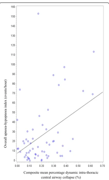
AHI, RDI, ODI and average SpO2 in supine and non-supine sleep (Tables 3 and 4). BMI and gender were consistently identified as relevant confounders for inclu-sion in the adjusted multivariate linear regresinclu-sion models; increased BMI and male gender were associated with increased dynamic intra-thoracic central airway col-lapse. The Pearson correlation coefficient for the un-adjusted linear model associating composite mean percentage dynamic intra-thoracic central airway col-lapse and overall AHI was 0.419 (p= 0.0010), represent-ing a statistically significant moderately strong association (Fig. 1). In the adjusted model, every 10% worsening in dynamic airway collapse was associated with an increase in AHI of 5 events/hour (95% confi-dence interval: 0.9–10 events/hour |P= 0.019).
No association was demonstrated between degree of dynamic intra-thoracic central airway collapse and symptoms or healthcare utilisation.
Discussion
Some expiratory narrowing of central airways is physio-logical. The equal pressure point (EPP) theory is key. Al-veolar pressure, the driving pressure for gas movement on expiration, is comprised of elastic recoil pressure plus pleural pressure. Alveolar pressure (‘intraluminal’ pres-sure outside alveoli) decreases on movement up the tra-cheobronchial tree because pressure is required to force gas across airway resistance; this is supported by the Bernoulli principle, which operates due to the decreasing total CSA of airways from periphery to mouth, and which states the perpendicular pressure exerted by a gas decreases as its velocity through a tube increases. Intra-luminal pressure also decreases as expiration progresses because elastic recoil falls, and airway resistance in-creases, as lung volume reduces.
There is a dynamic point along the tracheobronchial tree where the intraluminal pressure falls to become equal to the external pleural pressure (the EPP). The air-way is prone to collapse downstream (closer to mouth) of the EPP because pleural pressure can exceed intra-luminal pressure. The system functions as a Starling resistor so airway collapse downstream of the EPP does not contribute to airway resistance (West & Luks,2016). The EPP usually exists in lobar to subsegmental bronchi and from here to the mouth there is minimal reduction in intraluminal pressure and minimal resistance. EDAC does not reduce expiratory airflow (Kolakowski et al.,
2018; Macklem et al., 1965; Macklem & Wilson, 1965; Smaldone & Smith, 1985; Loring et al., 2007; Boiselle et al.,2012; Sverzellati et al.,2009).
EDAC may arise from diseases that drive the EPP closer to the alveolus plus, separately, increased collaps-ibility of the pars membranosa. Diseases with increased airway resistance (asthma and chronic bronchitis) will Table 1Rates of categorical symptoms, comorbidities and
healthcare utilisation
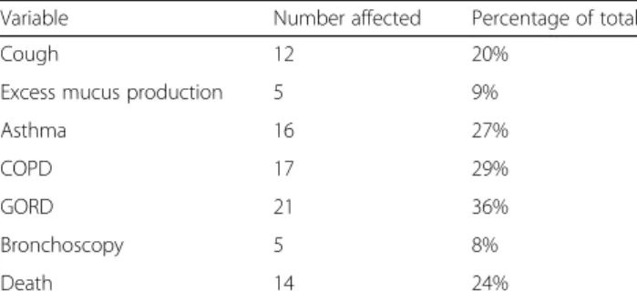
Variable Number affected Percentage of total
Cough 12 20%
Excess mucus production 5 9% Asthma 16 27%
COPD 17 29%
GORD 21 36%
Bronchoscopy 5 8%
Death 14 24%
Legend:COPDchronic obstructive pulmonary disease,GORD gastro-oesophageal reflux disease
Table 2Rates of ordinal and continuous symptoms, healthcare utilisation and pulmonary function test results
Variable Median Interquartile range Dyspnoea 2 2–3
ED presentations per year 0 0–0.6 Inpatient admission days per year 0 0–3.8 FEV1 as percentage of predicted 70% 52–93% FVC as percentage of predicted 78% 62–98% TLC as percentage of predicted 93% 75–107% DLCO as percentage of predicted 68% 55–83%
precipitate a more rapid fall in intraluminal pressure and thus drive the EPP further upstream. Loss of elastic re-coil (emphysema) does the same by reducing driving pressure for gas movement and, separately, diminishing tractional forces that promote airway patency. Obesity increases pleural pressure and forces the individual to respire at reduced lung volumes where elastic recoil is lower and airway resistance is higher (Behazin et al.,
1985). Conversely there are limited data to identify which diseases promote increased intrinsic collapsibility of the pars membranosa. These could include obesity (increased extra-luminal pressure and mediastinal fat),
chronic cough (repetitive tension, micro-trauma and re-duced elasticity), OSA (repetitive tension from inspira-tory efforts against occluded upper airway) and chronic airway inflammation (reduced elasticity) from any cause be it intrinsic or extrinsic (Kolakowski et al.,2018; Good et al., 2018). Indeed Majid et al. found 45% of 139 pa-tients with ECAC had GORD (Majid et al.,2019).
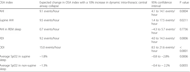
Defining a ‘pathological’ threshold for distinguishing normal dynamic central airway narrowing from EDAC is problematic. Historically a 50% reduction in airway CSA was set. Subsequent studies with spirometry-controlled dynamic CT scans with physiologist coaching have Table 3Unadjusted linear regression models assessing associations between intra-thoracic central airway collapse and obstructive sleep apnoea
OSA index Expected change in OSA index with a 10% increase in dynamic intra-thoracic central airway collapse
95% confidence interval
P value AHI 9.1 events/hour 4.1 to 14.1 events/
hour
0.0004 Supine AHI 9.5 events/hour 1.4 to 17.5 events/
hour
0.0211 AHI in REM sleep 0.7 events/hour −4.3 to 5.7 events/
hour
0.7736 RDI 9.2 events/hour 4.0 to 14.3 events/
hour
0.0006 ODI 15.0 events/hour 8.5 to 21.6 events/
hour
< 0.0001 Average SpO2 in supine
sleep
−1.8% −0.8 to−2.8% 0.0006 Average SpO2 in non-supine
sleep −
1.3% −0.4 to−2.2% 0.0055
Legend: Associations are described by the expected change in the obstructive sleep apnoea index given a 10% increase in dynamic intra-thoracic central airway collapse.OSAobstructive sleep apnoea,AHIapnoea hypopnoea index,REMrapid eye movement sleep,RDIrespiratory disturbance index,ODIoxygen 3% desaturation index,SpO2peripheral oxygen saturation
Table 4Adjusted linear regression models assessing associations between intra-thoracic central airway collapse and obstructive sleep apnoea
OSA index Confounders in adjusted model
Expected change in OSA index with a 10% increase in dynamic intra-thoracic airway collapse
95% confidence interval
P value AHI Gender, BMI 5.4 events/hour 0.9 to 10 events/
hour
0.0187 Supine AHI Gender, BMI 3.8 events/hour −3.5 to 11.1
events/hour
0.3129 AHI in REM sleep BMI −0.5 events/hour −5.4 to 4.4
events/hour
0.84 RDI Gender, BMI 5.5 events/hour 0.8 to 10.2
events/hour
0.0230 ODI Gender, BMI 10.6 events/hour 4.5 to 16.6
events/hour
0.0006 Average SpO2 in supine
sleep
BMI −1.5% −0.6 to−2.5% 0.001 Average SpO2 in
non-supine sleep
FEV1 −1.2% −0.4 to−2.0% 0.0038
Legend: Associations are described by the expected change in the obstructive sleep apnoea index given a 10% increase in dynamic intra-thoracic central airway collapse.OSAobstructive sleep apnoea,AHIapnoea hypopnoea index,BMIbody mass index,REMrapid eye movement sleep,RDIrespiratory disturbance index,
demonstrated healthy controls have approximately 50– 60% reduction in CSA with a 20% standard deviation (Boiselle et al.,2009; O’Donnell et al.,2012; Litmanovich et al.,2010; Boiselle et al.,2012). Thus 75% collapse may be a superior threshold for defining EDAC, provided im-aging is performed with the same robust methods. How-ever such scans are not readily available in most institutions and, furthermore, it is critical to appreciate that this definition of EDAC arises from limited assess-ments of healthy volunteers and has not been rigorously examined in symptomatic patients or in those with underlying lung disease (Murg & Colt,2013; Kolakowski et al.,2018). Differential thresholds of‘pathological’ cen-tral airway collapse may be applicable in different patient groups. Investigation of dynamic intra-thoracic central
airway collapse as a continuous rather than a categorical variable may help to address these issues.
Other methods of assessment of dynamic intra-thoracic central airway behaviour have limitations that likely outweigh those associated with dynamic CT im-aging. Bronchoscopy allows direct real-time visualisation of the intra-thoracic central airways however precise measurement of airway collapse is hampered by (a) par-allax error and other technical factors that can impede CSA calculation, (b) effects of sedation which may alter airway behaviour when compared with wakefulness, (c) continuous positive pressure transmitted to the central airways if nasal high-flow oxygen is utilised, thereby falsely reducing any dynamic collapse, (d) inability to spect all airway locations simultaneously and (e) in-creased cost and risk. Although other methods such as drug-induced sleep endoscopy and magnetic resonance imaging have value in examining upper airway function in OSA, they have not been rigorously assessed in dy-namic intra-thoracic central airway collapse (Ciscar et al.,2001; Moon et al.,2010; Cavaliere et al.,2013; Kim et al.,2014; Certal et al.,2016; Feng et al.,2018).
A disease should have attributable symptoms or sequa-lae and here, as outlined previously, EDAC remains par-ticularly poorly characterised. Boiselle and colleagues examined degree of dynamic central airway collapse in 100 patients with COPD and found only weak correl-ation with symptoms (Boiselle et al., 2012). Good et al. found 76% of 42 patients with chronic cough had ≥65% dynamic central airway collapse, providing indirect sup-port for the common belief that central airway collapse contributes to cough while also revealing the potential pitfalls of fixating on≥75% reduction in CSA as the only clinically relevant threshold for pathological dynamic central airway collapse (Good et al., 2018). No studies have thoroughly investigated associations between de-gree of dynamic intra-thoracic central airway collapse and respiratory complications or mortality.
Existing evidence for an association between EDAC and OSA is mixed. Ehtisham et al. reported a retrospect-ive case series in which 62% of 107 patients with ECAC had OSA, but neither degree of airway collapse required for EDAC diagnosis nor OSA severity were specified (Ehtisham et al., 2015). Good et al. found OSA (defined as AHI of ≥5 events/hour) in 78% of 32 patients with both chronic cough and≥65% dynamic change in cen-tral airway CSA, being their threshold for significant col-lapse (Good et al., 2018). Kolakowski and colleagues audited 200 patients who had undergone investigations for both EDAC (defined as ≥75% collapse) and OSA (Kolakowski et al., 2018). Patients were subdivided into those with and without OSA (defined as AHI of ≥5 events/hour). Prevalence of EDAC across both groups was approximately 25% but severe EDAC (arbitrarily
defined in their study as≥85% collapse) occurred signifi-cantly more often in patients with OSA (15% versus 6%). Degree of dynamic tracheal collapse was also assessed as a continuous variable, being maximal percentage CSA reduction at any location in the trachea, and this was weakly correlated with supine but not overall AHI. Al-though the work by Kolakowski et al. is valuable in this developing area, their analysis also exemplifies the limi-tations that can arise from a categorical statistical meth-odology; in their research this was applied not only to the degree of dynamic intra-thoracic central airway col-lapse but also OSA, which is another complex and in-herently continuous pathophysiological phenomenon.
Our study adds significantly to the existing literature because it is the first to demonstrate statistically robust associations between degree of dynamic intra-thoracic central airway collapse and multiple OSA indices reflect-ing both respiratory event-related sleep fragmentation and hypoxic burden. An important strength of our study is the introduction of a multi-site continuous composite index of airway collapse rather than the sole use of the arbitrary categorical presence of‘EDAC’ at any one site, thereby more accurately reflecting complex central air-way behaviour; this novel approach permitted our ana-lysis to elucidate, for the first time, the fractional impact of increasing dynamic intra-thoracic central airway col-lapse on upper airway collapsibility in sleep. This is also the first study to utilise regression models to adjust for confounders, which is important when diseases may share risk factors.
We stress that we do not claim to have proven an as-sociation between EDAC, as defined as a≥75% reduc-tion in airway CSA, and OSA; rather, our data support the theory that increasing dynamic intra-thoracic central airway collapse is associated with increasingly severe ob-structive sleep apnoea, as measured by obob-structive re-spiratory events and hypoxic burden. Failure to identify EDAC in our study was likely because the dynamic CT chest scans were all performed as part of high-resolution protocols with a focus on lung parenchyma rather than airway collapse, meaning there was none of the spirometry-control or physiologist coaching used in the research protocols that identified ≥75% collapse as ab-normal in healthy volunteers. This methodological flaw was unavoidable in a retrospective study and explains why only 34% of scans were graded as good quality for our purposes (evidence of adequate expiratory effort and optimal number of slices on expiratory phase). However, if anything, any bias arising from this weakness would have reduced the probability of finding a relationship be-tween dynamic intra-thoracic central airway collapse and OSA. Future prospective research with dedicated high-quality dynamic CT imaging is required to further explore the potential association.
Analysis of any association between airway collapse and symptoms / healthcare utilisation would have been confounded by the inclusion of dynamic CT chest scans for any indication, meaning all patients must have had some respiratory symptom to prompt investigation.
Conclusions
This study provides compelling evidence for a relation-ship between dynamic intra-thoracic central airway col-lapse and OSA especially because, even after adjusting for confounders, multiple OSA indices continued to demonstrate statistically and clinically significant associ-ations with degree of collapse. We feel that these find-ings should prompt clinicians to consider EDAC in patients with severe OSA and vice versa. The shared phenotype may be the obese middle-aged patient with underlying airways disease and chronic cough, although further work is clearly required to test these commonly held clinical beliefs. Continuous positive airway pressure offers the potential to improve both conditions while mucus retention due to EDAC may benefit from positive expiratory pressure techniques for sputum clearance (Murgu & Colt, 2007; Dedhia et al., 2015; Adliff et al.,
1997; Sirithangkul et al.,2010).
Future research could seek to prospectively confirm the association and test its extension to EDAC, clarify any association between airway collapse and symptoms / healthcare utilisation, assess whether differing thresholds of‘pathological’airway collapse may be applicable in dif-ferent patient populations, and determine if physiological tests like impulse oscillometry can identify EDAC with or without OSA (Fielding et al.,2018).
Abbreviations
AHI:Apnoea-hypopnoea index; COPD: Chronic obstructive pulmonary disease; CPAP: Continuous positive airway pressure; CSA: Cross-sectional area; CT: Computed tomography; ECAC: Excessive central airway collapse; EDAC: Excessive dynamic airway collapse; EPP: Equal pressure point; FEV1: Forced expiratory volume in 1 second; FVC: Forced vital capacity; GORD: Gastro-oesophageal reflux disease; IQR: Inter-quartile range; ODI: Oxygen 3% desaturation index; OSA: Obstructive sleep apnoea; RDI: Respiratory disturbance index; REM: Rapid eye movement; SpO2: Peripheral oxygen saturation
Acknowledgements
The authors sincerely thank Suzanne Edwards for her generous assistance with statistical design and analysis. The authors thank Dr. Mark Jurisevic and Dr. Sonya Johnston for assistance in collating polysomnography data.
Authors’contributions
contributed to the methodology, assisted in data analysis and reviewed the manuscript. The author(s) read and approved the final manuscript.
Funding
No external funding was received for this study.
Availability of data and materials
All data generated or analysed during this study are included in this published article.
Ethics approval and consent to participate
This multi-centre retrospective audit was approved by the local research eth-ics committee (Central Adelaide Local Health Network Research Etheth-ics Com-mittee approval number 137). The requirement for patient consent was waived by the ethics committee given the retrospective nature of the re-search involving existing clinical information and the negligible risk to patients.
Consent for publication Not applicable.
Competing interests
The authors declare that they have no completing interests.
Author details
1Discipline of Medicine, University of Adelaide, Adelaide, SA 5000, Australia. 2Central Adelaide Local Health Network, 1 Port Road, Adelaide, SA 5000,
Australia.3Department of Thoracic Medicine, Royal Adelaide Hospital, 1 Port
Road, Adelaide, SA 5000, Australia.
Received: 26 December 2019 Accepted: 30 April 2020
References
Adliff M, Ngato D, Keshavjee S, et al. Treatment of diffuse tracheomalacia secondary to relapsing polychronditis with continuous positive airway pressure. Chest. 1997;112(6):1701–4.
Behazin N, Jones S, Cohen R, et al. Respiratory restriction and elevated pleural and esophageal pressures in morbid obesity. J Appl Physiol. 1985;108(1):212– 8.
Boiselle P, Michaud G, Roberts D, et al. Dynamic expiratory tracheal collapse in COPD: correlation with clinical and physiologic parameters. Chest. 2012; 142(6):1539–44.
Boiselle P, O’Donnell C, Bankier A, et al. Tracheal collapsibility in healthy volunteers during forced expiration: assessment with multi-detector CT. Radiology. 2009;252(1):255–62.
Cavaliere M, Russo F, Iemma M, et al. Awake versus drug-induced sleep endoscopy: evaluation of airway obstruction in obstructive sleep apnea / hypopnoea syndrome. Laryngoscope. 2013;123(9):2315–8.
Certal V, Pratas R, Guimaraes L, et al. Awake examination versus DISE for surgical decision making in patients with OSA: a systematic review. Laryngoscope. 2016;126(3):768–74.
Ciscar M, Juan G, Martinez V, et al. Magnetic resonance imaging of the pharynx in OSA patients and healthy subjects. Eur Respir J. 2001;17(1):79–86. Dedhia R, Kapur V, Weaver E. Excessive dynamic airway collapse of the lower
airway: a cause for persistent sleep-disordered breathing after tracheostomy. J Clin Sleep Med. 2015;11(11):1337–9.
Ehtisham M, Azhar-Munir R, Klopper E, et al. Correlation between
tracheobronchomalacia / hyper dynamic airway collapse and obstructive sleep apnoea. Am J Respir Crit Care Med. 2015;191:A3075.
Feng Y, Keenan B, Wang S, et al. Dynamic upper airway imaging during wakefulness in obese subjects with and without sleep apnea. Am J Respir Crit Care Med. 2018;198(11):1435–43.
Fielding D, Travers J, Nguyen P, et al. Expiratory reactance abnormalities in patients with expiratory dynamic airway collapse: a new application of impulse oscillometry. ERJ Open Res. 2018;4(4):00080–2018.https://doi.org/10. 1183/23120541.00080-2018.
Good J, Rollins D, Kolakowski C, et al. New insights in the diagnosis of chronic refractory cough. Respir Med. 2018;141:103–10.
Kim A, Keenan B, Jackson N, et al. Tongue fat and its relationship to obstructive sleep apnea. Sleep. 2014;37(10):1639–48.
Kolakowski C, Rollins D, Jennermann T, et al. Clarifying the link between sleep disordered breathing and tracheal collapse: a retrospective analysis. Sleep Sci Pract. 2018;2:10.https://doi.org/10.1186/s41606-018-0030-2.
Litmanovich D, O’Donnell C, Bankier A. Bronchial collapsibility at forced expiration in healthy volunteers: assessment with multidetector CT. Radiology. 2010;257(2):560–7.
Loring S, O’Donnell C, Feller-Kopman D, et al. Central airway mechanics and flow limitation in acquired tracheobronchomalacia. Chest. 2007;131(4):1118–24. Macklem P, Fraser R, Brown W. The detection of the flow-limiting bronchi in
bronchitis and emphysema by airway pressure measurements. Med Thorac. 1965;22:220–30.
Macklem P, Wilson N. Measurement of intrabronchial pressure in man. J Appl Physiol. 1965;20(4):653–63.
Majid A, Kheir F, Alape D, et al. The prevalence of gastroeosophageal reflux in patients with excessive central airway collapse. Chest. 2019;155(3):540–5. Matus I, Richter W, Mani S. Awareness, competencies, and practice patterns in
tracheobronchomalacia: a survey of pulmonologists. J Bronchology Interv Pulmonol. 2016;23(2):131–7.
Moon I, Han D, Kim J, et al. Sleep magnetic resonance imaging as a new diagnostic method in obstructive sleep apnea syndrome. Laryngoscope. 2010;120(12):2546–54.
Murg S, Colt H. Tracheobronchomalacia and excessive dynamic airway collapse. Clin Chest Med. 2013;34(3):527–55.
Murgu S, Colt H. Description of multidimensional classification system for patients with expiratory central airway collapse. Respirology. 2007;12(4):543– 50.
O’Donnell C, Litmanovich D, Loring S, et al. Age and sex dependence of forced expiratory central airway collapse in healthy volunteers. Chest. 2012;142(1): 168–74.
Sirithangkul S, Ranganathan S, Robinson P, et al. Positive expiratory pressure to enhance cough effectiveness in tracheomalacia. J Med Assoc Thail. 2010; 93(6):S122–8.
Smaldone G, Smith P. Location of flow-limiting segments via airway catheters near residual volume in humans. J Appl Physiol. 1985;59(2):502–8. Sverzellati N, Rastelli A, Chetta A. Airway malacia in chronic obstructive
pulmonary disease: prevalence, morphology and relationship with emphysema, bronchiectasis and bronchial wall thickening. Eur Radiol. 2009; 19(7):1669–78.
West J, Luks A. West’s respiratory physiology: the essentials. Philadelphia, PA: Wolters Kluwer; 2016.
Publisher’s Note