CELLULAR & MOLECULAR BIOLOGY LETTERS http://www.cmbl.org.pl
Received: 25 March 2010 Volume 15 (2010) pp 611-629 Final form accepted: 07 September 2010 DOI: 10.2478/s11658-010-0032-2 Published online: 17 September 2010 © 2010 by the University of Wrocław, Poland
* Author for correspondence. e-mail: [email protected], tel.: + 48 32 2789806, fax: + 48 32 2789840
Abbreviations used: DAPI – 4’,6-diamidino-2-phenylindole; DMEM – Dulbecco’s Modified Eagle Medium; DMSO – dimethylsulfoxide; dNTP – deoxyribonucleotide triphosphate; GAPDH – glyceraldehyde-3-phosphate dehydrogenase; mRFP – monomeric red fluorescent protein; PCR – polymerase chain reaction; RACE – rapid amplification of cDNA ends; RPA – replication protein A; SNP – single nucleotide polymorphism; TDG – thymine-DNA glycosylase
Research article
A FUNCTIONAL ANALYSIS OF G23A POLYMORPHISM AND THE
ALTERNATIVE SPLICING IN THE EXPRESSION OF THE XPA GENE
DOROTA BUTKIEWICZ1, MAŁGORZATA KRZEŚNIAK1, RASA VAITIEKUNAITE1, BOŻENA SIKORA1, ELISE D. BOWMAN2,
CURTIS C. HARRIS2 and MAREK RUSIN1*
1Department of Tumour Biology, Maria Skłodowska-Curie Memorial Cancer Centre and Institute of Oncology, Gliwice Branch, Wybrzeże Armii Krajowej 15, 44-101 Gliwice, Poland, 2Laboratory of Human Carcinogenesis, National Cancer
Institute, NIH, 37 Convent Drive, Bethesda MD 20892-4258, USA
Abstract: The XPA gene has a commonly occurring polymorphism (G23A) associated with cancer risk. This study assessed the functional significance of this polymorphism, which is localised near the translation start codon. Lymphoblastoid cell lines with alternative homozygous genotypes showed no significant differences in their XPA levels. The luciferase reporter assay detected no functional difference between the two sequences. Unexpectedly, we found that the alternatively spliced form of XPA mRNA lacked a part of exon 1. Only the reading frame downstream of codon Met59 was preserved. The alternative mRNA is expressed in various human tissues. The analysis of the 5’cDNA ends showed similar transcription start sites for the two forms. The in vitro expression of the alternative XPA labelled with the red fluorescent protein (mRFP) showed a lack of preferential nuclear accumulation of the XPA isoform. The biological role of the alternative XPA mRNA form remains to be elucidated.
INTRODUCTION
The nucleotide excision repair (NER) pathway removes DNA lesions induced by ultraviolet light (UV), tobacco smoke and other chemicals, including environmental pollutants or chemotherapeutic agents, such as cisplatin. The XPA protein participates in DNA damage recognition and recruits other NER components to the lesion site [1]. In humans, defects in NER, which are caused by mutations in the NER genes, are associated with a rare, cancer-prone syndrome called xeroderma pigmentosum (XP). This syndrome is characterized by an impaired DNA repair capacity, extreme UV sensitivity, skin abnormalities, skin cancer, and in some cases, neurodegeneration. XPcomplementation group A patients, who have a mutated XPA gene, show the most severe symptoms of all the XP types [2-4].
Variations in the DNA repair capacity caused by functional polymorphisms of DNA repair genes may result in the modulation of an individual’s susceptibility to cancer [5, 6]. So far, reports on four infrequent single nucleotide substitutions in the coding sequence of the XPA gene have been published. These were not associated with XP [7-9]. However, the G23A polymorphism (rs1800975) present in the XPA 5’ untranslated region has proven to occur commonly enough that population-based, case-control studies could be performed.
The G23A polymorphism is characterized by a G-to-A substitution within the Kozak sequence in the fourth nucleotide upstream from the initiation codon (ATG; Fig. 2A). The carriers of the two G alleles are wild-type homozygotes. This polymorphism has been associated with lung, endometrial, oesophageal and oral carcinoma risk in several studies [10-15]. Kiyohara and Yoshimasu wrote a meta-analysis on genetic polymorphisms and lung cancer risk, concluding that the XPA 23 GG genotype protects against lung cancer [16]. This polymorphism was also shown to influence the course of the disease or therapy response in bladder, ovarian and non-small cell lung cancers [17-19]. Healthy individuals with one or two G alleles exhibited a more efficient DNA repair capacity than
AA genotype carriers [20].
MATERIALS AND METHODS
Cell culture and Western blotting for XPA
To identify Epstein-Barr virus-immortalized human lymphoblastoid cell lines (from Centre d’Etude du Polymorphisme Humain-CEPH and Utah pedigree families, Corriell Cell Repositories, Camden, NJ) as having the XPA AA or GG
homozygous genotypes, DNA was isolated from cell lines via standard methods, and the samples were used as the templates for the genotyping reaction described below. Subsequently, three selected cell lines with the AA genotype and three with the GG genotype were grown on RPMI 1640 medium (Gibco BRL) supplemented with 15% heat-inactivated foetal calf serum at 37ºC in 5% CO2. Logarithmically growing cells were centrifuged, and the cell pellet was washed with phosphate-buffered saline (PBS) and snap-frozen. The cells were lysed via treatment with a RIPA buffer supplemented with protease inhibitors. The total cell lysate was obtained by incubating the cells in the buffer for 20 min on ice and centrifuging (14000 rpm, 20 min, 4ºC). The supernatant was saved and an equal amount of protein extract (70 μg) was loaded and separated on a 12% denaturing polyacrylamide gel. After the electrotransfer, the nitrocellulose membrane was blocked with a 5% skim milk in PBS/0.1% Tween 20 solution and incubated with mouse monoclonal anti-XPA antibody, diluted to 1:400 (Ab-1; Lab Vision Corp.) at 4ºC overnight. To verify that there was equal protein loading in each lane, the nitrocellulose membrane was reprobed with the monoclonal anti-β-actin antibody (Chemicon Int.). To calculate the relative levels of the XPA protein in the studied cell lines, the pictures of the blots were scanned, and the areas of the peaks were measured in arbitrary units via semi-quantitative densitometric analysis (One-Dscan, Scanalytics). The levels of the XPA protein in each cell line were normalised to the corresponding β-actin levels.
Genotyping of the XPA G23A polymorphism
The polymorphism was detected via a PCR assay combined with restriction enzyme digestion (PCR-RFLP). The PCR primer sequences used were: (sense) 5’ TCAGAAAGGCCGCTGGGT 3’ and (anti-sense) 5’ CTGGCGCAGCAT CAGTGC 3’. For PCR, 50 ng of genomic DNA was used with 1 x PCR Buffer II (PE Applied Biosystems), 1.5 mM MgCl2, 0.2 mM of each dNTP (Pharmacia), 12.5 pmol of each primer (BioTez, Berlin), 2 U AmpliTaq Gold DNA polymerase (PE Applied Biosystems) and 5% DMSO in a total volume of
25 μl. The reaction was performed as follows: pre-denaturation and enzyme
(BMA). The XPA G23A polymorphism results in a loss of the MspI restriction site, and the digestion pattern observed was: 108-, 95-, and 30-bp fragments in homozygote GG (an additional control digestion site was present within the PCR product); 138-, 108-, 95-, and 30-bp fragments in heterozygote GA; and 138-, and 95-bp fragments in homozygote AA.
Molecular cloning of plasmids with the XPA variant 23G and variant 23A promoter
The XPA polymorphism is located within the Kozak consensus sequence. The wild-type and polymorphic versions of the XPA promoter and the 5’UTR region were inserted into the pGL3-Basic plasmid, which contains the firefly luciferase gene luc+ (GenBank U47295; Promega, Madison). This allows for reporter luciferase gene expression under the transcriptional and translational control of the XPA promoter, and 5’UTR in either the wild-type or polymorphic version using a reporter assay described below under “Functional assay for the XPA
G23A polymorphism”. The cloning was performed using the triple-ligation strategy, involving the ligation of two inserts (e.g. PCR products) into a plasmid at three mutually incompatible restriction sites (HindIII, PpuMI and BstBI in our experiment, Fig. 1). The first PCR fragment (from position 1228 to 1803 of the GeneBank U16815 sequence) was amplified from genomic DNA and encompassed the XPA promoter sequence and 5’UTR with the PpuMI restriction site located 17 residues upstream from the XPA translation start codon. This fragment was amplified using the primers: TTTTAAGCTTAAGGCTGTGTC TCTAGGCCG (sense), containing the HindIII restiction site (bold font), and CTGGCGCAGCATCAGTGC (anti-sense) located in the XPA exon 1. The fragment downstream of the PpuMI site was removed after the restriction digestion reaction and gel purification of the PCR product.
DNA polymerase (Applied Biosystems). The amplification of the XPA promoter region required the use of 1 mM betaine and 2.5 U AmpliTaq Gold DNA polymerase. The temperature profile of PCR for the XPA promoter fragment was 95ºC for 10 min (DNA denaturation and polymerase activation); then 40 cycles at 95ºC for 30 sec, 58ºC for 30 sec, and 72ºC for 45 sec; followed by a final extension in 72ºC for 7 min. For the G-luc or A-luc fragments, pre-denaturation was at 95ºC for 5 min; then 30 cycles of 95ºC for 30 sec, 50ºC for 30 sec, and 72ºC for 45 sec; and a final extension at 72ºC for 4 min. Next, the PCR products were gel-purified with a QIAEX II kit (Qiagen), and digested with HindIII and
PpuMI (XPA promoter) or with PpuMI and BstBI (G-luc, A-luc), according to the manufacturer’s instructions (New England BioLabs), then gel purified again. The pGL3-Basic plasmid was digested with HindIII and BstBI, and also gel purified. The digested PCR products were ligated into the HindIII and BstBI sites located in the plasmid. The ligation products were transferred into competent DH5α bacteria. After recombinant plasmid purification, the clones carrying the vectors with either the G variant (pGL3-XPApromG) or A variant (pGL3-XPApromA) were identified. The inserts of the selected clones were sequenced with a BigDyeTM Terminator Cycle Sequencing kit (Applied Biosystems), using the primers GTGATTTGTATTCAGCCCATATCG and TCTTCCAGCGGATAGAATG to check for the presence of the XPA G or A sequence and to verify that the inserts did not contain any mutations that may have been introduced during the PCR. Subsequently, the G and A clones were used to produce four batches of each plasmid DNA using a Qiagen QIAprep Spin Miniprep kit (Qiagen). After measuring the concentration of the plasmid solutions (Beckman DU 640B spectrophotometer), the DNA concentrations in all of the batches were equalised using Tris-HCl buffer at pH 8.0.
The DLR assay for the XPA G23A polymorphism
To study the influence of the XPA G23A polymorphism on the activity of the
XPA promoter/5’UTR region, we used the Dual-Luciferase ReporterTM (DLR) assay system (Promega), which measures the reporter gene (luc+) activity under
the transcriptional control of an inserted promoter. Two plasmid versions, containing the XPA promoter sequence with either the G or A nucleotide in the fourth position before a start codon of the luc+ gene (i.e., pGL3-XPApromG or
pGL3-XPApromA constructs, respectively), were transfected separately into human NCI H1299 cells in culture together with a control vector, pRL-TK (Promega). The pRL-TK control plasmid contains the herpes simplex virus thymidine kinase promoter, controlling the expression of the Rluc gene, which codes for a luciferase from Renilla reniformis. Both the firefly and Renilla
transfection and cell lysis efficiency. In each measurement, the normalised firefly luciferase activity (NFLA) was calculated by dividing the firefly luciferase activity reading by the Renilla luciferase activity reading. Four transfections and luciferase assays were performed for each of the plasmids, pGL3-XPApromG and pGL3-XPApromA. The control transfection with the pGL3-Basic plasmid was also performed. This experiment was repeated twice, using different batches of the plasmid preparations to avoid the variability that could be introduced due to differences in the DNA quality. The means and standard deviations for the NFLA were calculated for the experiments, and the statistical significance of the NFLA difference between cells transfected with G or A vectors was evaluated using the t-test.
The cell line used in this study (lung carcinoma cell line NCI H1299, from the American Type Culture Collection) was grown in DMEM medium supplemented with 10% foetal bovine serum (Gibco BRL), and was maintained at 37ºC in 5% CO2. The cells were grown in 1 ml of medium on 12-well plates (NUNC Brand Products). All of the transfections were performed on subconfluent cells (40% confluence) with the FuGene 6 reagent, according to the manufacturer’s instructions (Roche Applied Sciences). The experimental/control vector ratio was 10:1. The activity of the reporter gene was measured 24 h after the transfection.
RT-PCR of XPA
The amplification of the 5’ fragment of XPA cDNA was performed using the XPA12 anti-sense primer (GCTTGTGTTTATCATCAGCATC), located at the beginning of exon 4, and the XPA14 sense primer (TAGGTCCTCGGAGTG GGC), located in exon 1 upstream of the translation start site (Fig. 2C). The PCR was performed in a reaction volume of 25 μl, with 1x PCR buffer II (Applied Biosystem), 1.5 mM MgCl2, 10 pmols of each primer 200 μM of each dNTPs, and 1 unit of AmpliTaq Gold polymerase (Applied Biosystems). The enzyme was activated by 10 min incubation at 95ºC, which was followed by 35 cycles at 94ºC for 30 sec, 60ºC for 20 sec, and 72ºC for 40 sec, and a final extension at 72ºC for 7 min. Under these conditions, only the alternatively spliced XPA
The 5’-RACE analysis of XPA
The analysis was performed using the Gene Racer kit (Invitrogen). The synthesis of cDNA was primed by XPA-SEQ2 oligonucleotide (AATTTAAGAGGTG GCTCTC) using, as a template, RNA isolated from normal human fibroblasts GM08402, obtained from Coriell Cell Repositories. The first round of 5’-RACE PCR was performed using GeneRacer 5 primer from the kit and XPA-RACE2 primer (TTTACATTAGCCATGCCGAG) spanning the exon 1-exon 2 junction of the alternative transcript. This primer pair can only amplify the alternative sequence. The nested PCR was performed using the GeneRacer 5 Nested primer and XPA-RACE3 primer (TCCGCGGGTTGCTCTAAAGC) complementary to the exon 1 fragment. The product of the nested PCR was directly sequenced using the BigDye Terminator Cycle Sequencing Kit (Applied Biosystems) after purification by exonuclease I and shrimp alkaline phosphatase. Alternatively, the two rounds of PCR were performed with primers: GeneRacer 5 and XPA 11 (AAGATATTCTTGTTTTGCCTCTG); GeneRacer 5 Nested and XPA12 (Fig. 2C). These primer pairs can amplify both major and alternatively spliced sequences. The amplification reactions using these primers were performed either with or without 1 M of betaine solution. The resulting PCR products were cloned using the TOPO cloning kit for sequencing (Invitrogen) and the inserts of plasmid DNA isolated from several clones were sequenced.
The molecular cloning of XPA-mRFP and protein localisation analysis
The XPA cDNA was amplified using the primers XPA-Afl-S (TTTTCTTAAGCTGGGAGCTAGGTCCTCGGAG) and XPA-Afl-A (TTTTCTTAAGCATTTTTTCATATGTCAGTTCATGGCC), containing the
AflII restriction sites (underlined). The PCR product was gel purified and ligated into the AflII site of the pcDNA3.1/HisC expression vector (Invitrogen) yielding the XPA-AFL-HisC plasmid. Subsequently, the HindIII-BamHI fragment of the previously made XPA-EGFP plasmid was cut and gel-purified. The HindIII site is located in XPA cDNA (Fig. 2C and 3E), and the BamHI site was placed just downstream of the last amino acid codon of XPA. This fragment was ligated to the HindIII and BamHI sites of the XPA-AFL-HisC vector. This step removed the second AflII site, downstream of the XPA cDNA from the XPA-AFL-HisC vector, and the part of the pcDNA3.1/His C polylinker between the AflII and
for fusion protein detection via Western blotting. The plasmid, with the alternative form of XPA cDNA was constructed via PCR amplification, without the betaine of the alternative cDNA sequence but with the primers, XPA-Afl-S and XPA11, and ligation of the PCR product into the AflII and HindIII sites of the XPA-mRFP, ultimately generating the Alt-XPA-mRFP vector. All of the PCR-generated sequences were sequenced to ensure that there were no mutations in the plasmids.
The cellular localisation of the mRFP-labelled XPA forms was examined after the transfection with FuGene6 (Roche) of the expression vectors into the U-2 OS human osteosarcoma cell line cultured in DMEM medium supplemented with 10% FBS and penicillin-streptomycin solution. The cells were fixed in 3.7% formalin 28 h after transfection, and after ethanol dehydration, they were mounted in Vectashield with DAPI (Vector Laboratories). The cells were photographed using a Nikon Eclipse fluorescent microscope.
Statistical analysis
The Mann Whitney U-test and Student’s t-test were respectively used to compare the differences in the XPA protein levels between the cell lines with the
AA and GG genotypes, and in the mean NFLA values between the cell lines transfected with the pGL3-XPApromG and pGL3-XPApromA reporter vectors. Differences were considered statistically significant at P < 0.05. All of the statistical analyses were performed using STATISTICA 6.0 software.
RESULTS
The functional analysis of G23A polymorphism
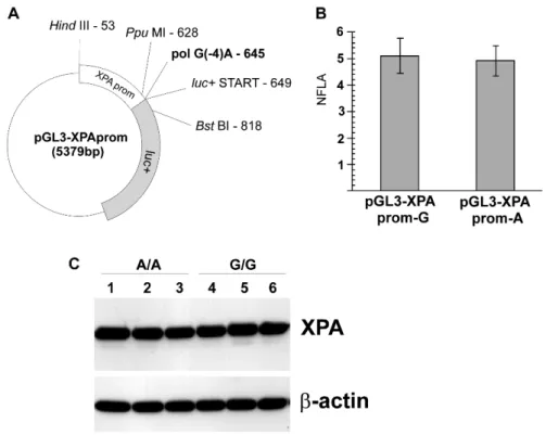
To find out whether the XPA polymorphism located within the Kozak consensus sequence in the 5’UTR had any influence on the XPA promoter/5’UTR activity, we inserted the relevant XPA region into the pGL3-Basic plasmid (Fig. 1A). The firefly luc+ gene carried by the plasmid was under the transcriptional and
translational control of the XPA promoter/5’UTR with or without the polymorphism (versions G or A, i.e. the XPApromG or pGL3-XPApromA vectors, respectively). The activity of the reporter luc+ gene is presented as normalised firefly luciferase activity (NFLA), which is calculated by dividing the reporter (firefly) luciferase activity reading by the control Renilla
luciferase activity reading (pRL-TK plasmid co-transfection – see the Materials and Methods section). The reporter gene activities were similar for the G and A versions of the sequence (respectively 5.10 ± 0.66 versus 4.91 ± 0.57, P = 0.67 by t-test; Fig. 1B). The results demonstrate that the G23A substitution does not influence the reporter gene expression in this assay.
Western blots. As illustrated in Fig. 1C, the two groups of cell lines had similar XPA protein levels (GG: 94.1 ± 1.5 versus AA: 89.8 ± 1.3, P = 0.13, via Mann Whitney U-test). Thus, in the examined cells growing under standard conditions, the polymorphism was not associated with detectable changes in the XPA protein level.
Fig. 1. The functional analysis of G23A polymorphism.A – The plasmid map showing the
XPA promoter inserted upstream of the luc+ gene, the position of the polymorphic
nucleotide pol G(-4)A, and the restriction sites used for the cloning. The start codon of the
luc+ open-reading frame is also marked. For image clarity, other plasmid features are not
shown. B – The normalised firefly luciferase activity (NFLA) in the NCI H1299 cell line transfected with the pGL3-XPApromG or pGL3-XPApromA reporter vectors, together with the control pRL-TK vector. Each bar shows the mean NFLA value from four separate transfections with the respective reporter plasmid and the control plasmid, while the whiskers show the standard deviation of the mean (± SD). C – Western blot analysis of XPA protein expression levels in three different lymphoblastoid cell lines with the
XPA G23A homozygous AA genotype and three lymphoblastoid cell lines with the
GG genotype. Lines: 1 – #6992, 2 – #7005, 3 – #6988, 4 – #7000, 5 – #7062, 6 – #7053 (CEPH lymphoblastoid cell lines).
The alternatively spliced form of XPA mRNA
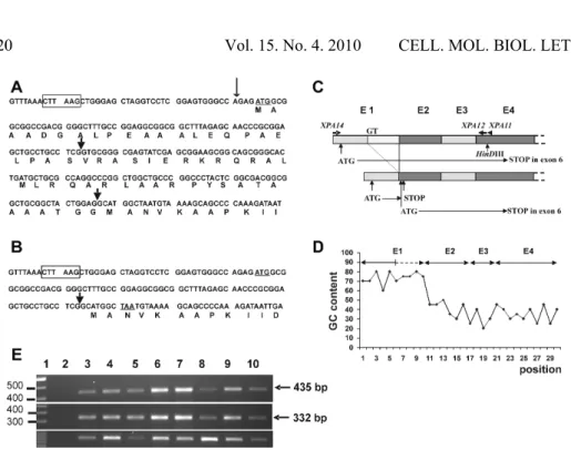
Fig. 2. The alternative splicing of XPA mRNA.A – The 5’ end sequence of the major XPA
cDNA form. The AflII cloning site of the pcDNA3.1/HisC expression vector is marked by the rectangle. The XPA amino acid sequence is shown below the cDNA sequence. The arrowheads flank the sequence spliced out from the alternative mRNA. The polymorphic residue is marked by the thin arrow. B – The 5’ end of the alternatively spliced form of
XPA cDNA. The alternative site of the exon junctions is marked by an arrow. The ATG translation start codon of the major XPA mRNA is underlined as well as the in-frame stop codon TAA. This mRNA can produce part of the XPA protein lacking the first 58 amino acids, provided that the translation starts from the second ATG codon corresponding to codon 59 of the major XPA mRNA. C – The positions of the exon-coded fragments of the major (longer) and alternatively spliced form (shorter) of XPA mRNA. E1-E4 represent exons 1-4. The last two exons are not shown. The XPA12 and XPA14 arrowheads mark the position of the PCR primers used to detect the mRNA forms by RT-PCR. The GT represents the position of the alternative splicing 5’ donor site. The ATG in exon 1 represents the first translation start codon of the XPA open-reading frame (ORF). This reading frame ends in exon 2 (STOP) in the alternative mRNA. The position of the methionine codon (ATG) in exon 2 that may drive the translation of the XPA protein without the first 58 amino acids is also shown. D – The GC content in the major form of
was about 100 bp shorter then expected (Fig. 2E, middle panel). The sequencing revealed that this DNA fragment is derived from XPA cDNA with a missing fragment of exon 1 (Fig. 2B). The analysis of the missing sequence indicated that the shorter fragment was formed by the use of the alternative 5’ donor splicing site located in exon 1, 70 residues downstream of the first nucleotide of the start codon (Fig. 2A, C). The major form of XPA mRNA could be amplified if 1 M betaine was present in the PCR reaction. In the presence of betaine, the amplification of the minor splicing form was not detectable. The betaine helps to amplify GC-rich sequences. The GC content in exon 1 is higher than in the rest of the XPA cDNA (Fig. 2D). The analysis of the alternatively spliced form indicated that the reading frame defined by the first ATG codon ends 27 codons downstream, at the beginning of exon 2-coded fragment (Fig. 2B, C). The alternative mRNA could direct the production of XPA if the translation started from codon 59, coding for methionine in the context of the Kozak sequence (Fig. 2A, B, C). In the alternatively spliced mRNA, there was no other upstream start codon in frame with the XPA ORF, assuming that both forms had the same 5’ end. To find out if this assumption was correct, we performed a 5’-RACE analysis of the alternative mRNA on RNA isolated from normal human fibroblasts. The first RACE-PCR amplification was performed with the primers GeneRacer 5 and XPA-RACE2 (a primer complementary to the alternative junction of exon 1-exon 2, amplifying only the alternative transcript) and the second, nested PCR was performed using the GeneRacer 5 Nested and XPA-RACE3 primers. The direct sequencing of the nested PCR product revealed that the major transcription start is located 32 nucleotides upstream from the translation site (TGGAGCTGGGAGC, transcription start site is underlined). When the 5’-RACE analysis was performed using XPA11 and XPA12 as anti-sense primers, and the betaine was not used in the PCR reaction mixture (Fig. 2C), the alternative mRNA end was preferentially amplified and the longest transcript did not go beyond the 35th residue upstream from the translation start codon. However, when the same primers were used as the anti-sense primers and the betaine was added to the PCR reaction mixtures at a concentration of 1 M, the two transcripts were amplified. This RACE-PCR product was cloned into the pCR4-TOPO vector and the inserts of six clones were sequenced. Two clones represented the alternative sequence starting 28 and 57 residues upstream from the start codon and four clones represented the major XPA mRNA form starting 27, 28, 35 and 87 residues upstream from the start codon. Thus, our 5’-RACE analysis showed transcription start heterogeneity and did not detect any alternative transcript that would go upstream of the 57th residue of the major translation start codon.
The expression of the alternative XPA mRNA and localisation of its protein
performed without betaine. As a template, we used a panel of first-strand cDNA solutions purchased from BD Biosciences, derived from RNA samples isolated from brain, heart, lung, liver, pancreas, skeletal muscle, kidney, and placenta tissue. Four independent amplifications were performed, and the amount of PCR product on the agarose gel is shown in Fig. 2E (middle panel). All of the amplifications yielded a similar pattern of the relative XPA expression, i.e. the highest expression of the alternative form can be detected in the RNA isolated from the liver and pancreas, whereas the lowest level is detected in the skeletal muscles. The expression pattern of the major XPA form is very similar to the expression of the alternative form, indicating that the expression of both mRNAs is coordinated (Fig. 2E, top panel). The control, housekeeping gene (GAPDH) also shows variable expression in the examined human organs (Fig. 2E, bottom panel). This is not unusual and was observed by others: the relatively high expression of the gene in the heart and skeletal muscles and low expression in the lung and placenta is consistent with the relative abundance of GAPDH
mRNA molecules in the respective human tissues [23].
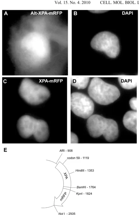
Next, we cloned the XPA cDNA sequence into an expression vector and fused it with the cDNA coding for the red fluorescent protein (mRFP). The map of the expression vector, named XPA-mRFP, is shown in Fig. 3E. The XPA cDNA sequence together with the 31 nucleotides upstream from the translation start site was inserted using PCR into the AflII site of the pcDNA3.1/HisC vector (Invitrogen). The six nucleotides forming the BamHI site were used to join the last codon of XPA with the sequence coding for the anti-Xpress antibody epitope, which was followed by the cDNA of the mRFP1 sequence ending with the NotI site of pcDNA3.1/HisC. The identity of the alternative sequence was confirmed via the sequencing of the XPA cDNA. The vector was named Alt-XPA-mRFP.
The localisation of XPA-mRFP- and Alt-XPA-mRFP-coded proteins was examined in the U-2 OS cells 28 h after the start of transfection. As expected, the major form of the XPA protein showed conspicuous nuclear localisation (Fig. 3C, D). However, the mRFP-labelled protein expressed from the Alt-XPA-mRFP vector showed both cytoplasmic and nuclear localisation patterns (Fig. 3A, B), which is not surprising considering the presence of the nuclear localisation signal within the first 58-amino acid sequence of XPA [24], i.e. the fragment that is lost due to the alternative splicing (Fig. 2A, B).
DISCUSSION
to an increased risk of cancer [16]. This may also modulate the response of an individual to the therapy of cancer [18]. Although the epidemiological data is quite consistent, especially in relation to the lung cancer risk [16], the mechanistic explanation of this phenomenon is only a matter of speculation. Our study was an attempt to better understand the molecular basis of the epidemiological observations. One report showed an increased cancer risk in heterozygous carriers of the xeroderma pigmentosum mutation [26]. We hypothesized that the XPA polymorphism could impair the expression of the
XPA gene, leading to a decreased DNA repair capacity and an increased cancer risk. However, in our experiments, we did not find evidence that the polymorphism modulated the expression level of the XPA protein. The G23A substitution is localised within the sequence surrounding the translation start codon in the 5’UTR, and it may influence the translation and/or transcription efficiency. Moreover, this substitution destroys a CpG site, and may interfere with proper methylation of the XPA promoter and proper regulation of XPA
expression. Those observations prompted us to study the influence of this SNP on the XPA promoter/5’UTR activity using the DLR assay, and its influence on the amount of XPA protein in cell lines exhibiting homozygous GG or AA
genotypes.
The sequence context of the start codon is assumed to affect the protein translation initiation process. Therefore, changes in translation efficiency may cause a variation in protein expression between individuals [27]. Although the XPA protein expression levels measured in the two XPA genotype groups of the lymphoblastoid cell lines were similar in our study, others observed that the polymorphism in the Kozak sequence of different genes modulated the amount of the expressed protein [28, 29].
Moreover, tissue and organ-specific differences in the mRNA levels observed for various NER genes including XPA [30, 31] may influence the repair rate of a particular tissue. Also, in this study, we detected differences in the expression levels of XPA mRNA between various human organs (Fig. 2E). Thus, a possible modulating effect of the XPA polymorphism may be dependent on the tissue type and may be different in white blood cells compared with lung epithelial cells. Alternatively, this XPA polymorphism may be in linkage disequilibrium with another functional polymorphism that is as yet unknown, and may also explain why we observed no effect of the XPA G23A polymorphism on the activity of the reporter luc+ gene.
Unexpectedly, we observed the existence of alternatively spliced form of XPA
mRNA indicated that in the alternative mRNA sequence, there was no translation start site upstream from codon 59 that would be in frame with the
XPA ORF downstream from codon 59. Thus, if the alternative form is translated, it produces XPA molecules with 58 amino acids deleted from the N-terminus. This truncated XPA isoform lacks a nuclear localisation signal and the RPA p34 subunit binding site [24]. Fused to the mRFP and expressed in cells, it shows both nuclear and cytoplasmic localisation (Fig. 3A, B). It remains to be determined whether it is produced in cells from the endogenous XPA locus, and if so, what function it has.
Previously, we showed the presence of the alternative splicing form of the TDG
gene with missing exon 2. Analysis of this alternative TDG sequence did not show clearly what protein it encodes [33]. Alternative splicing is a common phenomenon changing the diversity of protein molecules [34]. It also is common among genes coding DNA repair proteins [35, 36], enabling them, for instance, to code for protein isoforms able to enter the mitochondria and repair mitochondrial DNA [37]. To the best of our knowledge, the alternative splicing of XPA has not been reported so far. Frequently, the alternatively spliced mRNA molecules are less efficiently translated than the major mRNA forms [38], and sometimes they produce protein molecules showing a dominant-negative effect [39] or are aberrantly localised [40]. It remains to be determined whether the alternatively spliced form of XPA mRNA has physiological significance or if it is merely a product of the imperfect splicing. Interestingly, exon 1 is the only
XPA exon where xeroderma pigmentosum mutations have not yet been found. It is likely that the truncated protein produced from the alternative mRNA and translated from the Met59 coded by exon 2 is able to provide enough XPA activity that the exon 1 mutations do not show severe phenotypic effects. Thus, some alternative mRNA forms may be a source of residual protein activity protecting cells from the deleterious effects of mutations. A case of xeroderma
pigmentosum group A (XP-A) was reported with a novel XPA mutation
destroying the splice donor site in intron 1 [41]. The RT-PCR did not reveal the existence of the major XPA mRNA in samples isolated from the cells of this patient. Instead, the researchers could amplify only the truncated form of the
XPA mRNA sequence. This form (named def E1A) is identical to the alternatively spliced form that we amplified from normal human fibroblasts and other sources. Tanioka et al. [41] considered this form as aberrantly spliced, which was justified because under the PCR conditions used to amplify XPA
destroy XPA activity (usually mutations affecting the DNA-binding domain of XPA) [3]. Thus, some residual XPA activity is probably preserved in the cells of the newly detected XP-A case. This supposition is not contradicted by the lack of detection of the truncated XPA protein by the monoclonal antibody [41]. This form may be expressed below the detection threshold level of the antibody in the Western blot.
The results presented in this study and the observations reported by others [41] warrant further research on the alternatively spliced XPA form. This research may improve our understanding of XPA functioning and may help to explain the molecular pathology of some xeroderma pigmentosum cases.
Acknowledgements. The authors would like to thank Mrs. Iwona Matuszczyk and Ms. Helena Paterak for their excellent technical assistance. The editorial assistance of Mrs. Dorothea Dudek-Creaven is also appreciated. The study was supported by the Polish State Committee for Scientific Research (KBN) grant no. 4P05A 062 17 and grant no. 3P04A 004 23, Polish-American M. Sklodowska-Curie Fund grant no. MZ/HHS-97-313, UICC Fellowship Award 9/2000 to Marek Rusin and Fellowship Program at Department of Tumour Biology, supported by the National Cancer Institute – Office for International Affairs, NIH, Bethesda, MD, USA to Rasa Vaitiekunaite.
REFERENCES
1. Hoeijmakers, J.H.J. Genome maintenance mechanisms for preventing cancer. Nature 411 (2001) 366-374.
2. Cleaver, J.E. Common pathways for ultraviolet skin carcinogenesis in the repair and replication defective groups of xeroderma pigmentosum.
J. Dermatol. Sci. 23 (2000) 1-11.
3. Cleaver, J.E. Cancer in xeroderma pigmentosum and related disorders of DNA repair. Nat. Rev. Cancer 5 (2005) 564-573.
4. Kraemer, K.H., Patronas, N.J., Schiffmann, R., Brooks, B.P., Tamura, D. and DiGiovanna, J.J. Xeroderma pigmentosum, trichothiodystrophy and Cockayne syndrome: a complex genotype-phenotype relationship.
Neuroscience 145 (2007) 1388-1396.
5. Mohrenweiser, H.W. and Jones, I.M. Variation in DNA repair is a factor in cancer susceptibility: a paradigm for the promises and perils of individual and population risk estimation? Mutat. Res. 400 (1998) 15-24.
6. Goode, E.L., Ulrich, C.M. and Potter, J.D. Polymorphisms in DNA repair genes and associations with cancer risk. Cancer Epidemiol. Biomarkers Prev. 11 (2002) 1513-1530.
7. Richards, F.M., Goudie, D.R., Cooper, W.N., Jene, Q., Barroso, I., Wicking, C., Wainwright, B.J. and Ferguson-Smith, M.A. Mapping the multiple self-healing squamous epithelioma (MSSE) gene and investigation of xeroderma pigmentosum group A (XPA) and PATCHED (PTCH) as candidate genes.
8. Butkiewicz, D., Rusin, M., Harris, C.C. and Chorazy, M. Identification of four single nucleotide polymorphisms in DNA repair genes: XPA and XPB (ERCC3) in Polish population. Human Mut. 15 (2000) 577-578.
9. Mellon, I., Hock, T., Reid, R., Porter, P.C. and States, J.C. Polymorphisms in the human xeroderma pigmentosum group A gene and their impact on cell survival and nucleotide excision repair. DNA Repair 1 (2002) 531-546. 10.Park, J.Y., Park, S.H., Choi, J.E., Lee, S.Y., Jeon, H.S., Cha, S.I., Kim, C.H.,
Park, J.H., Kam, S., Park, R.W., Kim, I.S. and Jung, T.H. Polymorphisms of the DNA repair gene xeroderma pigmentosum group and risk of primary lung cancer. Cancer Epidemiol. Biomarkers Prev. 11 (2002) 993-997. 11.Butkiewicz, D., Popanda, O., Risch, A., Edler, L., Dienemann, H., Schulz, V.,
Kayser, K., Drings, P., Bartsch, H. and Schmezer, P. Association between the risk for lung adenocarcinoma and a (-4) G-to-A polymorphism in the XPA gene. Cancer Epidemiol. Biomarkers Prev. 13 (2004) 2242-2246. 12.Weiss, J.M., Weiss, N.S., Ulrich, C.M., Doherty, J.A., Voigt, L.F. and Chen, C.
Interindividual variation in nucleotide excision repair genes and risk of endometrial cancer. Cancer Epidemiol. Biomarkers Prev. 14 (2005) 2524-2530.
13.Zienolddiny, S., Campa, D., Lind, H., Ryberg, D., Skaug, V., Stangeland, L., Phillips, D.H., Canzian, F. and Haugen, A. Polymorphisms of DNA repair genes and risk of non-small cell lung cancer. Carcinogenesis 27 (2006) 560-567. 14.Sugimura, T., Kumimoto, H., Tohnai, I., Fukui, T., Matsuo, K., Tsurusako,
S., Mitsudo, K., Ueda, M., Tajima, K. and Ishizaki, K. Gene-environment interaction involved in oral carcinogenesis: molecular epidemiological study for metabolic and DNA repair gene polymorphisms. J. Oral. Pathol. Med.
35 (2006) 11-18.
15.Guo, W., Zhou, R.M., Wan, L.L., Wang, N., Li, Y., Zhang, X.J. and Dong, X.J. Polymorphisms of the DNA repair gene xeroderma pigmentosum groups A and C and risk of esophageal squamous cell carcinoma in a population of high incidence region of North China. J. Cancer Res. Clin. Oncol. 134 (2008) 263-270.
16.Kiyohara, C. and Yoshimasu, K. Genetic polymorphisms in the nucleotide excision repair pathway and lung cancer risk: a meta-analysis. Int. J. Med. Sci. 4 (2007) 59-71.
17.Gu, J., Zhao, H., Dinney, C.P., Zhu, Y., Leibovici, D., Bermejo, C.E., Grossman, H.B. and Wu, X. Nucleotide excision repair gene polymorphisms and recurrence after treatment for superficial bladder cancer. Clin. Cancer Res. 11 (2005) 1408-1415.
19.Saldivar, J.S., Lu, K.H., Liang, D., Gu, J., Huang, M., Vlastos, A.T., Follen, M. and Wu, X. Moving toward individualized therapy based on NER polymorphisms that predict platinum sensitivity in ovarian cancer patients.
Gynecol. Oncol. 107 (Suppl. 1), (2007) S223-S229.
20.Wu, X., Zhao, H., Wei, Q., Amos, C.I., Zhang, K., Guo, Z., Qiao, Y., Hong, W.K. and Spitz, M.R. XPA polymorphism associated with reduced lung cancer risk and a modulating effect on nucleotide excision repair capacity.
Carcinogenesis 24 (2003) 505-509.
21.Porter, P.C., Mellon, I. and States, J.C. XP-A cells complemented with Arg228Gln and Val234Leu polymorphic XPA alleles repair BPDE-induced DNA damage better than cells complemented with the wild type allele. DNA Repair 4 (2005) 341-349.
22.Campbell, R.E., Tour, O., Palmer, A.E., Steinbach, P.A., Baird, G.S., Zacharias, D.A. and Tsien, R.Y. A monomeric red fluorescent protein. Proc. Natl. Acad. Sci. USA 99 (2002) 7877-7882.
23.Barber, R.D., Harmer, D.W., Coleman, R.A. and Clark, B.J. GAPDH as a housekeeping gene: analysis of GAPDH mRNA expression in a panel of 72 human tissues. Physiol. Genomics 21 (2005) 389-395.
24.Cleaver, J.E. and States, J.C. The DNA damage-recognition problem in human and other eucaryotic cells: the XPA damage binding protein.
Biochem. J. 328 (1997) 1-12.
25.Dusinska, M., Dzupinkova, Z., Wsolova, L., Harrington, V. and Collins, A.R. Possible involvement of XPA in repair of oxidative DNA damage deduced from analysis of damage, repair and genotype in a human population study. Mutagenesis 21 (2006) 205-211.
26.Swift, M., Chase, C. Cancer in families with xeroderma pigmentosum.
J. Natl. Cancer Inst. 62 (1979) 1415-1421.
27.Kozak, M. Interpreting cDNA sequences: some insights from studies on translation. Mamm. Genome 7 (1996) 563-574.
28.Afshar-Khargan, V., Li, C.Q., Khoshnevis-Asl, M. and Lopez, J.A. Kozak sequence polymorphism of the glycoprotein (GP) Ibα gene is a major determinant of the plasma membrane levels of the platelet GP Ib-IX-V complex. Blood 94 (1999) 186-191.
29.Jacobson, E.M., Concepcion, E., Oashi, T. and Tomer Y. A Graves' disease-associated Kozak sequence single-nucleotide polymorphism enhances the efficiency of CD40 gene translation: a case for translational pathophysiology. Endocrinology 146 (2005) 2684-2691.
30.Layher, S.K. and Cleaver, J.E. Quantification of XPA gene expression levels in human and mouse cell lines by competitive RT-PCR. Mutat. Res. 383 (1997) 9-19.
transcription-polymerase chain reaction. Cancer Epidemiol. Biomarkers Prev. 8 (1999) 801-807.
32.Henke, W., Herdel, K., Jung, K., Schnorr, D. and Loening, S.A. Betaine improves the PCR amplification of GC-rich DNA sequences. Nucleic Acids Res. 25 (1997) 3957-3958.
33.Krzesniak, M., Butkiewicz, D., Samojedny, A., Chorazy, M. and Rusin M. Polymorphisms in TDG and MGMT genes - epidemiological and functional study in lung cancer patients from Poland. Ann. Hum. Genet. 68 (2004) 300-312.
34.Stetefeld, J. and Ruegg, M.A. Structural and functional diversity generated by alternative mRNA splicing. Trends Biochem. Sci. 30 (2005) 515-521. 35.Emmert, S., Schneider, T.D., Khan, S.G. and Kraemer, K.H. The human
XPG gene: gene architecture, alternative splicing and single nucleotide polymorphisms. Nucleic Acids Res. 29 (2001) 1443-1452.
36.Khan, S.G., Muniz-Medina, V., Shahlavi, T., Baker, C.C., Inui, H., Ueda, T., Emmert, S., Schneider, T.D. and Kraemer, K.H. The human XPC DNA repair gene: arrangement, splice site information content and influence of a single nucleotide polymorphism in a splice acceptor site on alternative splicing and function. Nucleic Acids Res. 30 (2002) 3624-3631.
37.Kang, D. and Hamasaki, N. Maintenance of mitochondrial DNA integrity: repair and degradation. Curr. Genet. 41 (2002) 311-322.
38.Yamaguchi, S., Shinmura, K., Saitoh, T., Takenoshita, S., Kuwano, H. and Yokota, J. A single nucleotide polymorphism at the splice donor site of the human MYH base excision repair genes results in reduced translation efficiency of its transcripts. Genes Cells 7 (2002) 461-474.
39.Inoki, T., Yamagami, S., Inoki, Y., Tsuru, T., Hamamoto, T., Kagawa, Y., Mori, T. and Endo, H. Human DDB2 splicing variants are dominant negative inhibitors of UV-damaged DNA repair. Biochem. Biophys. Res. Commun. 314 (2004) 1036-1043.
40.Tao, H., Shinmura, K., Hanaoka, T., Natsukawa, S., Shaura, K., Koizumi, Y., Kasuga, Y., Ozawa, T., Tsujinaka, T., Li, Z., Yamaguchi, S., Yokota, J., Sugimura, H. and Tsugane, S. A novel splice-site variant of the base excision repair gene MYH is associated with production of an aberrant mRNA transcript encoding a truncated MYH protein not localized in the nucleus. Carcinogenesis 25 (2004) 1859-1866.