Address for correspondence Dr. Md. Mostaque Mahmud, Consultant, Skin & VD, 250 Bed District Hospital, Manikganj, Bangladesh.
Original Article
Role of polymerase chain reaction in the diagnosis of
cutaneous tuberculosis
Introduction
Cutaneous tuberculosis (TB) is caused by
infection by Mycobacterium tuberculosis. Lesion caused by direct contact with an external source usually appears as tuberculous chancre, tuberculosis verrucosa cutis and occasionally lupus vulgaris. The skin infection originating from an endogenous site may appear as scrofuloderma, acute miliary TB, tuberculous Mohammmad Atiqur Rahman, ATM Asaduzzaman*, Md. Mostaque Mahmud**, Agha Masood Chowdhury*, Mohammad Mahmudur Rahman†, Mohammad Anowarul Haque*, Shawana Haque††
Department of Dermatology, Bashundhara Ad-din Medical College, Dhaka, Bangladesh *
Department of Dermatology & Venereology, Bangabandhu Sheikh Mujib Medical University, Dhaka, Bangladesh
** Consultant, Skin & VD, 250 Bed District Hospital, Manikganj, Bangladesh † MO, Sadar Hospital, Lakshmipur, Bangladesh
University, Dhaka, Bangladesh
†† Department of Biochemistry, BIRDEM Hospital, Dhaka, Bangladesh.
Abstract
Objective This study was designed to establish polymerase chain reaction (PCR) as a more effective tool for diagnose cutaneous tuberculosis (TB).Methods This cross-sectional study was conducted in the department of Dermatology & Venereology, Bangabandhu Sheikh Mujib Medical University (BSMMU), Dhaka, Bangladesh, over a duration of one year. Thirty clinically diagnosed patients of cutaneous tuberculosis were selected for this study. The selected skin lesion was biopsied and it was divided in two portions, one for histopathological examination and another for bacteriological Ziehl-Neelsen (Z-N) staining, culture and molecular PCR analysis.
Results In our study mean age was found 34.5±16.6 years, male to female ratio was 1.3:1. Tuberculosis verrucosa cutis (43%) was the commonest type of skin TB than lupus vulgaris (30%), erythema induratum of Bazin (13.3%) and others cutaneous types (13.3%). Concomitant pulmonary tuberculosis was detected in 10% of examined patients. In histopathological report, maximum 22 (73.3%) samples had typical granuloma without caseous necrosis, 5 (16.7%) cases had granuloma with caseous necrosis and 3 (10.0%) had only inflammatory cell infiltrate. Mantoux test was positive in 76.7% of the cutaneous tuberculosis patients. The evidence of Mycobacterium tuberculosis was negative in microscopy and culture media. PCR was positive in 3 (10.0%) patients. Sensitivity and specificity of PCR for M. tuberculosis was determined as 7.4% and 66.7%, respectively. Accuracy was found 13.3% whereas positive predictive value and negative predictive value were found 66.7% and 7.4% respectively.
Conclusion Although higher specificity of cutaneous tuberculosis cases, the low sensitivity of PCR in this study, discourages the use of this method in routine diagnosis of cutaneous tuberculosis .
Key words
gumma, lupus vulgaris and orificial TB. Orificial TB is usually observed in patients with involvement of the lungs, intestine, anogenital area and the mouth is the most commonly affected site. Lupus vulgaris is the most common clinical form of cutaneous TB in developed countries: up to 40% of patients present associated with pulmonary or osseous TB, supposedly the foci of primary infection. However, scrofuloderma is most commonly associated with active pulmonary TB.1 In addition to infectious lesions, there may be cutaneous eruptions resulting not from infection but rather from immune phenomena generated by distant infections. These lesions are known as tuberculids. Tuberculids are of three types: papulonecrotic lesions, lichen scrofulosorum and erythema induratum. With the exception of primary inoculation TB, tuberculosis cutis orificialis and miliary TB, all cutaneous TB are paucibacillary in nature.2 Its incidence is around 2% among all clinical form of tuberculosis. Despite long period of indolence, spontaneous healing can occur, the long-standing untreated lesions may lead to scarring, contracture, tissue destruction and disfiguration. Squamous cell carcinoma and less commonly basal cell carcinoma may develop in lupus vulgaris. Miliary TB is also reported.3
Cutaneous tuberculosis represents a real diagnostic challenge because the cutaneous tissue slightly favors reproduction of bacilli. The challenge increases because of the pathogenic status and minor damage to the affected skin compared to other extrapulmonary tuberculosis forms. Most of the time, diagnosis is done by clinical presentation in combination with corroborative histopathological evidence. Conventional methods for the diagnosis of cutaneous TB include microscopy, using histochemical stains e.g. Ziehl-Neelsen (Z-N) technique combined with isolation of Mycobacterium tuberculosis on culture, which
Study of 95 patients using skin biopsy tissue for diagnosis of cutaneous tuberculosis was done in 2012, which targeted IS6110 as M. tuberculosis genome sequence. Tuberculosis was confirmed in 65 out of 95 cases (68.4%) and 48 patients were positive for the PCR amplification (73.8%) with sensitivity 74% and specificity 91%. PCR, using suitable primers, is an efficient and sensitive method for the diagnosis of cutaneous tuberculosis. The sensitivity of PCR was compared to microscopy and culture and found that PCR had higher sensitivity (86%) yet variable specificity (95%) than microscopy 31% and 96%, respectively.5 The utility of PCR was evaluated as a tool for rapid diagnosis of cutaneous tuberculosis specially in cases negative by ZN staining and culture. It was found overall PCR positivity of 64% which is comparable to other series.
Methods
After obtaining approval from the ethical committee of the University hospital (BSMMU), this study was performed with written and informed consent of the patients or from authorized legal guardian in case of minor.
Patients were enrolled from Dept. of
dermatology, BSMMU, Dhaka, attending
between May 2014 to May 2015. According to structured proforma, their particulars and history were taken. They were selected on the basis of clinical examination, exclusion and inclusion criteria. Then the selected patients were biopsied with deep incision and biopsy specimen was cut into two pieces, one for histopathological examination that was preserved in 10% formaldehyde solution and second one preserved on ice in a sterile test tube for microbiological laboratory tests. Mantoux test (MT) was done in all patients with intradermal injection of 10 TU of purified protein derivatives (PPD) on the flexor surface of forearm and induration was measured after 72 hours. Baseline investigations
were done in all patients including complete blood count, liver function tests, renal function tests and X-ray chest PA view. Other
investigations to look for underlying
tuberculosis were done as directed by the clinical presentation.
For histopathological examination,
formaldehyde preserved specimen were sent to the pathology department of BSMMU. The specimens were considered tubercular when typical granuloma formation with caseous necrosis, hyaline capsule, fibrosis and epithelioid cells were present.
Bacteriological studies were performed on the
homogenized and concentrated specimens
(standard Petroff digestion-decontamination procedure) in the department of Microbiology, BSMMU.
After DNA extraction, aliquots of 5 microliter were loaded in the cartridge that contained mixer of suitable primer, Taq polymerase, buffer etc. The loaded cartridges were placed into the GeneXpert instrument as per manufacturer’s instruction (Cepheid gene Expert System). The results were visualized by computer analyzed UV transillumination method. In this method,
M. tuberculosis DNA amplification, as well as, rifampicin resistance gene can be detected. A PCR amplification was performed on the IS6110 fragment (97%sensitivity and 97% specificity) .
Results
1.3:1. Regarding duration of disease, the majority (43.3%) patients had disease for last 1-10 years. In 11 (36.7%) patients the duration was <6 months, in 4 (13.3%) it ranged from 6 months to one year and in 2 (6.7%) patients it was >10 years (Table 1).
Table 2 shows the clinical type of cutaneous TB, it was observed that majority 13 (43.3%) patients had tuberculosis verrucosa cutis, 9 (30.0%) had lupus vulgaris, 4 (13.3%) had erythema induratum of Bazin, 3 (10.0%) had scrofuloderma and 1 (3.3%) had other clinical type.
Table 3 shows site of skin involvement of the patients. It was observed that in one-third (16.7%) cases lesions were on face and scalp including neck. Other results depicted in the table.
It was observed that majority (83.3%) patients had normal chest x-ray, 3 (10.0%) had patchy opacity in lung fields and 2 (6.7%) had other X-ray chest P/A findings. Table 4 shows the Mantoux test result of cutaneous TB patients. It was observed that more than three fourth (76.7%) patients had positive Mantoux test. However, all samples were negative in microscopy and microbiological culture.
Table 1 Distribution of the patients by age (N=30)
N (%) Age (in year)
<20 7 (23.3)
20-40 13 (43.3)
41-60 8 (26.7)
>60 2 (6.7)
Sex
Male 56.7
Female 43.3
Duration of disease
<6 months 11 (36.7)
6 months to 1 year 4 (13.3) 1 year to 10 years 13 (43.3)
>10 years 2 (6.7)
Table 2 Distribution of the patients by clinical type of cutaneous tuberculosis (N=30).
Type of cutaneous tuberculosis N (%) Tuberculosis verrucosa cutis 13 (43.3)
Lupus vulgaris 9 (30.0)
Erythema induratum of Bazin 4 (13.4)
Scrofuloderma 3 (10.0)
Others 1 (3.3)
Table 3 Distribution of the patients by site of skin involvement (N=30).
Site of skin involvement Percentage
Upper extremities 10 (33.3)
Lower extremities 8 (26.7)
Face and scalp including neck 5 (16.7)
Trunk 2 (6.7)
Both (upper and lower) extremities 1 (3.3)
Others 4 (13.3)
Table 4 Results of Mantoux test result, microscopy with Z-N stain and microbiological culture in Lowenstein-Jansen medium (N=30).
Name of test Positive n (%)
Negative n (%) Mantoux test 23 (76.7) 7 (23.3) Microscopy with Z-N
stain 0 (0) 30 (100)
Microbiological
culture in L-J Medium 0 (0) 30 (100) Table 5 Distribution of the patients by skin histopathology (N=30)
Skin histopathology N (%)
Granuloma without caseous
necrosis 22 (73.3)
Granuloma with caseous necrosis 5 (16.7) Only inflammatory cell infiltrate 3 (10.0) Acid-fast bacilli demonstrated 0 (0.0) Table 6 Results of PCR of M. tuberculosis in skin samples (N=30).
PCR for M. tuberculosis N (%)
Positive 3 (10.0)
Negative 27 (90.0)
Table 7 Comparison between histopathological diagnosis and PCR for cutaneous tuberculosis (N=30)
PCR for M. tuberculosis
Histopathological findings Positive, n (%) Negative, n (%)
Positive (n=3) 2 (6.6) 1 (3.3)
Negative (n=27) 25 (83.4) 2 (6.6)
Total 27 (90) 3 (10)
Table 8 Distribution of the patients by antitubercular treatment response within 3 months (N=30)
Response N (%)
Positive response 20 (66.7)
No response 3 (10.0)
Response not monitored 7 (23.3)
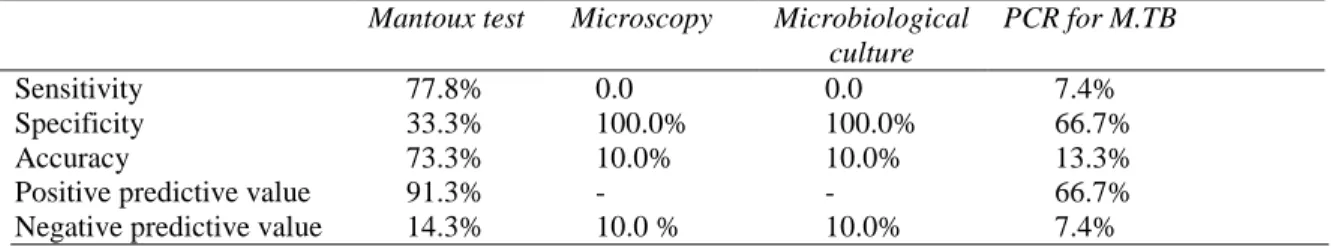
Table 9 Sensitivity, specificity, accuracy, positive and negative predictive values of the Mantoux test results, microscopy using Ziehl-Neelsen stain, microbiological culture using Lowenstein-Jensen medium and PCR for cutaneous tuberculosis (N=30).
Mantoux test Microscopy Microbiological culture
PCR for M.TB
Sensitivity 77.8% 0.0 0.0 7.4%
Specificity 33.3% 100.0% 100.0% 66.7%
Accuracy 73.3% 10.0% 10.0% 13.3%
Positive predictive value 91.3% - - 66.7%
Negative predictive value 14.3% 10.0 % 10.0% 7.4%
Table 6 shows PCR results for M. tuberculosis
of the patients. It was observed that 3 (10.0%) patients had positive PCR for M. tuberculosis
and 27 (90.0%) had negative PCR for M. tuberculosis.
Table 7 shows comparison between histopathological diagnosis and PCR for M.
tuberculosis in cutaneous tuberculosis. It was
observed that there were true positive 2 (6.6%) cases, false positive one (3.3%) case, false negative 25 (83.4%) cases and true negative 2 (6.6%) cases identified by skin histopathology.
Table 8 depicts antitubercular treatment response of the patients within 3 months. It was observed that more than two third (66.7%) patients responded to antitubercular treatment within 3 months, response was not monitored in 7 (23.3%) patients and 3 (10.0%) patients had no response.
Sensitivity, specificity, accuracy, positive and negative predictive values of the Mantoux test results, microscopy, microbiological culture and PCR are shown in Table 9.
Discussion
sensitivity and specificity values of PCR in the diagnosis of cutaneous TB vary widely in literature. In cutaneous TB, the sensitivity of PCR has varied from 54% to 100% and specificity varied from 80% to 100%.6 The sensitivity and specificity were found 7.4% and 66.7%, respectively in this study.
This study also shows that the most common cutaneous tuberculosis was tuberculosis verrucosa cutis (43.3%). 9 (30.0%) patients had lupus vulgaris, 4 (13.3%) erythema induratum of Bazin, 3 (10.0%) scrofuloderma and 1 (3.3%) patient had other clinical types. The most common cutaneous tuberculosis reported in literature is lupus vulgaris.7 This dissimilarity is difficult to explain.
The study found that most common location of cutaneous tuberculosis was an extremity followed by head and neck region including the face. Lupus vulgaris is common on the face and neck. Erythema induratum commonly occurs on the posterior lower calf and tuberculosis verrucosa cutis is frequently located on dorsa of the fingers and hands in adults, and the ankles and buttocks in children.
The study showed that most of the lesions were of solitary in nature. Concomitant pulmonary tuberculosis was detected in 10% of patients. Mantoux test was positive in 76.7% of the cutaneous TB patients, while 23.3 % of the cases showed negative. Solis et al. found that 26% of cutaneous tuberculosis and 10% of nontuberculous patients were M. tuberculosis positive. For cutaneous tuberculosis, TST has sensitivity between 33% and 96% and specificity of 62.5% with a cut off of 10 mm. In unvaccinated populations, the sensitivity is much higher, close to 97%.
Histopathological examination is essential to complement the investigation of cutaneous TB
cases, however, in this study, the most common histological finding in cutaneous TB was the granuloma in the dermis/ subcutis (73.3% cases), whereas granuloma with caseous necrosis was found in 16.7% cases. In the majority (55.6%) of the cases, the location of the granulomas was not specified; middermal granulomas were found in 7.4% cases and 18.5% of granulomas were in upper dermis, 18.5% of the granulomas were in the subcutis. Nevertheless, it should be remembered that other types of granuloma can also be found in cutaneous TB, such as sarcoid and suppurative granuloma. Therefore when there is other evidence for cutaneous TB and one of these histological elements is present, the diagnosis of cutaneous TB cannot be discarded.
group of researchers studied protein antigen b (Pab) based PCR test in diagnosis of pulmonary and extrapulmonary tuberculosis and found that PCR has much higher sensitivity (74%) than culture (47%).8
A similar study was done in Singapore to explore the role of the polymerase chain reaction (PCR) for the detection of M. tuberculosis DNA as a diagnostic aid in cutaneous tuberculosis using routinely processed skin biopsy specimen. Tan et al.9 concluded that PCR based detection of M. tuberculosis DNA in skin samples may extend and improve the diagnostic panel for cutaneous tuberculosis and may be also used to differentiate atypical mycobacterial infections in an immunocompromised patient with negative culture, if the technique is prudently and properly used. In their study, they did not find PCR to be a useful complement to the clinical and histologic diagnosis of paucibacillary forms of cutaneous tuberculosis.9 However, our results were not consistent with the above study. False negative PCR results were found in 90% of the cases, which could be due to nonuniform distribution of the M. tuberculosis or to the presence of inhibitory substances in the tissue specimen. M. tuberculosis complex include M. tuberculosis, M. bovis, M. canetti and BCG Pasteur. M. bovis is an important cause of cutaneous tuberculosis and it share 99.5% gene sequence of M. tuberculosis complex. IS 1081 method detected all of the M. tuberculosis complex strains. So in GeneXpert method, there had the possibility of false negative PCR results of cutaneous TB. Paucibacillary nature of most of the cutaneous TB is another reason for the low positivity rate.
Conclusion
Under the light of our study, we discourage the
use of PCR as a routine diagnostic tool of cutaneous tuberculosis for its low sensitivity. We recommend a large scale study to support this findings.
References
1. Narasimhan P, Wood J, MacIntyre CR, Mathai D. Risk factors for tuberculosis. Pulm Med. 2013;2013:828939.
2. Schlossberg D, editor. Tuberculosis and Nontuberculous Mycobacterial Infections. New York: American Society for Microbiology Press; 2011.
3. Yates VM, Walker SL. Mycobacterial Infections. In: Griffiths C, Barker J, Bleiker T, Chalmers R, Creamer D, editors. Rook's Textbook of Dermatology, 9th edn. Oxford: John Wiley & Sons; 2016. P. 27.1-27.46. 4. Abdalla CM, De Oliveira ZN, Sotto MN,
Leite KR, Canavez FC, De Carvalho CM. Polymerase chain reaction compared to other laboratory findings and to clinical evaluation in the diagnosis of cutaneous tuberculosis and atypical mycobacteria skin infection. Int J Dermatol. 2009;48:27-35. 5. Kolk AH, Kox LF, Van Leeuwen J, Kuijper
S, Jansen HM. Clinical utility of the polymerase chain reaction in the diagnosis of extrapulmonary tuberculosis. Eur Resp J. 1998;11:1222-6.
6. Padmavathy L, Rao L, Veliath A. Utility of polymerase chain reaction as a diagnostic tool in cutaneous tuberculosis. Indian J Dermatol Venereol Leprol. 2003;69 (3):214-6.
7. Salem JI, Gadelha AR, Maroja F, David HL. Non-cultivable mycobacteria in ulcers of the skin. Acta Leprologica. 1988;7:10-5. 8. Negi SS, Anand R, Basir SF, Pasha ST.
Protein antigen b (Pab) based PCR test in diagnosis of pulmonary and extra-pulmonary tuberculosis. Indian J Med Res. 2006;124:81.