Open Access
Review
Mutant mouse models and their contribution to our knowledge of
corpus luteum development, function and regression
Luiz E Henkes
1, John S Davis
2and Bo R Rueda*
1Address: 1Vincent Center for Reproductive Biology, Department of Obstetrics and Gynecology, Massachusetts General Hospital, Boston,
Massachusetts 02114, USA and 2Olson Center for Women's Health, Department of Obstetrics and Gynecology, University of Nebraska Medical
Center, Omaha, Nebraska 68198; VA Medical Center, Omaha, Nebraska 68105, USA
Email: Luiz E Henkes - [email protected]; John S Davis - [email protected]; Bo R Rueda* - [email protected] * Corresponding author
Abstract
The corpus luteum is a unique organ, which is transitory in nature. The development, maintenance and regression of the corpus luteum are regulated by endocrine, paracrine and autocrine signaling events. Defining the specific mediators of luteal development, maintenance and regression has been difficult and often perplexing due to the complexity that stems from the variety of cell types that make up the luteal tissue. Moreover, some regulators may serve dual functions as a luteotropic and luteolytic agent depending on the temporal and spatial environment in which they are expressed. As a result, some confusion is present in the interpretation of in vitro and in vivo studies. More recently investigators have utilized mutant mouse models to define the functional significance of specific gene products. The goal of this mini-review is to identify and discuss mutant mouse models that have luteal anomalies, which may provide some clues as to the significance of specific regulators of corpus luteum function.
Introduction
The corpus luteum is an important byproduct of the ovu-lating follicle. It is a transitory, hormonally regulated organ that consists of a heterogeneous cell population. Unlike the follicle, the different cell types are not segre-gated into distinct compartments, but are highly inte-grated. The steroidogenic cells synthesize progesterone for the establishment and maintenance of pregnancy. Other cell types include the endothelial cells and immune cells, typically thought to play supportive roles. There is evi-dence to suggest that the endothelial cells and the immune cells play an active role in luteal formation, maintenance and regression [1]. The vascular endothelial cells make up a large portion of the corpus luteum, whereas the immune cells vary in number dependent upon the stage of the luteal phase or pregnancy [1-3].
Studies designed to elucidate the contributions of one or more of the luteal cell types are often difficult to interpret. More often than not these studies are based on in vitro cell culture models. Primary cultures of dispersed luteal tissue or enriched populations of specific cell types provide some opportunity for investigators to delineate potential signaling pathways, which may be engaged in response to a specific stimulus. Outcomes derived from in vitro studies are important but are often subject to criticism. For exam-ple, in vitro studies tend to favor one cell type or another. Moreover, the cell-cell interactions that are present in a multidimensional environment in vivo are removed when experiments are performed in a two dimensional field in vitro (eg, tissue culture dish). What effect this has on an outcome is not always fully appreciated and cannot be directly extrapolated to the in vivo model. For example prostaglandin F2α (PGF2α) is primarily considered a Published: 10 November 2003
Reproductive Biology and Endocrinology 2003, 1:87
Received: 15 April 2003 Accepted: 10 November 2003
This article is available from: http://www.rbej.com/content/1/1/87
luteolytic agent in vivo, yet it has no direct lytic effect on endothelial cells or steroidogenic cells in vitro [1,4,5]. This raises a number of questions. Is the response observed in vitro an artifact of the static culture systems most often employed? Alternatively, are in vitro cultures lacking a luteolytic agent found in vivo, or is cell-to-cell communi-cation critical for the production of a luteolytic factor present only in the in vivo environment?
Alternatives to the current in vitro and in vivo strategies are necessary to fully understand the functional significance of putative mediators of luteal development and regres-sion. The development of various mutant mouse models has provided an invaluable knowledgebase for defining or possibly redefining the function and/or significance of many gene products. The mutant mouse models, whether they are hypermorphs, hypomorphs, conditional knock-outs or true knockknock-outs, provide a unique opportunity to define function of the genes or their products. However, these models have inherent caveats and have provided us with a new list of disclaimers to help interpret the unex-plainable findings. One such issue is redundancy. Often times there are built in safeguards within a cell type or alternatively there is system overlap to protect or compen-sate for the loss of a particular protein. Therefore when a protein is deficient, a gross phenotype is not always read-ily evident. Alternatively, the significance of a particular protein to corpus luteum function may be underestimated when a loss of the protein results in embryonic lethality. Of course this makes it very difficult to determine its func-tion or significance in events that control the cyclic nature of the mature female.
Some phenotypes are more controversial than others. The 'fertility' of female mice is subject to a number of biases. Some investigators will claim that if a female delivers one live offspring she is fertile. Others would argue that because the average mouse litter size is 7–8 pups, a mouse that delivers fewer than 7–8 pups has a fertility problem. For practical purposes herein a reduction in litter size will be described as sub-fertile. It is not so clear how to classify mutant mouse models that display erratic estrous cycles, because they may still be able to become pregnant and deliver a normal size litter. Although mutant mice may never reach the reproductive potential of their wild type siblings, they do deliver pups and under a strict definition of fertility they could be classified as fertile. In reality though the reproductive potential of a number of female mutant mice is sub-optimal suggesting that they are truly less fertile than their wild type counter parts.
Mutant mice models are often generated to investigate non-reproductive problems. Therefore, investigators who are not directly engaged in a reproductive study or inves-tigators not familiar with the reproductive field may miss
or dismiss a phenotype pertinent to reproduction. Repro-ductive anomalies are not limited to reduced fecundity or irregular estrous cycles, but include anovulation, hypoth-alamic or pituitary defects, implantation defects, sub-opti-mal hormone concentrations, and/or parturition defects. This article provides a brief review of mouse models that have defects affecting the development, function and regression of the corpus luteum.
Mutant mice models with preovulatory/luteal
development and/or luteal maintenance defects/
anomalies
It is important to recognize that in some mutant mouse models ovarian follicles fail to ovulate (Table 1); yet, the steroidogenic cells may undergo luteinization spontane-ously or in response to exogenous gonadotropins result-ing in a luteinized unruptured follicle. A number of anovular phenotypes have been reported: gonadotropin receptors: LH receptor, FSH receptor [6]; gonadotropins: FSHβ subunit [7], glycoprotein hormone α subunit [8]; steroid hormone receptors: ERα [9], ERα/ERβ [10], PR [11]: cell cycle regulatory proteins: cyclin D2 [12], p27(kip) [13]; enzymes for steroidogenesis and prostag-landin synthesis: aromatase [14], and COX-2 [15]. The ability of steroidogenic cells to undergo luteinization nat-urally would suggest that at least some signaling between the pituitary-hypothalamic-ovarian axis is intact. Alterna-tively, if luteinization does not occur, but is initiated only with exogenous gonadotropins it can be predicted that one or more signaling pathways have been interrupted. For example, preovulatory follicles of cyclin-D2-/- females
undergo arrest and do not ovulate, however their granu-losa cells undergo luteinization [16]. Similarly, inactiva-tion of the type 4 cAMP specific phosphodiesterase (PDE4D) gene results in infertile female mice. PDE4D is critical for feedback regulation of cAMP levels and PDE4D females have a high incidence of entrapped oocytes within the follicles and yet the steroidogenic cells undergo luteinization [17]. Another example includes the nuclear corepressor Nrip1 (a.k.a. RIP140) -/- mouse which is
infer-tile [18]. The infertility stems from a failure of the follicles to undergo maturation. As in the previous examples the inability to ovulate is independent of the ability to undergo luteinization [18]. Connexin 37-/- female mice
also fall into this category. Connexin 37 is normally present in gap junctions between oocyte and granulosa cells of the follicle and is critical for signaling [19]. Con-nexin 37-/- female mice lack mature preovulatory follicles
was achieved. Thus, cell-cell signaling through intercellu-lar channels critically regulates the highly coordinated set of cellular interactions required for successful oocyte development and ovulation [19]. In contrast, it is not nec-essarily obligatory for cell-cell signaling through intercel-lular channels to induce luteinization.
There are also mutant mouse models, which provide indi-rect evidence that luteinization can occur in the absence of ovulation (Table 1). For example, female mice lacking the gene for endothelial nitric oxide synthase (eNOS-/-) have
irregular estrous cycles and fewer pups per litter [20]. In response to gonadotropin stimulation the eNOS-/- females
have a significant reduction in ovulatory efficiency com-pared with wild type female, however there was no signif-icant difference in plasma progesterone concentrations [21]. It appears that the luteinization process is not inter-rupted although ovulation rate is compromised. This dif-ference may be due in part to unrecognized luteinized follicles.
Collectively the mutant models with ovulation defects described above provide evidence to suggest that luteini-zation is independent of ovulation and that ovulation of the oocyte is not obligatory for luteinization. There are also examples of mutant mice which have an ovulation defect but there is no evidence that the follicles undergo luteinization (Table 1). One example is the Progesterone Receptor (PRKO) and Progesterone Receptor alpha (PRAKO) knockout mice. The importance of progesterone derived from the corpus luteum in the establishment of pregnancy is well accepted. However the pervasive impact of progesterone on reproduction became more evident with the development of the PRKO mice [11,22]. The PRKO model was designed by targeting both the PRA and PRB isoforms. The females develop normally, however they have multiple reproductive defects including an ina-bility to ovulate, uterine hyperplasia, limited mammary development and an inability to exhibit sexual behavior [22]. All of these symptoms likely contribute to their reported infertility. The PRAKO mouse, generated by selective ablation of the PRA gene [23], are also infertile. Gonadotropin stimulation of PRKO and PRAKO mice results in the development of follicles, however only PRAKO mice ovulate. Pregnancy is not possible due a defect in decidualization [22,23]. Collectively, the availa-ble data indicate that progesterone is required for more than just the establishment of pregnancy; it is required for ovulation, a prerequisite for true CL formation. There are no data provided to determine whether or not the unrup-tured follicles become luteinized.
An additional example of an anovulatory mutant mouse would include the estrogen receptor mutant mice. There is no doubt that estrogen plays a significant physiological
role in folliculogenesis. Estrogen stimulates both granu-losa cell proliferation and differentiation [24,25]. Estro-gen is also responsible for the induction of follicle stimulating hormone (FSH) and luteinizing hormone (LH) receptors [24,26]. Estrogen binds both estrogen receptors; ERα [27,28] and ERβ [29]. Both are expressed in granulosa cells of preantral and antral follicles [25] and have a highly conserved DNA binding domain [29]. ERα is more prevalent in stromal and theca cells while ERβ is predominant in antral follicles [30]. ERα knockout mice (αERKO) females are acyclic, infertile and display enlarged, hemorrhagic and cystic follicles with a high inci-dence of ovarian tumors [10]. In contrast to the ERα female mice, which are completely infertile, the ERβ null mice females are subfertile. The ERβ-/- mice (βERKO) have
decreased ovulation rates, fewer litters, less pups per litter and sparse corpora lutea. The double knockouts (αERKO and βERKO) present with a phenotype similar to ERα knockout [10].
Luteinizing hormone (LH), obviously by its name, is well recognized as a luteotropic agent and is pivotal to mam-malian reproduction. LH contributes to the maintenance of gametogenesis and reproductive tract development in the female [31-34]. Receptors for LH are found predomi-nately in the ovary, but numerous reports over the past 15 years demonstrate expression of functional LH receptors in numerous extra-gonadal tissues [33]. Mutations in gonadotropin and gonadotropin receptor genes are very rare [35,36], however, these mutations have helped to define the physiology and pathophysiology of gonadotro-pin action [37]. Targeted disruption of the LHR gene causes infertility in both sexes [34,38-40]. Other pheno-types include gross underdevelopment of internal and external genitalia [38,40]. With respect to the mouse ovary, the adult LHR-/- female displays small ovaries and
follicular development up to the preantral stage [40]. The mutant mice had no discernable reductions in FSH recep-tor or progesterone receprecep-tor mRNA [38]. Furthermore, there was no apparent difference in the development of the theca layer surrounding the developing follicle. However, the theca in mutant mice displayed a marked reduction in the expression of mRNA for P450 17-hydrox-ylase [41] and steroidogenic acute regulatory (StAR) pro-tein [38]. As a result steroid hormone levels were markedly reduced, which can account for the observed hypoplastic uterus and elevated gonadotropin levels [38,41]. No evidence of preovulatory follicles or corpora lutea are observed in the LHR-/- mice [38-40]. Injections of
follicle requires the actions of LH. Recent studies using the LHR-/- mutant mouse model also provide evidence for
possible extra-gonadal roles for the LH receptor in uterine morphogenesis [39].
Another example of where mutant mice display an inabil-ity to develop corpora lutea includes the mice deficient in CATT/enhancer binding protein (C/EBPβ) [42]. CATT/ enhancer binding protein (C/EBPβ) is expressed in granu-losa cells of the ovary after LH stimulation, in vitro. Simi-larly, C/EBPβ is expressed in granulosa and not thecal cells of antral follicles derived from hCG treated wild type females. C/EBPβ is not evident in the intact corpus luteum in wild type mice suggesting a functional role for C/EBPβ in the granulosa cells. This role is apparently lost or severely down regulated during the luteinization of gran-ulosa cells. The obligatory role of C/EBPβ is demonstrated in the mice deficient in this protein [42]. C/EBPβ-/- female
mice fail to ovulate and therefore cannot initiate or main-tain a pregnancy. There were no gross abnormalities in the uterine tissue and uterine wet weights are similar between the C/EBPβ-/- and wild type females. Marked differences in
ovarian function were observed when females were sub-jected to gonadotropin-induced superovulation regime. The heterozygous females ovulated an average of 30 oocytes whereas the C/EBPβ-/- females ovulated 3 to 6
oocytes. The ovaries of the C/EBPβ-/-females mice had
evi-dence of large, often hemorrhagic antral follicles which were not evident in the wild type females [42]. These observations suggest that there is a transition failure in ovulation and luteinization. To verify that the infertility was intrinsic to the C/EBPβ-/- ovarian phenotype, ovaries
deemed to be normal were transplanted to homozygous null females and the infertility was resolved arguing that the pituitary, hypothalamus and uterus were hormonally responsive and intact. In contrast, corpora lutea never formed when ovaries of mutant mice were transplanted into normal females. Sterneck et al., [42] summarized that the morphology of superovulated ovaries of C/EBPβ
-/-females were indicative that these mice lacked the neces-sary mechanisms required to induce ovulation and sup-port luteinization. The ovarian transplant experiments further support the significance of C/EBPβ to luteal formation.
The final example of a mouse model with an ovulation/ luteinization defect is the Large tumor suppressor homolog 1 mutant mouse (Lats-1-/-) [43]. Lats1 is a tumor
suppressor originally identified in the Drosophila mela-nogaster. Lats1-/- mice display infertility, growth
retarda-tion and lack of mammary gland development. They also exhibit hyperplastic changes in the pituitary and decreased serum hormone levels (i.e. LH, prolactin (PRL) and growth hormone). Based on vaginal cytology Lats1
-/-mice do not exhibit an estrous cycle and remain in
mete-strus. The majority of the follicles are primary and second-ary follicles with few follicles if any attaining antrum formation. There is no evidence of CL formation. Gona-dotropins stimulate estrous cyclicity, although is reported to be prolonged [43]. It is not clear what phase of the cycle is prolonged. Ovaries from Lats-1-/-females contain fewer
follicles than wild type females of the same litter. The reproductive hormone defects of the Lat1 mutant mice are similar to that LH-hypogonadotropic hypogonadism and CL insufficiency in humans [43].
It is well recognized that luteinization of the steroidogenic cells of the follicle marks a significant point whereby the steroidogenic cells undergo hypertrophy and hyperplasia only to be followed by cellular differentiation and a dra-matic reduction in cellular proliferation. Concurrent with this process there are significant changes in the levels and actions of specific cyclins, their corresponding cyclin dependent kinases, and cell cycle inhibitors (i.e. p27 and p21). The cell cycle is regulated by cyclin interaction with cyclin dependent kinases (CDKs) [44]. Progression through G1 is regulated by the Cyclin D and E dependent kinases. In the G1 phase type D cyclins bind and activate CDK4 or CDK6. Cyclin E activates CDK2 in the late G1 phase. The CDKs can be inhibited by CDK inhibitors, which are classified into two groups, Kip/Cip and Ink4 inhibitors. Kip/Cip family includes p21, p27 and p57. The Ink4 inhibitor family includes p15, p16, p18 and p19 [44]. Many of the changes observed in these regulators of cell cycle are believed to be mediated in part by hormones also implicated in follicular growth, ovulation, luteal for-mation/luteinization [45].
The significance of cyclins, CDK and their inhibitors becomes readily apparent in the mutant mouse models. FSH or bromo-cAMP failed to induce proliferation of granulosa cells derived from cyclin D2-/- female mice
[12,16]. Moreover, the cyclin D2-/- female mice fail to
ovu-late, but undergo luteinization [12]. The p27-/- mice
exhibit a number of abnormalities including gigantism with multi-tissue hyperplasia, benign adenomas in the pituitary, and female infertility [46]. Of interest to this review is the fact that granulosa cells in the ovary of p27-/ - mice continue to proliferate beyond the LH surge,
sug-gesting that p27 plays a critical role in establishing quies-cence or differentiation of luteinizing granulosa cells. Cyclin dependent kinase 4-/- mice are also infertile and
females exhibit prolonged estrous cycles [47,48]. Although the CDK4-/- mice develop corpora lutea, the
Not all mutants are the product of human intervention. Hypothyroid (hyt) mice are autosomal recessive for hypothyroidism [49]. The hyt females display continuous diestrous contributing to their infertility [50]. Stimulation of immature female hyt and wild type mice with exoge-nous gonadotropins will induce follicle development at the same rate. However in gonadotropin stimulated mature female hyt mice, the number of oocytes ovulated were less than their wild type counterparts and pregnancy is never achieved. Mature hyt females have significantly fewer corpora lutea > 500 microns in diameter and signif-icantly lower progesterone. Thyroxine treatment before mating reverses the insufficiencies; the mice have well-developed corpora lutea and progesterone levels are increased [50].
Tissue inhibitor of metalloproteinase-1 (TIMP-1) has been implicated as a potential regulator of steroidogenesis [51]. This has been recently validated by evaluation of the luteal phenotype of TIMP-1 mutant mice [51]. To validate TIMP-1 functional significance to steroidogenesis in the corpora lutea, wild type and TIMP-/- mice were treated
with eCG, followed by hCG to induce ovulation. Proges-terone increased post hCG treatment in both genotypes, however, the progesterone concentrations in TIMP-/- were
less than that observed in wild type mice. The lack of pro-gesterone was not attributed to insufficient luteal forma-tion since a similar number of oocytes were harvested from both wild type and TIMP-/- mice suggesting a similar
number of corpora lutea were formed. Although the mean mass of the corpora lutea in the two genotypes was not
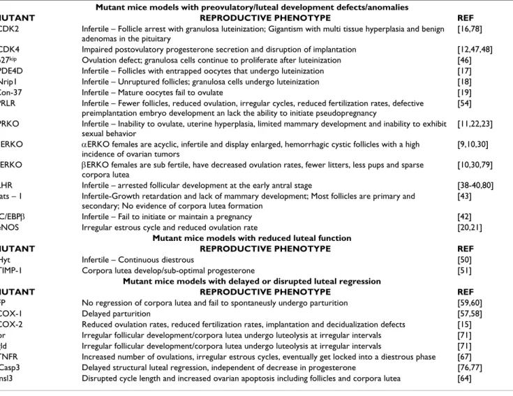
Table 1: Summary of Reproductive Female Phenotypes in Mutant Mice models.
Mutant mice models with preovulatory/luteal development defects/anomalies
MUTANT REPRODUCTIVE PHENOTYPE REF
aCDK2 Infertile – Follicle arrest with granulosa luteinization; Gigantism with multi tissue hyperplasia and benign
adenomas in the pituitary
[16,78]
bCDK4 Impaired postovulatory progesterone secretion and disruption of implantation [12,47,48] cp27kip Ovulation defect; granulosa cells continue to proliferate after luteinization [46] dPDE4D Infertile – Follicles with entrapped oocytes that undergo luteinization [17] eNrip1 Infertile – Unruptured follicles; granulosa cells undergo luteinization [18]
fCon-37 Infertile – Mature oocytes fail to ovulate [19]
gPRLR Infertile – Fewer follicles, reduced ovulation, irregular cycles, reduced fertilization rates, defective
preimplantation embryo development an lack the ability to initiate pseudopregnancy
[54]
hPRKO Infertile – Inability to ovulate, uterine hyperplasia, limited mammary development and inability to exhibit
sexual behavior
[11,22,23]
iαERKO αERKO females are acyclic, infertile and display enlarged, hemorrhagic cystic follicles with a high
incidence of ovarian tumors
[9,10,30]
jβERKO βERKO females are sub fertile, have decreased ovulation rates, fewer litters, less pups and sparse
corpora lutea
[10,30,79]
kLHR Infertile – arrested follicular development at the early antral stage [38-40,80] lLats – 1 Infertile-Growth retardation and lack of mammary development; Most follicles are primary and
secondary; No evidence of corpora lutea formation
[43]
mC/EBPβ Infertile – Fail to initiate or maintain a pregnancy [42] neNOS Irregular estrous cycle and reduced ovulation rate [20,21]
Mutant mice models with reduced luteal function
MUTANT REPRODUCTIVE PHENOTYPE REF
oHyt Infertile – Continuous diestrous [50]
pTIMP-1 Corpora lutea develop/sub-optimal progesterone [51]
Mutant mice models with delayed or disrupted luteal regression
MUTANT REPRODUCTIVE PHENOTYPE REF
qFP No regression of corpora lutea and fail to spontaneusly undergo parturition [59,60]
rCOX-1 Delayed parturition [57,58]
sCOX-2 Reduced ovulation rates, reduced fertilization rates, implantation and decidualization defects [15] tlpr Irregular follicular development/corpora lutea undergo luteolysis at irregular intervals [71] ugld Irregular follicular development/corpora lutea undergo luteolysis at irregular intervals [71] vTNFR Increased number of ovulations, irregular estrous cycles, eventually get locked into a diestrous phase [67] wCasp3 Delayed structural luteal regression, independent of decrease in progesterone [76,77] xInsl3 Disrupted cycle length and increased ovarian apoptosis including follicles and corpora lutea [64]
List of acronyms or abbreviations: aCyclin dependent kinase 2, bCyclin dependent kinase 4, cp27(kip), dType 4 cAMP-specific phosphodiesterase, eNuclear receptor co-repressor Nrip1 (a.k.a. RIP140), fConnexin-37, gProlactin receptor, hProgesterone receptor, iαEstrogen receptor, jβEstrogen
receptor, kLuteinizing hormone receptor, lLarge tumor suppressor homolog 1, mCCAAT/enhancer-binding protein β, nEndothelial nitric oxide
reported, the weights of the ovaries from TIMP-/- mice
were significantly larger than their wild type counterparts following gonadotropin stimulation [51]. These data pro-vide epro-vidence to support a significant role of TIMP-1 in steroidogenesis.
Mutant mice models with luteal regression
defects/anomalies
The prolactin receptor knockout mice provide some inter-esting insights into luteal function and regression. Prolac-tin (PRL) is a pituitary hormone recognized for its luteotropic and luteolytic actions [52,53]. More specifi-cally, prolactin regulates corpora lutea formation, ster-oidogenesis, gonadotropin receptors and luteal demise in rodents [52]. Therefore it is not unexpected that prolactin receptor null mice (PRLR-/-) have multiple reproductive
anomalies. PRLR-/- mice have fewer follicles, reduced
ovu-lation, irregular cycles, reduced fertilization rates, defec-tive preimplantation embryo development and lack the ability to initiate pseudopregnancy. The length of the estrous cycle does not appear to differ between the PRLR-/ - females and their wild type counterparts [54]. Moreover
the number of oocytes ovulated normally or in response to gonadotropin stimulus were the same between the two phenotypes [54]. These data suggest that PRL deficiency does not affect the ovarian responsiveness to gonadotro-pins. Corpora lutea form but display an elevated level of apoptosis. Moreover, there is little evidence of PECAM/ CD31, an indirect index for vascularization. These data suggest that the corpora lutea of PRLR-/- mice have reduced
vascularization [54]. Collectively, the reproductive anom-alies observed in the PRLR-/- mice have been attributed to
impaired luteal function resulting in insufficient levels of progesterone to support implantation.
Cyclooxygenase (COX) catalyzes the conversion of arachi-donic acid into prostaglandin H2 (PGH2) a substrate required for the generation of other prostaglandins including PGF2α. Prostaglandin F2α is especially impor-tant in the process of luteolysis [1-3,55]. The distribution and varied levels of COX expression in different tissues suggest that the biological actions of cyclooxygenase may be tissue specific [56]. COX activity is considered a rate-limiting step and disruption of COX activity and subse-quent diminished prostaglandin levels was hypothesized to have a significant negative effect on reproductive func-tion. COX-1-/- female mice have multiple defects [57,58].
Of importance herein, the COX-1-/- mice have a delayed
parturition. In a normal pregnant wild type mouse there is an increase in uterine PGF2α production on day 19 associated with luteolysis and parturition. This increase is not evident in the COX-1-/- pregnant females [57,58].
Administration of PGF2α will reverse the parturition defect. These data support an obligatory role for COX-1 in parturition and hence luteal regression. The reproductive
defects displayed by COX-1 deficient mice are similar to that displayed by FP-/- mice [59,60]. The COX-2-/- females
have evidence of disrupted ovulation, reduced fertiliza-tion rates, implantafertiliza-tion and decidualizafertiliza-tion defects [15,58]. Simultaneous inhibition of COX-1 and COX-2 resulted in more severe effects than either isoform alone [61].
Prostaglandin F2α has long been implicated as a primary luteolytic agent, however the development of the PGF2α receptor mutant mice (FP-/-) provides additional insight
into the overall significance of PGF2α to the regression of the corpora lutea. Sugimoto and colleagues [59] demon-strated that homozygous females cycled normally and achieved pregnancy. Interestingly, FP-/- pregnant females
failed to undergo spontaneous parturition similar to that observed in the COX mutant females [58]. There was no decline in progesterone levels and no morphological evi-dence of regression. Parturition could only be induced by an ovariectomy on day 19; likely the result of a fall in pro-gesterone levels. It is interesting that the effect on the cor-pus luteum is limited to the corpora lutea of pregnancy. There is no evidence that the lack of PGF2α signaling had any effect on the corpora lutea of the estrous cycle or in the corpora lutea formed in response to pseudopregnancy.
The like factor 3 (Insl3), a member of the insulin-like hormone family or relaxin family [62] is also impor-tant for gonadal function. In the female, low amounts of RLF (Insl3) are produced in both the uterus and ovary, particularly in the theca cells of small antral follicle, where expression of the hormone is correlated with the selection of the follicles to become preovulatory [63]. In knockout mice, there is a altered female phenotype, with disturbed cycle length and increased ovarian apoptosis, particularly in follicles and corpora lutea [64]. This was demonstrated following the collection of ovaries from 40-day-old- and 6-month-old wild type and Insl3-/- mice littermates, which
were serially sectioned and assessed. It was determined that the number of zonae pellucidae is higher in Insl3
-/-ovaries of both ages than in -/-ovaries of wild-type sisters. Wild type mice of both ages possess threefold more cor-pora lutea than their Insl3-/- littermates. In general,
wild-type corpora lutea appear healthy, show GS I-positive endothelial cells and no apoptotic cells whereas corpora lutea from mutants are rich in regressing GS I luteal cells, and an increased number of apoptotic cells. It was con-cluded that follicular atresia and luteolysis are accelerated in ovaries of Insl3-/- mice probably because of increased
apoptosis. The Insl3 function may provide survival signals to rescue endocrine cells from the apoptotic pathway.
mouse has irregular estrous cycles what exactly does that mean? Is the irregular estrous cycle attributed to only dis-rupted follicle development, delayed luteal development or disruption of luteal regression or can it be a combina-tion of all three. Examples of mutant mice models with irregular estrous cycles other than those discussed above would include the tumor necrosis factor receptor mutants (TNFR1-/-), generalized lymphoproliferative disease (gld)
mutants and lymphoproliferation (lpr) mutant mice (Table 1). Evidence for the involvement of TNFα in ovar-ian function is provided in recent reviews [1,65,66] which was further supported by Roby et al [67] who described the reproductive anomalies associated with the TNFRI
-/-female mice. Prepubertal TNFRI-/- mice stimulated with
gonadotropins ovulate more ova compared to their wild type controls. This increase in number of ovulations by TNFRI-/- mice was associated with higher serum levels of
progesterone. The increased ovulatory response was lost when the mutant females matured. At an early age the TNFRI-/- female mice have the same length of estrous cycle
as their wild type counterparts. However the TNFRI
-/-females spent more time in diestrous than did the control mice. By 6 months of age only 40% percent of the females remained cyclic and those that did not cycle appeared to be 'locked' into a diestrous phase. Also of interest was that an increased number of TNFRI-/- females failed to deliver
and pups suggesting that there was a higher incidence of infertility [67]. This study implicates TNFα as a critical reg-ulator of luteal regression. These results are supported indirectly by an earlier study in which anti-thymocyte antiserum was injected in rats to inhibit immune function [68]. Similar to the TNFRI-/- mice these rats failed to
progress past the diestrous phase. Although this study does not directly implicate TNFα, it does provide addi-tional support that the immune system plays an integral role in the physical regression of the corpora lutea.
The homozygous gld mice have a non-functional Fas lig-and (FasL) lig-and lpr mice have reduced expression of FAS (receptor) [69,70]. The corpora lutea of these mice undergo luteolysis but at irregular intervals. Moreover they have irregular follicle development. The lpr mice have increased numbers of secondary follicles [71]. There-fore it is not clear as to where the defect lies. Regardless, these studies do provide evidence to suggest that FAS mediated events are critical to the cyclicity of the female mouse. FasL or FAS activating antibodies can induce luteal cell death in the human, mouse, rat, and cow [71-75] and induce luteal regression in wild type mice [71,76]. FAS-mediated cell death results in the activation of caspase-3, a primary effector caspase [76]. More interestingly, the onset of FAS mediated cell death is attenuated in caspase-3-/- mice when compared to wild type mice [76].
Carambula et al., [77] predicted that corpora lutea derived from caspase-3-/- mice would exhibit a delayed onset of
apoptosis during luteal regression when compared with corpora lutea derived from wild type mice. Upon exami-nation of ovaries of wild type mice stimulated with gona-dotropins only residual luteal tissue at day 6 post-ovulation, ovaries collected from caspase-3-/- mice
retained many corpora lutea at day 6 post-ovulation that were similar in size to those observed in the early luteal phase of wild type mice. Notably, there was no dramatic increase in apoptosis in corpora lutea of caspase-3-/- mice
at any time point examined post-ovulation, indicating that luteal involution had been delayed. On the contrary, the levels of progesterone declined regardless of genotype. These data provide evidence that caspase-3 is functionally required for apoptosis to proceed normally during luteal regression. Moreover, these data suggest caspase-3 is not a direct mediator of the decrease in steroidogenesis associ-ated with luteolysis [77]. Using this same model it was demonstrated that caspase-3 was downstream of PGF2α and FAS mediated luteal regression [76]. Treatment with PGF2α or Jo2 post-ovulation induced caspase-3 activation and increased the number of apoptotic cells when com-pared to IgG treated controls. In contrast, corpora lutea in ovaries collected from caspase-3-/- mice, whether treated
with PGF2α, Jo2 or control IgG, showed little evidence of active caspase-3 or apoptosis. Corpora lutea of wild type mice treated with Jo2 had increased the caspase-8 activity, an activator of caspase-3 that is coupled to the FAS death receptor. Treatment of wild type mice with PGF2α or Jo2 resulted in a increase in caspase-8 activity in the corpora lutea [76]. Based on these data it is suggested that luteoly-sis, at least in part, can be mediated by increasing the bio-activity or bioavailability of cytokines, such as FasL and that multiple endocrine factors can activate caspase-3-driven apoptosis during luteolysis [76].
Conclusions
tradi-tional views. Regardless, a better understanding of the sig-nificance of specific proteins and/or their receptors in corpora lutea development, function and regression can be gained from information obtained from mutant mice.
Acknowledgements
Dr. Rueda is supported in part by NIH HD35934 and the Vincent Memorial Hospital. Dr. Davis is supported by NIH HD38813 and the DVA.
References
1. Davis JS, Rueda BR: The corpus luteum: an ovarian structure with maternal instincts and suicidal tendencies. Front Biosci
2002, 7:d1949-1978.
2. Niswender GD, Juengel JL, Silva PJ, Rollyson MK, McIntush EW:
Mechanisms controlling the function and life span of the cor-pus luteum.Physiol Rev 2000, 80:1-29.
3. McCracken JA, Custer EE, Lamsa JC: Luteolysis: a neuroendo-crine-mediated event.Physiol Rev 1999, 79:263-323.
4. Cavicchio VA, Pru JK, Davis BS, Davis JS, Rueda BR, Townson DH:
Secretion of monocyte chemoattractant protein-1 (MCP-1) by endothelial cells of the bovine corpus luteum: Regulation by cytokines but not prostaglandin F2 alpha. Endocrinology
2002, 143:3582-3589.
5. Pru JK, Lynch MP, Davis JS, Rueda BR: Signaling mechanisms in tumor necrosis factor alpha-induced death of microvascular endothelial cells of the corpus luteum.Reprod Biol Endocrinol
2003, 1:17.
6. Danilovich N, Babu PS, Xing W, Gerdes M, Krishnamurthy H, Sairam MR: Estrogen deficiency, obesity, and skeletal abnormalities in follicle-stimulating hormone receptor knockout (FORKO) female mice.Endocrinology 2000, 141:4295-4308.
7. Burns KH, Yan C, Kumar TR, Matzuk MM: Analysis of ovarian gene expression in follicle-stimulating hormone beta knock-out mice.Endocrinology 2001, 142:2742-2751.
8. Kendall SK, Samuelson LC, Saunders TL, Wood RI, Camper SA: Tar-geted disruption of the pituitary glycoprotein hormone alpha-subunit produces hypogonadal and hypothyroid mice.
Genes Dev 1995, 9:2007-2019.
9. Couse JF, Korach KS: Reproductive phenotypes in the estrogen receptor-alpha knockout mouse. Ann Endocrinol (Paris) 1999,
60:143-148.
10. Couse JF, Korach KS: Estrogen receptor null mice: what have we learned and where will they lead us? Endocr Rev 1999,
20:358-417.
11. Lydon JP, DeMayo FJ, Conneely OM, O'Malley BW: Reproductive phenotpes of the progesterone receptor null mutant mouse.
J Steroid Biochem Mol Biol 1996, 56:67-77.
12. Sicinski P, Donaher JL, Geng Y, Parker SB, Gardner H, Park MY, Rob-ker RL, Richards JS, McGinnis LK et al.: Cyclin D2 is an FSH-responsive gene involved in gonadal cell proliferation and oncogenesis.Nature 1996, 384:470-474.
13. Fero ML, Rivkin M, Tasch M, Porter P, Carow CE, Firpo E, Polyak K, Tsai LH, Broudy V, Perlmutter RM et al.: A syndrome of multior-gan hyperplasia with features of gimultior-gantism, tumorigenesis, and female sterility in p27(Kip1)-deficient mice. Cell 1996,
85:733-744.
14. Fisher CR, Graves KH, Parlow AF, Simpson ER: Characterization of mice deficient in aromatase (ArKO) because of targeted disruption of the cyp19 gene.Proc Natl Acad Sci U S A 1998,
95:6965-6970.
15. Lim H, Paria BC, Das SK, Dinchuk JE, Langenbach R, Trzaskos JM, Dey SK: Multiple female reproductive failures in cyclooxygenase 2-deficient mice.Cell 1997, 91:197-208.
16. Robker RL, Richards JS: Hormone-induced proliferation and dif-ferentiation of granulosa cells: a coordinated balance of the cell cycle regulators cyclin D2 and p27Kip1. Mol Endocrinol
1998, 12:924-940.
17. Jin SL, Richard FJ, Kuo WP, D'Ercole AJ, Conti M: Impaired growth and fertility of cAMP-specific phosphodiesterase PDE4D-deficient mice.Proc Natl Acad Sci U S A 1999, 96:11998-12003. 18. White R, Leonardsson G, Rosewell I, Ann Jacobs M, Milligan S, Parker
M: The nuclear receptor co-repressor nrip1 (RIP140) is essential for female fertility.Nat Med 2000, 6:1368-1374.
19. Simon AM, Goodenough DA, Li E, Paul DL: Female infertility in mice lacking connexin 37.Nature 1997, 385:525-529.
20. Drazen DL, Klein SL, Burnett AL, Wallach EE, Crone JK, Huang PL, Nelson RJ: Reproductive function in female mice lacking the gene for endothelial nitric oxide synthase.Nitric Oxide 1999,
3:366-374.
21. Jablonka-Shariff A, Olson LM: The role of nitric oxide in oocyte meiotic maturation and ovulation: meiotic abnormalities of endothelial nitric oxide synthase knock-out mouse oocytes.
Endocrinology 1998, 139:2944-2954.
22. Chappell PE, Lydon JP, Conneely OM, O'Malley BW, Levine JE: Endo-crine defects in mice carrying a null mutation for the proges-terone receptor gene.Endocrinology 1997, 138:4147-4152. 23. Mulac-Jericevic B, Mullinax RA, DeMayo FJ, Lydon JP, Conneely OM:
Subgroup of reproductive functions of progesterone medi-ated by progesterone receptor-B isoform. Science 2000,
289:1751-1754.
24. Drummond AE, Findlay JK: The role of estrogen in folliculogenesis.Mol Cell Endocrinol 1999, 151:57-64.
25. Britt KL, Findlay JK: Estrogen actions in the ovary revisited.J Endocrinol 2002, 175:269-276.
26. Drummond AE, Britt KL, Dyson M, Jones ME, Kerr JB, O'Donnell L, Simpson ER, Findlay JK: Ovarian steroid receptors and their role in ovarian function.Mol Cell Endocrinol 2002, 191:27-33. 27. Green S, Walter P, Greene G, Krust A, Goffin C, Jensen E, Scrace G,
Waterfield M, Chambon P: Cloning of the human oestrogen receptor cDNA.J Steroid Biochem 1986, 24:77-83.
28. Green S, Walter P, Kumar V, Krust A, Bornert JM, Argos P, Chambon P: Human oestrogen receptor cDNA: sequence, expression and homology to v-erb-A.Nature 1986, 320:134-139.
29. Kuiper GG, Enmark E, Pelto-Huikko M, Nilsson S, Gustafsson JA:
Cloning of a novel receptor expressed in rat prostate and ovary.Proc Natl Acad Sci U S A 1996, 93:5925-5930.
30. Couse JF, Hewitt SC, Bunch DO, Sar M, Walker VR, Davis BJ, Korach KS: Postnatal sex reversal of the ovaries in mice lacking estrogen receptors alpha and beta. Science 1999,
286:2328-2331.
31. Dufau ML: The luteinizing hormone receptor.Annu Rev Physiol
1998, 60:461-496.
32. Hillier SG: Gonadotropic control of ovarian follicular growth and development.Mol Cell Endocrinol 2001, 179:39-46.
33. Ziecik AJ, Derecka-Reszka K, Rzucidlo SJ: Extragonadal gonado-tropin receptors, their distribution and function. J Physiol Pharmacol 1992, 43:33-49.
34. Zhang M, Shi H, Segaloff DL, Van Voorhis BJ, Zheng M: Expression and localization of luteinizing hormone receptor in the female mouse reproductive tract.Biol Reprod 2001, 64:179-187. 35. Jameson JL: Inherited disorders of the gonadotropin
hormones.Mol Cell Endocrinol 1996, 125:143-149.
36. Huhtaniemi I: The Parkes lecture. Mutations of gonadotrophin and gonadotrophin receptor genes: what do they teach us about reproductive physiology? J Reprod Fertil 2000,
119:173-186.
37. Themmen APN, Huhtaniemi IT: Mutations of gonadotropins and gonadotropin receptors: elucidating the physiology and pathophysiology of pituitary-gonadal function. Endocr Rev
2000, 21:551-583.
38. Lei ZM, Mishra S, Zou W, Xu B, Foltz M, Li X, Rao CV: Targeted disruption of luteinizing hormone/human chorionic gonado-tropin receptor gene.Mol Endocrinol 2001, 15:184-200.
39. Rao CV, Lei ZM: Consequences of targeted inactivation of LH receptors.Mol Cell Endocrinol 2002, 187:57-67.
40. Huhtaniemi I, Zhang FP, Kero J, Hamalainen T, Poutanen M: Trans-genic and knockout mouse models for the study of luteiniz-ing hormone and luteinizluteiniz-ing hormone receptor function.Mol Cell Endocrinol 2002, 187:49-56.
41. Zhang FP, Poutanen M, Wilbertz J, Huhtaniemi I: Normal prenatal but arrested postnatal sexual development of luteinizing hormone receptor knockout (LuRKO) mice. Mol Endocrinol
2001, 15:172-183.
42. Sterneck E, Tessarollo L, Johnson PF: An essential role for C/EBP-beta in female reproduction.Genes Dev 1997, 11:2153-2162. 43. St John MA, Tao W, Fei X, Fukumoto R, Carcangiu ML, Brownstein
Publish with BioMed Central and every scientist can read your work free of charge
"BioMed Central will be the most significant development for disseminating the results of biomedical researc h in our lifetime."
Sir Paul Nurse, Cancer Research UK
Your research papers will be:
available free of charge to the entire biomedical community
peer reviewed and published immediately upon acceptance
cited in PubMed and archived on PubMed Central
yours — you keep the copyright
Submit your manuscript here:
http://www.biomedcentral.com/info/publishing_adv.asp
BioMedcentral
44. Sherr CJ: The Pezcoller lecture: cancer cell cycles revisited.
Cancer Res 2000, 60:3689-3695.
45. Richards JS, Russell DL, Robker RL, Dajee M, Alliston TN: Molecular mechanisms of ovulation and luteinization.Mol Cell Endocrinol
1998, 145:47-54.
46. Tong W, Kiyokawa H, Soos TJ, Park MS, Soares VC, Manova K, Pol-lard JW, Koff A: The absence of p27Kip1, an inhibitor of G1 cyclin-dependent kinases, uncouples differentiation and growth arrest during the granulosa->luteal transition. Cell Growth Differ 1998, 9:787-794.
47. Moons DS, Jirawatnotai S, Tsutsui T, Franks R, Parlow AF, Hales DB, Gibori G, Fazleabas AT, Kiyokawa H: Intact follicular maturation and defective luteal function in mice deficient for cyclin-dependent kinase-4.Endocrinology 2002, 143:647-654.
48. Rane SG, Dubus P, Mettus RV, Galbreath EJ, Boden G, Reddy EP, Bar-bacid M: Loss of Cdk4 expression causes insulin-deficient dia-betes and Cdk4 activation results in beta-islet cell hyperplasia.Nat Genet 1999, 22:44-52.
49. Stein SA, Oates EL, Hall CR, Grumbles RM, Fernandez LM, Taylor NA, Puett D, Jin S: Identification of a point mutation in the thy-rotropin receptor of the hyt/hyt hypothyroid mouse. Mol Endocrinol 1994, 8:129-138.
50. Jiang JY, Imai Y, Umezu M, Sato E: Characteristics of infertility in female hypothyroid (hyt) mice.Reproduction 2001, 122:695-700. 51. Nothnick WB: Tissue inhibitor of metalloproteinase-1 (TIMP-1) deficient mice display reduced serum progesterone levels during corpus luteum development. Endocrinology 2003,
144:5-8.
52. Risk M, Gibori G: Mechanisms of luteal cell regulation by prolactinBoston: Kluwer Academic Publishers; 2001.
53. Bowen JM, Keyes PL, Warren JS, Townson DH: Prolactin-induced regression of the rat corpus luteum: expression of monocyte chemoattractant protein-1 and invasion of macrophages.Biol Reprod 1996, 54:1120-1127.
54. Grosdemouge I, Bachelot A, Lucas A, Baran N, Kelly PA, Binart N:
Effects of deletion of the prolactin receptor on ovarian gene expression.Reprod Biol Endocrinol 2003, 1:12.
55. Bazer FW: Mediators of maternal recognition of pregnancy in mammals.Proc Soc Exp Biol Med 1992, 199:373-384.
56. Morita I: Distinct functions of COX-1 and COX-2.Prostaglandins Other Lipid Mediat 2002, 68–69:165-175.
57. Langenbach R, Morham SG, Tiano HF, Loftin CD, Ghanayem BI, Chu-lada PC, Mahler JF, Lee CA, Goulding EH, Kluckman KD et al.: Pros-taglandin synthase 1 gene disruption in mice reduces arachidonic acid-induced inflammation and indomethacin-induced gastric ulceration.Cell 1995, 83:483-492.
58. Gross GA, Imamura T, Luedke C, Vogt SK, Olson LM, Nelson DM, Sadovsky Y, Muglia LJ: Opposing actions of prostaglandins and oxytocin determine the onset of murine labor.Proc Natl Acad Sci U S A 1998, 95:11875-11879.
59. Sugimoto Y, Yamasaki A, Segi E, Tsuboi K, Aze Y, Nishimura T, Oida H, Yoshida N, Tanaka T, Katsuyama M, Hasumoto K, Murata T, Hirata M, Ushikubi F, Negishi M, Ichikawa A, Narumiya S: Failure of partu-rition in mice lacking the prostaglandin F receptor.Science
1997, 277:681-683.
60. Sugimoto Y, Segi E, Tsuboi K, Ichikawa A, Narumiya S: Female reproduction in mice lacking the prostaglandin F receptor. Roles of prostaglandin and oxytocin receptors in parturition.
Adv Exp Med Biol 1998, 449:317-321.
61. Reese J, Zhao X, Ma WG, Brown N, Maziasz TJ, Dey SK: Compara-tive analysis of pharmacologic and/or genetic disruption of cyclooxygenase-1 and cyclooxygenase-2 function in female reproduction in mice.Endocrinology 2001, 142:3198-3206. 62. Ivell R, Bathgate RA: Reproductive biology of the relaxin-like
factor (RLF/INSL3).Biol Reprod 2002, 67:699-705.
63. Irving-Rodgers HF, Bathgate RA, Ivell R, Domagalski R, Rodgers RJ:
Dynamic changes in the expression of relaxin-like factor (INSL3), cholesterol side-chain cleavage cytochrome p450, and 3beta-hydroxysteroid dehydrogenase in bovine ovarian follicles during growth and atresia.Biol Reprod 2002, 66:934-943. 64. Spanel-Borowski K, Schafer I, Zimmermann S, Engel W, Adham IM:
Increase in final stages of follicular atresia and premature decay of corpora lutea in Insl3-deficient mice.Mol Reprod Dev
2001, 58:281-286.
65. Pate JL, Landis Keyes P: Immune cells in the corpus luteum: friends or foes?Reproduction 2001, 122:665-676.
66. Pate JL: Involvement of immune cells in regulation of ovarian function.J Reprod Fertil Suppl 1995, 49:365-377.
67. Roby KF, Son DS, Terranova PF: Alterations of events related to ovarian function in tumor necrosis factor receptor type I knockout mice.Biol Reprod 1999, 61:1616-1621.
68. Bukovsky A, Presl J, Krabec Z, Bednarik T: Ovarian function in adult rats treated with antithymocyte serum.Experientia 1977,
33:280-281.
69. Nagata S: Human autoimmune lymphoproliferative syn-drome, a defect in the apoptosis-inducing Fas receptor: a les-son from the mouse model.J Hum Genet 1998, 43:2-8. 70. Takahashi T, Tanaka M, Brannan CI, Jenkins NA, Copeland NG, Suda
T, Nagata S: Generalized lymphoproliferative disease in mice, caused by a point mutation in the Fas ligand. Cell 1994,
76:969-976.
71. Sakamaki K, Yoshida H, Nishimura Y, Nishikawa S, Manabe N, Yone-hara S: Involvement of Fas antigen in ovarian follicular atresia and luteolysis.Mol Reprod Dev 1997, 47:11-18.
72. Pru JK, Hendry IR, Davis JS, Rueda BR: Soluble Fas ligand acti-vates the sphingomyelin pathway and induces apoptosis in luteal steroidogenic cells independently of stress-activated p38MAPK.Endocrinology 2002, 143:4350-4357.
73. Quirk SM, Harman RM, Huber SC, Cowan RG: Responsiveness of mouse corpora luteal cells to Fas antigen (CD95)-mediated apoptosis.Biol Reprod 2000, 63:49-56.
74. Quirk SM, Cowan RG, Joshi SG, Henrikson KP: Fas antigen-medi-ated apoptosis in human granulosa/luteal cells. Biol Reprod
1995, 52:279-287.
75. Roughton SA, Lareu RR, Bittles AH, Dharmarajan AM: Fas and Fas ligand messenger ribonucleic acid and protein expression in the rat corpus luteum during apoptosis-mediated luteolysis.
Biol Reprod 1999, 60:797-804.
76. Carambula SF, Pru JK, Lynch MP, Matikainen T, Goncalves PB, Flavell RA, Tilly JL, Rueda BR: Prostaglandin F2alpha- and FAS-activat-ing antibody-induced regression of the corpus luteum involves caspase-8 and is defective in caspase-3 deficient mice.Reprod Biol Endocrinol 2003, 1:15.
77. Carambula SF, Matikainen T, Lynch MP, Flavell RA, Goncalves PB, Tilly JL, Rueda BR: Caspase-3 is a pivotal mediator of apoptosis dur-ing regression of the ovarian corpus luteum. Endocrinology
2002, 143:1495-1501.
78. Robker RL, Richards JS: Hormonal control of the cell cycle in ovarian cells: proliferation versus differentiation.Biol Reprod
1998, 59:476-482.
79. Krege JH, Hodgin JB, Couse JF, Enmark E, Warner M, Mahler JF, Sar M, Korach KS, Gustafsson JA, Smithies O: Generation and repro-ductive phenotypes of mice lacking estrogen receptor beta.
Proc Natl Acad Sci U S A 1998, 95:15677-15682.