MECHANISM-BASED INHIBITION OF BACTERIAL AND MITOCHONDRIAL TRYPTOPHANYL-TRNA SYNTHESASES IS POTENTIATED BY MG2+•ATP
Tishan L. Williams
A dissertation submitted to the faculty of the University of North Carolina at Chapel Hill in partial fulfillment of the requirements for the degree of Doctor of Philosophy in the
Department of Biochemistry and Biophysics, School of Medicine.
Chapel Hill 2015
Approved by:
Charles Carter, Jr.
Hengming Ke
Howard Fried
ii ABSTRACT
Tishan L. Williams: Mechanism-based Inhibition of Bacterial and Mitochondrial Tryptophanyl-tRNA Synthesases is Potentiated by Mg2+•ATP
(Under the direction of Charles W. Carter, Jr.)
Eukaryotes have distinct nuclear genes for tryptophanyl-tRNA synthetase (TrpRS)
enzymes targeted by N-terminal sequence variations to the cytoplasm (Hc) and mitochondria (Hmt) that share only 14% sequence identity. Indolmycin, a natural tryptophan analog, competes with tryptophan for binding to tryptophanyl-tRNA synthetase (TrpRS) enzymes. Although
bacterial and eukaryotic cytosolic TrpRSs have comparable affinities for tryptophan, KM ~2 µM, eukaryotic cytosolic TrpRS enzymes are able to evade inhibition by indolmycin. Tryptophan
binding to Bacillus stearothermophilus (Bs) TrpRS is largely promoted by hydrophobic
interactions and recognition of the indole nitrogen by the side chain of Asp 132. By contrast,
HcTrpRS complements non polar interactions for tryptophan binding with electrostatic and hydrogen bonding interactions, which we show by modelling are inconsistent with indolmycin
binding. Our crystallographic and inhibition kinetics data show the non-reactive analog
indolmycin can recruit unique polar interactions to form an active-site metal coordination that
lies off the normal mechanistic path, enhancing affinity to BsTrpRS and other prokaryotic TrpRS
enzymes by 1500-fold over its tryptophan substrate. The Mg2+ ion in the inhibited complex forms significantly closer contacts with triphosphate oxygen atoms of ATP and three water molecules
than occur in the catalytically-competent pre-transition state (preTS). Indolmycin binding also
leads to weakened interactions between ATP and active-site lysine side-chains. Confirmation of
iii
structure of an indolmycin-inhibited HmtTrpRS complex. This structure unequivocally
demonstrates the use of similar determinants by mitochondrial and bacterial TrpRS enzymes to
bind both ATP and indolmycin, with the mitochondrial enzyme forming similar ATP-enzyme,
ATP-metal and indolmycin-enzyme interactions. Indolmycin binds HmtTrpRS ~700-times tighter than tryptophan and Mg2+•ATP leads to an ~80-fold enhancement in indolmycin binding affinity. The oxazolinone- Mg2+•ATP interaction contributes ~-2.2 kcal/mol to the Gibbs free energy of the fully-liganded indolmycin inhibited HmtTrpRS complex. Together, our complementary structural, kinetic and thermodynamic characterization of BsTrpRS and HmtTrpRS establish a shared mechanism for indolmycin inhibition of mitochondrial and prokaryotic TrpRS enzymes,
iv
ACKNOWLEDGEMENTS
I would like to thank Charlie for his support, encouragement and mentorship. I am
grateful to my committee members and lab mates for aiding in the successful completion of my
dissertation. Thanks to Vita, Li, Martha, and Niranj for creating a productive and
intellectually-stimulating lab environment. I thank you for your contributions and for allowing me to
contribute to your research. I am appreciative of the various people and labs that shared
knowledge, resources, reagents and equipment. I am grateful to D. Söll and to J. Sello for
discussions and for unpublished data on indolmycin-resistant mutants of E. coli and
indolmycin-resistant TrpRS variants in S. griseus. Shubin Liu kindly performed quantum-mechanical
simulations of Mg2+•ATP.
Finally, I would like to acknowledge my friends and family. Thanks to Ozgun for sharing
in my accomplishments and frustrations. We have shared so much over many cups of tea and
endless hours of knitting. Thank you for being a great friend. I am grateful for the love and
support of my family, especially that of my grandmother Alice and my siblings. You are my
v
I dedicate this work to my siblings, Steven, Lamont, Tyrone, Lenny (in memoriam), Brandon,
vi
TABLE OF CONTENTS
LIST OF FIGURES ... x
LIST OF TABLES ... xii
LIST OF ABBREVIATIONS ... xiii
CHAPTER I: INTRODUCTION ... 1
The never-ending cycle of drug development and antibiotic resistance ... 1
aaRS as antibiotic targets ... 2
Prokaryotic versus eukaryotic TrpRS ... 3
Differential structural responses to ligand binding ... 3
Tryptophan recognition ... 6
Bent versus extended ATP configuration ... 6
Pre-transition states and Mg2+ coordination ... 7
Indolmycin Inhibition ... 8
CHAPTER II: SELECTIVE INHIBITION OF BACTERIAL TRYPTOPHANYL- TRNA SYNTHETASES BY INDOLMYCIN IS MECHANISM-BASED ... 10
Introduction ... 10
Experimental procedures ... 14
Construction of pet28-His-BsTrpRS vector ... 14
vii
Active Site Titration ... 15
Michaelis-Menten Kinetics ... 15
Indolmycin Inhibition Assays ... 15
Differential scanning fluorimetry (DSF; Thermofluor) ... 16
Crystallization, Data Collection, Structure Determination ... 17
Results ... 18
BsTrpRS binds indolmycin ~1500x more tightly than tryptophan ... 18
Indolmycin and ATP form a ternary complex with BsTrpRS ... 19
Indolmycin induces new contacts with active-site side-chains ... 20
Structural modifications induced by indolmycin to the Mg2+•ATP configuration ... 24
Ligand-induced stability changes imply cooperative sources of high indolmycin affinity ... 28
Mutations to residues in the D1 Switch alter the indolmycin:tryptophan selectivity ratio ... 32
Discussion ... 34
Why is indolmycin a high-affinity inhibitor of bacterial TrpRS? ... 34
Mutation of His 43 results in indolmycin-resistance ... 36
Modeling reveals why indolmycin is a weak inhibitor of eukaryotic cytosolic TrpRS ... 37
Conclusion ... 40
CHAPTER III: HUMAN MITOCHONDRIAL TRYPTOPHANYL-TRNA SYNTHETASE UTILIZES CONSERVED ELEMENTS TO BIND INDOLMYCIN AND ATP ... 42
viii
Experimental procedures ... 44
Construction of HmtTrpRS expression plasmids and mutagenesis ... 44
Heterologous Expression of 6xHis-HmtTrpRS ... 44
Purification of His-HmtTrpRS ... 47
Active Site Titration ... 47
Indolmycin Inhibition Assays ... 48
Differential scanning fluorimetry (DSF; Thermofluor) ... 48
Circular Dichroism (CD) ... 49
Isothermal Titration Calorimetry (ITC) ... 49
Crystallization, Data Collection, Structure Determination ... 49
Results ... 50
Co-expression with GroEL/ES enhances both expression and solubility of dimeric HmtTrpRS ... 50
N-terminal truncations have minimal effect on catalytic efficiency ... 53
Indolmycin is a tight-binding inhibitor of HmtTrpRS ... 53
Indolmycin-induced thermal stability is not enhanced by Mg2+•ATP ... 54
Indolmycin affinity is enhanced by Mg2+•ATP binding ... 55
Oxazolinone-Mg2+•ATP interaction stabilizes the indolmycin-inhibited complex ... 56
Indolmycin binds to HmtTrpRS as a ternary complex with ATP ... 58
Indolmycin interactions with active-site side-chains mimic those observed for BsTrpRS ... 61
ix
Mutations to active site residues have greater impact on kcat than KM ATP ... 65
Discussion ... 70
Shared inhibition mechanism of BsTrpRS and HmtTrpRS by indolmycin ... 70
Conserved aspartic acid recognizes indole nitrogen ... 71
Role of active-site lysines in ATP binding and catalysis ... 71
Overall structural agreement between HmtTrpRS and BsTrpRS, but not HcTrpRS ... 73
Implications for tryptophanyl-5’ AMP formation by HmtTrpRS ... 74
Conclusion ... 77
CHAPTER IV: CONCLUDING REMARKS AND FUTURE DIRECTIONS ... 79
x
LIST OF FIGURES
Figure 1: Varied structural consequences of substrate binding to prokaryotic and
eukaryotic TrpRS. 5
Figure 2: Differential modes of tryptophan recognition. 7
Figure 3: Functional equivalences of tryptophan and indolmycin. 12
Figure 4: BsTrpRS forms a ternary complex with indolmycin and Mg2+•ATP in a closed
conformation. 22
Figure 5: Hydrogen bonding and electrostatic interactions promote indolmycin
binding and Mg2+ coordination within the active site. 23
Figure 6: Comparison of ligand-free, preTS, and inhibited BsTrpRS structures. 26
Figure 7: Differential BsTrpRS side-chain interactions with the water molecules
electrostatically coordinated with Mg2+ in the PreTS and inhibited structures. 27 Figure 8: Contributions of Mg2+, ATP, indolmycin and tryptophanamide to the thermal
stability of BsTrpRS. 31
Figure 9: D1 switch residues contribute to tryptophan versus indolmycin selectivity. 33
Figure 10: Steric hindrance and altered hydrogen bonding pattern allow HcTrpRS to
discriminate between tryptophan and indolmycin. 39
Figure 11: Structure-based sequence alignment of BsTrpRS, HmtTrpRS and HcTrpRS. 45 Figure 12: Auto-induction and chaperone co-expression increase the expression and
solubility of dimeric HmtTrpRS. 51
Figure 13: HmtTrpRS is a dimer in solution with ~100% active sites participating
in catalysis. 52
Figure 14: Pre-binding with Mg2+•ATP enhances the affinity of HmtTrpRS for indolmycin. 57 Figure 15: The potentiation effect of Mg2+•ATP is mediated via the methylamino-
xi
Figure 16: Dimeric HmtTrpRS binds one molecule of ATP and indolmycin per active site. 64 Figure 17: Molecular determinants of indolmycin and ATP binding are conserved in
HmtTrpRS and BsTrpRS. 67
Figure 18: Mutations to active site residues have varying effects on catalytic efficiency. 69
xii
LIST OF TABLES
Table 1: Antibiotics targeting the translation pathway. 2
Table 2: Steady-state kinetic analysis of indolmycin inhibition. 19
Table 3: Data collection and refinement Statistics. 21
Table 4: Thermofluor analysis of ligand-dependent stability. 29
Table 5: Steady-state kinetic analysis reveals differential indolmycin and tryptophan
binding by D1 switch mutants. 32
Table 6: Primers and oligos used for gene amplification and mutagenesis by pcr. 46
Table 7: Catalytic efficiency of HmtTrpRS is independent of residues 1-33. 53 Table 8: Indolmycin inhibits HmtTrpRS and its binding is potentiated by Mg2+•ATP. 54
Table 9: Ligand-dependent thermal stability of HmtTrpRS. 55
xiii
LIST OF ABBREVIATIONS
aa Amino acid
aa-AMP Aminoacyl-adenylate
aaRS Aminoacyl-tRNA synthetase
AI Auto-induction
CD Circular dichroism
DSF Differential scanning fluorimetry
Hc Human cytosolic
Hmt Human mitochondrial
IND Indolmycin
IPTG Isopropyl β-D-1-thiogalactopyranoside
ITC Isothermal titration calorimetry
LF Ligand-free
LTN Tryptophanamide
OXA Methylamino-substituted oxazolinone
PCR Polymerase chain reaction
PEI Polyethylenimine
PPi Pyrophosphate
preTS Pre-transition state
SEC Size exclusion chromatography
Sol Soluble fraction
TEV Tobacco Etch Virus
xiv Tm Melting temperature
Trp Tryptophan
TrpRS Tryptophanyl-tRNA synthetase
TYM Tryptophanyl-5’AMP
Wcl Whole-cell lysate
1
CHAPTER I: INTRODUCTION
The never-ending cycle of drug development and antibiotic resistance
Antibiotic selective pressure ensures that only microbes that have either acquired or
evolved methods by which to evade drug action will survive and propagate. Adaptive responses
by pathogenic bacteria over time and following prolonged use of anti-infectives to treat infected
individuals allow bacteria to develop resistance. Despite the great variety among bacterial
species and antibiotics, resistance emerges primarily via three well-characterized mechanisms; i)
enzyme-catalyzed inactivation of antibiotic (1-3), ii) reduced affinity for antibiotic by
reprogramming of drug target structure (4, 5), and iii) reduced intracellular drug concentration
due to increased efflux or decreased cellular import (6-8). The accumulation of resistance
through genomic alterations in pathogenic organisms necessitates the perpetual development of
new anti-infective therapeutics. A common method employed to increase the available arsenal of
antibiotics is to chemically modify existing compounds that bind to exploited targets but can, due
to the modification, counteract current resistance mechanisms.
One over-utilized target for anti-infective action is the ribosome (Table 1). Drugs
targeting the ribosome have been around since the beginning of the antibiotic era with the latest
drug class targeting the 50S ribosomal subunit becoming clinically-relevant in 2000. The
emergence of resistance in response to compounds from each class belies the need for new
compounds targeting other essential translation components (9). Divergent evolution between
2
for selective inhibition of prokaryotic translation (10-13). One validated but under-exploited
class of targets is the aminoacyl-tRNA synthetase family (14).
Drug Class Year of Clinical Introduction Target
Aminoglycosides 1946 30S ribosomal subunit
Phenicols 1948 50S ribosomal subunit
Macrolides 1951 50S ribosomal subunit
Tetracyclines 1952 30S ribosomal subunit
Streptogramins 1999 50S ribosomal subunit
Oxazolidinones 2000 50S ribosomal subunit
Table 1: Antibiotics targeting the translation pathway. Many antibiotics make use of divergent elements involved in prokaryotic and eukaryotic translation for selective inhibition of the prokaryotic pathway. The 30S and 50S ribosomal subunits are the targets of multiple drug classes.
aaRS as antibiotic targets
Protein synthesis is essential for life and fidelity of the genetic code depends on the specific
pairing of tRNA with cognate amino acid by the suite of aminoacyl-tRNA synthetases (aaRS).
Organisms in all kingdoms of life utilize aaRS enzymes to catalyze the following two-step
tRNA-charging reaction:
1. Amino acid activation: aaRS + ATP + aa ⇄ aaRS•aa-AMP + PPi
2. Aminoacyl transfer: aaRS•aa-AMP + tRNA ⇄ aaRS + aa-tRNA + AMP
In the first step, aaRS binds both ATP and the amino acid (aa) for which it is selective such that
the nucleophilic carboxylate of the amino acid can attack the ATP α-phosphate. Pyrophosphate
(PPi) is released in the process and the enzyme retains an aminoacyl-adenylate (aa-AMP)
intermediate to be used as a substrate for the second step. Aminoacyl transfer occurs when either
3
ester bond links the amino acid to the 3’ adenosine of tRNA (aa-tRNA) and AMP is released.
While all aaRS bind ATP, they utilize distinct structural and sequence motifs that allow for their
grouping into one of two classes. Class I synthetases employ HIGH and KMSKS signature
sequences contained within a characteristic Rossmann fold to bind ATP in an extended
configuration. TRNA-charging by members of this class occurs exclusively via the 2’hydroxyl.
Class II aaRS have three signature sequences, Motifs 1-3, and bind a bent ATP conformer at an
active site that consists of anti-parallel β-sheets (15, 16).
It is evident that targeted inhibition of aaRS enzymes can be achieved by blocking either
step of the two-step charging reaction. Amino acid activation of all aaRSs should be inhibited by
non-reactive ATP analogues whereas individual aaRS can be targeted by amino acid analogues
as is the case for TrpRS inhibition by indolmycin (17-19). Similarly, structural analogues of the
aminoacyl-adenylate intermediate are potent aaRS inhibitors. Mupirocin, used in the treatment of
methicillin-resistant Staphylococcus aureus (MRSA) infections, inhibits Isoleucyl-tRNA
synthetase (IleRS) by blocking the isoleucyl-adenylate (Ile-AMP) binding site (10). Sequence
variation among prokaryotic and eukaryotic IleRS homologs in active-site residues allows for
selective inhibition of bacterial over eukaryotic IleRS by mupirocin (10). Mupirocin validates the
aaRS enzyme family as a druggable target for antibiotic development and demonstrates the
pivotal role of structural and sequence differences between eukaryotic and prokaryotic aaRS in
achieving selective inhibition of prokaryotic synthetases by novel anti-infective therapeutics.
Prokaryotic versus eukaryotic TrpRS
Differential structural responses to ligand binding
Extensive structural and kinetic characterization of tryptophanyl-tRNA synthetases from
4
the catalysis of tryptophan activation (17, 20-25). Comparison of primary TrpRS sequences
identified N-terminal eukaryote-specific (ESE) and vertebrate-specific extensions (VSE) that are
absent from all bacterial TrpRS enzymes (25). Furthermore, two lysine residues (Lys 111 and
Lys 195) and an aspartic acid (Asp 132) shown to interact with the PPi leaving group of ATP and
NE1 atom of tryptophan by Bacillus stearothermophilus (Bs)TrpRS, respectively, are missing
from eukaryotic TrpRS. Together, these sequence variations point to the use of non-conserved
active-site residues in the recognition and binding of both substrates by eukaryotic TrpRS.
The structural changes accompanying tryptophan activation (Trp + ATP → TYM + PPi)
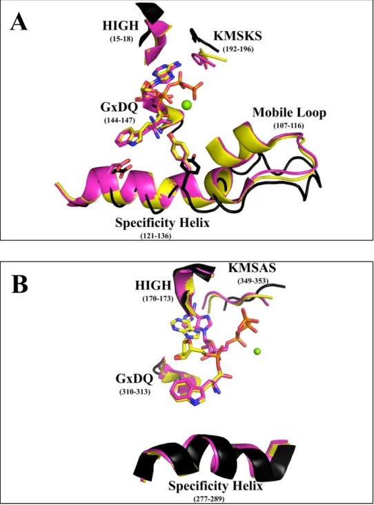
by BsTrpRS are well-characterized (Fig. 1A). In the absence of ligands (1D2R), the enzyme
exists in an open conformation (26). This same open state is observed when either ATP (1MAW)
or tryptophan (1MB2) separately occupies the active site (22). Induced-fit closing of the active
site to form the pre-transition state (preTS) results from full-ligation with ATP and tryptophan
(1MAU; (22)). The transition from open to closed is facilitated by movements of the mobile loop
containing Lys 111 (~3 Å), the specificity helix (~3 Å), GxDQ (~1.5 Å), HIGH (~3 Å) and the
KMSKS loop (~4 Å). At high concentrations ATP can drive closing of the active site to form the
pre-transition state (1M83).
In contrast, human cytosolic (Hc) TrpRS, which does not engage residues in the specificity helix for tryptophan recognition, adopts a closed state in response to tryptophan
binding (2QUH; (23)). The apo (1O5T; (27)) and tryptophan-bound HcTrpRS complexes differ by 0.8 Å (316 Cα pairs) compared to the 0.3 Å RMSD (301 Cα pairs) for the corresponding
BsTrpRS structures (Fig. 1B). Structural changes accompanying tryptophan binding include
movements toward the active site by the KMSAS loop (0.8 Å), GxDQ (~2 Å) and the loopcontaining Arg
5
Figure 1: Varied structural consequences of substrate binding to prokaryotic and eukaryotic TrpRS. (A)BsTrpRS resides in an open state conformation in the absence of ligands (black) and transitions to a closed, preTS conformation when both ATP and tryptophan(amide) are bound (pink). Tryptophan activation is accompanied by a slight opening of the KMSKS loop (yellow). (B) Smaller global movements occur when HcTrpRS goes from open (apo; black) to closed, preTS (pink) to closed,
product (yellow) conformation. Tryptophan-bound BsTrpRS favors the open apo-BsTrpRS structure whereas tryptophan-HcTrpRS favors the closed, preTS HcTrpRS complex.
A
6
complexes showed a high degree of structural similarity with an RMSD of 0.3 Å for 298 Cα
pairs. The addition of ATP in the HcTrpRS preTS complex closes the KMSAS loop by an additional 2 Å. Overall, less dramatic structural changes accompany the transition from open,
ligand-free state to closed, pre-transition state by HcTrpRS (0.7 Å) than BsTrpRS (2.5 Å).
Tryptophan recognition
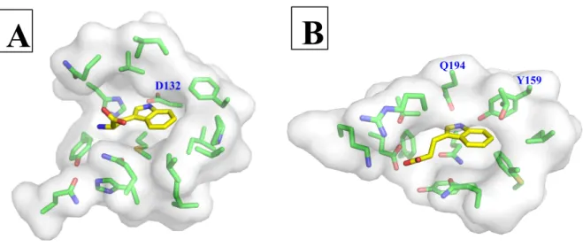
Tryptophan binds in a hydrophobic pocket within the BsTrpRS active site (Fig. 2A).
BsTrpRS utilizes the carboxylate side-chain of Asp 132 to recognize the indole nitrogen of
substrate tryptophan. Other stabilizing interactions include hydrophobic van der Waals (Ile 8,
Val 40, Val 141, Val 143, Ile 133, and Ile 151), π-π (Phe 5) and π-sulfur (Met 129) interactions.
In contrast, HcTrpRS relies more heavily on hydrogen bonding interactions with Tyr 159 and Gln 194 for tryptophan recognition (Fig. 2B). Additional direct polar interactions with four
active-site side-chains (Glu 199, Lys 200, Gln 284 and Gln 313) and π-π (Phe 317 and Tyr 159)
interactions promote tryptophan binding to HcTrpRS.
Bent versus extended ATP configuration
HcTrpRS and BsTrpRS bind different ATP conformers, with BsTrpRS binding the extended conformer that is characteristic for Class I aminoacyl-tRNA synthetases. BsTrpRS
engages three active-site lysine residues to stabilize the triphosphate group of ATP. Two of these
lysines (192 and 111) compete with the catalytic Mg2+ ion electrostatically coupled to each phosphate group for negatively charged oxygen atoms in the eventual PPi leaving group. Lysine
195 forms a salt bridge with an α-phosphate oxygen atom. While Lys 192 is conserved in all
TrpRS enzymes, Lys 111 and Lys 195 are missing from eukaryotic cytosolic TrpRSs. Instead,
7
Figure 2: Differential modes of tryptophan recognition. (A) Prokaryotic TrpRS enzymes use a strictly conserved aspartic acid to recognize the NE1 atom of tryptophan. (B) The tryptophan binding pocket of eukaryotic cytosolic TrpRS is more polar and less hydrophobic than that of prokaryotic homologs.
The γ-phosphate group interacts with Arg 162 and His 173 (HIGH) side-chains. Arg 162 is close
in sequence to the eukaryotic-specific extension (ESE) which Yang et al. showed was involved
in ATP binding (25). Deletion of the ESE reduces ATP binding ~25-fold.
Pre-transition states and Mg2+ coordination
From Figure 1 it is clear that BsTrpRS-bound ATP and tryptophan need minimal
movements for nucleophilic attack of the α-phosphate by the tryptophan carboxylate; i.e. the
indole and AMP moieties occupy similar positions in the pre-transition state and product state
structures. Induced-fit closing of the active site in response to ATP binding is responsible for the
proper placement of the ATP for catalysis to proceed. Contrastingly, the preTS complex for
HcTrpRS, which closes in response to tryptophan instead of ATP binding, shows the ATP α -phosphate must move closer to tryptophan by ~5 Å before catalysis can occur. Interestingly,
ATP movement is not facilitated by HcTrpRS structural changes.
B
8
In addition to differences in ATP conformation and ATP-enzyme interactions,
comparison of the bacterial and eukaryotic pre-transition state complexes reveals a distinct
difference in Mg2+ ion placement and coordination. In H
cTrpRS, the metal lies between the α- and β-phosphate groups. The bent ATP configuration places the γ-phosphate group too far away
for electrostatic coupling of this group with Mg2+. The coordination sphere for the Mg2+ ion consists of oxygen atoms from the α- and β-phosphate groups, the side-chain hydroxyl group of
Ser 165 and a stable water molecule. The precise role of Mg2+ in assisting tryptophan activation by HcTrpRS and other eukaryotic TrpRS enzymes remains undefined.
Contrastingly, BsTrpRS does not form any direct interactions with Mg2+ and the metal forms electrostatic interactions with an oxygen atom from each phosphate group of ATP and one
stable water molecule. As will be discussed in Chapter II the placement and coordination of the
Mg2+ ion within the BsTrpRS active site is dependent on ATP and the identity of the ligand occupying the tryptophan binding pocket. BsTrpRS couples the metal to an allosteric center
remote from the active site called the D1 switch (20, 21). Side-chain rearrangements between
residues of the D1 switch are responsible for activating the catalytic metal. Activation involves
movement of Mg2+ toward the eventual PPi leaving group, allowing for stabilization of the negative charge during the catalytic transition to tryptophanyl-5’AMP formation.
Indolmycin Inhibition
Varied modes of substrate binding, utilization of non-conserved residues in substrate
recognition, and differential catalytic assist by Mg2+ all point toward tryptophan activation proceeding via different mechanisms for bacterial and eukaryotic cytosolic TrpRS enzymes.
9
inhibitors, thereby making TrpRS a suitable target for novel anti-infective therapies to treat
infected individuals. The relatively minor structural differences between tryptophan and
indolmycin (Fig. 3), a naturally-occurring prokaryotic-specific TrpRS inhibitor produced by
Streptomyces griseus, allow for comparable tryptophan affinities but drastically different
indolmycin affinities between prokaryotic and eukaryotic cytosolic TrpRS homologs (19).
Binding of indolmycin to prokaryotic and eukaryotic cytosolic TrpRS enzymes differs by six
orders of magnitude (18, 19).
In addition to cytosolic TrpRS, eukaryotes express a second TrpRS required for
translation of the mitochondrial genome. Mitochondrial TrpRS is nuclear-encoded and translated
in the cytosol prior to mitochondrial import (28). Any potential clinically-useful TrpRS
inhibitors need to avoid inhibition of both cytosolic and mitochondrial TrpRS to prevent
deleterious side-effects. Lack of structural information about mitochondrial TrpRS has hindered
our understanding of the binding determinants used for substrate recognition by these enzymes.
Sequence comparison shows conservation among ATP- and tryptophan-interacting residues
between human mitochondrial TrpRS and BsTrpRS. A shared mode of tryptophan recognition
and binding might be indicative of shared susceptibility to inhibition by indolmycin.
The following chapters detail our efforts to uncover the structural and mechanistic basis
for selective inhibition of bacterial TrpRS over eukaryotic TrpRS, accounting for the 106-fold difference in affinity. Specifically, we account for the 103-fold stronger preference of BsTrpRS for indolmycin than its tryptophan substrate and uncover potential means employed by
eukaryotic cytosolic TrpRS to evade inhibition by indolmycin. Finally, we establish
mitochondrial TrpRS as a target for indolmycin inhibition via a shared inhibition mechanism
10
CHAPTER II: SELECTIVE INHIBITION OF BACTERIAL TRYPTOPHANYL-TRNA
SYNTHETASES BY INDOLMYCIN IS MECHANISM-BASED1
Introduction
The accumulation of resistance in pathogenic organisms over time and with prolonged
drug use necessitates the continued development of new anti-infective therapeutics. Such
developments can include modifications to current drugs that are active against exploited targets
while counteracting current resistance mechanisms or novel compounds targeted against
underexploited targets. One group of enzyme targets that has been validated but remains
underexploited is the class of aminoacyl-tRNA synthetases (aaRS). Aminoacyl-tRNA
synthetases maintain the fidelity of the genetic code by ensuring the charging of tRNA with its
cognate amino acid via the following two- step reaction:
1) aaRS + ATP + aa aaRS•aa-AMP + PPi
2) aaRS•aa-AMP + tRNA aaRS + aa-tRNA +AMP.
All aaRS enzymes bind ATP and activate a specific amino acid by catalyzing the
formation of an aminoacyl-5’adenylate (aa—AMP) during the first step. This is followed by
transfer of the activated amino acid to the 3’ end of the correct tRNA. Structural and mechanistic
differences among the different aaRS enzymes as well as orthologs of individual synthetases
make it possible to selectively modulate the activity of specific synthetases; e.g. prokaryotic over
11
eukaryotic TrpRS (19). This makes the aaRS enzymes attractive targets for novel anti-infective
therapeutics.
Any compounds intended for clinical use must be much less inhibitory against the
eukaryotic orthologs of its intended target. Naturally occurring aminoacyl-tRNA synthetase
inhibitors include indolmycin (TrpRS), granaticin (LeuRS), mupirocin (IleRS), and ochratoxin A
(PheRS) (10, 19, 29, 30). Of these, mupirocin displays the required selectivity for prokaryotic
over eukaryotic IleRS and has been developed for the treatment of infections in humans (14).
Indolmycin produced by Streptomyces griseus displays selective inhibition for
prokaryotic TrpRS (9 nM; E. coli) over eukaryotic TrpRS (4 mM; B. taurus) (31). Problems with
off-target effects on tryptophan metabolism have prevented its clinical use (32). However, if we
could understand the molecular basis for the observed inhibition and selectivity we could exploit
this information for the rational design of antibiotics targeted against TrpRS on pathogens.
Structurally, tryptophan and indolmycin are quite similar, with a heterocyclic indole
moiety at the root of each ligand (Fig. 3). Indolmycin differs from tryptophan in the following
ways: i) the carbon that is functionally equivalent to Cβ is substituted with a methyl group, ii)
the carbonyl carbon is part of an oxazolinone ring, iii) the hydroxyl and amine groups of
tryptophan are replaced by the nitrogen and oxygen atoms of the oxazolinone ring, respectively,
and iv) the -NH-CH3 moiety attached to the oxazolinone ring does not have functionally
equivalent atoms in tryptophan.
BsTrpRS is one of the most extensively characterized TrpRS enzymes. Mechanistically a
12
Figure 3: Functional equivalences of tryptophan and indolmycin. Indolmycin differs from tryptophan in three key ways: 1) the incorporation of Cα-constituents into an oxazolinone ring, 2) a methylamino group extending from the oxazolinone ring, and 3) replacement of a hydrogen on Cβ with a hydrophobic methyl group.
ion must move to be catalytically competent, yet no protein-metal interactions have been
observed in any of the BsTrpRS crystal structures determined. Instead a remote allosteric
location, the D1 switch, must undergo significant conformational change in order to promote the
Mg2+ ion to a catalytically competent position. The metal moves closer to the PPi leaving group, whose charge is further stabilized in the transition state by the KMSKS loop. ATP binding is
required for the conformational switching between the open and closed states that allows for
catalysis. ATP-dependent induced-fit closing of the active site brings ATP ~4 Å closer to
tryptophan in a predominantly translational movement mediated by relative movement of the
catalytic and anticodon-binding domains.
In the absence of ATP, tryptophan binding is promoted by hydrophobic Van der Waals
interactions, π- π interactions with Phe 5 and a hydrogen bond between the indole nitrogen and
Asp 132 of the specificity helix. When both substrates bind, the tryptophan substrate undergoes a
rotational movement that brings the indole ring deeper into the binding pocket and results in
13
facilitated by the inward movement of the specificity helix that is not observed when only
tryptophan is bound.
ATP-dependent induced fit rearrangement of the active site facilitates proper ATP
positioning in BsTrpRS, whereas molecular dynamics simulations demonstrate that tryptophan is
required to achieve the requisite movement of the αP in HcTrpRS(22, 24). Even a modest substitution of tryptophanamide in place of tryptophan prevents the re-positioning of ATP.
These findings support the idea that HcTrpRS is intrinsically better at discriminating between tryptophan and its structural analogues than is BsTrpRS.
HcTrpRS uses different structural elements for substrate recognition than its prokaryotic orthologs (23). Such elements include an extended N-terminus with a β1-β2 hairpin structure
shown to have a role in ATP binding as well as the amino acid activation reaction in HcTrpRS (25). In contrast to BsTrpRS, it is tryptophan binding that leads to induced-fit rearrangement of
the active site in HcTrpRS. There are a greater number of binding determinants for tryptophan recognition as eight direct and water-mediated hydrogen bonds with polar side-chains stabilize
tryptophan in the active site. It has been proposed that amino acid activation proceeds via an
associative transition state in HcTrpRS with an unclear role of Mg2+ in the catalytic transition state (24). However, comparison of the pre-transition (2QUI) and product states (2QUJ) shows
that, as with BsTrpRS, the Pα of ATP must move 5.3 Å to be in a position for nucleophilic attack
by tryptophan.
Despite mechanistic and structural differences, BsTrpRS and HcTrpRS have comparable tryptophan binding affinities. Yet, these inherent differences between prokaryotic and eukaryotic
TrpRS enzymes promote the binding of indolmycin to prokaryotic TrpRSs ~1500 fold while
14
structure of BsTrpRS bound by Mg2+•ATP and indolmycin allowed us to probe the structural basis for indolmycin inhibition and selectivity. Specifically, we examined this structure along
with the catalytically-relevant structures of BsTrpRS and HcTrpRS deposited in the Protein Data Bank to answer the following questions: 1) what are the structural consequences of binding
indolmycin; 2) why is indolmycin a tight inhibitor of prokaryotic TrpRS; and 3) why isn’t
indolmycin an inhibitor of eukaryotic cytosolic TrpRSs?
Experimental procedures
Construction of pet28-His-BsTrpRS vector
The full-length BsTrpRS sequence was pcr-amplified from a pet11 construct made
previously in the lab. Pcr primers contained restriction sites for BamHI and HindIII. The
resultant pcr product was digested with BamHI and HindIII. A three-way ligation between the
pcr product (BamHI/HindIII), dsoligo encoding for the TEV site (NdeI/BamHI) and pet28b
(NdeI/HindIII) yielded an expression vector for His-tev-BsTrpRS.
Expression and Purification of His-BsTrpRS
BsTrpRS was expressed by auto-induction with BL21(DE3)pLysS cells at 37°C (33).The
cells were pelleted at 4500 rpm for 30 minutes, resuspended in lysis buffer and frozen at -20°C.
Upon thawing cells were sonicated and centrifuged (16000 rpm, 4°C, and 1 hour). His-BsTrpRS
was captured from the lysate on Ni-NTA resin and eluted with 0.3 M imidazole. Purified protein
was cleaved overnight with TEV while dialyzing out the imidazole. The cleaved protein mixture
was passed back over a Ni-NTA column to capture both uncleaved protein and his-TEV
15
Active Site Titration
Active sites were titrated by following the loss of ATP to determine the fraction of
molecules competent for catalysis (34, 35). The reaction was performed at 37°C in a final
reaction mix containing 50 mM Hepes pH 7.5, 5000 cpm/µl (γ32P)-ATP, 10 µM ATP, 0.5 mM tryptophan, 5 mM MgCl2 and 0.05 U/µl pyrophosphatase. The reaction was initiated with enzyme at a final concentration of 3 µM. At various time points between 10 seconds and 30
minutes, 3 µl of the reaction were added to 6 µl of quench buffer (sodium acetate pH 5.3, 1%
SDS) and placed on ice. Three microliters of each quenched reaction were spotted onto a
cellulose-PEI TLC plate and run in 0.75 M KH2PO4 pH 3.5 with 4 M urea to separate 32Pi and (γ32P)-ATP. Plates were developed using a Typhoon Imager and analyzed with ImageJ (36) and JMP (37).
Michaelis-Menten Kinetics
The incorporation of (32P)-PPi into ATP was tracked either by TLC or filter-binding after purification on charcoal (35). Reactions contained (32P)-PPi (5000 cpm/µl for TLC and 400 cpm/µl for filter assay), 0.1 M Tris pH 8.0, 70 mM BME, 5 mM MgCl2, 10 mM KF, 2 mM PPi, 2 mM ATP and tryptophan ranging from 0.3 – 100 µM. Reactions were initiated with enzyme at
a final concentration of 30 nM.
Indolmycin Inhibition Assays
Inhibition assays were performed as described above; Michaelis-Menten experiments for
tryptophan were performed in the presence of stoichiometric amounts of indolmycin to enzyme.
Indolmycin to enzyme ratios of 1:5, 1:1 and 5:1 were used and results fitted to a competitive
inhibition model (1) using JMP (37). Non-linear regression to eq. (1) allowed for determination
16
[
]
[
] [
]
⎟⎟ ⎠ ⎞ ⎜⎜ ⎝ ⎛ + + ∗ = Tryptophan K Indolmycin k Tryptophan Rate i cat 1 (1)Differential scanning fluorimetry (DSF; Thermofluor)
The effects of ATP, tryptophanamide (LTN), and indolmycin on the thermal stability of
BsTrpRS were assessed by Thermofluor. We showed separately (Weinreb, V, Weinreb, G,
unpublished) that DSF detects a conversion of TrpRS into a molten globule form that fully
denatures only at higher temperature. The following saturating ligand concentrations were used
to ensure a predominance of conformations corresponding to those observed in crystal structures:
5 mM ATP, 5 mM MgCl2, 10 mM LTN, and 600 µM indolmycin. All reactions contained 8 µM BsTrpRS, 50 mM NaCl, 5 mM BME, 50 mM Hepes pH 7.5, and 0.15% Sypro-Orange in a final
volume of 20 µl. Fluorescence intensities were determined using an Applied Biosystems
7900HTFast Real Time (RT)PCR instrument, and data were analyzed with MATLAB
(Mathworks) with routines developed by Visinets, Inc. The software was built as a pipeline of
several m-files, connected to provide full analysis of the data, including thermodynamic
characterization and presentation of statistics. Fluorescence at each data point along a melting
curve is assumed to be the sum of contributions from two states with probabilities P1, P2 established by thermodynamic equilibrium between the two states:
€
F t
( )
=(
a1+b1T)
*P1+(
a2+b2T)
*P2 (2)where a1, and a2 are adjustable parameters representing intercepts and b1 and b2 the slopes of the linear dependences of the initial and final states, and T is the Kelvin temperature. The pipeline
17
A. Reading the data from high-throughput, 384 well, RT PCR) files and transforming them into a
matrix consisting of four columns: i) number of the well from which temperature-dependent
readings were taken; ii) an index representing the protein variant; and finally the data, iii)
temperature and iv) fluorescence readings;
B. Fitting the Thermofluor data to a thermodynamic model (eq. 3, 4).
€
F =F0+a1+b1T+(a2+b2T)e
−ΔG T( )/RT
1+e−ΔG T( )/RT
€
=F0+
(
a2+b2T)
+a1−a2
(
)
+(
b1−b2)
T1+e−ΔG T( )/RT (3)
where ΔG is the Gibbs energy difference between the two states, and e-ΔG(T)/RT is the Boltzmann factor that determines the state probabilities, P1 and P2:
€
ΔG=ΔH T
( )
m +ΔCp *(
T −Tm)
−T ΔS T( )
m +cpln T Tm ⎛ ⎝ ⎜ ⎞ ⎠ ⎟ ⎡ ⎣ ⎢ ⎤ ⎦ ⎥ (4)where ΔH and ΔS are the enthalpy and entropy changes between the states, cp is the heat capacity at temperature T and Δcp is the heat capacity change between the two states at the melting
temperature, Tm.
C. Independent determination of Tm assuming that the state probabilities, P1, P2 can be estimated from distances between the intersection of the melting curve with vertical lines connecting the
extrapolated linear final and initial slopes. Data initially worked up using both methods (B) and
(C) agreed closely, and the analysis reported here follows (C).
Crystallization, Data Collection, Structure Determination
Crystals of seleno-Met- substituted BsTrpRS in complex with ATP, Mg2+, and
indolmycin were grown by vapor diffusion against a reservoir of 1.4 M potassium citrate and 0.1
18
before plunging into liquid nitrogen. Data were collected remotely at SER-CAT (Beamline
ID-22) using inverse beam geometry at 0.979 Å to obtain experimental phases from the Bijvoet
differences, and processed with XDS (38). Phenix (39) and Coot (40) were used for phase
determination, to interpret the map, and iteratively refine the final structure (PDB accession code
5DK4).
Results
BsTrpRS binds indolmycin ~1500x more tightly than tryptophan
Indolmycin is a competitive inhibitor of Bacillus stearothermophilus and other bacterial
TrpRS enzymes that competes with tryptophan for binding to the enzyme’s active site. By
conducting Michaelis-Menten experiments at increasing tryptophan concentrations in the
presence of different indolmycin concentrations and fitting all 64 data points simultaneously to
Eq. (1) we were able to determine KM, tryptophan, 3 µM, and Ki, indolmycin, 2 nM (Table 2). As these experiments were carried out under exchange conditions (41), we determined the standard free
energy, ΔG° = -RTlnK, at 310 K for tryptophan and indolmycin binding to be 7.8 and 12.3
kcal/mol, respectively. This translates to a free energy difference of 4.5 kcal/mol between the
affinities of the catalytic and inhibited complexes for the indole-containing ligand. In order to
determine what factors account for the observed difference in binding free energy we determined
the structure of the BsTrpRS•ATP•indolmycin ternary complex and then conducted differential
19
kcat (s-1) ΔGkcat
(kcal/mol) KM,tryptophan (M)
ΔGKm
(kcal/mol) Ki,indolmycin (M)
ΔGKi (kcal/mol)
KM,tryptophan/ Ki,indolmycin 31.6 +/- 0.8 -2.1 +/- 0.01 3.0E-06 +/- 6.4E-07 7.8 +/- 0.1 2.0E-09 +/- 5.2E-10 12.3 +/- 0.1 1.5E+03
Table 2: Steady-state kinetic analysis of indolmycin inhibition. PPi exchange assays performed in the presence of saturating Mg2+•ATP with varying concentrations of tryptophan and indolmycin show
BsTrpRS binds indolmycin ~1450 times tighter than tryptophan. The difference in free energy between the catalytic and inhibited complexes is ~4.5 kcal/mol.
Indolmycin and ATP form a ternary complex with BsTrpRS
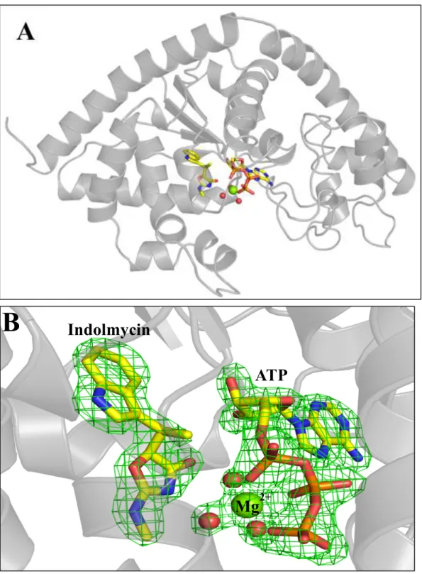
Extensive crystallization studies conducted on BsTrpRS have revealed three distinct
conformational states; an open conformation (ligand-free, tryptophan, low ATP (22, 26)), a
closed pre-transition state [high ATP, ATP + tryptophanamide; (17, 22)], and a closed product
conformation [Trp-5’AMP; (42, 43)]. A previously unpublished structure of BsTrpRS bound to
Mg2+•ATP and indolmycin was never deposited (44). Nevertheless, that structure was the first example of a series of subsequently solved structures that have been described as “Pre-transition
state” (PreTS) structures (1MAU, 1M83; (22)). In these structures the initial ATP binding site in
the small domain composed of the N-terminal alpha helix and the anticodon-binding domain
closes on the remainder of the Rossmann fold, bringing the nucleotide alpha phosphate from 6.7
Å away to within van der Waals contact distance of the tryptophan carboxyl oxygen (22).
The new structure presented here is at higher resolution (1.9 Å vs 2.4 Å) and the
experimental phases greatly enhanced the quality of electron density maps (Table 3, Fig. 4).
Details of the new structure, such as the orientation of the ribose and the metal position, are quite
similar to those observed in deposited PreTS structures, 1MAU and 1M83. Detailed differences
20
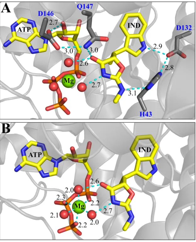
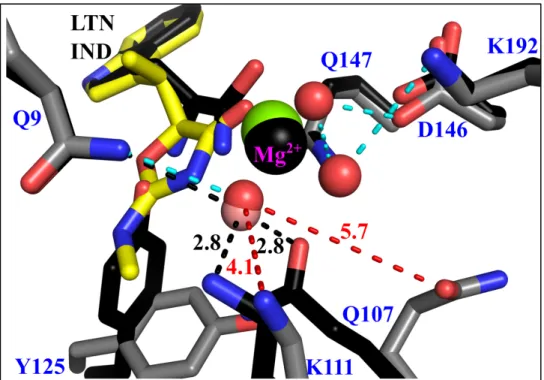
Indolmycin induces new contacts with active-site side-chains
Indolmycin makes contacts with the side chains of His 43, Asp 132 and Gln 147 as well
as two water molecules (Fig. 5A). The interaction between OD2 of Asp 132 in the specificity
helix and the nitrogen atom of the indole ring is observed when tryptophan (3.1 Å),
tryptophanamide (3.0 Å), or indolmycin (2.9 Å) is bound. The addition of the oxazolinone group
to the ligand allows for stabilizing interactions with His 43, Gln 147, with functionalities on
either side of the ring, which have the effect of fixing the rotation of the ring (Fig. 5B). ND1 of
His 43 can donate and/or accept a hydrogen bond from N2 (methylamino group) of indolmycin.
In addition to these hydrogen bonds His 43 can form a salt bridge with OD2 of Asp 132 (2.8 Å).
The amide group of Gln 147 forms two hydrogen bonds, one with the carbonyl oxygen of the
oxazolinone ring (3.0 Å) and another with OD2 of Asp 146 (3.0 Å). OD1 of D146 makes a
highly conserved hydrogen bond with the 2’OH group of ATP (2.7 Å). These side-chain
interactions in the conserved GEDQ motif link the indolmycin and ATP binding sites, while
reinforcing the linkage between opposite sides of the indole-binding pocket.
Structurally, indolmycin binding also prevents the Tyr 125 rotamer switch that occurs
upon the enzyme going from its open to closed conformation (Fig. 6A, B). During the catalytic
cycle Tyr 125 changes hydrogen-bonding partners from His 150 (2.7 Å) in the open
conformation to the alpha amino group (2.4 Å) of the tryptophan substrate in the closed PreTS.
The tryptophanyl-adenylate intermediate is stabilized by two polar contacts with the hydroxyl
group of Tyr 125 (1I6K). The inhibited state maintains the side-chain interaction between Tyr
21
Data Collection _
Space group
Cell Constants
a, b, c, α, β, γ
Resolution (Å)
Completeness (%)a
CC1/2 (%)a,b
Rmeas (%)a
Mean I/σI a
Number of Observations
Multiplicity
P43212
62.04 Å 62.04 Å 219.06 Å
90.00° 90.00° 90.00°
43.02 – 1.9
99.7 (97.9) 99.6 (93.2) 15.3 (61.9) 12.6 (3.3) 464942 15.5
Refinement _
Rwork/Rfree (%)
Fo, Fc correlation
RMS (bonds)
RMS (angles)
Ramachandran favored (%)
Ramachandran outliers (%)
Average B, all atoms (Å2)
Clash score
PDB Entry ID
16.6/18.7 0.96 0.005 0.994 97.0 0.0 32.0 2.2 5DK4
aHighest resolution shell is shown in parentheses.
bCC1/2 is the percentage of correlation between intensities from random half-datasets.
22
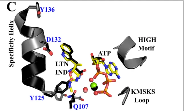
Figure 4: BsTrpRS forms a ternary complex with indolmycin and Mg2+•ATP in a closed
conformation. (A) Functionally dimeric BsTrpRS crystallizes with one monomer in the asymmetric unit. (B) In addition to indolmycin and ATP, the active site contains a Mg2+ ion and three stable water
molecules. The Fo-Fc omit map, derived by omitting indolmycin, ATP, Mg2+ and three water molecules
from a final round of refinement, contoured to 4.0 σ is depicted in green.
ATP
Indolmycin
23
24
Structural modifications induced by indolmycin to the Mg2+•ATP configuration
Superposition of the inhibited structure onto the closed PreTS structure (1MAU) gives a root
mean-square deviation of 0.28 Å for 323 Cα pairs. For comparison the two closed PreTS
structures, 1MAU and 1M83, have an RMSD 0.17 Å for 328 Cα pairs. The greatest structural
difference between the PreTS and inhibited states occurs around Glu 103 – Ala 120, with an
RMSD of 0.63 Å for these 18 residues. This mobile loop, which contains Gln 107 and Lys 111,
is more open by ~0.5 Å in the inhibited structure, as measured from the γP of ATP to the alpha
carbons of residues Gln 107, Lys 111, and Lys 115. The carbonyl oxygen of the Gln 107 side-
chain accepts a hydrogen bond from the water molecule coordinated to the Mg2+ ion (2.8 Å) and another from NE2 of Gln 147 (3.0 Å) in the pre-transition state. Steric clashing with the
constrained Tyr 125 rotamer prevents Gln 107 from switching rotamers in the inhibited state. As
such, the interaction with Gln 147 is not observed. Instead, the side-chain of Gln 107 accepts a
hydrogen bond at OE1 from a water molecule (3.0 Å) and donates a hydrogen bond at NE2 to
another water molecule (3.3 Å).
In the pre-transition state, the NZ atom of Lys 111 forms a salt bridge with the O1G atom
of ATP (2.9 Å), which also forms a strong electrostatic interaction with the catalytic Mg2+ ion (2.4 Å). This interaction presumably is important for stabilizing the developing charge on the PPi
leaving group released after tryptophan activation. Additionally, in the pre-transition state Lys
111 is in position to act as a hydrogen bond donor to the one water molecule coordinated to the
Mg2+ ion (Fig. 7). The subtle opening of this loop in the inhibited state weakens the salt bridge between the Lys 111 NZ atom and the O1G atom of ATP (3.4 Å) from that observed in the
preTS. Now the closest interactions are with three water molecules (2.6, 2.8, and 2.8 Å), none of
26
Figure 6: Comparison of ligand-free, preTS, and inhibited BsTrpRS structures. (A) Compared to the apo-form (pink; 1D2R), the fully occupied PreTS structure (black; 1MAU) assumes a closed
conformation. The Cα of Tyr 125 is shifted inward by 2.4 Å and the side-chain is flipped ~45° (measured from OH-Ca-OH). A Mg2+ ion (black sphere) forms electrostatic interactions with ATP and one water
molecule (salmon sphere). (B) Binding of indolmycin and ATP causes similar shifts in the backbone (grey) as the enzyme adopts a closed conformation. However, due to the addition of the methylamino-substituted oxazolinone ring this movement to the closed conformation is not accompanied by a rotamer change of Tyr 125 in the inhibited structure. (C) Consequently, Gln 107 is constrained and is rotated 106° around Cβ away from the specificity helix in the inhibited state compared to the pre-transition state. Finally, the Mg2+ (green sphere) is shifted toward the αPO4 and has hexavalent coordination to ATP and
three water molecules (red spheres) as compared to the preTS structure.
Replacement of tryptophan(amide) with indolmycin in the active site alters the
coordination and placement of the Mg2+ ion used during the activation step of the aminoacylation reaction. Presumably because its orientation is fixed by the hydrogen bonding network described
above, the oxazolinone forms hydrogen bonds with two water molecules. Introduction of these
two water molecules is associated with the movement of the Mg2+ ion into a hexavalent coordination that closely resembles stable configurations generated in quantum mechanical
27
As is also true in the PreTS structure, the Mg2+ ion in the inhibited BsTrpRS structure coordinates with a non-bridging oxygen from each phosphate group and three water molecules
(Fig. 5B, 6B), two of which are further stabilized by the presence of indolmycin. In addition to
the three electrostatic interactions with ATP only one water molecule was seen to coordinate
with the Mg2+ ion in the PreTS structure (1MAU) (Fig. 6A, 7). The side-chain residues that accept and donate hydrogen bonds to this water molecule differ between these two states (Fig.
7). Due to the different Gln 107 rotamer and slight opening of the mobile loop around Lys 111
these two residues no longer interact with this water molecule in the inhibited state.
Figure 7: Differential BsTrpRS side-chain interactions with the water molecules electrostatically coordinated with Mg2+ in the PreTS and inhibited structures. Gln 9, Gln 107 and Lys 111 are in
position to accept (Gln 107) or donate (Gln 9 and Lys 111) hydrogen bonds (black dashed lines) to the water molecule (salmon sphere) coordinated with Mg2+ (black sphere) in the PreTS (black sticks). As indolmycin binding leads to opening of the mobile loop containing Gln 107 and Lys 111, these residues are too far (red dashed lines) to form stabilizing interactions with the equivalent water molecule (red sphere) in the inhibited complex (grey sticks). The three water molecules (red spheres) electrostatically coordinated to Mg2+ (green sphere) in the inhibited complex are stabilized by interactions (cyan dashed
28
The Mg2+ ion is closer and more central to the triphosphate moiety in the inhibited structure than in the PreTS structure (Fig. 6C), and makes equivalent interactions with the
phosphate oxygen atoms. The significance of the differences in metal positions is evident from
several measurements. (i) Metal to oxygen distances are significantly shorter (0.2 Å; P = 0.002)
in the inhibited complexes. (ii) Movement of the divalent metal ion into closer contact with the
ATP phosphate oxygen atoms is associated with a subtle, but statistically significant opening of
the active site crevice between the N-terminal helix of the second crossover connection (the
GXDQ motif), the KMSKS signature, and the mobile loop containing Lys 111.
Ligand-induced stability changes imply cooperative sources of high indolmycin affinity
The nature of the ligands within the active site has a significant effect on an enzyme’s
conformation and thermal stability. For small perturbations, the fractional change in melting
temperature (ΔTm/Tm) induced by ligand binding is proportional to the free energy change in stability, the proportionality constant being the enthalpy change, ΔH (45). We use this implicit
relationship to assess the stabilizing or destabilizing effects of various ligands on the BsTrpRS
enzyme. Binding of ATP, tryptophan or tryptophanamide stabilizes the thermal transition of
molten globule formation by 3%, 7% and 7%, respectively (Table 4). Indolmycin enhances the
thermal stability of BsTrpRS by 20%, increasing Tm by 13.5°C. The enhanced affinity for indolmycin over tryptophan results in a shift by 8°C to higher temperature in the thermal
transition due to molten globule formation in the presence of indolmycin compared to
tryptophan.
The linkage between protein stability and ligand binding (46-48) implies that we can
attribute differences in stability changes to binding affinity. Two of the stabilizing interactions
29
in the metal position, relative to the ATP phosphate oxygen atoms. A key implication of the
structural observations in Fig. 5A and Fig. 6C is that binding of indolmycin to BsTrpRS should
be potentiated by the presence of Mg2+•ATP.
Ligand Mg2+ Tm (°C) ΔTm (°C) (ΔTm/Tm)*100
LF - 69.0 ± 0.2 n/a n/a
ATP - 71.1 ± 0.1 2.1 ± 0.2 3.0 ± 0.2%
TRP - 74.7 ± 0.1 5.7 ± 0.3 8.2 ± 0.4%
LTN - 74.1 ± 0.1 5.1 ± 0.3 7.4 ± 0.4%
LTN + ATP - 74.7 ± 0.2 5.7 ± 0.2 8.3 ± 0.3%
IND - 82.5 ± 0.2 13.5 ± 0.3 19.6 ± 0.5%
IND + ATP - 83.3 ± 0.3 14.3 ± 0.4 20.7 ± 0.6%
LF + 69.0 ± 0.1 n/a n/a
ATP + 71.2 ± 0.03 2.2 ± 0.1 3.1 ± 0.2%
TRP + 74.1 ± 0.1 5.1 ± 0.1 7.4 ± 0.1%
LTN + 74.2 ± 0.2 5.1 ± 0.2 7.4 ± 0.3%
LTN + ATP + 76.8 ± 0.2 7.7 ± 0.1 11.2 ± 0.2%
IND + 82.7 ± 0.04 13.6 ± 0.2 19.7 ± 0.3%
IND + ATP + 87.5 ± 0.1 18.4 ± 0.2 26.7 ± 0.4%
Table 4: Thermofluor analysis of ligand-dependent stability. Substrates (ATP and tryptophan) and substrate analogs (LTN and IND) induce conformational changes in BsTrpRS. These changes vary with ligand type and confer varying degrees of thermal stability in the transition to molten globule formation. ATP, tryptophan, tryptophanamide, and indolmycin separately enhance the thermal stability of BsTrpRS. Mg2+ is required for additional stabilization by ATP of both LTN-bound and IND-bound BsTrpRS. The inhibited complex (BsTrpRS bound with IND and Mg2+•ATP) is the most thermal stable, with a Tm 18.4°C and 10.7°C higher than that of ligand-free (LF) enzyme and PreTS complex, respectively.
The presence/position of Mg2+ in the active site is strictly dependent on ATP, because the protein makes no contacts with the metal. As no direct ATP-indolmycin interactions are
observed, we therefore expected that ATP would enhance the thermal stability of the
30
expected, the DSF measurements show that the BsTrpRS in complex with indolmycin and
Mg2+•ATP has a 27% increase in melting temperature compared to ligand-free enzyme, with Mg2+•ATP contributing an additional 5°C of thermal stability on top of the 13.5°C provided by indolmycin binding. By contrast, binding of indolmycin, indolmycin+Mg2+, or indolmycin+ATP all elicit far smaller changes of ~20% in thermal stability, demonstrating that both Mg2+ and ATP are required to confer additional thermal stability to the BsTrpRS•IND complex.
The conclusion that the metal is essential to the enhanced affinity of indolmycin to the
pre-transition state complex can also be derived using the 3D thermodynamic cycle of
contributions to stability from ATP, the methylamino-substituted oxazolinone ring (OXA) and
the presence/absence of Mg2+ (Fig. 8). Differences in binding and thermal stability between tryptophanamide and indolmycin were attributed to the methylamino-substituted oxazolinone
ring as this is the major structural difference between these two ligands.
Stabilizing and destabilizing interactions are distinguished by positive and negative
non-additivity, respectively. If thermal stability were unaffected by interactions between the ligands,
then we expect the effects of binding multiple ligands, e.g. ATP and LTN, to be additive and
binding one ligand not to be affected by the presence of a second ligand. Thus:
€
ΔT
m(
ATP
)
+
ΔT
m(
LTN
)
=
ΔT
m(
ATP
+
LTN
)
(5)€
ΔT
m(
LTN
)
=
T
m(
LTN
)
−
T
m( )
LF
=Tm(
ATP+LTN)
−Tm(
ATP)
(6)An interaction between the ligands would introduce a term, ΔTm,int, to describe the non-additivity (49), and:
€
31
Figure 8: Contributions of Mg2+, ATP, indolmycin and tryptophanamide to the thermal stability of
BsTrpRS. TrpRS enzymes require a Mg2+ ion for tryptophan activation. While Mg2+ does not change the thermal stability of BsTrpRS on its own, its presence or absence significantly impacts the interaction of ATP with both tryptophanamide (LTN) and indolmycin (IND). In the absence of Mg2+ (top row) both the ATP-LTN and ATP-IND interactions lower the fractional change in Tm by 2% from its expected value. In the presence of Mg2+ (bottom row) the interaction between ATP and LTN is insignificant, while the
ATP-IND interaction raises the Tm 4% higher than expected. As expected from the crystal structure, the Mg2+ -dependent ATP-IND interaction is mediated through the oxazolinone moiety of indolmycin.
In the absence of Mg2+ there is no significant ATP•OXA interaction, and binding either IND or LTN reduces the effect of ATP on Tm by ~1.5°C. The ATP•LTN and ATP•IND
interactions are both destabilizing, i.e. the doubly-liganded complexes melt at lower
temperatures. In contrast, addition of Mg2+ stabilizes the interactions of ATP with LTN and IND to varying degrees. In the case of LTN•ATP, addition of Mg2+ compensates for the destabilizing ATP•LTN interaction such that the interaction is no longer significant. The metal compensates
for the -1.3°C destabilizing ATP•IND interaction and allows for an additional stabilizing
32
orientation is, in turn, stabilized by hydrogen bonds to His 43 and Gln 147 as discussed in a
previous section.
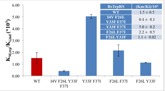
Mutations to residues in the D1 Switch alter the indolmycin:tryptophan selectivity ratio
Residues I4, F26, L29, Y33, C35, F37 and I140 within the D1 switch region of BsTrpRS
rearrange to form new Delaunay tetrahedra during Mg2+-assisted catalysis and are critical for protein-metal interactions in the transition state for tryptophan activation (50). Furthermore,
mutations to these residues lead to altered tryptophan-tyrosine specificity ratios. In light of the
observed stabilizing oxazolinone-ATP interaction that is mediated via Mg2+ we wondered if D1 switch mutants displayed differential indolmycin inhibition kinetics. Steady-state PPi-exchange
assays conducted with varying indolmycin and tryptophan concentrations allowed us to
determine kcat, KM tryptophan, and Ki indolmycin for four BsTrpRS variants (Table 5).
BsTrpRS kcat (s-1) KM, trp (M) Ki, ind (M)
I4V F26L
Y33F F37I 35.3 ± 1.2 2.2E-06 ± 6.0E-07 5.5E-09 ± 2.2E-09
Y33F F37I 21.6 ± 1.3 2.2E-05 ± 6.3E-06 4.5E-09 ± 1.3E-09
F26L F37I 29.9 ± 1.0 1.5E-05 ± 2.8E-06 7.0E-09 ± 1.5E-09
F26L Y33F 26.4 ± 0.6 9.0E-06 ± 2.2E-06 8.0E-09 ± 2.4E-09
Table 5: Steady-state kinetic analysis reveals differential indolmycin and tryptophan binding by D1 switch mutants.
All four variants tested had altered catalytic efficiencies compared to wild-type BsTrpRS. The
Y33F F37I and I4VF26LY33FF37I mutants were the least and most efficient variants,
respectively. Both double mutants containing the F37I substitution bound tryptophan an order of
magnitude weaker than native BsTrpRS. While the affinity of the quadruple mutant for
33
observed ~2-fold weaker indolmycin binding of the Y33F F37I BsTrpRS variant was
accompanied by a 10-fold reduction in tryptophan affinity. The F26LF37I and F26LY33F double
mutants had comparable changes in their affinities for tryptophan and indolmycin such that their
selectivity ratio did not differ from wild-type (Fig. 9).
Figure 9: D1 switch residues contribute to tryptophan versus indolmycin selectivity. Compared to native BsTrpRS (WT; red bar), variants containing substitutions at residues involved in long-range coupling to the catalytic Mg2+ ion display differential inhibition kinetics. The Y33FF37I variant has the
highest preference for indolmycin due mainly to decreased tryptophan affinity. Reducedindolmycin binding results in the quadruple mutant having the smallest (400-fold) difference between tryptophan and indolmycin affinities.
0.00 1.00 2.00 3.00 4.00 5.00 6.00
WT I4V F26L Y33F F37I
Y33F F37I F26L F37I F26L Y33F
K
M,tr
p
/K
i,in
d
(*10
34 Discussion
An array of crystal structures of both BsTrpRS and HcTrpRS provide snapshots of the enzymes along their catalytic paths and demonstrate the conformational changes that result from
binding of various ligands (22, 23, 26). From these structures it is evident that HcTrpRS uses a greater number of binding determinants for tryptophan recognition and that binding of
tryptophan causes an induced-fit rearrangement of the active site in HcTrpRS, but not BsTrpRS. Here we discuss possible structural and mechanistic reasons for the tight binding of indolmycin
to BsTrpRS and the inability of indolmycin to inhibit eukaryotic TrpRSs.
Why is indolmycin a high-affinity inhibitor of bacterial TrpRS?
There are no drastic global changes between pre-transition state (1MAU) and the
indolmycin-inhibited (5DK4) BsTrpRS structures. We propose that subtle, mechanistically
relevant differences in the active-site metal configuration account for the ability of indolmycin to
inhibit BsTrpRS as tightly as it does. We observe stronger Mg2+--ATP and weaker BsTrpRS--ATP interactions (Fig. 8), as well as altered Mg2+ coordination and placement in the inhibited state (indolmycin + Mg2+•ATP; Figs. 5B, 6), compared to the pre-transition state
(tryptophanamide + Mg2+•ATP) structure. We attribute these differences to the replacement of tryptophanamide with indolmycin which varies mainly at the methylamino-substituted
oxazolinone ring of indolmycin. Interactions between His 43, Gln 147, and indolmycin restrict
the oxazolinone ring orientation, thereby reducing the entropy of the α-carbon mimic in the
inhibited complex compared to the pre-transition state complex. This unfavorable entropy
change is compensated by the enthalpy from additional hydrogen bonds formed between the
35
These hydrogen bonds stabilize the water molecules that are also tightly coordinated to
the catalytic Mg2+ ion. Functional groups of the α-carbon atoms of tryptophan and
tryptophanamide can adopt alternative conformations that are similar in energy, none of which
allow for completion of the Mg2+ coordination sphere. We conclude from these observations that completion of that coordination sphere allows the metal to form significantly tighter interactions
with all three phosphate oxygen atoms, and hence that indolmycin stabilizes a ground-state
Mg2+•ATP configuration, opposing the tendency of the PreTS state to promote the metal to a high energy state that assists in transition-state stabilization.
Further, the oxazolinone ring of indolmycin, stabilized by hydrogen bonds with His 43
and Gln 147, prevents the rotamer switch of Tyr 125 in the specificity helix that is part of the
structural transition from the open to closed state. To avoid a steric clash with the constrained
Tyr 125 residue Gln 107 likewise does not switch rotamers in the presence of indolmycin. Gln
107 is part of a highly mobile loop that shows a subtle but significant opening in the inhibited
state compared to the pre-transition state. This opening results in the weakening of
ATP--BsTrpRS interactions, specifically those between Lys 111 and the γ-phosphate group.
In the catalytically-competent PreTS configuration, coordination by lysine residues of the
phosphate oxygen atoms promotes the metal to an activated, less stable state with weaker
interactions to the three phosphate oxygen atoms and prevents the Mg2+ ion from assuming a lower energy position with stronger contacts to ATP. The positively charged NZ atom of Lys
36
Substitution by indolmycin for tryptophanamide simultaneously weakens the Lys 111—
Oγ (3.4 Å) interaction and strengthens that between that oxygen atom and the Mg2+ ion (2.2 Å). Additionally, the 0.4 Å shift in Mg2+ placement along with the opening of the mobile loop around Lys 111 allows for tight, hexavalent Mg2+ coordination, accompanied by stronger, more nearly equivalent interactions between Mg2+ and the three ATP phosphate groups.
Mutation of His 43 results in indolmycin-resistance
When both tryptophanamide and ATP are bound, the His 43 side-chain switches from
one rotamer to another in the transition from open to closed, PreTS state and back again in the
closed, product state. This rotamer switch does not occur when the active site is bound to AMP,
PPi and tryptophan (51). In the PreTS (Mg2+•ATP + LTN) and inhibited (Mg2+•ATP + IND) states, NE2 of His 43 interacts with OD2 of Asp 132. In all other observed states, NE2 of His 43
forms an interaction with the carbonyl oxygen of Tyr 125. His 43 also contributes to indolmycin
binding via ND1. This re-orientation of His 43 appears correlated with the succession of ligands
most similar to the putative catalytic reaction path, and it may thus also be functional.
Several groups have identified mutations that confer high-level indolmycin resistance
(52-54). One of the mutant sites, His 43, is of direct interest in the context of the present
inhibited structure, which furnishes a semi-quantitative explanation for the mutational effects at
position 43. We have implicated His 43 in a hydrogen bond network that requires hydrogen
bonds to both indole nitrogen atoms that stabilize the orientation of the plane of the oxazolinone
ring of indolmycin. Fixing the orientation of the ring consequently allows formation of a full