DETECTION OF CARIES ADJACENT TO TOOTH COLORED PROXIMAL RESTORATIONS USING STATIONARY INTRAORAL TOMOSYNTHESIS
Robert L Hilton
A thesis submitted to the faculty at the University of North Carolina at Chapel Hill in partial fulfillment of the requirements for the degree of Master of Science in Oral and Maxillofacial Radiology in the School of
Dentistry.
Chapel Hill 2019
Approved by:
Angela Broome
André Mol
Andrea Ferreira Zandona
iii ABSTRACT
Robert L Hilton: DETECTION OF CARIES ADJACENT TO TOOTH COLORED PROXIMAL RESTORATIONS USING STATIONARY INTRAORAL TOMOSYNTHESIS
(Under the direction of André Mol)
Caries adjacent to restorations (CAR) is the most common reason for replacing restorations. This
study compared the ability of stationary intraoral tomosynthesis (s-IOT) and conventional bitewings in
detecting CAR. Extracted teeth (N=77) with 113 proximal tooth-colored restorations were used.
Observers (N=7) utilized a 5-point scale to rate their confidence that CAR was present and
stereomicroscopy was used to establish ground truth. S-IOT had a statistically higher (ANOVA p<0.05)
observer Az than conventional bitewings. S-IOT and conventional bitewings had a sensitivity of 0.48 and
0.44, respectively, which was statistically significant (ANOVA p<0.05) and a specificity of 0.57 and 0.61
respectively, which was not statistically significant (ANOVA p>0.05). S-IOT showed higher diagnostic
accuracy and sensitivity than conventional bitewings and is thus better in detecting caries around
proximal composite restorations. While the clinical effect size is small, s-IOT is a promising imaging
iv
ACKNOWLEDGEMENTS
Special thanks are due to Christy Inscoe and Connor Puett who generously offered many hours of
expertise regarding tomosynthesis image acquisition and processing. Thanks also to Pooja Saha and Dr.
v
TABLE OF CONTENTS
LIST OF TABLES……….vi
LIST OF FIGURES……….vii
LIST OF ABBREVIATIONS………viii
INTRODUCTION………..…1
PILOT STUDY………6
SPECIFIC AIMS………7
MATERIALS AND METHODS……….8
RESULTS………..12
DISCUSSION………..14
CONCLUSIONS………..21
TABLES AND FIGURES………..22
vi
LIST OF TABLES
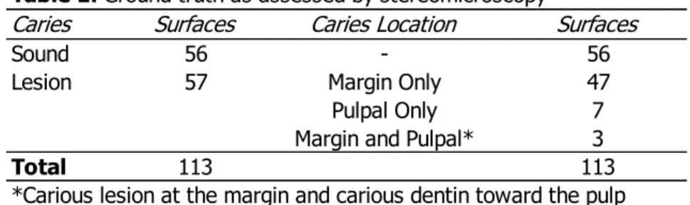
TABLE 1 – Ground truth as assessed by stereomicroscopy
TABLE 2 – Caries detection Az by modality and observer
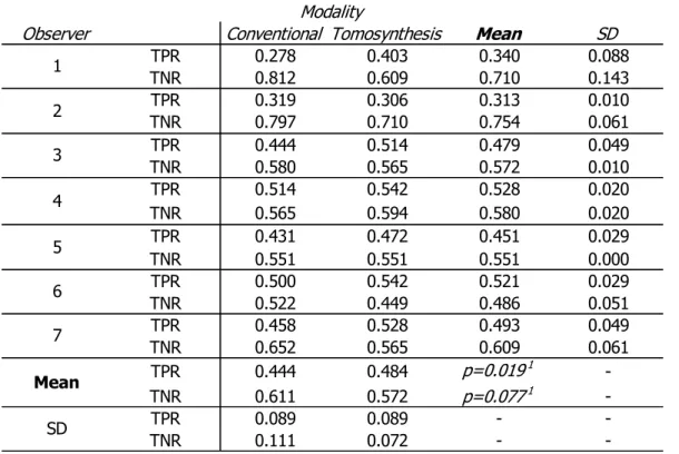
TABLE 3 – Sensitivity and specificity by modality and observer
TABLE 4 – Summery statistical findings from two-way ANOVA
TABLE 5 – Interobserver agreement using intraclass correlation coefficients
vii
LIST OF FIGURES
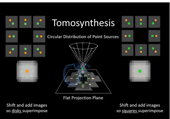
Figure 1 – Tomosynthesis geometrical concept
Figure 2 – Reconstructed s-IOT image showing a proximal composite restoration and a dark artifact
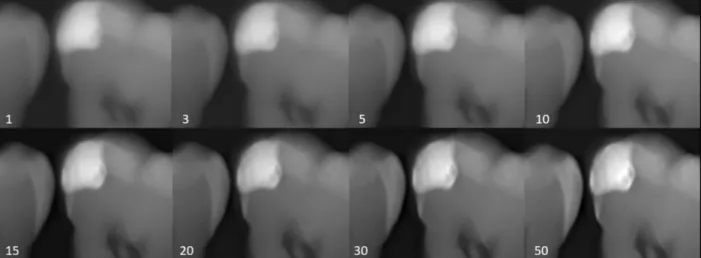
Figure 3 – Pilot study image comparing various numbers of iterative reconstructions.
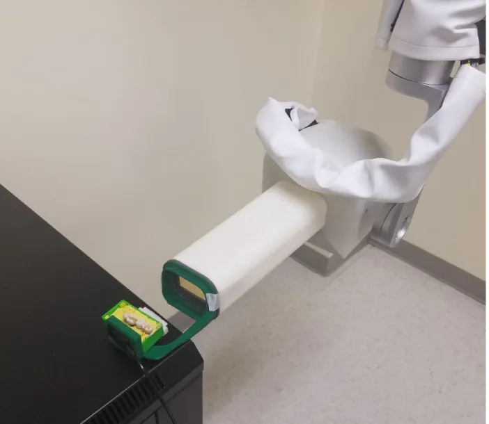
Figure 4 – The s-IOT unit at the UNC School of Dentistry.
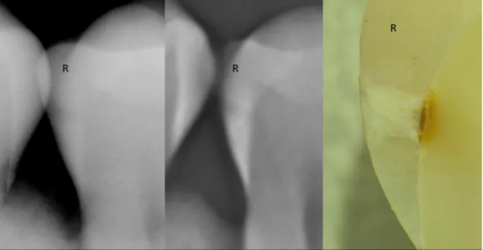
Figure 5 – Comparison of conventional and s-IOT bitewings with stereomicroscopic image.
Figure 6 – ROC curves by observer and modality
viii
LIST OF ABBREVIATIONS
ANOVA analysis of variance
Az area under the ROC curve
CAR caries adjacent to restorations
CBCT cone-beam computed tomography
CNT carbon nanotube
ICDAS international caries detection and assessment system
IRB institutional review board
MAR metal artifact reduction
MDCT multi-detector computed tomography
micro-CT micro-computed tomography
PI principal investigator
ROC receiver operating characteristic
SD standard deviation
s-IOT stationary intraoral tomosynthesis
TPR true positive rate
TNR true negative rate
TACT tuned-aperture computed tomography
UNC University of North Carolina at Chapel Hill
1
INTRODUCTION
Caries adjacent to restorations (CAR) is the most common cause for repair or replacement of
dental restorations.1–4 Each additional operative intervention creates costs for the patient and loss of
tooth structure or the tooth itself. Research supports the validity of the increasingly popular philosophy
of minimally invasive dentistry.5–8 This approach emphasizes early detection of carious lesions, tracking
progression or regression, and utilizing the least invasive intervention or procedure to achieve the
therapeutic goal. Early and accurate detection of CAR helps clinicians choose appropriate interventions
for the health of the tooth.
Past literature regarding CAR has used terminology that led to confusion with certain terms being
utilized differently by different authors.9,10 Secondary caries or recurrent caries typically refers to new
carious lesions forming next to a restoration at either the “outer wall” or the “inner wall”. Outer wall
lesions form at the exterior tooth surface in the same manner as primary lesions and are considered
analogous. Inner wall lesions form in the gap or micro-gap between the restoration and the prepared
tooth surface. Residual caries commonly refers to carious tooth structure that has been left behind,
intentionally or not, and is either sealed under the restoration or is present at the restoration margin. In
epidemiological surveys, there is no distinction between new and residual caries, and the term CAR is
intended to refer to any carious lesion adjacent to a restoration.10
Microscopically, three zones of dentin can be identified in the presence of a carious lesion: zone
1, normal unaffected dentin; zone 2: affected dentin, which is demineralized, but not infected and can be
remineralized; zone 3: infected dentin, which has bacterial invasion and is incapable of remineralizing.
Frequently, because of carious lesions, odontoblasts in dentinal tubules will die, leaving empty tubules
called dead tracts. These dead tracts are often dark when visualized on ground sections but they do not
2
Residual caries is often left behind intentionally in accordance with evidence-based
recommendations that depend on the proximity of the carious lesion to the pulp chamber and the nature
of the carious dentin.9 Bacterially contaminated and demineralized tissues near the pulp should not be
removed in asymptomatic patients with a vital pulp. New terminology has been recommended to help
characterize dentin according to the clinical consequences of leaving the dentin behind. While Knoops
hardness measurements can help distinguish between normal, affected, and infected dentin, this is not
clinically practical and no other clinical techniques correlate histologically. The terms soft, leathery, firm,
and hard have been defined to help guide clinicians in caries excavation. Soft dentin will deform when a
hard instrument is pressed unto it and can be easily scooped up with little force. Leathery dentin does
not deform when pressed, but can still be easily lifted without much force. Firm dentin is resistant to
hand excavation and some pressure is needed to lift it. Hard dentine requires a pushing force with a
sharp instrument or a bur to lift it. Hard dentin also makes a scratchy sound when a probe is taken
across it. It has not been demonstrated decisively how these clinical presentations of dentin relate to the
histopathology of carious lesions.9
The most effective methods of detecting caries adjacent to restorations include visual,
radiographic, and laser fluorescence, which all demonstrate a similar sensitivity (0.50 to 0.59) and
specificity (0.78 to 0.83).2 However, radiographs are more accurate than visual assessment at proximal
surfaces, and more accurate than laser fluorescence around amalgam restorations.2,12–14 In addition,
radiographs are able to provide information about the proximity of the carious lesion to the pulp.
Unfortunately, the recent technological advances in radiology have not led to a substantial improvement
in the detection of primary caries or CAR.
Digital radiography has introduced image enhancement and manipulation with flexibility in
storage and display that was not previously possible with film imaging. Despite these advantages,
studies have not demonstrated a concomitant improvement in caries detection.15,16 Cone-beam
computed tomography (CBCT) provides improved assessment of interproximal caries depth and detection
3
intraoral radiographs.12,17 However, with even small composite or metal restorations near the area of
interest, beam hardening and streak artifacts severely degrade CBCT image quality and the ability to
detect caries. Thus, the detection of caries next to restorations remains a diagnostic challenge.
Tomosynthesis and tuned-aperture computed tomography (TACT) are related imaging systems
that have improved primary or secondary caries detection in some studies.18–20 Both of these techniques
obtain multiple image projections at different angles and then mathematically reconstruct them into a
stack of images that provide 3D depth information (See figure 1). Conventional tomosynthesis utilizes a
known imaging geometry by mechanically coupling a moving x-ray source and a detector, while TACT
calculates the imaging geometry after image acquisition using a standardized fiduciary marker next to the
object being imaged. The fiducial marker in TACT allows the x-ray source to be moved to custom
locations as desired by an operator without needing to know the precise geometry. Whether TACT or
tomosynthesis, the individual projections acquired at differing angles are shifted so that an object of
interest viewed in each projection is superimposed on itself. The plane including the object of interest is
in focus while objects outside that plane are blurred. In theory and in practice this can be done with film,
however, with modern computers and digital imaging an entire stack of image planes from the volume of
interest can be generated in seconds. The resulting image stacks for both TACT and tomosynthesis allow
the clinician to scroll through different 2D planes to focus on a specific 2D plane of interest. The out of
plane objects are blurred but are not removed from the image. Many computational approaches are
available to reconstruct these images but typically filtered back projection or algebraic iterative
reconstruction is used. Iterative reconstructions are often preferred to further remove the out-of-plane
objects from the image and increase image sharpness. This approach uses a high-pass filter to suppress
blurred out-of-plane objects from the plane containing the object of interest. With each successive
iteration, the out-of-plane objects are further suppressed relative to the in-plane objects. In theory, this
process, with sufficient iterations, will completely de-blur the entire stack of images. In reality, random
4
A study conducted by Nair et al (1998) demonstrated that TACT images created with a circular
x-ray source arx-ray and reconstructed with three iterations had superior diagnostic efficacy in detecting
secondary caries (artificial caries) compared to film and digital imaging (Az for TACT iteratively restored
images = 0.9171, film = 0.6608, direct digital images = 0.5979).19 This study makes no mention of
metal or beam hardening artifacts, and was completed with both amalgam and composite restorations.
The same authors published a related paper that year investigating the effect of the restorative material
and lesion location on detection by TACT, film, and digital radiography. It was found for all modalities
that caries diagnosis was most efficacious when the lesion was adjacent to an amalgam restoration,
followed by a radiopaque composite, and least efficacious next to radiolucent composite restorations.
Lesions located at a point angle were easier to detect than lesions located at the mid-gingival floor.20
Other studies conducted in the late 1990’s and early 2000’s demonstrated that TACT enhances
root fracture detection, bony periodontal defect characterization, impacted tooth evaluation, and the
assessment of implant sites.23–26 However, neither TACT nor tomosynthesis gained a foothold in the
practice of dentistry due to the time intensive process of adjusting the x-ray source position for TACT and
speed limitations in detector acquisition and image reconstruction for both modalities. In addition, CBCT
started to enter the world of dentistry at this time, providing dentists with 3D information and dominating
oral radiology research for the next decade.
While CBCT was growing within dentistry, tomosynthesis found broad applications within
medicine. Compared to conventional medical radiography, tomosynthesis is more effective when
detecting breast cancer, lung nodules, and fractures.27 It has found applications in imaging paranasal
sinus and gastrointestinal disease. It also presents advantages over MDCT, including lower patient dose,
higher in-plane resolution, and more options for positioning the patient.27–31
Recent advances in carbon nanotube (CNT) x-ray emission, digital sensor speed, and computer
processing have addressed most of the hurdles limiting the application of tomosynthesis in dentistry and
it has been demonstrated that an intraoral image can be acquired and reconstructed in less than 10
5
radiology clinic. This unit utilizes seven CNT x-rays sources arranged in a horizontal linear array and
compact enough to fit in a standard size dental tube head. These stationary CNT sources eliminate
cumbersome image acquisition techniques as well as motion blur that plagued other tomosynthesis
systems using a moving x-ray source. It is expected that a commercially viable s-IOT system will be
brought to market soon; however, there are only a few published studies demonstrating the diagnostic
efficacy of a CNT s-IOT system.
Whereas s-IOT overcomes some of the limitations of early tomosynthesis techniques, some
issues related to image reconstruction remain challenging. Of particular concern are the dark “shadow”
artifacts that appear next to radiopaque objects in the direction of the linear x-ray source array. These
artifacts are most pronounced next to metal restorations, but are still readily apparent next to composite
restorations and even enamel (see figure 2).22 The intensity of these artifacts is proportional to the
radiodensity of the object and the number of iterations used in the tomosynthetic reconstruction. There
are no artifacts seen after one iteration. A light grey artifact can be seen around composite restorations
at 5 iterations, and the artifact becomes a dark black after 15 iterations. These artifacts have the
potential to obscure the area of interest and hide caries next to restorations or periodontal defects next
to implants.22 The false positive rate may also increase in CAR caries detection, due to the potential for
confusing artifact for caries. Early TACT and tomosynthesis researchers suggested the optimal number of
iterations to be between 3 and 5 to maximize image sharpness without introducing too much noise and
computational time. However, the most recent work published about s-IOT has used as many as 20
iterations, helping improve the visibility of primary caries and fractures while keeping the noise at a
6 PILOT STUDY
A pilot study was conducted evaluating s-IOT images of composite restorations using 1, 3, 5, 10,
15, 30, and 50 iterations (see figure 3) and comparing them against conventional bitewings. Two
observers chose the number of iterations that produced the best subjective image quality while producing
the least confusion about what was artifact and what was a radiolucency attributable to a caries lesion.
It was unanimously agreed that 1 and 3 iterations had too much blur, making caries lesions less visible
than on the conventional bitewings. 5, 10, and 15 iterations made the caries easier to see, however, the
artifact produced by restorations was too close in appearance to caries, making false positives likely. The
artifact produced by 30 and 50 iterations was deemed too dark to be confused with caries to a trained
observer and therefore not likely to produce many false positive responses. 50 iterations was dismissed
as it produced far too much noise degrading image quality relative to 30 iterations. Therefore, 30
iterations were selected based on the subjective ease of caries visualization without the potential of
confusing artifact with caries. The proper number of iterations to maximize the diagnostic efficacy for
7
SPECIFIC AIMS
The objectives of this ex vivo study are to compare stationary intraoral tomosynthesis (s-IOT)
against conventional 2D digital radiography (conventional bitewings) in its accuracy and reliability
(sensitivity, specificity, area under the ROC curve, and intraobserver and interobserver reliability) for
8
MATERIALS AND METHODS
Institutional review board (IRB) approval was sought to collect de-identified extracted human
teeth from existing tooth repositories and to perform observer sessions at the UNC School of Dentistry
(Study # 18-0306). The submission was reviewed by the Office of Human Research Ethics, which
determined that the submission does not constitute human subjects research as defined under federal
regulations [45 CFR 46.102 (d or f) and 21 CFR 56.102(c)(e)(l)] and does not require IRB approval.
Posterior molar and premolar teeth with tooth-colored proximal restorations were selected and sorted
into groups using visual examination according to the International Caries Detection and Assessment
Criteria (ICDAS) and sorted into groups as follows: (1) ICDAS 0, no visually detectable caries lesion; (2)
ICDAS 1&2, an enamel lesion only; (3) ICDAS 3&4, an enamel cavitation or a dark shadow; (4)
ICDAS(5), a cavitation extending to the dentin.32,33 Teeth with proximal caries lesions at any proximal
surface were included. Teeth with a caries lesion at the occlusal surface were excluded. Also, surfaces
with a cavitation larger than 2mm were excluded from the study. To simulate the situation where the
clinician left residual caries, either intentionally or not, teeth with large carious lesions were collected and
then prepared by leaving various amounts of carious dentin within a class II preparation prior to
restoration with Filtek Supreme Ultra composite (3M ESPE, St. Paul, MN, USA). Caries was left either at
the margin or at the deepest part of the preparation. For controls, 37 teeth were prepared using a class
II preparation and restored using composite resin (Filtek Supreme Ultra) so no carious or discolored
enamel or dentin was left behind. All teeth were stored in a 0.1% thymol solution. 77 teeth were
included in the study with a total of 113 proximal restorations to be imaged and evaluated.
The sample teeth were mounted individually with three other randomly selected posterior teeth
that had no restorations. The teeth were mounted in Play-Doh within a radiolucent plastic Lego block
9
soft tissues of the cheek, a 1cm thick slab of wax was placed between the x-ray source and the mounted
teeth at a distance of 1 cm from the Lego block. Each quadrant was then imaged using the
tomosynthesis unit and a conventional x-ray tube and CMOS detector. The geometry was standardized
such that the relationship between the tube head, sample, and detector could be reproduced between
the tomosynthesis unit and the conventional intraoral unit. The x-ray sources were directed so the
central x-rays traveled parallel to the floor creating an orthogonal relationship to the long axis of the
mounted teeth. This was done to simulate a typical bitewing geometry even though teeth were imaged
without an opposing arch in a similar fashion to a periapical projection. If the image showed overlapping
contacts extending more than halfway through the enamel of the adjacent tooth at the interproximal
surface of interest, the image was retaken until less than half of the enamel was overlapped.
The conventional bitewings were acquired using a Schick 33 CMOS digital sensor. They were
taken at the UNC School of Dentistry radiology clinic using the school’s standard intraoral source
(Instrumentarium Dental, Tuusula, Finland) at 70kVp, 7mA, 0.08s, at 40cm SID with 30cm
rectangular collimation. A stationary tomosynthesis system at the UNC School of Dentistry was used
to image all the samples. The system had a 7 CNT source array (model 2008-08-L75-002; XinRay
Systems Inc., Research Triangle Park, NC), and an intraoral digital CMOS sensor (SuniRay2; Suni
Medical Imaging Inc., San Jose, CA).22 The CNT source array used 70 kVp and 100mAs. The
intraoral sensor was a size 2 sensor with a field-of-view of 35.2 X 25.2 mm and a pixel size of 33 X 33
µm.
Once all the teeth had been imaged, they were sectioned in the mesial-distal plane, using a
diamond saw. The first section was made in the area of any visually detectable carious lesion, or in the
absence of a lesion, at the middle of the restoration. The teeth were then serially sectioned in 1mm
increments until either a carious lesion or discolored dentin was found. If no caries was found under
stereomicroscopy, the tooth was sectioned until the entire restoration was removed, demonstrating that
there was no caries adjacent to the restoration. The sections were viewed under a stereomicroscope at
10
the ground sections for the presence of either caries at the margin or infected/affected dentin that did
not communicate with the margin. Particular attention was given to distinguish dead tracts, which form a
comet-tail shape and run parallel to the course of the dentin tubules, from infected/affected dentin.
Although micro-hardness testing of the dentin was not used, an explorer was utilized to assess
restoration margins to distinguish caries from a defective margin. If there was any disagreement
between the histologic assessments of the two observers, a third expert observer with training in oral
microbiology was used as a tiebreaker.
A total of 7 observers were recruited from the UNC School of Dentistry and were either faculty
members or residents in a graduate training program. All 7 observers had at least 4 years of experience
as dentists evaluating intraoral radiographs for caries. Observers attended calibration sessions to discuss
the purpose of the study and to learn interpretation principles for detecting caries with both imaging
systems. Observers were taught the proper use of a 5-point scale for scoring their confidence level
regarding the presence or absence of caries adjacent to the restoration in question. Observers were
allowed to view sample images of teeth with CAR and discuss the images with the PI. The observers
then rated the likelihood of caries presence on the 5-point scale where 1 = caries definitely not present, 2
= caries probably not present, 3 = unsure, 4 = caries probably present, and 5 = caries definitely present.
All observation sessions were conducted in the UNC School of Dentistry’s radiology consultation
room where the PI was present at each session to clarify questions and troubleshoot any issues. There
were two workstations with three monitors each for viewing the images. Each monitor underwent quality
control checks prior to the observation sessions using test group 18 test patterns from the American
Association of Physicists in Medicine. Images from both modalities were viewed using the trial version of
the RadiAnt Dicom Viewer under subdued lighting conditions. Observers were permitted to use contrast,
brightness, and zoom functions while all other image processing parameters were held constant between
observers. To establish intraobserver reliability, all 7 observers repeated the observation session no less
11
Scores from the observers for each imaging modality were entered into a spreadsheet (Microsoft
Excel 2013, Redmond, WA) along with the ground truth value. Receiver operating characteristic (ROC)
curves and the corresponding areas under the curve (Az) were generated through an internet based ROC
analysis tool made available through the Johns Hopkins University School of Medicine (www.jrocfit.org).
Sensitivity and specificity were calculated by collapsing observer confidence ratings of 4 and 5 to a
positive response and ratings of 1 to 3 as a negative response. Az, sensitivity, and specificity were
calculated for each imaging modality-observer combination.
Two-way analysis of variance (ANOVA) tests were performed on the responses (Az,
sensitivity, and specificity) respectively, using modality and observer as main covariates. A p-value <
0.05 was considered a statistically significant test result. Interobserver agreement was assessed by
calculating the intraclass correlation coefficient for each modality. The observation scores from the
second session were used to determine the intraobserver agreement. Weighted kappa statistics were
calculated and a chi-squared test was used with statistical significance set at p<0.05. Intraclass
correlation and kappa values between 0.01 – 0.20 had slight agreement, 0.21 – 0.40 had Fair
agreement, 0.41 – 0.60 had moderate agreement, 0.61 – 0.80 had substantial agreement, and 0.81 –
12 RESULTS
An examples of a carious lesion imaged using 2D digital intraoral radiography, s-IOT, and
stereomicroscopy can be seen in figure 5. After sectioning and stereomicroscopic analysis, 57 of the 113
restorations had either caries adjacent to the restoration (table 1). 47 of these restorations had CAR at
the margin, whether they were outer wall or inner wall lesions. Seven of the teeth showed only affected
or infected dentin under the restoration that did not communicate with the surface margin. Three of the
teeth had both caries adjacent to the restoration at the margin and a separate area of carious dentin
under the restoration. All 37 control restorations were negative for caries adjacent to the restoration
under stereomicroscopy. Overall there were 56 restored surfaces without caries adjacent to the
restoration and 57 with caries adjacent to the restoration.
A summary of statistical findings for CAR detection is provided in Table 4. ROC analysis was
conducted for each observer-modality combination and Az scores for each combination are provided
in Table 2, while sensitivity and specificity scores are provided in table 3. Diagnostic accuracy as
measured by Az scores was higher for s-IOT (0.720) than for conventional bitewings (0.684) and was
found to be statistically significant (p=0.021) using a two-way ANOVA test. S-IOT demonstrated a
higher mean sensitivity (0.48) than conventional bitewings (0.44), which was also statistically
significant (p=0.019). There was no statistically significant difference (p=0.077) in mean specificity
for s-IOT (0.57) and conventional bitewings (0.61).
Interobserver agreement as measured by intraclass correlation coefficients was fair for s-IOT
(0.374) and moderate for conventional bitewings (0.459). The intraobserver agreement as
measured by mean weighted kappa coefficients for was moderate for s-IOT (0.584) and substantial
13
observer 7 had a statistically significant difference (p=0.016) in intraobserver agreement between
14 DISCUSSION
This study used ROC analysis and derived Az values to discern differences in caries detection
around restorations between conventional digital radiography and s-IOT. The advantage of ROC analysis
is that it permits assessment of diagnostic accuracy despite varying decision thresholds between
observers and the resulting differences in observer sensitivity and specificity values.34 With 6 out of 7
observers having a higher Az for tomosynthesis, statistical analysis showed that there was a significant
difference between the modalities’ mean Az values, indicating better performance in detection of caries
around composite restorations for s-IOT compared to conventional bitewings.
Sensitivity and specificity were also analyzed in this study because a false positive and a false
negative result often do not carry the same weight in clinical dentistry. Some clinicians feel that
maintaining specificity is valued over increasing the sensitivity because the slow advance of caries makes
it likely that a carious lesion will eventually be detected before becoming too large.35 Also, a false
positive leads to needless loss of tooth structure at additional expense to the patient.36 However, early
non-surgical treatments are becoming more popular in dentistry with many researchers emphasizing the
importance of minimally invasive dentistry. This trend may place more emphasis on increasing sensitivity
if the practitioner utilizes early interventions that are less expensive and preserve tooth structure. In the
case of interproximal CAR the same logic applies, however, there is the additional consideration that the
restoration may obscure early radiographic detection of a carious lesion and may also permit an easy
pathway for caries-causing bacteria to spread deeper before a lesion is detected.37 To our knowledge,
there are no patient or society level studies regarding the tradeoff between radiographic sensitivity and
specificity of CAR that was left behind intentionally.
The absolute mean sensitivity for tomosynthesis was 4 percent higher than bitewings (a 9
15
sensitivity was statistically significant. The apparent equality in specificity was a slightly surprising result
in light of the artifacts that composite restorations produce in tomosynthesis images. Depending on the
radiodensity of the composite and the buccal-lingual thickness of the restoration, the intensity of the
artifact varies from black to light grey. It was thought to be inevitable that some of the artifacts would
have a shade of gray indistinguishable from caries, leading to false positive responses from observers.
That there was no difference in specificity is likely due to the observers being well-trained in recognizing
the signs of this artifact.
The precise reason for the increase in sensitivity for tomosynthesis is not as clear. In
radiography, a carious lesion is detectable due to its lower mineral content relative to adjacent healthy
tooth structure, thereby permitting less attenuation of x-rays. This differential attenuation within the
tooth represents subject contrast and is common between both imaging modalities. However, s-IOT
images teeth at different angles. Some angles have x-ray beams that pass through less superimposed
healthy tooth structure and more carious demineralized tooth structure. This results in greater
differential attenuation between healthy and carious tooth structure and gives the lesion more contrast in
some of the projections. However, the exposure for each individual projection is approximately a seventh
of what is used in conventional digital radiography and the images produced by a single projection likely
lacks the contrast that a conventional digital projection taken at the same angle would have. The
hypothesized reason for the increase in image contrast seen in s-IOT is the ability to reduce
superimposition by mathematically blurring out-of-plane objects and focusing on in-plane objects.
Therefore, the differential attenuation between the carious lesion and the in-plane healthy tooth structure
does not get washed out by superimposed planes in which there is no differential attenuation. Caries
detection has long been understood to be a contrast limited diagnostic task and this increase in contrast
is very likely to produce an increase in sensitivity.38,39
It has been proposed that some carious lesions that would go undetected in both conventional
and tomosynthetic imaging are correctly identified in tomosynthesis for the incorrect reason: artifact next
16
always interprets artifact as caries this would lead to perfect sensitivity for the wrong reasons, and at the
unacceptable cost of having no specificity. The extent to which observers have a false rationale for
correctly identifying a lesion could be answered with studies that track eye movements or allow the
observer to record the rationale behind their caries detection decisions. On the other hand, it has also
been argued that s-IOT will lead to higher specificity from observers with a more conservative approach
who may interpret a radiolucency as artifact, when it is actually a void; again, making the correct choice
for the wrong reason. It is the opinion of the author that this scenario would occur less often because
voids have a very different appearance than artifacts produced by tomosynthesis, whereas artifacts quite
frequently mimic caries (see figure 2). Teaching correct principles in tomosynthesis interpretation would
minimize the effect of making the correct diagnosis for the wrong reason; however, it is unlikely that
even the best observers would be immune to these errors. The small increase in sensitivity for
tomosynthesis seen in this study may be the result of the observers’ tendency to call an artifact a carious
lesion, whether or not a lesion is present. However, this argument is not supported by the fact that we
do not observe a statistically significant decrease in the specificity from s-IOT. While it was assumed that
observers are identifying artifacts as carious lesions, this assumption is not supported by statistical
analysis. The extent to which an observer will err on the side of either aggressive or conservative caries
diagnoses will vary for each observer and is accounted for in ROC analysis. Since s-IOT had a statistically
higher mean Az and mean sensitivity, without a loss in specificity, it may be presumed that s-IOT is the
more effective diagnostic tool.
Interobserver agreement was moderate for conventional bitewings, while it was only fair for
tomosynthesis. Intraobserver agreement was substantial for conventional bitewings, while it was only
moderate for tomosynthesis. This suggests that observers were less consistent with each other and with
themselves when observing tomosynthesis images. This is likely due to the limited experience observers
had with tomosynthesis prior to the study. All observers had at least 4 years of experience evaluating
conventional radiographs. In contrast, the brief calibration and training session prior to observations was
the only experience observers had using s-IOT to evaluate caries adjacent to restorations. There was
17
This suggests that some observers felt more confident and were more consistent with s-IOT than were
other observers. Additional training and better calibration of the observers in the interpretation of s-IOT
images could have led to different results, and perhaps better diagnostic performance for s-IOT.
The mean sensitivity (0.44) and specificity (0.61) for conventional bitewings found in this study is
lower than most studies reported in a 2016 meta-analysis, which showed a mean sensitivity of 0.53 and a
mean specificity of 0.83 across 13 studies. The lower sensitivity and specificity values in this study
suggests that the carious lesions were diagnostically more challenging than the lesions in the studies that
were included in the meta-analysis. In this study, it was attempted to use an even distribution of lesions
across the ICDAS categories. Reporting sensitivity and specificity values according to ICDAS categories
has not been done consistently in the literature. However, reporting this information will allow better
comparisons between studies in the future. It is not feasible at this time to report that data for this
study, but this information will be included in any future publications of this study.
Stereomicroscopy was used for establishing ground truth in this study and has a long history in
published caries literature as a reliable reference standard.40 There is no consensus gold standard for
assessing CAR as microradiography, stereomicroscopy, clinical visualization, and tactile assessment all
have advantages and disadvantages.2 Micro-CT would have been an ideal reference standard for this
study as x-ray attenuation related to levels of mineralization in carious lesions is common to 2D digital
radiography, tomosynthesis, and micro-CT. However, access to micro-CT for imaging 77 teeth was cost
prohibitive in this investigation. Removal of the composite restorations for visual and tactile assessment
could result in accidental removal of a carious lesion prior to assessment. Sectioning and using
stereomicroscopy for the assessment of CAR is not always straightforward and discrepancies between
microradiography and stereomicroscopy have been reported.41 Discolorations of enamel and dentin do
not always correlate to a specific zone of the caries process due to different staining that may result from
various diets and microbial flora. Discolored dentin may represent either infected dentin, affected dentin,
sclerotic dentin, or dead tracts. Distinguishing between infected dentin and affected dentin is not always
18
as positive for caries, even though affected dentin is often left behind by clinicians intentionally when
trying to preserve tooth structure.
Weaknesses in this study include a heterogeneous observer group that consisted of residents and
faculty from different specialties. Ideally, the observers should all be experts in the diagnosis of caries
for both modalities so that variations in education and experience level do not confound the results. Most
of the observers had no experience with tomosynthesis prior to this study. The example images shown
in the calibration sessions did not have ground truth for the teeth shown. This may have favored
conventional bitewings because all of the observers had at least four years of experience using 2D
intraoral radiography. Another weakness was the ex vivo design of the study. When imaging real
patients, the scatter radiation from the soft tissues may be slightly different from the soft-tissue
equivalent material used in this study. It is not clear how this might benefit one modality over the other
in terms of contrast or diagnostic performance. In addition, it may be more difficult to manipulate one of
the imaging systems to open contacts and obtain diagnostic quality radiographs when dealing with a real
patient. Imaging real patients using s-IOT has a learning curve, is somewhat cumbersome, and takes
some additional time. These difficulties may be overcome in subsequent versions of s-IOT systems
making the coupling of the tube head and the XCP device more intuitive and less prone to error. Despite
these difficulties, it has been suggested that TACT and tomosynthesis are better at obtaining open
contacts than 2D intraoral radiography. This is based on the idea that at least one projection will be at
an angle where the contacts are open. In this study, the imaging geometry was standardized between
both modalities ensuring that contacts would be open.
Another considerable limitation to this study was the choice to use tooth colored restorations and
to exclude metallic restorations. Despite the fact that a previous study on TACT showed that caries was
more easily detectable next to amalgam restorations20, that study made no mention of metal or beam
hardening artifacts. It is not clear why those investigators did not run into metal artifacts but it is likely
19
projections (9) with a non-linear source array. These artifacts would likely play a larger role in detecting
caries next to metallic restorations had they been included in this study.
A circular or conical source array has been shown to provide better depth discrimination than
linear sources arrays. Studies still need to be conducted to determine whether this increased depth
discrimination would aid in CAR diagnosis. It has also been proposed that a circular or conical source
array may decrease the amount of artifact produced around a restoration by “spreading it around”. This
does not seem supported by the fact that the artifact produced is a limited-angle artifact and the limited
angle will still remain in a circular geometry. However, no published papers were found which
investigated the effect of circular and linear source arrays on the extent of artifact production.
With a horizontal linear source array, objects that are oriented in a horizontal direction are less
amenable to the depth discrimination capabilities of tomosynthesis. If the source array is linear in a
vertical direction, vertical objects such as implants and tooth roots would be less amenable to depth
discrimination. In this study, if a carious lesion at a gingival point angle was obscured by the restoration
in one projection, then it would be obscured in all of the projections due to the horizontal linear source
array. If a vertical source array had been used, it is more likely that one or more of the projections
would have shown the carious lesion at the gingival floor. However, a vertical linear source array would
also have produced artifacts that would likely be superimposed on that same carious lesion at the gingival
floor. It is not clear whether a vertical linear source array would have performed better or worse than a
horizontal source array.
Many papers have been published concerning the reduction of metal artifacts in both medical and
dental applications of tomography in general but also specifically for tomosynthesis. These papers have
demonstrated significant reduction in artifacts size and intensity and have restored fine hard-tissue detail
next to metal objects. However, the MAR techniques available in s-IOT are quite time consuming,
require some customization depending on exposure settings and patient factors, and are not easily
applied for all images.22 The choice was made to not use a MAR in this investigation because no clinically
20
active area of research that shows promise in the medical and dental literature.22,42–44 Many of the
techniques focus on segmenting the metal restorations out of the projection images and replacing them
with grey values similar to neighbor structures prior to reconstruction. After reconstruction, the metallic
object is re-inserted back into the reconstructed stack of images. This is an active area of research and
studies ought to be conducted to compare new MAR reconstruction methods.
Further investigation is also needed to determine the optimal number of iterations in the
reconstruction for detecting CAR. In this study, a subjective decision was made by the investigators
based on optimizing subjective image contrast while making artifact as easy to distinguish from caries as
possible. It is entirely reasonable that the ideal number of iterations for CAR detection is much lower
than the 30 iterations used in this study and closer to the 3 iterations used in the TACT study by Nair et
al (1998). While the images appear sharper and there is less blurring, noise and artifacts increase with
the number of iteratons.21 The ideal number of iterations suitable for one diagnostic task may not be the
21 CONCLUSIONS
S-IOT is a promising imaging modality for advancing the detection of CAR as seen in this ex vivo
study. S-IOT showed a statistically significant increase in sensitivity compared to conventional bitewings
without a corresponding change in specificity. There was also a statistically significant increase in the
diagnostic accuracy of s-IOT in the detection of caries adjacent to proximal composite restorations.
More research should be completed to investigate how to optimize s-IOT, including effects of metal
artifact reduction, iterative reconstruction techniques, source array geometries, and restorative material
type. The reference standard in this study could not distinguish between infected and affected dentin,
nor did it distinguish between soft, leathery, firm, and hard dentin. Therefore, no conclusions could be
drawn regarding the radiographic appearance of carious lesions on s-IOT images and the clinical extent
of the carious lesions. In a field that is moving toward conservative non-invasive therapies, an increase
in the sensitivity of caries detection from s-IOT imaging may improve the care the dental profession can
give its patients.
22
TABLES AND FIGURES
Caries
Surfaces
Caries Location
Surfaces
Sound 56 - 56
Lesion 57 Margin Only 47
Pulpal Only 7
Margin and Pulpal* 3
Total 113 113
Table 1. Ground truth as assessed by stereomicroscopy
*Carious lesion at the margin and carious dentin toward the pulp separated by healthy dentin.
Modality
1
2
3
4
5
6
7
Mean
SD
p-value
1Conventional 0.653 0.649 0.704 0.685 0.675 0.705 0.720 0.684 0.027 0.021
Tomo 0.692 0.739 0.732 0.712 0.721 0.691 0.756 0.720 0.024
Mean 0.673 0.694 0.718 0.699 0.698 0.698 0.738
SD 0.028 0.064 0.020 0.019 0.033 0.010 0.025
Abbreviations: SD standard deviation, Az area under the curve.
1ANOVA testing comparing modality Az values with statistical significance set at p<0.05
Observer
23
Table 3. Sensitivity and specificity by modality and observer
Observer
Conventional Tomosynthesis
Mean
SD
TPR 0.278 0.403 0.340 0.088
TNR 0.812 0.609 0.710 0.143
TPR 0.319 0.306 0.313 0.010
TNR 0.797 0.710 0.754 0.061
TPR 0.444 0.514 0.479 0.049
TNR 0.580 0.565 0.572 0.010
TPR 0.514 0.542 0.528 0.020
TNR 0.565 0.594 0.580 0.020
TPR 0.431 0.472 0.451 0.029
TNR 0.551 0.551 0.551 0.000
TPR 0.500 0.542 0.521 0.029
TNR 0.522 0.449 0.486 0.051
TPR 0.458 0.528 0.493 0.049
TNR 0.652 0.565 0.609 0.061
TPR 0.444 0.484
p=0.019
1-TNR 0.611 0.572
p=0.077
1-TPR 0.089 0.089 -
-TNR 0.111 0.072 -
-Abbreviations: TPR, true positive rate (sensitivity); TNR, true negative rate (specificity)
1ANOVA test of modality TPR and TNR values with statistical significance set at p<0.05
3 4 5 6 7 Mean SD
Modality
1 2Az, sensitivity, specificity scores
Measure
Effect
p-value
Fitted Az Observer 0.25
Modality 0.021*
Sensitivity Observer 0.002*
Modality 0.019*
Specificity Observer 0.025*
Modality 0.077
Table 4. Summary statistical findings from two-way ANOVA comparing
*denotes statistically significant effect
Table 5. Interobserver agreement using intraclass correlation coefficient
Modality
Intraclass Correlation*
24
Figure 1. Tomosynthesis geometrical concept. A disc and a square at located within two different coronal planes. Individual projections taken at different angles from a circular x-ray source array arranged at the top left and top right. Projections are shifted and overlapped so that the disc(left) and square on the right are overlapped and objects from different planes are blurred.
Table 6. Intraobserver agreement between session 1 and 2
using weighted kappa coefficients
Observer
Conv kappa
Tomo kappa
p-value
11 0.514 0.746 0.084
2 0.701 0.563 0.385
3 0.698 0.666 0.822
4 0.694 0.626 0.603
5 0.645 0.702 0.676
6 0.646 0.479 0.308
7 0.708 0.305 0.016*
Mean 0.658 0.584 0.480
1Chi-squared test for difference in kappa between modalities
25
26
27
28
31 REFERENCES
1. Burke FJ, Cheung SW, Mjör IA, Wilson NH. Reasons for the placement and replacement of restorations in vocational training practices. Prim Dent Care. 1999;6(1):17-20.
http://www.ncbi.nlm.nih.gov/pubmed/10752459. Accessed March 8, 2019.
2. Brouwer F, Askar H, Paris S, Schwendicke F. Detecting Secondary Caries Lesions. J Dent Res. 2016;95(2):143-151. doi:10.1177/0022034515611041
3. Forss H, Widström E. Reasons for restorative therapy and the longevity of restorations in adults.
Acta Odontol Scand. 2004;62(2):82-86. http://www.ncbi.nlm.nih.gov/pubmed/15198387. Accessed March 8, 2019.
4. Gordan V V, Riley JL, Geraldeli S, et al. Repair or replacement of defective restorations by dentists in The Dental Practice-Based Research Network. J Am Dent Assoc. 2012;143(6):593-601.
http://www.ncbi.nlm.nih.gov/pubmed/22653939. Accessed March 8, 2019.
5. Lallam C, Decup F. Minimal intervention dentistry II: part 2. Management of caries and periodontal risks in general dental practice. Br Dent J. 2014;216(4):179-185.
doi:10.1038/sj.bdj.2014.143
6. Holmgren C, Gaucher C, Decerle N, Doméjean S. Minimal intervention dentistry II: part 3. Management of non-cavitated (initial) occlusal caries lesions – non-invasive approaches through remineralisation and therapeutic sealants. Br Dent J. 2014;216(5):237-243.
doi:10.1038/sj.bdj.2014.147
7. Holmgren CJ, Roux D, Doméjean S. Minimal intervention dentistry: part 5. Atraumatic restorative treatment (ART) – a minimum intervention and minimally invasive approach for the management of dental caries. Br Dent J. 2013;214(1):11-18. doi:10.1038/sj.bdj.2012.1175
8. Colon P, Lussi A. Minimal intervention dentistry: part 5. Ultra-conservative approach to the treatment of erosive and abrasive lesions. Br Dent J. 2014;216(8):463-468.
doi:10.1038/sj.bdj.2014.328
9. Innes NPT, Frencken JE, Bjørndal L, et al. Managing Carious Lesions: Consensus Recommendations on Terminology. Adv Dent Res. 2016;28(2):49-57.
doi:10.1177/0022034516639276
10. Shivakumar K, Prasad S, Chandu G. International Caries Detection and Assessment System: A new paradigm in detection of dental caries. J Conserv Dent. 2009;12(1):10-16. doi:10.4103/0972-0707.53335
11. Heymann H, Swift EJ, Ritter A V., et al. Sturdevant’s Art and Science of Operative Dentistry. 6th ed. Elsevier/Mosby; 2013.
12. Wenzel A. Bitewing and digital bitewing radiography for detection of caries lesions. J Dent Res. 2004;83(1):72-75. doi:10.1177/154405910408301S14
13. Gordan V V., Riley JL, Geraldeli S, et al. Repair or replacement of defective restorations by dentists in the dental practice-based research network. J Am Dent Assoc. 2012;143(6):593-601. doi:10.14219/jada.archive.2012.0238
32
use of bitewing radiographs. Oral Surgery, Oral Med Oral Pathol. 1989;68(5):661-665. doi:10.1016/0030-4220(89)90257-0
15. Syriopoulos K, Sanderink GC, Velders XL, van der Stelt PF. Radiographic detection of approximal caries: a comparison of dental films and digital imaging systems. Dentomaxillofac Radiol. 2000;29(5):312-318. doi:10.1038/sj/dmfr/4600553
16. Wenzel A. Digital imaging for dental caries. Dent Clin North Am. 2000;44(2):319-38, vi. http://www.ncbi.nlm.nih.gov/pubmed/10740771. Accessed April 6, 2019.
17. Valizadeh S, Tavakkoli MA, Karimi Vasigh H, Azizi Z, Zarrabian T. Evaluation of Cone Beam Computed Tomography (CBCT) System: Comparison with Intraoral Periapical Radiography in Proximal Caries Detection. J Dent Res Dent Clin Dent Prospects. 2012;6(1):1-5.
doi:10.5681/joddd.2012.001
18. Shan J, Tucker AW, Gaalaas LR, et al. Stationary intraoral digital tomosynthesis using a carbon nanotube X-ray source array. Dentomaxillofacial Radiol. 2015;44(9):1-9.
doi:10.1259/dmfr.20150098
19. Nair MK, Tyndall DA, Ludlow JB, May K. Tuned Aperture Computed Tomography and Detection of Recurrent Caries. Caries Res. 1998;32(1):23-30. doi:10.1159/000016426
20. Nair MK, Tyndall DA, Ludlow JB, May K, Ye F. The effects of restorative material and location on the detection of simulated recurrent caries. A comparison of dental film, direct digital radiography and tuned aperture computed tomography. Dentomaxillofacial Radiol. 1998;27(2):80-84.
doi:10.1038/sj.dmfr.4600323
21. van der Stelt PF, Ruttimann UE, Webber RLL. Restoration and enhancement of tomosynthetic images and its applications in dentistry. Proc Annu Symp Comput Appl Med Care. 1985;(May):673. http://www.ncbi.nlm.nih.gov/pmc/articles/PMC2578107/.
22. Puett C, Inscoe C, Hilton R, et al. Stationary digital intraoral tomosynthesis: Demonstrating the clinical potential of the first-generation system. In: Progress in Biomedical Optics and Imaging - Proceedings of SPIE. Vol 10573. ; 2018. doi:10.1117/12.2293722
23. Nair MK, Bezik J. Tuned-Aperture Computed Tomography for Detection of Induced Mid-Buccal/Lingual Alveolar Bone Defects. J Periodontol. 2006;77(11):1833-1838.
doi:10.1902/jop.2006.050452
24. Limrachtamorn S, Edge MJ, Gettleman L, Scheetz JP, Farman AG. Array geometry for assessment of mandibular implant position using tuned aperture computed tomography (TACTTM).
Dentomaxillofacial Radiol. 2004;33(1):25-31. doi:10.1259/dmfr/18567887
25. Harase Y, Araki K, Okano T. Diagnostic ability of extraoral tuned aperture computed tomography (TACT) for impacted third molars. Oral Surgery, Oral Med Oral Pathol Oral Radiol Endodontology. 2005;100(1):84-91. doi:10.1016/j.tripleo.2004.12.001
26. Nair MK, Nair UDP, Gröndahl HG, Webber RL, Wallace JA. Detection of artificially induced vertical radicular fractures using tuned aperture computed tomography. Eur J Oral Sci. 2001;109(6):375-379. http://www.ncbi.nlm.nih.gov/pubmed/11767273. Accessed March 8, 2019.
27. Machida H, Yuhara T, Tamura M, et al. Whole-Body Clinical Applications of Digital Tomosynthesis.
33
28. Yoo JY, Chung MJ, Choi B, et al. Digital Tomosynthesis for PNS Evaluation: Comparisons of Patient Exposure and Image Quality with Plain Radiography. Korean J Radiol. 2012;13(2):136.
doi:10.3348/kjr.2012.13.2.136
29. Wells ITP, Raju VM, Rowberry BK, Johns S, Freeman SJ, Wells IP. Digital tomosynthesis--a new lease of life for the intravenous urogram? Br J Radiol. 2011;84(1001):464-468.
doi:10.1259/bjr/95862259
30. Ha AS, Lee AY, Hippe DS, Chou S-HS, Chew FS. Digital Tomosynthesis to Evaluate Fracture Healing: Prospective Comparison With Radiography and CT. AJR Am J Roentgenol.
2015;205(1):136-141. doi:10.2214/AJR.14.13833
31. Quaia E, Baratella E, Cernic S, et al. Analysis of the impact of digital tomosynthesis on the radiological investigation of patients with suspected pulmonary lesions on chest radiography. Eur Radiol. 2012;22(9):1912-1922. doi:10.1007/s00330-012-2440-3
32. International Caries Detection and Assessment System Coordinating Committee. Rationale and Evidence for the International Caries Detection and Assessment System (ICDAS II).; 2005. https://www.iccms-web.com/uploads/asset/592848be55d87564970232.pdf. Accessed April 6, 2019.
33. Dikmen B. Icdas II criteria (international caries detection and assessment system). J Istanbul Univ Fac Dent. 2015;49(3):63-72. doi:10.17096/jiufd.38691
34. Metz CE. ROC analysis in medical imaging: a tutorial review of the literature. Radiol Phys Technol. 2008;1(1):2-12. doi:10.1007/s12194-007-0002-1
35. Dove SB. Radiographic diagnosis of dental caries. J Dent Educ. 2001;65(10):985-990. http://www.ncbi.nlm.nih.gov/pubmed/11700001. Accessed March 8, 2019.
36. Dove SB. Radiographic diagnosis of dental caries. J Dent Educ. 2001;65(10):985-990. http://www.ncbi.nlm.nih.gov/pubmed/11700001.
37. González-Cabezas C, Fontana M, Gomes-Moosbauer D, Stookey GK. Early detection of secondary caries using quantitative, light-induced fluorescence. Oper Dent. 2003;28(4):415-422.
http://www.ncbi.nlm.nih.gov/pubmed/12877427. Accessed March 8, 2019.
38. Huda W, Rill LN, Benn DK, Pettigrew JC. Comparison of a photostimulable phosphor system with film for dental radiology. Oral Surg Oral Med Oral Pathol Oral Radiol Endod. 1997;83(6):725-731. http://www.ncbi.nlm.nih.gov/pubmed/9195631. Accessed March 8, 2019.
39. Kashima I. Computed radiography with photostimulable phosphor in oral and maxillofacial radiology. Oral Surg Oral Med Oral Pathol Oral Radiol Endod. 1995;80(5):577-598. http://www.ncbi.nlm.nih.gov/pubmed/8556466. Accessed March 8, 2019.
40. Hintze H, Wenzel A LM. Stereomicroscopy, film radiography, microradiography and naked-eye inspection of tooth sections as validation for occlusal caries diagnosis. Caries Res. 1995;29(5):359-363.
41. Meira KRS, Forte FDS, Silva PF, de Sousa FB, de Holanda Ferreira DA, Chaves AMB. Dentin
34
42. Lu Y, Chan H-P, Wei J, Hadjiiski LM, Samala RK. Improving image quality for digital breast tomosynthesis: an automated detection and diffusion-based method for metal artifact reduction.
Phys Med Biol. 2017;62(19):7765-7783. doi:10.1088/1361-6560/aa8803
43. Guo S, Tang H, Zhou Y, Huang Y, Shao H, Yang D. Accuracy of Digital Tomosynthesis With Metal Artifact Reduction for Detecting Osteointegration in Cementless Hip Arthroplasty. J Arthroplasty. 2018;33(5):1579-1587. doi:10.1016/j.arth.2017.12.037