INFLUENCE OF CASTRATION AND A HIGH PROTEIN DIET ON LOAD-MEDIATED HYPERTROPHY AND SKELETAL MUSCLE FORCE PRODUCTION IN RATS
FOLLOWING IMMOBILIZATION
Ryan J. Viverette
A thesis submitted to the faculty at the University of North Carolina at Chapel Hill in partial fulfillment of the requirements for the degree of Masters of Arts in the Department of Exercise
and Sports Science (Exercise Physiology).
Chapel Hill 2017
Approved by:
Erik Hanson
Alan Hayes
Eric Ryan
iii ABSTRACT
Ryan J. Viverette: Influence of Castration and and a High Protein Diet on Load-Mediated Hypertrophy and Skeletal Muscle Force Production in Rats Following Immobilization
(Under the direction of Erik Hanson)
The purpose of the present study to was to to examine the effects of low testosterone on
muscle mass and function in rats following 10 days of hindlimb immobilization and determine if
a high protein diet enhances load-mediated hypertrophy. Average SOL CSA was significantly
lower for immobilized vs. control legs at 0 (30.3%, p < 0.001) and 14 days of reloading (15.9%, p
= 0.006). Significant differences in average soleus mass were present at all days of reloading (all
p < 0.001). Immobilized SOL Po followed a similar pattern (all p < 0.001), and castrated animals
showed lower Po at 14 days (p < 0.050). At present, functional overload in a testosterone-deprived
state appears to reduce the regrowth of skeletal muscle size, suggesting that testosterone may play
a role in load-mediated hypertrophy following immobilization. As well, a high protein diet did not
iv
TABLE OF CONTENTS
LIST OF FIGURES ... vi
CHAPTER I ... 1
INTRODUCTION ... 1
Basis for Study: ... 1
Research Questions... 5
Research Hypothesis... 5
Limitations ... 5
Significance of Study... 6
CHAPTER II ... 7
REVIEW OF LITERATURE... 7
Hypogonadism via Chemical Castration ... 7
Hypogonadism via Surgical Castration ... 9
Hindlimb Immobilization ... 10
Strength Training to reduce effects of Hypogonadism ... 13
High Protein Diets ... 16
CHAPTER III ... 19
v
CHAPTER IV ... 22
RESULTS... 22
CHAPTER V ... 32
DISCUSSION ... 32
vi
LIST OF FIGURES
Figure 1. Body weight...23
Figure 2. EDL Wet Weight ...24
Figure 3. Gastrocnemius Wet Weight ...25
Figure 4. Plantaris wet weight ...26
Figure 5. Soleus wet weight ...27
Figure 6. EDL Peak Force ...28
Figure 7. Soleus Peak Force...29
Figure 8. Soleus CSA ...30
Figure 9. Average Food Intake ...31
1
CHAPTER I
INTRODUCTION
Basis for Study:
The loss of endogenous testosterone, as a result of normal aging or pharmacological
intervention for prostate cancer therapy, has adverse effects on skeletal muscle mass, strength,
function, and physical activity levels (Basaria et al., 2008). It is estimated that hypogonadism
affects between 2.1 - 12.8% of adult males in the general population (Zarotsky et al., 2014).
Primary hypogonadism is a result of a defect within the Leydig cells of the testes in males, while
secondary hypogonadism occurs elsewhere, often within the hypothalamic-pituitary gonadal
(HPG) axis. Androgen deprivation therapy (ADT) is a form of prostate cancer treatment that
suppresses male sex hormone production to help slow the progression of prostate cancer by
preventing the pituitary from releasing lutenizing hormone (LH) or through the use of
antiandrogens antagonists (Sharifi et al., 2010). Regardless of the pathology, hypogonadism can
induce changes similar to the effects of aging but at an accelerated rate (Bylow et al. 2007, Hanson
et al., 2011 such as significant reductions in muscle mass, increases in fat mass (van Londen et al.,
2008), and reduced bone mass (Basaria et al., 2002), and physical inactivity (Galvao et al., 2009).
Suppression of endogenous testosterone levels is also associated with a decrease in
fractional muscle protein synthesis in younger men (Mauras et al., 1998) and older prostate cancer
patients (Hanson et al., 2017). Resistance training may be a beneficial strategy to mitigate these
2
function in men with hypogonadism show mixed results; with some studies showing attenuated
regrowth (Kvorning et al., 2006; Nilsen et al., 2014, Winters-Stone et al. 2014), and other studies
showing increases in muscle strength, muscle function, and lean mass (Hanson et al. 2013, Galvao
et al. 2010). These mixed results in human research may due to a number of confounding factors
influencing the responses, such as diet and physical activity levels. To better control for these factors and to investigate this relationship further, an animal model is a feasible alternative
approach.
Animal models are a justifiable and potential alternative as they have demonstrated
increased fat mass and decreased muscle mass similar results and behaviors to castration as
humans do to hypogonadism. Eleven weeks of castration in aged rats showed increased fat mass,
reduced muscle mass, strength, and bone mass compared to age- and weight-matched controls
(Gentile et al., 2010). Young castrated rats also exhibit an attenuated weight gain, decreased
muscle mass (Jiao et al., 2009), decreased food intake, and increased bone resorption (Borst &
Conover 2006; Borst et al., 2007) compared to sham controls. Castration has also been shown to
increase expression of ubiquitin ligases, markers of skeletal muscle proteolysis (Pires-Oliveira et
al., 2010). Moreover, Ibebunjo et al. (2011) showed that castration results in almost complete
cessation in voluntary wheel running that was restored to normal levels following testosterone
replacement therapy.
Hindlimb immobilization is a model widely used to induce muscle atrophy that has been
shown to significantly reduce muscle mass, muscle fiber size, muscle function, and body weight
compared to control groups (Brown et al., 2001; Murphy et al., 2011; Childs et al., 2003; Morris
et al., 2004?). Aged rats typically display less atrophy than younger rats, suggesting that older
3
immobilization period (Hepple et al., 2004; Blough et al., 2000). Following 10 days of
immobilization and 15 days of reloading, young rats appear experienced more atrophy from
immobilization and exhibited a trend towards increasing soleus mass while older rats lacked the
ability to completely regrow the atrophied soleus (Childs et al., 2003; Morris et al., 2004). This
diminished capacity for skeletal muscle regrowth in aged rats may be as a result of decreased
growth factors, such as testosterone, as a result of aging. Heterochronic parabiosis (surgical pairing
of young and old animals) restored physiologically normal levels of testosterone and showed
improvements in muscle weight, and muscle fiber cross-sectional area compared to controls,
indicating testosterone may have a critical role in mediating the improvements (Sinha et al., 2014).
Castration results in loss of peak tetanic tension regardless of muscle fiber type or function,
however, it does not exacerbate hindlimb unloading-related atrophy or peak tetanic tension (Brown
et al. 2001). While aged rat skeletal muscle typically shows less atrophy as well as diminished
capacity for skeletal muscle regrowth than young rats, some of which may be due to decreased
testosterone (Sinha et al., 2014) due to aging, data are lacking that directly assess the role of
testosterone on load-mediated hypertrophy.
Muscle mass is regulated by the balance between protein synthesis and degradation
(Phillips and Van Loon, 2011), with hypogonadism (Mauras et al. 1998; Hanson et al. 2017) and
immobilization (Cholewa et al. 2017) favoring an environment that promotes muscle loss.High
protein diets are a common approach used to increase muscle protein synthesis and may be a
beneficial strategy to help attenuate losses resulting from hypogonadism, immobilization, and
decreased physical activity. For example, BCAA supplementation attenuates the increase in
markers of skeletal muscle proteolysis (Jang et al., 2015; Maki et al. 2012) and to partly preserve
4
immobilized rats (Bajotto et al. 2011). Specifically, leucine supplementation has been shown to
attenuate muscle mass loss in hindlimb immobilized young rats (Baptista et al., 2010) and to
suppress reductions in muscle mass in the presence of an otherwise protein-free diet (Sugawara et
al., 2009). To our knowledge there are currently no studies in rats that examine the ability of a
high protein diet to overcome the detrimental effects resulting from castration, however
leucine-rich diets have been shown to help increase muscle protein synthesis (Ventrucci et al., 2004),
modulate proteasome subunits (Salomao et al., 2010), and attenuate decreases in lean body mass
(Gomes-Marcondes et al., 2003) in tumor-bearing cachectic rats. The peripheral wasting of muscle
mass associated with cachexia may be a result of increased protein degradation or decreased
protein synthesis. The ability of high protein diets to mitigate losses in cachectic rats indicates
there may be the ability to mitigate declines resulting from hypogonadism as well.
Therefore, the aims of this study were two-fold: 1) to examine the effects of testosterone
status on muscle mass, force, and cross-sectional area during reloading following 10 days of
immobilization and 2) to determine if a high protein diet attenuates immobilization- and
castration-induced atrophy and enhances load-mediated hypertrophy. We hypothesized combined castration
and immobilization will not act synergistically to induce greater atrophy than just immobilization
alone and that suppression of endogenous testosterone will not attenuate adaptions to the reloading
protocol. Further, a high protein diet will attenuate atrophy during the immobilization period and
facilitate better adaptations to reloading compared to castrated mice with a traditional diet. The
recovery of muscle mass, body weight, muscle force, and cross-sectional area will be similar
5
Research Questions
RQ1: Will combined castration and immobilization act synergistically to induce greater
muscle atrophy and loss of function than immobilization alone?
RQ2: Is the muscloskeletal function with reloading following immobilization influenced
by testsosteone levels?
RQ3: Does a high protein diet attenuate castrationan- and immobilization-induced atrophy
and provide a greater anabolic stimulus for recovery during reloading?
Research Hypothesis
H1: Combined castration and immobilization will act synergistically to induce greater
atrophy than just immobilization alone.
H2: Suppression of endogenous testosterone via orchidectomy will not attenuate adaptions
to the reloading protocol.
H3: A high protein diet will attenuate atrophy during the immobilization period and
facilitate better adaptations to reloading compared to castrated mice with a traditional diet.
Limitations
One limitation to this study is it is a retrospective analysis of previously collected data.
Retrospective studies are vulnerable as the investigators have no control over exposure or outcome
measures and must rely on accurate record keeping. Another limitation is the lack of a true control
group. The non-immobilzed contralateral limb served as the control for each animal, however as
these rats are young they may still be growing as a part of normal development which potentially
6
from Sprague-Dawley rats (Krawiec et al. 2005) indicates that unilateral immobilization had no
effect on the contralateral noncasted leg and served as the justification for this approach in the
current study.
Significance of Study
It is well established that functional overload (i.e. strength training) elicits neural and
morphological adaptations that may lead to increased muscle strength and muscle hypertrophy but
relatively few studies have examined the hypertrophic response in the absence of testosterone.
Moreover, many of these studies produce conflicting results. Protein and amino acid
supplementation has been widely used as a nutritional countermeasure to increase muscle protein
synthesis, as well as attenuate muscle protein degradation, but to our knowledge there are currently
no studies in that examine the ability of a high protein diet to overcome the detriments resulting
from hypogonadism and reduced activity/immobilization. Functional overload and combined
protein supplementation may provide additional treatment options for those suffering from
hypogonadism, as well as provide a potential therapy for prostate cancer patients undergoing
7
CHAPTER II
REVIEW OF LITERATURE
In this review, the effects of hypogonadism on skeletal muscle, body composition, and
contractile properties of muscle of both humans and rats will be examined. It will also examine
the use of hindlimb immobilization in rats as a model of muscle atrophy, and subsequent reloading
as a model of functional overload (e.g.strength training). Finally, the potential anabolic effects of
increased protein intake on recovery from muscle atrophy and castration will be examined. It has
been established that functional overload facilitates neural and morphological adaptations that can
lead to increased muscle force and hypertrophy in the eugonodal state; however studies present
conflicting results as to the effectiveness of strength training in the hypogonadal state and none to
our knowledge have included protein supplementation in patients at high risk for declines in
musculoskeletal function.
Hypogonadism via Chemical Castration
It is estimated that hypogonadism affects between 2.1 - 12.8% of adult males in the general
population (Zarotsky 2014). Primary hypogonadism is a result of a defect within the Leydig cells
of the testes in males, while secondary hypogonadism occurs elsewhere, often within the
hypothalamic-pituitary gonadal (HPG) axis. Androgen deprivation therapy (ADT) is a form of
prostate cancer treatment that suppresses male sex hormone production in the body, namely
8
the use of antiandrogens antagonists that prevent activation of the androgen receptors (Sharifi
2010).
While ADT is an effective treatment for prostate cancer, it can have adverse effects on
cancer patients in both the short-term and long-term. Hypogonadism results in the abrupt loss of
endogenous testosterone which resembles the symptoms of aging but presents at an accelerated
rate (Bylow et al. 2007, Hanson et al. 2011). Galvao et al. (2009) assessed the effects of 36 weeks
of ADT on whole body and regional muscle, fat, and bone mass and physical activity in men with
PCa. Their results showed that upper limb, lower limb, trunk, and whole-body lean mass decreased
by [5.6 (0.6)%, 3.7 (0.5)%, 1.4 (0.5)% and 2.4 (0.4)% (p < 0.01) respectively], while fat mass
increased in those respective areas [20.7 (3.3)%, 18.7 (2.7)%, 12.0 (2.5)% and 13.8 (2.3)% (p <
0.001)]. Hip, spine, whole-body, upper limb bone mineral density, and physical activity levels
decreased as well but not lower limb density as a result of ADT. Van Londen et al. (2008)
examined the long-term effects of ADT on body composition over a 2 year period. They showed
that men undergoing acute ADT treatment (mean 3 months) had significant gains in body fat mass
(1499.56±322.28 g after 12 months, 2167.15±676.45 g after 24 months) as well as losses in lean
body mass (929.74±296.36 g after 12 months, 1785.81±501.31 g after 24 months) over the 2 year
period. Men undergoing chronic ADT (mean 31 months) had smaller but still significant changes
over the 2 year period. These results show that men undergoing ADT experience significant gains
in body fat mass and losses in lean body mass and suggests that these changes are most pronounced
at the initiation of ADT. Finally, Basaria et al. (2002) conducted a cross-sectional study to
determine the effects of ADT on lean body mass, muscle strength, and bone mineral density. They
showed that men on ADT had significantly lower total-body and lumbar spine bone mineral
9
mass (p = 0.0001) and significantly reduced upper body strength (p = 0.001) compared to the other
groups. Suppression of endogenous testosterone levels is also associated with a decrease in
fractional muscle protein synthesis in younger men (Mauras et al. 1998) and older prostate cancer
patients (Hanson et al. 2017).
Hypogonadism via Surgical Castration
Animal models have demonstrated similar results to hypogonadism as humans. The most
common method to induce hypogonadism in animal models is through orchidectomy, a procedure
in which both testes are surgically removed, to remove endogenous testosterone production.
Gentile et al. (2010) showed that 21 week old Sprague Dawley castrated rats develop increased fat
mass (+15g), reduced muscle mass (-50g) and strength, as well as lower bone mass (-1.0g) as a
result of castration when compared to age and weight-matched sham rats. 12 month old Wistar
rats that were 15 weeks post-castration also had lower bone mineral content (-7.9%), bone mineral
density (-5.7%) and fat-free-mass (-10.8%) compared to rats who underwent sham surgery
(Vanderschueren et al. 2000). Borst et al. (2006) found that orchiectomized Fischer 344 male rats
display many of the catabolic effects exhibited in humans with hypogonadism such as significant
decreases in food intake, body weight, and muscle mass accompanied with increased adiposity and
bone resorption when compared with Brown-Norway and Wistar rat strains. The orchidectomized
F344 strain is thus found to be the best rat model for studying the androgenic pathway and the
reversal of the effects of hypogonadism.
Hypogonadism not only affects body composition and skeletal muscle mass in animal
models, it also affects skeletal muscle function as well. Harjola et al. (2000) examined whether
testosterone administration (40mm silastic implant) or hypogonadism affects the recovery of
10
recovery groups were allowed free cage activity for 2 weeks after immobilization. In all groups
but the testosterone group the body masses after immobilization were lower than in the control
group; however, gastrocnemius mass was significantly smaller in all groups after immobilization
when compared to controls. Further, relative gastrocnemius mass was regained during the 2 week
recovery period independent of testosterone status. These results suggest that the lack of
testosterone does not significantly affect the recovery of muscle mass from immobilization using
a reloading protocol. Brown et al. (2001) examined 6 month old Sprague-Dawley rats that
underwent either sham or castration surgery, 2 weeks of recovery and then 2 weeks of hindlimb
unloading to determine the effects of testosterone loss on skeletal muscle contractile properties
and physical inactivity. They showed that body weight and muscle mass were similar in the
castrated and sham groups, as well the ratio of peak tetanic tension to muscle mass was
significantly reduced in the castrated group (p < 0.05). They concluded that orchidectomy results
in loss of peak tetanic tension regardless of muscle fiber type or function but it does not exacerbate
HLU-related atrophy and peak tetanic loss. Finally, removal of endogenous testosterone by
orchidectomy and 7 days of recovery resulted in an almost complete cessation in voluntary wheel
running but only a small decline in muscle mass in 4 month old C57Bl/6NTac mice. Testosterone
replacement restored running behavior and muscle mass to normal levels (Ibebunjo et al. 2011).
Hindlimb Immobilization
Hindlimb immobilization of mouse and rat models using plaster casting techniques have
widely been used a model to induce muscle atrophy. Hindlimb suspension is another technique
used for limiting use, activity, or movement by restraining rats from hindlimbs or tails. Although
both hindlimb suspension and hindlimb immobilization produce significant atrophy in skeletal
11
hindlimb suspension allows for unloaded isotonic contractions while immobilization restricts all
muscle movement. In addition, both models produce significant atrophy in slow-twitch muscles
but hindlimb suspension fails to produce significant atrophy in fast-twitch muscle in male rats
(Bricout et al., 1999; Wineski et al., 2002) while hindlimb immobilization produces atrophy in
fast-twitch muscle that approaches that of slow-twitch. Therefore in order to fully immobilize the
hindlimb and investigate the recovery of both slow-twitch and fast-twitch muscles a hindlimb
immobilization model will be used. Booth et al (1973), examined the effects of a hindlimb
immobilization using 80 day old Sprague-Dawley rats. After 4 weeks of casting, significant
reductions in body (-12%), gastrocnemius (-32%), soleus (-26%), and plantaris (-27%) weights of
rats were shown compared to body weights and estimated muscle weights prior to casting. Murphy
et al. (2011) showed 12 week old C57Bl/10 mice who underwent 2 weeks of hindlimb
immobilization had reduced muscle mass (27% and 28%, respectively) and CSA (37% and
-34%, respectively) of soleus and extensor digitorum longus muscles when compared to control
mice. Chakravathy et al. (2000) found significant loss (-31-40%) in gastrocnemius mass after 10
days of hindlimb immobilization in aged FBN rats. Further, gastrocnemius muscle mass did not
recover from atrophied values after either the first 3-week or later 9-week recovery periods. Booth
et al (1977), examined the time course of muscle atrophy during 4 weeks of immobilization using
female Wistar rats. Muscular atrophy was attenuated when a muscle is stretched (loaded) rather
than shortened and about 60% of the total atrophy of the gastrocnemius and of the plantaris
occurred within the first 10 days of immobilization. Finally, Lawler et al (2012), investigated the
stress response during reloading after 4 weeks of hindlimb immobilization in 4 month old
Sprague-Dawley rats. They showed soleus mass decreased (-55%) with HU and remained depressed (-41%)
12
It is important to note that just as rat strains have differential responses to castration, aged
rats respond differently than young rats to hindlimb immobilization. First, aged rats experience
significant age-related atrophy when compared to younger rats prior to immobilization. Thomson
et al. (2004) showed significant age-related atrophy in plantaris (-16%) and soleus (-14%)
compared to control muscles. Hepple et al. (2004) examined young and late middle-aged male
Fischer 344 x Brown Norway (FBN) hybrid rats to determine the effects of aging on gastrocnemius
and soleus muscle. Significant age-related atrophy was noted in the gastrocnemius (-10%) and
triceps surae (-9%) muscles. In addition, the duration of physical activity over a 70 hour period
was 34% lower in late middle-aged FBN rats than the young FBN rats. A comparison of old
(Morris et al., 2003) and young rats (Childs et al., 2003) showed old rats experienced less atrophy
(19.2%) than young rats (38.5%) in response to 10 days of hindlimb immobilization. Finally,
Zarzhevsky et al. (2000) examined the recovery from hindlimb immobilization in 24 month old
female Wistar rats. They found 4 weeks of hindlimb immobilization caused reductions in wet
weights of the gastrocnemius (-41%), plantaris (-47%), quadriceps (-44%) and soleus (-40%) of
rats and after 4 weeks of reloading the muscles failed to return to the control weights. A comparison
of a similar hindlimb immobilization and reloading in young (6 months old) rats showed
immobilization resulted in atrophy [gastrocnemius (-58%), plantaris (-47%), quadriceps (-62%)
and soleus (-47%)] compared to the aged rats and that reloading caused a substantial recovery in
the hindlimb muscles with them reaching almost complete recovery to control levels. These
studies suggest that aged rats experience significant reductions in muscle mass as a result of aging
which results in aged rats experiencing less atrophy in response to immobilization compared to
13
Strength Training to reduce effects of Hypogonadism
It has been established that strength training facilitates neural and morphological
adaptations that can lead to increased muscle strength and muscle hypertrophy in the presence of
endogenous testosterone and the eugonadal state (Kraemer et al. 2003, Tipton et al. 2001).
However, studies examining the effects of resistance training to recover muscle atrophy and
function in men with induced hypogonadism show mixed results. Certain groups show in
hypogonadal men that strength training is associated with the preservation of lean mass but the
regrowth is attenuated compared to controls. Kvorning et al. (2006), investigated the role of
testosterone for increasing muscle mass and muscle strength in a group of young men with minor
strength training experience. The subjects were randomized to treatment with the GnRH agonist
goserelin or placebo subcutaneously every 4 weeks for 12 weeks. The goserelin group showed no
changes in isometric knee extension strength after training, whereas the placebo group increased
isometric knee strength (p = 0.05). Lean mass of the legs increased in the goserelin (0.37 ± 0.13kg)
and placebo groups (0.57 ± 0.30 kg) and body fat mass increased in the goserelin group (1.4 ± 1.0
kg) and decreased in the placebo (0.6 ± 1.2 kg) group. Winters-Stone et al. (2014) investigated
whether 1 year of resistance training could reverse ADT-related declines in bone mineral density
among prostate cancer patients. They showed no differences in hip or spine bone mineral
density with the exception of L4 BMD which was preserved in the resistance training group
(-0.4%) compared with a loss in the control group (-3.1%). Nilsen et al. (2015) examined the effects
of a 16-week high-load strength training program on prostate cancer patients on ADT. They
showed no statistically significant effect on total lean body mass but significant effects were found
14
respectively).This data suggests that endogenous testosterone is of paramount importance to the
adaptation to strength training.
Separate studies in men undergoing ADT show similar adaptations to strength training
when compared healthy controls. Hanson et al (2013) examined the effects of 12 weeks of strength
training on black men undergoing ADT for prostate cancer. Strength training significantly
increased total body muscle mass (2.7%), thigh muscle volume (6.4%), power (17%), strength
(28%), functional performance (20%), and muscle endurance (110%). Improved muscle function was associated with higher functional performance as well (R2 = 0.54). Galvao et al. (2006)
examined the effects of progressive resistance training in men receiving androgen deprivation
therapy for prostate cancer. Their results showed that muscle strength (chest press, 40.5%; seated
row, 41.9%; leg press, 96.3%; p < 0.001), muscle endurance (chest press, 114.9%; leg press,
167.1%; p < 0.001), and quadriceps muscle thickness increased (15.7%, p < 0.05) significantly
after training. Significant improvements (+7-27%, p < 0.05) ino measures of physical functioning
and balance were shown as well. Alberga et al. (2011) examined the effects of age and ADT on
body composition and fitness following a 24 weeks of resistance or aerobic exercise program in
prostate cancer patients. For men over the age of 65 lean body mass decreased in the aerobic
exercise and control groups but it was preserved in the resistance exercise group. The same
response was noted in those undergoing ADT, as lean body mass decreased in aerobic exercise
and control groups but not in the resistance exercise group. Galvao et al. (2010) examined the
combined resistance and aerobic exercise program as a countermeasure to AST-related declines.
They showed increases in total body lean mass (+1.2%), upper body lean mass (+3.2%) and lower
15
that strength training may influence muscle hypertrophy and strength even in the absence of
testosterone and could be an effective treatment for the adverse functional consequences of ADT.
Animal models present a unique challenge as they cannot undergo the same strength
training protocols as humans. Therefore researchers use different strategies to achieve functional
overload in animal models. One such strategy is through surgical ablation of synergistic muscle as
this technique can be used to induce hypertrophy in intact muscle as the remaining muscle must
compensate for the loss of the synergist. Blough et al. (2000) performed bilateral ablation of the
gastrocnemius to functionally overload the fast-twitch plantaris for 8 weeks to examine the effects
of old age on muscle plasticity. Plantaris wet weight, muscle CSA, average fiber CSA, and peak
isometric tetanic tension were 44%, 42%, 40%, and 83% lower respectively in old (36 month)
compared to young (6 month old) Fischer 344 x Brown Norway (FBN) hybrid rats. As well,
Thomson et al., (2004) used unilateral gastrocnemius ablation in young (8 month) and old (30
month) Fischer 344 x Brown Norway hybrid rats to examine age-attenuated overload-induced
hypertrophy. In fast-twitch plantaris muscles, percent hypertrophy with overload was significantly
attenuated with age (9.7% in old v. 30.0% change in young). Surgical ablation is an invasive
procedure induces muscle hypertrophy but is a rather extreme and therefore that may not best
represent normal overload stimulus.
Another strategy to achieve functional overload is through the use of reloading following
immobilization. This overload stimulus is achieved by allowing the rat to undergo normal activity
after atrophy has been induced via immobilization or suspension. Reloading atrophied rat skeletal
muscle through normal cage activity for has been shown to be sufficient to restore soleus muscle
weight to control values (Litvinova et al., 2007; Fluck et al., 2003) and increase fiber CSA (Fluck
16
soleus muscle mass in young rats after 10 days of immobilization and 15 days of reloading through
normal cage activity and found that younger rats experienced a trend towards increasing soleus
mass with a 15% increase over the course of the experiment; while Morris et al. (2004) examined
older rats after a similar immobilization protocol and found that they had attenuated regrowth.This
model is also effective in achieving muscle hypertrophy but may be better suited for this study as
it represents a non-invasive and realistic approach to achieving functional overload.
High Protein Diets
Muscle atrophy is characterized by a shift in the balance between protein synthesis and
degradation. During limb immobilization, the rate of protein synthesis and degradation is affected
through unloading of the tissue thereby triggering the ubiquitin proteasome system (UPS),
specifically the muscle-specific E3 ligases atrogin-1 (Gomes et al. 2001) and MuRF-1 (Bodine et
al. 2001) to carry out skeletal muscle proteolysis (Taillandier et al. 1996, Goldberg et al. 1996).
Castration has also been shown to increase expression of atrogin-1 and MuRF-1, markers of
skeletal muscle proteolysis (Pires-Oliveira et al., 2010). Studies in rat models show that BCAA
supplementation attenuates the increase in atrogin-1 and MuRF1 mRNA driven by immobilization.
Jang et al. (2015) investigated the effects of BCAA administration on muscle atrophy during
growth in male Wistar rats. Rats underwent hindlimb suspension and half were given oral BCAA
administration (600mg/kg) daily. After 14 days of hindlimb suspension, BCAA prevented a
decrease in soleus muscle weight and attenuated atrogin-1 and MuRF1 MRNA expression. Maki
et al. (2012) examined the effects of BCAA administration (600mg/kg) on hindlimb
immobilization-induced atrophy in young male Sprague-Dawley rats. Hindlimb suspension
significantly reduced soleus muscle weight and muscle fiber CSA and increased atrogin-1 and
17
attenuated the increases in atrogin-1 and MuRF1 in soleus muscles. As well, Bajotto et al. (2011)
found that BCAA supplementation partly preserves specific transduction proteins, such as cyclin
D1, that act as regulators of protein synthesis and cell growth, and reduced the loss myofibrillar
proteins in the soleus of hindlimb immobilized rats.
Specifically, leucine supplementation has been shown to attenuate muscle mass loss in
hindlimb immobilized young rats and suppress reductions in muscle mass in the presence of an
otherwise protein-free diet. Baptista et al. (2009) assessed the effects of leucine supplementation
on elements of ubiquitin-proteasome system (UPS) in rat skeletal muscle during immobilization.
They showed that leucine supplementation attenuates soleus muscle mass loss driven by
immobilization (p < 0.05). Leucine supplementation also abrogated the transient increase in
MuRF1 and atrogin-1 gene expression during immobilization (p < 0.05). Sugawara et al. (2009)
examined the effects of long-term leucine intake in dietary protein malnutrition on muscle protein
synthesis and degradation. They showed that a reduction in muscle mass was suppressed by leucine
supplementation in rats fed a protein-free diet for 7 days. Ubiquitin ligase mRNA (atrogin-1 and
MuRF1) expression were not suppressed with leucine supplementation, but protein light chain 3
active form (LC3-II), an autophagy marker, expression was significantly decreased. These results
suggest that BCAA and leucine supplementation attenuates muscle wasting induced by
immobilization and may reduce markers of skeletal muscle protein degradation. However, no
studies were found that examine the influence of a high protein diet and measures of muscle force,
suggesting that there is a lack of data exploring this relationship.
In summary, the loss of endogenous testosterone, as a result of normal aging or
pharmacological intervention for prostate cancer therapy, has adverse effects on skeletal muscle
18
either as strength training in humans or synergistic ablation or immobilization with reloading in
rodent models, has been used to help restore muscle mass and function following significant
atrophy. However studies examining the ability of strength training to recover muscle atrophy and
function in men with induced hypogonadism show mixed results. Combined castration and
hindlimb immobilization has been used as a model to investigate the effects of hypogonadism and
skeletal muscle atrophy in rats. As well, BCAA and leucine supplementation have been shown to
help decrease the amount of protein degradation by attenuating increases in ligases of the
ubiquitin-proteasome system in rats. However, to our knowledge there are currently no studies that
examine the ability of a high-protein diet overcome detriments as a result of castration or the ability
of combined reloading and a high-protein diet to reverse the effects of immobilization-induced
atrophy. The results of this study will help elucidate the role of testosterone on load-mediated
hypertrophy, as well as examine the ability of combined reloading and a high protein diet to help
19
CHAPTER III
METHODOLOGY
Animals. All experiments and procedures were approved by the Animal Ethics Committee at
Victoria University and in accordance with the Australian code of practice for the care and use of
animals in scientific research. 110 postpubertal male Fischer 344 rats (~8 weeks, Animal Resource
Centre, Canning Vale, WA, Australia) were housed in the Centre for Health Research and
Education at Sunshine Hospital in pairs in a light- and climate-controlled room (12:12 hours of
light and dark, 20-22°C). The F344 rat has previously been shown to be an appropriate model for
the adverse effects of hypogonadism (Borst & Conover, 2006). Animals were allowed to acclimate
for one week before being randomly allocated into castration + chow (N=32), castration + protein
(N=23), or sham + chow groups (N=36). Testosterone levels were manipulated by performing a
bilateral orchiectomy or sham surgery via a scrotal incision under sterile conditions (Jiao et al.,
2009). Additional pain relief was administered via intraperitoneal injection 30 minutes prior to
surgery (0.5mg/kg, Meloxicam, Therapon, Burwood, VIC, Australia). After surgery, animals
recovered for one week.
Experimental Protocols. To induce muscle atrophy, the unilateral immobilisation of the right
hindlimb was utilised for 10 days. Under 2-4% isoflurane anaesthesia, tape stirrups were attached
to the top and bottom of the foot and the leg was then wrapped in cast padding and compression
tape. The leg was immobilised by a thermoplastic splint (3’ Vet-lite casting material, Therapon,
20
strapping tape with the foot in a neutral position. The splint was inspected and repaired daily, as
required. After the immobilisation period, animals were anaesthetised and the splints were
removed. The atrophied muscles were reloaded for 0, 6, or 14 days under normal cage activity
before muscles were harvested. To establish a baseline group, 14 animals (7 castrated, 7 sham)
were also sacrificed prior to immobilisation.
Diet. At the onset of immobilisation, animals were placed on two different diets. Castration + chow
and sham + chow continued on the standard rodent growth diet (20% protein, AIN93G, Specialty
Feeds, Glenn Forrest, WA, Australia) whereas the castrate + protein initiated a high protein diet
(52% protein, SF00-252, Specialty Feeds, Glenn Forrest, WA, Australia) along with branched
chain amino acids dissolved in their drinking water (~600mg/kg/day, Pro Performance RapidDrive
BCAA 5000, General Nutrition Company, Pittsburgh, PA, USA). All groups maintained their
respective diets for the remainder of the study.
Ex Vivo Contractions. After 0, 6, or 14 days of reloading, animals were anaesthetised with sodium
pentobarbital (60mg/kg, Therapon, Burwood, VIC, Australia) via intraperitoneal injection. The
EDL and SOL muscles from both legs were excised tendon to tendon and placed in custom-made
organ baths (Zultek Engineering, Bayside, VIC, Australia) with Krebs Ringer solution at 30°C.
Muscles were attached to a force transducer via hooks and optimal length (L0) was established via
a series of twitch contractions (pulse duration 0.2 msec). L0 was the muscle length producing the
maximal twitch contraction. Maximal isometric force (P0) was established using a force frequency
relationship via a series of supramximal contractions (EDL: train duration 350 msec, 15 volts;
SOL: train duration 500 msec, 12 volts) of increasing frequencies (10 – 200 Hz) separated by 3
mins of recovery. After completing the contractile experiments, the EDL and SOL were removed
21
separate portions and either snap frozen in liquid nitrogen or mounted in embedding medium and
frozen in isopentane for future analyses. The remaining plantaris and gastrocnemius muscles were
then removed from both legs, weighed, and snap frozen in liquid nitrogen. Rats were enthuanised
via heart excision and any excess fat or vasculature was removed before the heart was weighed
and frozen.
Serum Analysis. Serum samples were collected for each animal and stored at -80°C. Total
testosterone was determined in duplicate via ELISA following the manufacturer instructions
(Crystal Chem, Downers Grove, IL, USA).
Muscle Cross Sectional Area. The SOL muscle was cut transversely in a refrigerated cryostat
(-20°C) and stained using haemotoxylin and eosin to determine individual muscle fibre cross
sectional area. Images were obtained on an automated scanning mircroscope and images were
quantified using ImageJ software (version 1.49v; Rasband, 1997-2014).
Statistical Analysis. Analysis were done for muscle wet weights, soleus CSA, and soleus and EDL
peak tension at 0 days, 6 days, and 14 days of reloading to determine specific differences between
groups. Groups were compared using a two-way ANOVA (group, casting) with a Tukey HSD post
hoc for to probe for specific differences within the main effects. The contralateral control limb
serves as the internal control, thus all comparisons to the immobilized limb are made with the
contralateral control. Data were analyzed using SPSS version 24.0 statistical software (SPSS Inc.,
22
CHAPTER IV
RESULTS
Body Mass
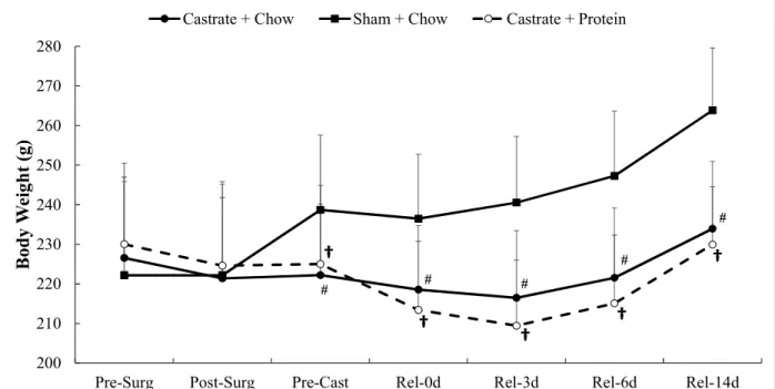
Figure 1. Body mass for sham + chow, castrate + chow, and castrate + protein groups beginning with surgery and continuing out to 14 days of reloading. Data are displayed as means ±SD. † Significant difference between sham + chow and castrate + protein groups.
# Significant difference between sham + chow and castrate + chow groups.
Body Weight. There were significant group differences beginning at the pre-casting time
point (p = 0.001) which persisted throughout the entire reloading period (p < 0.001) with both
castration conditions exhibiting lower body weight than the sham animals.
200 210 220 230 240 250 260 270 280
Pre-Surg Post-Surg Pre-Cast Rel-0d Rel-3d Rel-6d Rel-14d
B o d y W ei g h t (g )
Castrate + Chow Sham + Chow Castrate + Protein
23
Muscle Wet Weight
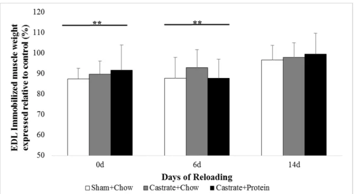
Figure 2. EDL muscle wet weight at baseline (prior to immobilization) and after 0 days, 6 days, or 14 days of reloading via normal cage ambulation.
* Significant difference from control limb p <0.05. ** Significant difference from control limb p < 0.001.
EDL. There were no significant group x cast interactions. Muscle wet weight was 10.6% and
10.4% lower in the immobilized vs. the contralateral limb at 0d and 6d (both p < 0.001; Figure
2), respectively. By 14 days there was no significant difference between the limbs. There were
24
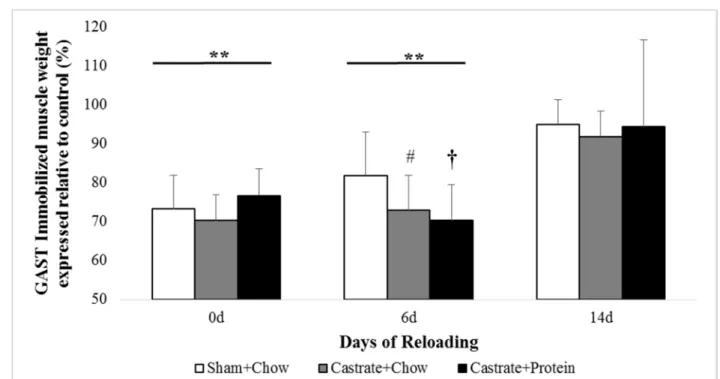
Figure 3. Gastrocnemius (GAST) muscle wet weight at baseline (prior to immobilization) and after 0 days, 6 days, or 14 days of reloading via normal cage ambulation.
* Significant difference from control limb p <0.05 ** Significant difference from control limb p < 0.001
† Significant difference between sham + chow and castrate + protein groups # Significant difference between sham + chow and castrate + chow groups
Gastrocnemius. There were no significant group x cast interactions. Similar to EDL,
casting reduced wet weight significant at 0 days and 6 days (26.6% and 24.6%, both p < 0.001;
Figure 3) and was no longer statistically different at 14 days (p = 0.064). There was a trend for
group differences at 14 days (p = 0.056) where both castration conditions exhibited lower muscle
than the sham + chow group. No differences between castration + chow and castration + protein
25
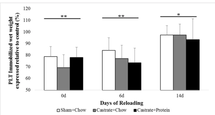
Figure 4. Plantaris (PLT) muscle wet weight at baseline (prior to immobilization) and after 0 days, 6 days, or 14 days of reloading via normal cage ambulation.
* Significant difference from control limb p <0.05 ** Significant difference from control limb p < 0.001
Plantaris. There were no significant group x cast interactions. Immobilization
significantly reduced muscle wet weight at all time points (all p < 0.05; Figure 4), with the casted
limb still being 4% less than control at 14 days. At baseline, castration reduced muscle weight (p
26
Figure 5. Soleus (SOL) muscle wet weight at baseline (prior to immobilization) and after 0 days, 6 days, or 14 days of reloading via normal cage ambulation.
* Significant difference from control limb p <0.05 ** Significant difference from control limb p < 0.001
† Significant difference between sham + chow and castrate + protein groups
Soleus. There were no significant group x cast interactions. Immobilization significantly
reduced muscle wet weight at all time points (all p < 0.001; Figure 5), with only modest recovery
being observed by 14 days. No group differences were present at baseline or 0 days, however sham
+ chow showed a trend to be greater than castration + protein at 6d (p = 0.067) that was significant
27
Figure 6. EDL peak muscle force at baseline (prior to immobilization) and after 0 days, 6 days, or 14 days of reloading via normal cage ambulation. SC = Sham + Chow, CC = Castrate + Chow, CP = Castrate + Protein.
* Significant difference from control limb p <0.05 ** Significant difference from control limb p < 0.001
Peak tension (force)
EDL. There were no significant group x cast interactions. Ten days of immobilization led
to decreased maximal force production (20.8%, p = 0.007; Figure 6) that was also observed at 6
days (p = 0.003) but not 14 days. There was a significantly overall effect of group at 6 days (p =
0.034) on force production. However, post hoc analysis indicated only a trend for reduced force
28
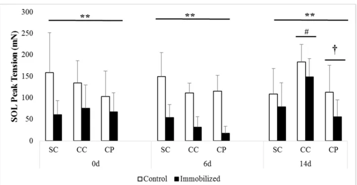
Figure 7. SOL peak muscle force at baseline (prior to immobilization) and after 0 days, 6 days, or 14 days of reloading via normal cage ambulation. SC = Sham + Chow, CC = Castrate + Chow, CP = Castrate + Protein.
* Significant difference from control limb p <0.05 ** Significant difference from control limb p < 0.001
† Significant difference between sham + chow and castrate + protein groups. # Significant difference between sham + chow and castrate + chow groups.
Soleus. The loss of force with immobilization was highly pronounced in soleus muscle,
with large deficits being reported at all three recovery time points (all p < 0.001; Figure 7).
Significant group differences were observed at 6 days (p = 0.050) and 14d (p = 0.009). Post hoc
analysis revealed that sham force tended to be greater than castrate + protein (p = 0.063) at 6 days
and this difference increased by 14 days with both castration groups demonstrating significantly
less force than sham (both p < 0.05). There were no other group differences or significant
29
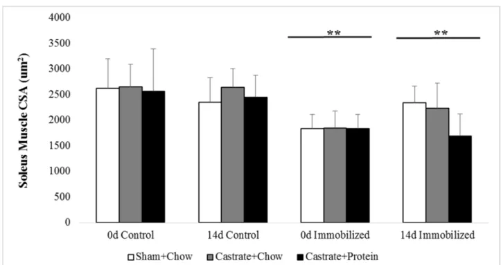
Figure 8. SOL cross-sectional area (CSA) at baseline (prior to immobilization) and after 0 days, 6 days, or 14 days of reloading via normal cage ambulation.
* Significant difference from control limb p <0.05 ** Significant difference from control limb p < 0.001
Soleus Cross-Sectional Area
Immobilization decreased SOL cross-sectional area (30.3%, p < 0.001: Figure 8) at 0d and
while cross-sectional area remained reduced at 14 days, the deficit was smaller (15.9%, p = 0.006).
Castration appeared to blunt the regrowth of the muscle fibers, but this difference did not reach
significance. There was a trend for a weak group x cast interaction (p = 0.083), but this failed to
30
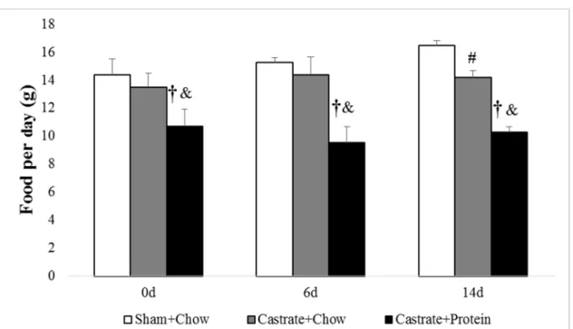
Figure 9. Average food intake for sham + chow, castrate + chow, and castrate + protein groups beginning with 0 days of reloading and continuing out to 14 days of reloading.
† Significant difference between sham + chow and castrate + protein groups # Significant difference between sham + chow and castrate + chow groups & Significant difference between castrate + chow and castrate + protein groups
Average Food Intake
There were significant group differences present at all time points (all p < .001). Post hoc
analysis showed the castrate + protein group consistently had lower food intake than sham +
chow (p < .001 for all time points: Figure 9) and castrate + chow (p = .001 at 0d, p < .001 for 6d
and 14d) for all time points, and sham + chow had greater food intake that castrate + chow (p
<.001) at 14 days of reloading. Further analysis by adjujsting average food weight intake into
average caloric intake showed that castrate + protein consumed significantly less calories
throughout compared to both castrate + chow (p = .002) and sham + chow (p < .001). However,
when adjusting for protein intake in grams per day the castrate + protein group consumed
significantly more protein throughout compared to castrate + chow and sham + chow groups
31
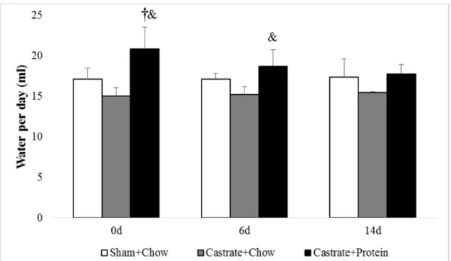
Figure 10. Average water intake for sham + chow, castrate + chow, and castrate + protein groups beginning with 0 days of reloading and continuing out to 14 days of reloading. † Significant difference between sham + chow and castrate + protein groups
& Significant difference between castrate + chow and castrate + protein groups
Average Water Intake
There were significant group differences shown at 0 days (p < .001: Figure 10) at and 6
days (p = .017) of reloading. Post hoc analysis revealed the castrate + protein group had greater
water intake than sham + chow (p = .003) and castrate + chow (p < .001) at 0 days of reloading,
32
CHAPTER V
DISCUSSION
The loss of endogenous testosterone has adverse effects on skeletal muscle mass, strength,
function, and physical activity levels. Functional overload (i.e. strength training) elicits adaptations
that may attenuate or reverse these effects but relatively few studies have examined the
hypertrophic response in the absence of testosterone. The current study showed that 10 days of
hindlimb immobilization significantly reduced muscle wet weights, force, and CSA and these
changes occurred independent of testosterone levels. During the 14 day reloading period, both
castration groups displayed a blunted total body weight growth. Locally, significantly lower
plantaris and gastrocnemius wet weight with 6 days of reloading, and diminished soleus peak
tension after 14 days of reloading compared to sham animals. Contrary to some of our work in
humans, functional overload in a testosterone-deprived state appears to attenuate the regrowth of
skeletal muscle mass and recovery of peak tension, suggesting that testosterone may play a role in
load-mediated hypertrophy following immobilization.
We hypothesized that castration combined with immobilization would act synergistically
to induce greater atrophy than just immobilization alone, however this was not shown in the present
study. Ten days of immobilization decreased soleus muscle weight by 34.8% in the castration +
chow group, 33.8% in the castrate + protein group, and by 36.5% in the sham + chow group, with
33
which showed that 10 days of immobilization decreased soleus muscle weight by 38.5% in young
rats. There were no significant group differences shown in EDL, gastrocnemius, plantaris or soleus
muscle wet weight, soleus muscle CSA, or EDL and soleus peak tension at the conclusion of
immobilization (0 days of reloading in Figures 1-4), indicating that castration did not act
synergistically to induce greater atrophy and that immobilization appears to be the greater
atrophy-inducing stimulus. This finding agrees with findings from an earlier study by Brown et al. (2001)
where they showed that castration did not exacerbate hindlimb unloading-related atrophy on
skeletal muscle contractile properties and physical inactivity. Immobilization also significantly
reduced force and CSA. Interestingly, soleus appears to have been more affected as mass and peak
tension both failed to recover compared to the control leg even with 14 days of reloading. This
data conflicts with findings from Fluck et al. (2003) that showed that by 14 days of reloading,
soleus mass had recovered to control values following 14 days of hindlimb suspension.
We hypothesized that muscle mass recovery after immobilization would occur independent
of testosterone status. Contrary to our hypothesis, castration attenuated muscle mass recovery
following both 6 and 14 days of reloading and protein supplementation did not augment the
response. Both castration groups exhibited attenuated gastrocnemius mass recovery with 6 days of
reloading (-12% lower than sham), and soleus mass of the castration + protein group remained was
significantly lower than sham after 14 days of reloading (-17% lower than sham). Soleus muscle
recovery at 6 days of reloading is similar to previous work (Childs et al., 2003), with 42% recovery
in the current study vs. 37% recovery. We also hypothesized that musculoskeletal function would
recover independent of testosterone status, however after 14 days of reloading soleus peak tension
34
testosterone may play a role in musculoskeletal function and mass recovery following
immobilization.
The attenuated muscle mass recovery is in agreeance with Kvorning et al. (2006) that found
attenuated increases in lean mass following strength training in hypogonadal men. However, it
should be noted the studies that show significant increases in lean mass with strength training, such
as Hanson et al. (2013) and Nilsen et al. (2014), utilized relatively high intensity training protocols;
with Hanson using a high-intensity protocol combined with heavy resistance at high volume which
elicited near-maximal effort on all repetitions, and Nilsen using a daily undulating periodization
model, with a linear progression in training volume through the intervention period. Therefore, the
significant increases in lean mass in the absence of testosterone may be a result of higher intensity
training protocols while those that show attenuated increases use lower intensity protocols. This
may explain the attenuated increases in the present study as well, as the immobilization and
subsequent reloading model is intended to act as an overload stimulus to induce muscle growth
following atrophy. However, this model may not place a large enough load on the muscle to be
considered high intensity and to induce significant atrophy as seen in Hanson et al. (2013) and
Nilsen et al. (2014).
The lack of group differences after 14 days of reloading, in all muscles except the soleus,
represent conflicting findings between muscle mass, CSA, and function within the present study.
While it seems that muscle mass had recovered with 14 days of reloading, although at an attenuated
rate, soleus cross-sectional area and peak tension failed to recover compared to the control leg in
all groups even with 14 days of reloading. This is unexpected as muscle mass and cross-sectional
area have been shown to be correlated (Kim et al., 2015; Hansen et al., 2007), therefore increases
35
immobilization protocol may have damaged the muscle tissue as immobilization been shown to
increase oxidative stress which can cause muscle damage (Moylan et al., 2007; Powers et al.,
2011), and as a result increased blood flow to damaged muscle. This means that at the time of
muscle harvesting, edema could have accumulated within the tissue and increased muscle wet
weight. However, if edema was accumulating within the tissue as a result of muscle damage
centrally located nuclei would be seen indicating the presence of regenerating fibers, but none
were found. Another possibility is that mass may be increasing as a result of these rats being young
and therefore still developing into mature rats. An increase in muscle fiber length as a result of
normal growth could increase muscle mass, but would not serve to increase muscle cross-sectional
area or peak tension
We hypothesized a high protein diet would attenuate atrophy during the immobilization
period and facilitate better adaptations to reloading compared to castrated rats with a traditional
diet, however this was not shown in the present study. In fact, castrate + protein group had
significantly lower soleus peak tension and soleus mass after 14 days of recovery compared to the
sham + chow groups,and the castrate + protein group exhibited a trend towards worse adaptations
to reloading than the castrate + chow group in some instances. The standard chow diet (AIN93G)
provides 17.5% of the total calories protein whereas the high protein diet (SF00-252) provides
45.83% of the total calories from protein. Castrate + protein consumed significantly more protein
per day compared to the other groups by approximately 40-50%. However, the castrate + protein
group consistently consumed significant less calories per day compared to both the castrate + chow
(21-34%), and sham + chow (26-38%) groups. A lower daily caloric intake could stunt body
weight and muscle regrowth as there is potentially a lack of energy available from the diet to
36
hunger in humans (Martens et al., 201l; Marmonier et al., 2000) and rats (Maurer et al., 2009).
This means that despite this group having a larger daily intake of protein compared to the other
groups, they were likely eating less as a result of increased satiety from high protein diet.
Moreover, increased protein intake increases thirst and increased water intake as result of this
increased thirst may result in lowered food intake. Additionally, the high protein diet is composed
of primarily casein, which has been shown to be less effective in promoting protein (or muscle)
accretion when compared to whey (Pennings et al., 2011).
There are limitations to this study, one being that is it is a retrospective analysis of
previously collected data. Retrospective studies are vulnerable as the investigators have no control
over exposure or outcome measures.. Another limitation is the lack of a true control group. The
use of a non-immobilized contralateral limb served as the control for each animal in the present
study, however this is somewhat controversial. In young mice who are still be growing, it was
argued previously that unilateral immobilization may skew the results (Murphy et al. 2011). Other
data from Krawiec et al. (2005) in rats indicates that unilateral immobilization had no effect on the
contralateral non-casted leg. Lastly, immobilization induce muscle atrophy via inactivity that is
reversed during by overloading the atrophied muscle via normal cage ambulation. As such, this
stimulus may promote a muscular endurance phenotype rather than muscular strength and may not
be an appropriate analog to study the effects of strength training. However, there are inherent
limitations in studying the adaptions to functional overload in animals, given they cannot perform
traditional strength training protocols that would be analogous to human exercise interventions.
In summary, our data suggests that testosterone influences the hypertrophic response as
castrated rats exhibited attenuated muscle regrowth following immobilization compared to sham
37
result of increased muscle mass resulting from increased fiber length as opposed to increased
muscle thickness. Further, the high protein diet did not attenuate immobilization-induced atrophy,
nor did it facilitate greater adaptations to reloading in castrated rats compared to chow diet. This
appears to be the result of decreased daily food intake (and lower total calories) from increased
satiety and water intake as a result of the high protein diet. Future studies in rats using a high
protein diet should aim to control for daily food intake and may explore possibility of a high protein
38
REFERENCES
1. Alberga, A. S., Khandwala, F., Scott, C. G., Kenny, G. P., Segal, R. J., Wells, G. A., James, J., et al. (2012). Age and androgen-deprivation therapy on exercise outcomes in men with prostate cancer. Supportive Care in Cancer, 20(5), 971–981.
2. Bajotto, G., Sato, Y., Kitaura, Y., & Shimomura, Y. (2011). Effect of branched-chain amino acid supplementation during unloading on regulatory components of protein synthesis in atrophied soleus muscles. European Journal of Applied Physiology, 111, 1815–1828.
3. Baptista, I. L., Leal, M. L., Artoli, G. G., Aoki, M. S., Fiamoncini, J., & Turri, A. O. (2009). Leucine attenuates Skeletal Muscle Wasting via Ihibition of Ubiquitin Ligases. Muscle and Nerve, 808.
4. Basaria, S., Lleb II, J., Tang, A. M., DeWeese, T., Carducci, M., Eisenberger, M., & Dobs, A. (2002). Long-term effects of androgen deprivation therapy in prostate cancer patients. Clinical Endocrinology, 56, 779–786.
5. Basaria, S. (2008). Androgen Deprivation Therapy, Review Insulin Resistance, and Cardiovascular Mortality: An Inconvenient Truth. Journal of Andrology, 29(5), 534–539.
6. Blough, E. R., & Linderman, J. K. (2000). Lack of skeletal muscle hypertrophy in very aged male Fischer 344 x Brown Norway rats. Journal of Applied Physiology, 88, 1265– 1270.
7. Bodine, S. C., Latres, E., Baumhueter, S., Lai, V., Goldberg, A. L., Nunez, L., & Clarke, B. A. (2001). Identification of Ubiquitin Ligases Required for Skeletal Muscle Atrophy. Science, 294(5547), 1704–1708.
39
9. Booth, F. W. (1988). Time course of muscular atrophy during immobilization of hindlimbs in rats. Journal of Applied Physiology Respiratory Environmental and Exercise Physiology, 43(4), 656–661.
10.Borst, S. E., & Conover, C. F. (2006). Orchiectomized Fischer 344 male rat models body composition in hypogonadal state. Life Sciences, 79, 411–415.
11.Borst, S. E., Conover, C. F., Carter, C. S., Gregory, C. M., Marzetti, E., Leeuwenburgh, C., Vandenborne, K., et al. (2007). Anabolic effects of testosterone are preserved during inhibition of 5α-reductase. American Journal of Physiology: Endocrinology and Metabolism, 293(2), 507–514.
12.Bricout, V. A., Serrurier, B. D., Bigard, A. X., & Guezennec, C. Y. (1999). Effects of hindlimb suspension and androgen treatment on testosterone receptors in rat skeletal muscles. European Journal of Applied Physiology and Occupational Physiology, 79(5), 443–448.
13.Brown, M., Fisher, J. S., & Hasser, E. M. (2001). Gonadectomy and Reduced Physical Activity: Effects on Skeletal Muscle. Archives of Physical Medicine and Rehabilitation, 82, 93–97.
14.Bylow, K., Mohile, S., Stadler, W. M., & Dale, W. (2007). Does Androgen-deprivation Therapy Accelerate the Development of Frailty in Older Men With Prostate Cancer? Cancer, 110(12), 2604–2613.
15.Chakravarthy, M. V., Davis, B. S., & Booth, F. W. (2000). IGF-I restores satellite cell proliferative potential in immobilized old skeletal muscle. Journal of Applied Physiology, 89, 1365–1379.
16.Childs, T. E., Spangenburg, E. E., Vyas, D. R., & Booth, F. W. (2003). Temporal alterations in protein signaling cascades during recovery from muscle atrophy. American Journal of Physiology: Cell Physiology, 285, 391–398.
40
18.Fluck, M., Schmutz, S., Wittwer, M., Hoppeler, H., & Desplanches, D. (2005). Transcriptional reprogramming during reloading of atrophied rat soleus muscle. American Journal of Physiology: Regulatory, Integrative and Comparative Physiology , 284, 14.
19.Fluck, M., Schmutz, S., Chiquet, M., Mayet-Sornay, M. H., & Desplanches, D. (2003). Reloading of atrophied rat soleus muscle induces tenascin-C expression around damaged muscle fibers. American Journal of Physiology: Regulatory, Integrative and Comparative Physiology , 284, 792–801.
20.Galvao, D. A., Nosaka, K., Taaffe, D. R., Spry, N., Kristjanson, L. J., McGuigan, M. R., Suzuki, K., et al. (2006). Resistance Training and Reduction of Treatment Side Effects in Prostate Cancer Patients. Medicine and Science in Sports and Exercise, 2045–2052.
21.Galvao, D. A., Spry, N. A., Davies, R., La Bianca, S., Joseph, D., Davidson, A., & Prince, R. (2009). Long-term effects of intermittent androgen suppression on testosterone recovery and bone mineral density: results of a 33-month observational study. British Journal of Urology, 104, 806–812.
22.Galvao, D. A., Newton, R. U., Taaffe, D. R., Joseph, D., & Spry, N. (2010). Combined Resistance and Aerobic Exercise Program Reverses Muscle Loss in Men Undergoing Androgen Suppression Therapy for Prostate Cancer Without Bone Metastases: A Randomized Controlled Trial. Journals of Clinical Oncology, 28(2), 340–347.
23.Gentile, M. A., Ray, W. J., Nanterment, P. V., Vogel, R. L., Phillips, R., Hodor, P., Freedman, L. P., et al. (2010). Androgen-mediated improvement of body composition and muscle function involves a novel early transcriptional program including IGF1, mechano growth factor, and induction of β-catenin. Journal of Molecular Endocrinology, 44(1), 55– 73.
24.Gomes, M. D., Lecker, S. H., Jagoe, R. T., Navon, A., & Goldberg, A. L. (2001). Atrogin-1, a muscle-specific F-box protein highly expressed during muscle atrophy. PNAS, 98(25), 14440–14445.
![[(R) 4,4′ Bis(diphenylphosphino) 2,2′,6,6′ tetramethoxy 3,3′ bipyridine κ2P,P′]dichloropalladium(II)](data:image/gif;base64,R0lGODlhAQABAIAAAP///wAAACH5BAEAAAAALAAAAAABAAEAAAICRAEAOw==)