part of Epigenomics
Review 2017/02/28 9
3
2017
Exposure to environmental contaminants during pregnancy has been linked to adverse outcomes at birth and later in life. The link between prenatal exposures and latent health outcomes suggests that these exposures may result in long-term epigenetic reprogramming. Toxic metals and endocrine disruptors are two major classes of contaminants that are ubiquitously present in the environment and represent threats to human health. In this review, we present evidence that prenatal exposures to these contaminants result in fetal epigenomic changes, including altered global DNA methylation, gene-specific CpG methylation and microRNA expression. Importantly, these changes may have functional cellular consequences, impacting health outcomes later in life. Therefore, these epigenetic changes represent a critical mechanism that warrants further study.
First draft submitted: 2 September 2016; Accepted for publication: 8 December 2016; Published online: 17 February 2017
Keywords: DNA methylation • endocrine disruptors • environmental exposure • epigenetics • fetal epigenome • in utero • prenatal • toxic metals
The fetal epigenome represents a critical developmental target, as epigenetic modi-fications are thought to underlie a range of disease outcomes, from cancer to neurode-velopmental disease [1,2]. There is increasing evidence that epigenetic reprogramming in early life may persist, influencing both devel-opment and susceptibility to disease later in life. In relation to the prenatal period, expo-sures are of particular concern as this time frame represents a susceptible developmental window during which critical patterns of DNA methylation and gene expression are being established [1]. Evidence that exposures during the prenatal period may result in epi-genetic alterations supports the Developmen-tal Origins of Health and Disease (DOHaD) hypothesis. This hypothesis, originally pro-posed by David Barker, posits that condi-tions experienced in utero play a role in fetal programming with important consequences for later life outcomes [3–5]. Critically,
epi-genetic modifications, particularly CpG methylation, are responsive to environmen-tal factors, allowing these exposures to leave ‘ environmental footprints’ on DNA [6–9].
There are three major forms of epigenetic modifications, namely DNA methylation, microRNA (miRNA) expression, and his-tone modifications [10]. This review focuses primarily on two of these modifications, namely, DNA methylation and miRNAs. Briefly, DNA methylation involves ‘tag-ging’ nucleotide bases with methyl groups, particularly at the 5′ position of cytosine (5′-mC). Depending on the genomic loca-tion, such methylation can either activate or silence transcription. A proposed mechanism by which this occurs is via transcription fac-tor binding to DNA. In contrast, miRNAs are short, nontranslated RNAs that bind to and inhibit translation of mRNAs with reciprocal sequences [11,12]. Notably, these epigenetic modifications may ‘cross-talk’
Effects of prenatal exposure to endocrine
disruptors and toxic metals on the fetal
epigenome
Paige A Bommarito1, Elizabeth Martin1 & Rebecca C Fry*,1,2
1Department of Environmental Sciences & Engineering, Gillings School of Global Public Health, University of North Carolina at Chapel Hill, Chapel Hill, NC 27599, USA
2Curriculum in Toxicology, University of North Carolina School of Medicine, University of North Carolina at Chapel Hill, Chapel Hill, NC 27516, USA *Author for correspondence: Tel.: +1 919 843 6864 [email protected]
with one another. For instance, evidence suggests that there is a relationship between DNA methylation and histone modifications, such that sites of histone modi-fications and DNA methylation are highly associated with one another [13]. Additionally, experimental evi-dence has demonstrated that manipulating histone acetylation induces alterations in DNA methylation, and vice versa [14,15]. Together, these epigenetic regula-tors control the timing and level of gene expression, f undamentally controlling cellular and physiologic function.
Toxic metals and endocrine disruptors are ubiqui-tously found in the environment and have been mea-sured and quantified in the fetus, supporting that in utero exposure to these chemicals occurs [16,17]. Given the link between prenatal exposures to toxic metals, endocrine disruptors and diseases occurring later in life, epigenetic modifications are believed, at least in part, to underlie these effects [18]. This review summa-rizes the current literature related to four toxic met-als where prenatal exposure is common and early life exposure yields both early developmental and later life effects, namely arsenic (As), cadmium (Cd), lead (Pb) and mercury (Hg). It also details studies related to five endocrine disruptors, specifically bisphenol-A (BPA), dichlorodiphenyltrichloroethane (DDT), poly-brominated diphenyl ethers (PBDE), polychlorinated biphenyls (PCB) and phthalates. This review provides an overview of existing research that explores the rela-tionship between prenatal exposure to both metals and endocrine disruptors in relation to the fetal epigenome, along with connections between observed epigenetic reprogramming and adverse health outcomes.
Toxic metals
Toxic metals, in contrast to essential metals, are those that are harmful to human health. For the toxic met-als reviewed here, there is substantive literature link-ing early life exposures to persistent adverse health effects. In the following section, we summarize studies detailing the relationship between prenatal exposure to four toxic metals – As, Cd, Pb and Hg – and the fetal epigenome. Notably, all of the toxic metals dis-cussed are known to cross the placental barrier, to some extent [19–21]. This suggests that the placenta serves as an incomplete barrier, resulting in direct fetal exposure to these compounds. The studies detailed below are summarized in Supplementary Table 1.
Arsenic
Inorganic arsenic (iAs) is a toxic metalloid ubiquitous in the environment with exposure related to both can-cer and noncancan-cer end points [22]. Notably, exposure via contaminated drinking water is a major route of
concern. More than 100 million people are at risk of exposure that exceeds the World Health Organization’s recommended limit of 10 parts per billion (ppb) [22]. iAs and its methylated metabolites are known to cross the placenta and are found within cord blood at similar levels as those in the mother [19]. Prenatal iAs exposure is linked to a wide range of adverse health outcomes that may be present at birth or those that emerge later in life, the latter suggesting long-term epigenomic reprogramming of the fetus [23]. It has been hypoth-esized that iAs impacts DNA methylation by creating competition for methyl donors, leading to dysregula-tion of DNA methyladysregula-tion processes [24,25]. However, this theory cannot account for the observation of gene-specific methylation patterns following iAs exposure. Instead, a separate hypothesis suggests that iAs, and other environmental contaminants, may disrupt tran-scription factor occupancy, including access of the methylation machinery to areas of the genome, giving rise to gene-specific patterning of CpG methylation [9].
The relationship between prenatal iAs exposure and global DNA methylation has been examined in several different populations, including those in Bangladesh, Mexico and the USA. Global methylation is often mea-sured by examining methylation at CpG sites located within transposable elements, such as LINE1 or Alu, which comprise up to 30% of the human genome [26]. Changes in global hypomethylation are of concern as it is thought to create genomic instability, yielding an environment in which mutations may arise from the activities of transposable elements or where tumor-promoting genes may be overexpressed. In Bangladesh, where iAs exposures can range up to 2.5 parts per mil-lion (ppm), mixed results have been noted with respect to LINE1 methylation and no relationships have been noted between iAs exposure and luminometric meth-ylation assay (LUMA) methmeth-ylation or Alu methyla-tion [22,27–29]. Similarly, results from an exposed popu-lation in Thailand indicate no repopu-lationship between cord blood LINE1 methylation and iAs exposure [30]. However, results from a Bangladesh-based nutritional intervention suggest a sex-dependent effect of maternal urinary As on cord blood methylation, noting nonsig-nificant hypermethylation in Alu, LINE1 and LUMA methylation in males, but hypomethylation in females. Results from this same study have also reported a positive association between maternal urinary total As and global methylation of cord blood, measured using a methyl incorporation assay [29]. These results suggest that impact of prenatal iAs exposure on global methylation may depend on fetal sex.
have identified differential methylation of p16 and p53 promoter regions, providing evidence that iAs dys-regulates critical tumor suppressor pathways in cord blood [27,30]. Several studies examining genome-wide methylation have reported few differentially methyl-ated CpG sites following false discovery rate correc-tion [28,31,32]. However, among the most significant probes identified in these analyses, enrichment for genes involved in pathways related to juvenile dia-betes, cancers, infectious diseases and inflammatory disorders has been noted, providing evidence, at the molecular level, for the relationship between prenatal iAs exposure and such outcomes [28,32]. Yet, other stud-ies have identified more robust changes in the newborn epigenome. For instance, research from the Biomark-ers of Exposure to ARsenic (BEAR) pregnancy cohort in Mexico identified over 2000 differentially meth-ylated CpG sites. Methylation for a set of these was functional, predicting altered gene expression in the cord blood as well as showing an association with birth outcomes. Notably, among the genes identified was the imprinted gene KCNQ1 [8]. When considering meth-ylation changes across a variety of fetal tissues, Carde-nas et al. identified numerous differentially methylated genes associated with iAs in the placenta and umbili-cal artery. In particular, the genes identified here were enriched for pathways similar to those noted above, reinforcing these previous observations [33]. Similarly, while a recent study from the New Hampshire Birth Cohort did not find significant relationships between maternal urinary total As and placental methylation, and only one significant probe when considering mater-nal toenail iAs, they reported over 100 differentially methylated CpG sites in the placenta associated with placental As [34]. Taken together, these results suggest that there are tissue-specific effects of iAs exposure on the fetal epigenome. Moreover, the effects of iAs expo-sure vary across individuals and populations, likely as a result of genotype and/or nutritional factors [35,36]. Future research should investigate whether differen-tial susceptibility to iAs is linked to the epigenome, contributing to the differences observed between the various Bangaldeshi, Mexican and USA populations.
As observed with studies focusing on global DNA methylation, sex-based differences have also been noted for gene-specific methylation. In particular, increas-ing iAs exposure in male infants was more strongly associated with cord blood CpG methylation than in females. Moreover, iAs exposure in males was associ-ated with DNA hypomethylation, while female infants tended to display hypermethylation. Pathway analysis of the most significantly affected genes also revealed that male infants displayed gene-specific methyla-tion at sites that were most significantly enriched for
cancer pathways, while females were most signifi-cantly enriched for inflammatory diseases [28]. Future research should be carried out to further elucidate the mechanistic underpinnings of sex-based differences in iAs-induced disease.
Two studies have examined the relationship between prenatal iAs exposure and miRNA expression in the placenta. Research from the formerly established National Children’s Study (NCS), a prospective cohort of children across the US, did not note a relationship between placental iAs and miRNA expression [37]. In contrast, results from the BEAR cohort included dif-ferential expression of miRNAs that mediate signaling pathways related to iAs-associated diseases, including cancer, inflammatory disease, respiratory disease and metabolic disease, among others [38]. Moreover, these miRNAs were predicted to target 20% of the observed differential mRNA expression in this population, indi-cating that iAs-associated miRNA dysregulation may have functional consequences for downstream gene expression that may be related to disease outcomes [38].
Cadmium
Cd is found in the environment both as a natural com-ponent of the earth’s crust and as a result of anthropo-genic activities. Specifically, Cd may be found in bat-teries, electronic waste, pesticides and tobacco smoke, among other sources [39]. Cd emissions have been declining globally, although areas of concern remain near smelters, e-waste sites, and other areas polluted by industry [39]. While Cd crosses the placenta, it does not cross as readily as other toxic metals because metallo-thionein sequesters Cd within the placenta. There-fore, the placenta provides a partial barrier against this metal [20]. As a result, Cd levels in cord blood are typically lower than maternal Cd levels [40]. Neverthe-less, in utero Cd exposure has been linked to a range of adverse health effects, including impaired growth and neurodevelopment [41–43]. To date, one study has examined the relationship between global DNA meth-ylation and prenatal Cd exposure. Boeke et al. dem-onstrated that there is an inverse relationship between maternal Cd exposure and LINE1 methylation in cord blood [44]. These observations have also been made in adult populations, suggesting that similar changes could be occurring within fetuses following prenatal Cd exposure [45].
major-ity of them exhibiting hypermethylation. Overlap between differentially methylated genes identified within the cord blood and within maternal blood were also observed [7]. Taken together with the relation-ship between Cd and global methylation, these results suggest that prenatal Cd exposure is associated with both global hypomethylation and gene-specific hyper-methylation. However, in the Maternal and Infant Nutrition Interventions, Matlab (MINIMat) cohort in Bangladesh, no CpG sites were identified as signifi-cant following multiple test corrections [46]. Instead, sex-specific effects were noted with the most signifi-cant probes displaying hypermethylation in males, but hypomethylation in females. Interestingly, in girls, the most significant probes were enriched for genes relating to bone mineralization and morphology, possibly shed-ding light on the sex-specific effects of Cd exposure, especially as it relates to the susceptibility of female populations to Cd-induced bone outcomes [46]. Other studies on Cd-associated fetal epigenomic alterations have also observed sex-specific effects [47,48]. Of note, the relationship between maternal blood Cd and the methylation of imprinted genes was altered in a sex-dependent manner in infants from the Newborn Epi-genetic STudy (NEST) in North Carolina [47]. These relationships were also dependent on levels of circulat-ing zinc and iron, indicatcirculat-ing that Cd-associated epig-enomic disruptions may be modified by maternal levels of essential metals.
Lastly, several studies have reported on Cd-induced miRNA dysregulation. Placental Cd levels were sig-nificantly associated with increased placental expres-sion of miR-1537 [37]. Little is known about this miRNA, however, it has been suggested to play a role in cancer [49,50]. Interestingly, a recent study examin-ing the relationship between preeclampsia and Cd exposure identified a set of miRNAs common to both preeclampsia and Cd-exposure. These miRNAs also regulate genes involved in the TGF-β signaling path-way [51]. Given the epidemiological links between pla-cental Cd levels and preeclampsia, these results pro-vide epro-vidence, at the molecular level, for a relationship between exposure and disease [52].
Lead
As observed with the ongoing struggles in Flint, Michigan, Pb continues to be a toxic metal of criti-cal importance in children’s health [53,54]. In urban centers, children are often exposed via Pb-based paint that remains in homes, particularly within low socio-economic areas [55]. However, maternal exposure is also of importance as Pb is known to cross the placenta, although the mechanisms underlying placental trans-fer remain unknown [20]. While exposures have
gener-ally been decreasing over the course of the last century, even levels below the established regulatory limits (5 μg/dL) have been associated with impaired neurode-velopment in children [56,57]. Given the latent and per-sistent effects of early life exposure to Pb, epigenetic reprogramming has been posited to contribute to these effects. Maternal bone Pb levels have been significantly associated with LINE1 and Alu methylation, although cord blood Pb has not [58,59]. Maternal bone Pb rep-resents cumulative exposure, suggesting that mater-nal cumulative Pb exposure is associated with altered global methylation. Supporting these observations, it has been previously noted that maternal bone Pb is better at predicting adverse health outcomes in infants and children than cord blood Pb [60].
In genic regions, results from the Early Life Exposures in Mexico to Environmental Toxicants (ELEMENT) study suggest that methylation of the imprinted genes IGF2 and HSD11B2 was positively associated with in utero Pb exposure, with a sex-dependent effect and stronger relationship in girls. However, results were not significant following multiple test correction [59]. Addi-tional studies have focused on the sex-specific effects of Pb-induced epigenetic disruptions of the fetus. Using cord blood and blood spots, Sen et al. examined dif-ferentially methylated regions of the genome that may serve as sex-dependent or independent biomarkers of Pb exposure [61,62]. Interestingly, different patterns were noted when observing CpG methylation in cord blood versus infant blood spots. In cord blood, males showed a greater number of sites with altered CpG methylation [61]. However, in blood spots, females showed greater disruption in CpG methylation. Given that males tend to be more susceptible to the effects of Pb exposure, researchers concluded that disruptions in CpG methylation may be adaptive in blood [62]. Notably, Sen et al. has also reported that maternal Pb exposure induces epigenetic changes in the germ line of their offspring, impacting DNA methylation in the F2 in humans. These results provide evidence that Pb exposure may induce multigenerational epigenomic changes [63].
Mercury
Long known to be linked to adverse neurological out-comes, Hg exposure during pregnancy is associated with impaired attention, visuospatial and motor func-tioning, among other outcomes [65]. Within the US, approximately 15% of childbearing-age women have elevated blood Hg levels of concern, suggesting that over 600,000 children may be born with increased risks of such neurological outcomes [66]. Importantly, Hg bioaccumulates within the fetus, suggesting an active transport mechanism across the placenta, although the mechanisms for such transport are unknown [21]. No studies were identified that examined the relationship between prenatal exposures to Hg and global DNA methylation. However, two studies from birth cohorts in the USA have explored the relationship between in utero Hg exposure and gene-specific methylation in cord blood. Using independent test and validation populations, the relationship between cord blood Hg and CpG methylation was examined using several dif-ferent methods [67]. It was shown that TCEANC2 was significantly differentially methylated in both the test and validation populations. However, given the associ-ation between TCEANC2 and blood cell composition, these results could be attributed to either Hg-induced changes in methylation at that loci or Hg-induced changes in blood cell composition [67]. Therefore, these observations should be interpreted with caution. In a second study, prenatal Hg exposure was significantly associated with genome-wide CpG methylation, with a majority of significant probes displaying a nonmono-tonic relationship between exposure and methylation. Notably, a subset of the Hg-associated probes were also significantly associated with high-risk status for adverse neurobehavioral outcomes, indicating that dysregulated CpG methylation may have functional consequence for Hg-associated outcomes [68].
Lastly, results from the NCS indicate that placen-tal Hg levels are significantly associated with changes in miRNA expression in the placenta. In particular, Hg levels were associated with a large number of miR-NAs within the let-7 family, which is critical for proper developmental timing in animal models [69]. Further examination of the relationship between let-7 miRNAs and Hg exposure may shed further light on the terato-genic and other developmental effects of prenatal and early life Hg exposure.
Endocrine disruptors
Early life exposure to endocrine disruptors, which interfere with endocrine signaling and development, remains a topic of concern. Endocrine disruptors are ubiquitous in the environment, representing a wide range of chemicals, such as pesticides, plasticizers or
other synthetic compounds used in industrial settings. While we have separated metals and endocrine disrup-tors in this review, it is important to note that some toxic metals also act as endocrine disruptors [70]. In the following section, the literature focusing on the relationship between prenatal exposure to five endo-crine disruptors and the fetal epigenome is reviewed. Specifically, exposures to BPA, DDT, PBDE, PCB and phthalates are highlighted. As noted above with respect to toxic metals, all of the endocrine disruptors discussed here are known to cross the placental bar-rier [71–74]. The studies detailed below are summarized in Supplementary Table 2.
Bisphenol-A
suggest that indicators of global methylation, namely repetitive DNA elements, are associated with prenatal BPA exposure in a dose- and tissue-specific manner. It is also important to note that repeat DNA elements respond differentially to environmental exposures, therefore, changes in Alu and LINE1 may represent methylation changes occurring in different parts of the methylome [26,83].
In contrast to global methylation detailed above, Faulk et al. also examined genome-wide CpG meth-ylation in relation to BPA exposure, identifying and validating differential methylation within the snRNA cluster around SNORD116, a maternally imprinted locus [77]. One study has tested the relationship between BPA exposure and miRNA expression, but no significant relationships were observed [37].
Dichlorodiphenyltrichloroethane
DDT is a persistent organic pollutant. While DDT was used extensively from the 1940s until 1973, it is no lon-ger in commercial use in the US, although it remains in the environment as a legacy contaminant [84]. Dichlo-rodiphenyldichloroethylene (DDE), a common break-down product of DDT that may form in the environ-ment or within the body, is often examined alongside DDT as it has been associated with adverse health outcomes in human populations [85,86]. While DDT and DDE both have long half-lives in the environment and the human body, DDE was observed to be the pre-dominant form found in pregnant women participat-ing in the National Health and Nutrition Examination Survey [87]. While levels of DDT are lower within fetal than maternal tissues, there is evidence that DDT is actively transported across the placenta as fetal levels are higher than expected via passive diffusion [72]. Pre-natal exposure to DDT and DDE has been associated with a range of early and later life health outcomes, including fetal growth measures, adolescent neuro-development, indicators of female reproductive func-tioning, and lung functioning [88–93]. When examin-ing the association between DDT, DDE, and global methylation of the fetal epigenome, results from the Center for Health Assessment of Mothers and Chil-dren of Salinas (CHAMACOS) study indicate that maternal serum DDT and DDE levels are inversely associated with Alu methylation in cord blood [83]. However, additional research from this cohort, using LUMA to measure global CpG methylation, did not report a relationship between placental DDE levels and global methylation [94]. The inconsistency between these results originating from the same cohort sug-gest that DDT has tissue-specific effects on the fetal epigenome, as was noted previously with BPA. Alter-natively, the discrepancy may also be due to the use of
different methods of assessing DNA methylation. As previously mentioned, LINE1 and Alu elements may be differentially impacted by environmental exposures and may capture changes occurring at only a portion of sites within the genome [26]. Thus results from LINE1 and Alu elements may differ from LUMA methyla-tion, which instead measures a ratio of unmethylated to methylated CpG sites across the genome.
In relation to DDE exposure, the CHAMACOS investigators have also examined placental methylation at CpG islands affiliated with two imprinted genes, IGF2 and H19. No significant associations were noted between placental DDE levels and methylation at these loci [94]. Additionally, no relationship has been found between exposure to DDE and placental miRNA expression [37].
Polybrominated diphenyl ethers
Two studies have examined fetal global methylation in association with exposure to PBDE, a class of com-pounds used as flame retardants in products ranging from clothing to household building materials [95]. Evi-dence suggests that there is relatively free transfer of PBDE across the placenta, particularly for low-bromi-nated congeners [73]. Notably, infants and young chil-dren have been observed to have higher body burden of PBDE than adults as a result of this placental transfer, as well as breast feeding and exposure to household dust [96]. Research originating from the CHAMA-COS study assayed LINE1 and Alu methylation in DNA derived from cord blood. No association was noted between these indicators of global methylation and third trimester maternal blood PBDE. However, interactions between maternal blood PBDE and blood DDT and DDE were noted [83]. These results will be discussed in more detail, below, in the context of co-exposures. A second study, examined placental LUMA methylation in correlation with placental PBDE levels. Here, a significantly positive relationship between pla-cental PBDE and global methylation was observed [94]. Additionally, loss of imprinting at IGF2 and H19 was also examined in relationship to PBDE, although no significant results were reported [94].
meth-ylation of TNF-α. Moreover, this effect appeared to be modified by the sex of the fetus, with the same pattern noted in females, but not males [97].
A single study has examined the relationship between placental PBDE levels and miRNA expres-sion. No relationship was found with total placental PBDE, however, PBDE 209 was positively associated with miR-188–5p expression, while PBDE 99 was negatively associated with let-7c expression [37].
Polychlorinated biphenyls
As with DDT, PCB are chemicals that are no longer in commercial production in the USA. Previously used widely as coolants, these chemicals persist in the envi-ronment [98]. Research has demonstrated that PCB lev-els are higher than expected in fetal tissues based on passive diffusion alone, suggesting an active transport mechanism across the placenta [72]. Moreover, biomon-itoring of pregnant women suggests that serum levels of PCB may increase over the course of pregnancy as lipid stores are mobilized [99]. Results from the NCS did not identify a relationship between LUMA meth-ylation in the placenta and placental PCB levels [94]. At a gene-specific level, no relationship was noted between PCB levels and methylation at the IGF2 and H19 loci, although an inverse relationship between H19 expres-sion and PCB levels was reported [94]. These results suggest that in utero exposure to PCB may influ-ence the expression of H19 via mechanisms that are i ndependent from CpG methylation.
Additional studies from the NCS examined miRNA expression in the placenta in relation to placental PCB levels. Notably, total PCB, PCB 52 and PCB 101 were significantly positively associated with miR-1537 expression [37].
Phthalates
Phthalates, like BPA, represent a class of ubiquitously used industrial compounds that are found widely in personal care items and household products [100–103]. There are numerous routes of exposure to phthalates, with the compounds being inhaled, ingested and der-mally absorbed. Urinary metabolites are often used as indicators of exposure representing all of these pos-sible routes, although their stability as a biomarker is short because phthalates are quickly metabolized and excreted from the body. Despite this rapid metabolism and excretion, phthalates are still detected in the amni-otic fluid, indicating that it crosses the placenta and enters the fetal compartment [74]. However, perfusion studies indicate that placental transfer of phthalates may be slow [104]. As with BPA, the MIREC study has also demonstrated that phthalate levels in pregnant women are comparable to those measured in the
gen-eral population [76]. There are many different phthal-ates used in industry, but several are more commonly detected and used in epidemiological studies. These include di(2-ethylhexyl) phthalate, diethyl phthalate and dibutyl phthalate, among others [105].
With respect to the fetal methylome, several stud-ies have assessed the impact of prenatal exposure to phthalates on global methylation, gene-specific meth-ylation and miRNA expression. Urinary levels of low-molecular-weight (LMW) phthalate metabolites and a diethyl phthalate metabolite, mono-ethyl phthalate, have been negatively associated with Alu methylation in cord blood using maternal-infant dyads from the CHAMACOS study [106]. These results suggest global hypomethylation of the cord blood following phthal-ate exposure and are supported by previous research originating from a Chinese cohort of pregnant women, which demonstrates that maternal urinary levels of di(2-ethylhexyl) phthalate were associated with hypo-methylation of LINE1 in the placenta [107]. Taken together, these results suggest that prenatal phthalate exposures result in global hypomethylation of fetal DNA as assessed within placental tissue and cord blood.
Only one study was identified that examined gene-specific methylation in relationship to prenatal phthal-ate exposure [108]. Namely, CpG methylation of H19 and IGF2 was examined in relationship to first-trimes-ter mafirst-trimes-ternal urinary phthalate measures. Both cumu-lative and LMW phthalates were inversely associated with H19 and IGF2 methylation. These results indi-cate a possible dysregulation of these imprinted genes that play a critical role in development [108].
A single study has examined the impact of in utero phthalate exposure and placental miRNA expression. Namely, maternal urinary LMW phthalates were significantly associated with decreased expression of miR-185. However, in silico predicted downstream mRNA targets were not differentially expressed, indicating that other mechanisms contribute to gene e xpression [109].
Mixtures
Toxic metal mixtures
Among infants exposed prenatally to toxic metals, a single study has examined the effects of exposure to iAs and Hg on the fetal epigenome. While no signifi-cant probes were identified after Bonferroni correction, a greater number of probes were observed with a p < 0.0001 when considering the interaction than when considering Hg alone [111]. These results may indicate that there are relationships between exposure to toxic metals and the fetal epigenome that may not be identi-fied until co-exposures are considered. More research into metals mixtures is warranted. For instance, while no studies have examined the impact of co-exposures to iAs and Cd on the fetal epigenome, recent research has identified that both contaminants significantly disrupt genes involved in the innate and adaptive immune system, particularly those involved in gluco-corticoid signaling [114]. These results suggest that iAs and Cd may similarly affect immune-related pathways, supporting the possibility of synergism.
Endocrine disruptor mixtures
As mentioned previously, the CHAMACOS study reported interactions between prenatal DDE and PBDE exposure [83]. When stratified based on mater-nal serum DDE, PBDE was associated with hypo-methylation of LINE1 in the fetus among those with low maternal DDE exposure. In those subjects with high maternal DDE exposure, PBDE levels were associated with LINE1 hypermethylation. The same relationships were noted for DDE exposure when the study population was stratified by PBDE levels. Nota-bly, no associations were found between DDE, PBDE and LINE1 methylation until they were considered as co-exposures [83]. These results suggest that relation-ships between prenatal exposures and the fetal epig-enome may not be fully elucidated until co-exposures are considered and underscore the need for continued research into how endocrine disruptors may interact with one another.
A number of endocrine disruptors exhibit estrogenic activity, including BPA, DDT/DDE and PCB [115]. Total effective xenoestrogen burden (TEXB) repre-sents a biomarker for cumulative xenoestrogen burden and has been used to assess co-exposures to such chem-icals [116]. TEXB has been associated with LINE meth-ylation in the placenta, with males displaying hypo-methylation of LINE and Alu elements [112]. However, when assessing the relationship between TEXB and CpG methylation, no significantly differentially methylated probes were identified [113].
Lastly, in placental samples from the Harvard Epi-genetic Birth Cohort (HEBC) and the Predictors of Preeclampsia Study (POPS), the sum of maternal
uri-nary phenols was significantly negatively associated with expression of miR-142 [109]. Cumulative exposure to nonparaben phenols, such as BPA and triclosan, was also significantly inversely associated with miR-15a-5p expression. Interestingly, this relationship exhibited a sex-dependent effect, with female infants exhibiting a significant negative relationship between total phenol exposure and miR-15–5p expression in the placenta. However, downstream gene expression analysis revealed that none of the in silico predicted mRNA targets were differentially expressed, nor were these miRNAs noted to be associated with any birth outcomes [109]. These results indicate that miRNA expression in the placenta is affected by cumulative phenol exposure, however, it is likely that multiple mechanisms are responsible for controlling downstream gene expression.
Contributions of animal models & in vitro
experiments
The bulk of this review focuses on evidence that in utero exposure to toxic metals and endocrine disrup-tors are associated with epigenomic reprogramming of human fetal tissues that, ultimately, provide mechanis-tic evidence for the DOHaD hypothesis. Animal mod-els have the potential to contribute substantially to our understanding of the mechanisms underlying these effects. First, animal models facilitate the examination of epigenomic changes that occur within tissues where disease originates, rather than accessible fetal tissues like the placenta or cord blood. For instance, if expo-sure occurs following zygote formation, tissue-specific epigenomic changes result in susceptible tissues. How-ever, if exposure directly impacts gametes, then whole-organism epigenomic changes may result. Only tissues susceptible to these changes are thought to go on to develop disease during development. Thus, animal models may be used to examine epigenomic changes occurring within disease-relevant tissues in order to determine the role of environmentally induced repro-gramming in disease. Moreover, they may aid in deter-mining whether epigenomic reprogramming occurring in readily accessible tissues serves only as a biomarker of disease, or whether they may be directly related to disease development.
example, in human cell lines, PCB exposure has been shown to modulate the activity of histone demethyl-ases via androgen receptor binding [117]. Aside from direct activity on steroid hormone signaling, toxic met-als and endocrine disruptors have met-also been shown to interact with DNA methyltransferases and ten-eleven translocation enzymes, which are responsible for meth-ylating and demethmeth-ylating CpG sites. For instance, Cd and As have been demonstrated to interact with DNA methyltransferases and ten-eleven translocations, alter-ing their activity in in vitro experiments [118,119]. Much of this mechanistic evidence is reviewed in greater depth, elsewhere [120]. Additionally, some toxic met-als and endocrine disruptors are known to interact with one-carbon metabolism, which produces methyl donors used for DNA methylation. For instance, iAs metabolism uses S-adenosylmethionine as a methyl donor [36]. Given that S-adenosylmethionine is also a methyl donor utilized for DNA methylation, it has been posited that iAs metabolism results in dysregula-tion of DNA methyladysregula-tion [36]. Importantly, while these mechanisms describe how toxic metals and endocrine disruptors may impact epigenetic machinery, they do not explain how such exposures result in gene-specific patterning of epigenomic markers [9]. Instead, the tran-scription factor occupancy theory provides an expla-nation by positing that environmental exposures alter transcription factor activity, changing the availability of DNA to epigenetic machinery and giving rise to gene-specific patterning of epigenomic markers [9].
When considering the persistence of the impacts of in utero exposure to toxic metals and endocrine disrup-tors, it is critical to consider the relative contribution of and interaction between pre- and postnatal exposures. As mentioned, the toxic metals and endocrine disrup-tors reviewed here represent ubiquitous exposures. Therefore, it is likely that populations experiencing in utero exposures to toxic metals and endocrine dis-ruptors also experience chronic exposure throughout the life course. Animal models represent important model organisms in which exposure during defined developmental windows can be investigated [121]. With respect to iAs exposure, prenatal exposure paradigms have been used to establish the sensitivity of the pre-natal developmental window compared with other periods of exposure [122,123]. In human populations, on the other hand, it is often impossible to differentiate between prenatal and postnatal exposures and their contributions to health outcomes [121]. Related, animal models also foster research examining the transgenera-tional impacts of exposure. As Sen et al. demonstrated, maternal Pb exposure may be related to DNA meth-ylation in the blood of their grandchildren (F2), sug-gesting that Pb impacts the germ line of a developing
fetus [63]. However, assessing truly transgenerational effects is unlikely to be feasible in human populations. In animal models, transgenerational impacts can be assessed by examining epigenomic changes occurring in the F3 generation, following exposure during the F1 prenatal period. While little work has been done with the toxic metals and endocrine disruptors reviewed here, exposure to a mixture of BPA and phthalates dur-ing pregnancy in rats has been observed to induce a range of adverse health outcomes, including ovarian/ testis disease and obesity, in the F3 generation. Inter-estingly, the F3 sperm also had altered DNA methyla-tion in promoter regions previously associated with the onset of obesity [124].
Discussion & future perspective
Global methylation is often measured when assess-ing the impact of environmental exposures on the epigenome. Much of the literature reviewed here uses transposable elements (i.e., LINE1 or Alu) as indica-tors of global methylation. However, there is evidence that these elements respond differentially to environ-mental exposures and in a sequence-specific manner, suggesting that they may represent only a specific part of the methylome. For instance, evidence demonstrates that LINE1 methylation may only represent weak CpG island methylation, rather than methylation occurring in other areas of the genome [26]. Moreover, Price et al. maintains that the only true measure of global meth-ylation is total 5′-mC content and that results between studies should only be compared when methylation has been assayed using the same technique [26]. This creates difficulties when comparing the existing data on prenatal exposure to endocrine disruptors, as few studies have utilized the same method and little repli-cation has been conducted. Faulk et al. has examined the relationship between BPA and global methyla-tion using next-generamethyla-tion sequencing in two separate studies, with both reporting hypomethylation based on transposable elements or overall genomic meth-ylation [77,78]. Additionally, with respect to maternal phthalate exposure, Alu hypomethylation was noted in cord blood, while LINE1 hypomethylation was found in the placenta [106,107]. Taken together, prenatal BPA and phthalate exposure may lead to global hypometh-ylation and genomic instability in the fetal epigenome. Additionally, these results suggest that prenatal phthal-ate exposure may have tissue-specific effects on indica-tors of global methylation. We reviewed no other stud-ies on endocrine disruptors that used the same method of assessing global methylation, and thus, we caution against cross-comparisons.
Unlike the literature on endocrine disruptors, research on toxic metals has used more consistent methods to measure both global and site-specific DNA methylation. Notably, high-throughput methods, such as the Illumina 450K BeadChip®, have been used routinely in the study of toxic metals-induced disease. Results from these assays facilitate gene-specific analy-sis and better enable analyanaly-sis of downstream affected biological pathways, shedding light on cell functions that may be dysregulated. For instance, studies from multiple Bangladeshi cohorts have identified differen-tial methylation in genes related to juvenile diabetes, cancers, immunodeficiency and neurological dys-function, among others related to iAs-associated dis-eases [28,32,33]. Moreover, in a cross-study analysis of genes targeted for altered CpG methylation in infants with in utero contaminant exposure, many of the gene targets were identified as having common enriched
transcription factor binding sites, providing evidence for common transcriptional controls in epigenetic patterning following contaminant exposures [9]. The identification of enriched transcription factor binding sites also provides further evidence for the transcrip-tion factor occupancy theory, indicating that altered binding of transcription factors to DNA may result in patterns of gene-specific hyper- and hypomethylation noted following environmental exposures [9].
Far less research has been conducted regarding the impacts of prenatal environmental exposures on fetal miRNA expression. Only four studies were identi-fied that examined the relationship between prenatal exposure to metals and endocrine disruptors on fetal miRNA expression. However, the results reviewed here demonstrate their utility. For instance, prenatal iAs exposure was associated with 12 dysregulated miR-NAs that were enriched for disease pathways related to iAs-associated disease (e.g., immune function) and predicted functional changes in downstream gene expression [38]. With respect to prenatal Pb exposure, dysregulated miRNAs in infants was significantly associated with neurobehavior-related signaling, pro-viding further evidence for the role of epigenetics in Pb-induced neurodevelopmental outcomes [37,64]. Taken together, miRNA dysregulation has explana-tory value when considered in the context of in utero exposures and research should be expanded upon. It is of particular importance given the observation, in some studies, that DNA methylation only corresponds to functional changes in gene expression at a subset of probes [8].
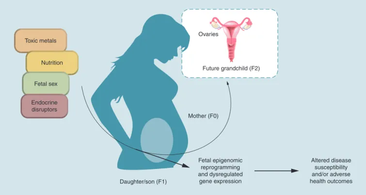
funda-Figure 1. Schematic of environmentally-induced fetal epigenomic reprogramming. Exposure to toxic metals and endocrine disruptors occurring during the prenatal period may result in fetal epigenomic reprogramming. The impacts of exposure on the fetal epigenome depend, in part, on factors such as nutrition and fetal sex. There is evidence that fetal epigenomic reprogramming is associated with altered disease susceptibility and may increase the incidence of adverse health outcomes in exposed populations. Finally, if germ cells are exposed to toxic metals and/or endocrine disruptors and undergo epigenomic reprogramming, multi- and transgenerational effects may be noted following prenatal exposure in the F1.
Toxic metals
Nutrition
Ovaries
Future grandchild (F2)
Mother (F0)
Daughter/son (F1)
Fetal epigenomic reprogramming and dysregulated
gene expression
Altered disease susceptibility and/or adverse health outcomes Fetal sex
Endocrine disruptors
mental differences between placentas derived from male or female fetuses may result in sexually dimor-phic responses to environmental exposures [132]. It has recently been shown that gene-specific methylation differs between male and female placentas and these genes are enriched for immune function, transport of substances across the placenta, and transcription fac-tors [133]. Further substantiating these findings, female- and male-derived placentas have differential expres-sion levels of immune-related genes. Specifically, male placentas have enriched expression of genes related to inflammation and immune functioning, while female placentas have enriched expression of immune-regu-lating genes [134,135]. These immunologic differences between male and female fetuses may result in greater vulnerability in male fetuses when faced with adverse environmental exposures in utero [132]. Additionally, differences in the expression of glucocorticoid receptors have been observed based on fetal sex, which may result in differential fetal susceptibility to the maternal stress and/or immune response following exposure to envi-ronmental contaminants [136,137]. Together, these may ultimately contribute to sex-specific epigenomic repro-gramming and health outcomes. While the mechanism underlying sex-dependent epigenetic reprogramming
As discussed above, humans are exposed to multi-contaminant mixtures, including metals and endocrine disruptors. In support of this, these compounds have been identified in cord blood together [16,110]. Thus, research focusing on exposure to a single contaminant does not adequately represent the true human experi-ence. Notably, Huen et al. did not identify a relation-ship between DNA methylation and exposures to sin-gle classes of persistent organic pollutants. However, when considered together, significant relationships between exposure to DDT/DDE, PBDE and LINE1 methylation emerged [106]. These results indicate that the impact of environmental exposures on the epig-enome may depend on simultaneous exposures, under-scoring the need for continued research into mixtures. Additionally, it is important to point out that while described as two separate classes of contaminants in this review, toxic metals and endocrine disruptors are
not mutually exclusive. Several toxic metals, includ-ing iAs, Cd, Pb and Hg have complex modes of action that include endocrine disrupting effects [70]. Despite the overlaps between these toxic metals and endocrine disruptors, no studies were identified that examined co-exposure to metals and endocrine disruptors.
In addition to considering co-exposures, another important factor underlying epigenomic reprogram-ming is nutritional status. Imbalances in micronutrients involved in one-carbon metabolism have been tied to dysregulated CpG methylation, resulting from their role as methyl donors [143]. In line with these observations, maternal nutrition also has a significant impact on the fetal epigenome, as well as developmental outcomes in offspring [144]. The relationship between micronutri-ents, environmental exposures and epigenomic repro-gramming has been well-documented with respect to iAs exposure in human populations. For instance, in
Executive summary Background
• There are three major epigenetic modifications including: DNA (CpG) methylation, microRNA expression and histone modifications.
• The fetal epigenome is impacted by environmental exposures, including toxic metals and endocrine disruptors.
Toxic metals
• Prenatal exposure to arsenic, cadmium, lead and mercury is associated with epigenomic alterations in the offspring.
• The epigenetic changes resulting from in utero toxic metal exposure are associated with functional changes in gene expression, birth outcomes and later life health outcomes.
Endocrine disruptors
• Exposure to bisphenol-a, dichlorodiphenyltrichloroethane, polybrominated diphenyl ethers, polychlorinated biphenyls and phthalates is associated with epigenetic changes in the offspring.
• Exposure to endocrine disruptors has been investigated with respect to global methylation, with tissue-specific effects identified. Less research is available with respect to gene-tissue-specific CpG methylation and miRNA expression.
Co-exposures/mixtures
• In human populations, exposures are likely to occur in mixtures.
• Relationships between exposure and the fetal epigenome may not be fully elucidated until mixtures are considered.
• Few studies have examined the effects of co-exposures to metals and/or endocrine disruptors on the fetal epigenome and further research is needed.
Contributions of animal models & in vitro experiments
• Animal models provide an important tool for discerning between the contributions of pre- and postnatal exposure to toxic metals and endocrine disruptors to epigenomic reprogramming.
• Animal models allow for assessment of target tissues and serve as model organisms in which transgenerational impacts of environmental exposures can be more readily examined.
• Animal models and in vitro experiments enable examination of mechanistic underpinnings of epigenomic reprogramming induced by exposure to toxic metals and endocrine disruption.
Conclusion & future perspective
• Prenatal exposures to toxic metals and endocrine disruptors are associated with birth outcomes and later life health effects.
• Exposure-induced epigenetic changes may underlie these effects as these compounds have been associated with changes to global methylation, CpG methylation and miRNA expression.
populations with chronic iAs exposure, nutrition is a sig-nificant modifier of the relationship between exposure, epigenomic reprogramming, and health outcomes [145– 147]. Importantly, folate supplementation has also been observed to reduce levels of more harmful arsenical species, indicating its potential as an intervention to mitigate the harms of iAs exposure [148]. Relationships between exposure to nutritional factors, such as folate and Vitamin D, have also been observed with respect to endocrine disruptors [149,150]. These relationships sug-gest that such nutritional factors may also modify the relationship between exposure to other toxic metals, endocrine disruptors and epigenomic reprogramming. This complicates the relationship between exposure to toxic metals, endocrine disruptors and the fetal epig-enome and the ability to disentangle these effects. How-ever, it also demonstrates that nutrition is an impor-tant factor to consider when studying the relationship between toxic metals, endocrine disruptors and the fetal epigenome. Moreover, it is especially important to con-sider when generalizing these results across populations. Changes to the epigenome provide an explanation for the persistent health effects that are observed following in utero exposures to environmental contaminants, such as toxic metals and endocrine disruptors. The research reviewed here demonstrates that the fetal epigenome displays changes associated with such exposures and is summarized in Figure 1. Some of these changes have also been associated with dysregulated downstream signaling and adverse birth outcomes, demonstrating the link between environmentally induced epigenetic
reprogramming and adverse outcomes. However, ques-tions remain about the functional consequences of these observations. Only a few studies reviewed here examined the relationship between differentially regulated epigen-etic marks and downstream gene expression. Moreover, further research is needed to determine how CpG meth-ylation and/or miRNA expression within the placenta or cord blood relate to disorders emerging within sepa-rate tissues. More work is needed to strengthen the links between exposure-associated epigenomic changes and adverse health outcomes.
Supplementary data
To view the supplementary data that accompany this paper please visit the journal website at: www.futuremedicine.com/ doi/full/10.2217/epi-2016-0112
Acknowledgements
The authors would like to thank C Reed for her assistance with the figures.
Financial & competing interests disclosure
This research was supported by grants from the National In-stitute of Environmental Health Sciences (T32ES007018 and P42ES005948). The authors have no other relevant affiliations or financial involvement with any organization or entity with a financial interest in or financial conflict with the subject mat-ter or materials discussed in the manuscript apart from those disclosed.
No writing assistance was utilized in the production of this manuscript.
References
Papers of special note have been highlighted as: • of interest; •• of considerable interest
1 Perera F, Herbstman J. Prenatal environmental exposures, epigenetics, and disease. Reprod. Toxicol. 31(3), 363–373 (2011).
2 Dolinoy DC, Weidman JR, Jirtle RL. Epigenetic gene regulation: linking early developmental environment to adult disease. Reprod. Toxicol. 23(3), 297–307 (2007).
3 Wadhwa PD, Buss C, Entringer S, Swanson JM.
Developmental origins of health and disease: brief history of the approach and current focus on epigenetic mechanisms.
Semin. Reprod. Med. 27(5), 358–368 (2009).
4 Barker DJP. The developmental origins of adult disease.
J. Am. Coll. Nutr. 23(Suppl. 6), S588–S595 (2004).
5 Barker D, Eriksson J, Forsén T, Osmond C. Fetal origins of adult disease: strength of effects and biological basis. Int. J.
Epidemiol. 31(6), 1235–1239 (2002).
6 Ray PD, Yosim A, Fry RC. Incorporating epigenetic data into the risk assessment process for the toxic metals arsenic, cadmium, chromium, lead, and mercury: strategies and challenges. Front. Genet. 5, 201 (2014).
7 Sanders AP, Smeester L, Rojas D et al. Cadmium exposure and the epigenome: Exposure-associated patterns of DNA methylation in leukocytes from mother-baby pairs.
Epigenetics 9(2), 212–221 (2014).
8 Rojas D, Rager JE, Smeester L et al. Prenatal arsenic exposure and the epigenome: identifying sites of
5-methylcytosine alterations that predict functional changes in gene expression in newborn cord blood and subsequent birth outcomes. Toxicol. Sci. 143(1), 97–106 (2015). 9 Martin EM, Fry RC. A cross-study analysis of prenatal
exposures to environmental contaminants and the epigenome: support for stress-responsive transcription factor occupancy as a mediator of gene-specific CpG methylation patterning. Environ. Epigenet. 2(1), dvv011 (2016).
• This study highlights a mechanism for environmental exposure-induced gene-specific patterning of DNA methylation.
11 Tammen SA, Friso S, Choi SW. Epigenetics: the link between nature and nurture. Mol. Aspects Med. 34(4), 753–764 (2013).
12 Bartel DP. MicroRNAs: target recognition and regulatory functions. Cell 136(2), 215–233 (2009).
13 Vaissière T, Sawan C, Herceg Z. Epigenetic interplay between histone modifications and DNA methylation in gene silencing. Mutat. Res. 659(1–2), 40–48 (2008).
14 Dong E, Guidotti A, Grayson DR, Costa E. Histone hyperacetylation induces demethylation of reelin and 67-kDa glutamic acid decarboxylase promoters. Proc. Natl Acad. Sci.
USA 104(11), 4676–4681 (2007).
15 Kawamoto K, Okino ST, Place RF et al. Epigenetic modifications of RASSF1A gene through chromatin remodeling in prostate cancer. Clin. Cancer Res. 13(9), 2541–2548 (2007).
16 Ünüvar T, Büyükgebiz A. Fetal and neonatal endocrine disruptors. J. Clin. Res. Pediatr. Endocrinol. 4(2), 51–60 (2012).
17 Al-Saleh I, Shinwari N, Mashhour A, Mohamed Gel D, Rabah A. Heavy metals (lead, cadmium and mercury) in maternal, cord blood and placenta of healthy women. Int. J.
Hyg. Environ. Health 214(2), 79–101 (2011).
18 Lo C-L, Zhou FC. Environmental alterations of epigenetics prior to the birth. Int. Rev. Neurobiol. 115, 1–49 (2014).
19 Concha G, Vogler G, Lezcano D, Nermell B, Vahter M. Exposure to inorganic arsenic metabolites during early human development. Toxicol. Sci. 44(2), 185–190 (1998).
20 Gundacker C, Hengstschlager M. The role of the placenta in fetal exposure to heavy metals. Wein. Med. Wochenschr. 162(9–10), 201–206 (2012).
21 Straka E, Ellinger I, Balthasar C et al. Mercury
toxicokinetics of the healthy human term placenta involve amino acid transporters and ABC transporters. Toxicology 340, 34–42 (2016).
22 Naujokas MF, Anderson B, Ahsan H et al. The broad scope of health effects from chronic arsenic exposure: update on a worldwide public health problem. Environ. Health Perspect. 121(3), 295–302 (2013).
23 Bailey K, Fry RC. Long-term health consequences of prenatal arsenic exposure: links to the genome and the epigenome.
Rev. Environ. Health 29(1–2), 9–12 (2014).
24 Paul S, Giri AK. Epimutagenesis: a prospective mechanism to remediate arsenic-induced toxicity. Environ. Int. 81, 8–17 (2015).
25 Niedzwiecki MM, Hall MN, Liu X et al. A dose-response study of arsenic exposure and global methylation of peripheral blood mononuclear cell DNA in Bangladeshi adults. Environ. Health Perspect. 121(11–12), 1306–1312 (2013).
26 Price EM, Cotton AM, Peñaherrera MS, Mcfadden DE, Kobor MS, Robinson W. Different measures of “genome-wide” DNA methylation exhibit unique properties in placental and somatic tissues. Epigenetics 7(6), 652–663 (2012).
27 Kile ML, Baccarelli A, Hoffman E et al. Prenatal arsenic exposure and DNA methylation in maternal and umbilical cord blood leukocytes. Environ. Health Perspect. 120(7), 1061–1066 (2012).
28 Broberg K, Ahmed S, Engstrom K et al. Arsenic exposure in early pregnancy alters genome-wide DNA methylation in cord blood, particularly in boys. J. Dev. Origins Health Dis. 5(4), 288–298 (2014).
29 Pilsner JR, Hall MN, Liu X et al. Influence of prenatal arsenic exposure and newborn sex on global methylation of cord blood DNA. PLoS ONE 7(5), e37147 (2012). 30 Intarasunanont P, Navasumrit P, Waraprasit S et al. Effects
of arsenic exposure on DNA methylation in cord blood samples from newborn babies and in a human lymphoblast cell line. Environ. Health 11, 31 (2012).
31 Koestler DC, Avissar-Whiting M, Houseman EA, Karagas MR, Marsit CJ. Differential DNA methylation in umbilical cord blood of infants exposed to low levels of arsenic in utero.
Environ. Health Perspect. 121(8), 971–977 (2013).
32 Kile ML, Houseman EA, Baccarelli AA et al. Effect of prenatal arsenic exposure on DNA methylation and leukocyte subpopulations in cord blood. Epigenetics 9(5), 774–782 (2014).
33 Cardenas A, Houseman EA, Baccarelli AA et al. In utero arsenic exposure and epigenome-wide associations in placenta, umbilical artery, and human umbilical vein endothelial cells. Epigenetics 10(11), 1054–1063 (2015). 34 Green BB, Karagas MR, Punshon T et al.
Epigenome-wide assessment of DNA methylation in the placenta and arsenic exposure in the New Hampshire Birth Cohort Study (USA). Environ. Health Perspect. 124(8), 1253–1260 (2016).
35 Drobna Z, Martin E, Kim KS et al. Analysis of maternal polymorphisms in arsenic (+3 oxidation state)-methyltransferase AS3MT and fetal sex in relation to arsenic metabolism and infant birth outcomes: Implications for risk analysis. Reprod. Toxicol. 61, 28–38 (2016).
• This study demonstrates the importance of the inclusion of genotype when considering susceptibility to environmental exposures across populations.
36 Pilsner JR, Liu X, Ahsan H et al. Genomic methylation of peripheral blood leukocyte DNA: influences of arsenic and folate in Bangladeshi adults. Am. J. Clin. Nutr. 86(4), 1179–1186 (2007).
37 Li Q, Kappil MA, Li A et al. Exploring the associations between microRNA expression profiles and environmental pollutants in human placenta from the National Children’s Study (NCS). Epigenetics 10(9), 793–802 (2015).
• This is the most comprehensive study on exposure to environmental contaminants and miRNA in a representative USA cohort of children.
38 Rager JE, Bailey KA, Smeester L et al. Prenatal arsenic exposure and the epigenome: altered microRNAs associated with innate and adaptive immune signaling in newborn cord blood. Environ. Mol. Mutagen. 55(3), 196–208 (2014). 39 ATSDR. Toxicological profile for cadmium (2012).
40 Arbuckle TE, Liang CL, Morisset A-S et al. Maternal and fetal exposure to cadmium, lead, manganese and mercury: the MIREC study. Chemosphere 163, 270–282 (2016). 41 Gardner RM, Kippler M, Tofail F et al. Environmental
exposure to metals and children’s growth to age 5 years: a prospective cohort study. Am. J. Epidemiol. 177(12), 1356–1367 (2013).
42 Kippler M, Tofail F, Gardner R et al. Maternal cadmium exposure during pregnancy and size at birth: a prospective cohort study. Environ. Health. Perspect. 120(2), 284–289 (2012).
43 Kippler M, Bottai M, Georgiou V et al. Impact of prenatal exposure to cadmium on cognitive development at preschool age and the importance of selenium and iodine. Eur. J.
Epidemiol. doi:10.1007/s10654-016-0151-9 (2016) (Epub
ahead of print).
44 Boeke CE, Baccarelli A, Kleinman KP et al. Gestational intake of methyl donors and global LINE-1 DNA methylation in maternal and cord blood: prospective results from a folate-replete population. Epigenetics 7(3), 253–260 (2012).
45 Hossain MB, Vahter M, Concha G, Broberg K. Low-level environmental cadmium exposure is associated with DNA hypomethylation in Argentinean women. Environ. Health
Perspect. 120(6), 879–884 (2012).
46 Kippler M, Engstrom K, Mlakar SJ et al. Sex-specific effects of early life cadmium exposure on DNA methylation and implications for birth weight. Epigenetics 8(5), 494–503 (2013).
47 Vidal AC, Semenova V, Darrah T et al. Maternal cadmium, iron and zinc levels, DNA methylation and birth weight.
BMC Pharmacol. Toxicol. 16, 20 (2015).
48 Mohanty AF, Farin FM, Bammler TK et al. Infant sex-specific placental cadmium and DNA methylation associations. Environ. Res. 138 74–81 (2015).
49 Voigtlander T, Gupta SK, Thum S et al. MicroRNAs in serum and bile of patients with primary sclerosing cholangitis and/or cholangiocarcinoma. PLoS ONE 10(10), e0139305 (2015).
50 Fieuw A, Kumps C, Schramm A et al. Identification of a novel recurrent 1q42.2–1qter deletion in high risk MYCN single copy 11q deleted neuroblastomas. Int. J. Cancer 130(11), 2599–2606 (2012).
51 Brooks SA, Martin E, Smeester L, Grace MR, Boggess K, Fry RC. miRNAs as common regulators of the transforming growth factor (TGF)-β pathway in the preeclamptic placenta and cadmium-treated trophoblasts: Links between the environment, the epigenome and preeclampsia. Food Chem. Toxicol. 98(Pt A), 50–57 (2016). 52 Laine JE, Ray P, Bodnar W et al. Placental cadmium levels
are associated with increased preeclampsia risk. PLoS ONE 10(9), e0139341 (2015).
53 Hanna-Attisha M, Lachance J, Sadler RC, Champney Schnepp A. Elevated blood lead levels in children associated with the flint drinking water crisis: a spatial analysis of risk and public health response. Am. J. Public Health 106(2), 283–290 (2016).
54 Kennedy C, Yard E, Dignam T et al. Blood lead levels among children aged <6 Years – Flint, Michigan, 2013–2016.
MMWR Morb. Mortal. Wkly Rep. 65(25), 650–654 (2016).
55 Clark S, Galke W, Succop P et al. Effects of HUD-supported lead hazard control interventions in housing on children’s blood lead. Environ. Res. 111(2), 301–311 (2011).
56 Tsoi MF, Cheung CL, Cheung TT, Cheung BM. Continual decrease in blood lead level in Americans: United States National Health Nutrition and Examination Survey 1999–2014. Am. J. Med. 129(11), 1213–1218 (2016). 57 Bellinger DC. Very low lead exposures and children’s
neurodevelopment. Curr. Opin. Pediatr. 20(2), 172–177 (2008).
58 Pilsner JR, Hu H, Ettinger A et al. Influence of prenatal lead exposure on genomic methylation of cord blood DNA.
Environ. Health Perspect. 117(9), 1466–1471 (2009).
59 Goodrich JM, Sanchez BN, Dolinoy DC et al. Quality control and statistical modeling for environmental epigenetics: a study on in utero lead exposure and DNA methylation at birth. Epigenetics 10(1), 19–30 (2015). 60 Gonzalez-Cossio T, Peterson KE, Sanin LH et al. Decrease
in birth weight in relation to maternal bone-lead burden.
Pediatrics 100(5), 856–862 (1997).
61 Sen A, Cingolani P, Senut MC et al. Lead exposure induces changes in 5-hydroxymethylcytosine clusters in CpG islands in human embryonic stem cells and umbilical cord blood.
Epigenetics 10(7), 607–621 (2015).
62 Sen A, Heredia N, Senut M-C et al. Early life lead exposure causes gender-specific changes in the DNA methylation profile of DNA extracted from dried blood spots. Epigenomics 7(3), 379–393 (2015).
63 Sen A, Heredia N, Senut MC et al. Multigenerational epigenetic inheritance in humans: DNA methylation changes associated with maternal exposure to lead can be transmitted to the grandchildren. Sci. Rep. 5, 14466 (2015).
• This is the first study to examine multigenerational effects of exposure to toxic metals on the human epigenome.
64 Pietrzykowski AZ, Spijker S. Impulsivity and comorbid traits: a multi-step approach for finding putative responsible microRNAs in the amygdala. Front. Neurosci. 8, 389 (2014). 65 Bose-O’reilly S, Mccarty KM, Steckling N, Lettmeier B.
Mercury exposure and children’s health. Curr. Probl. Pediatr.
Adolesc. Health Care 40(8), 186–215 (2010).
66 Mahaffey KR. Mercury exposure: medical and public health issues. Trans. Am. Clin. Climatol. Assoc. 116, 127–154 (2005).
67 Bakulski KM, Lee H, Feinberg JI et al. Prenatal mercury concentration is associated with changes in DNA methylation at TCEANC2 in newborns. Int. J. Epidemiol. 44(4), 1249–1262 (2015).
68 Maccani JZ, Koestler DC, Lester B et al. Placental DNA methylation related to both infant toenail mercury and adverse neurobehavioral outcomes. Environ. Health Perspect. 123(7), 723–729 (2015).
69 Lee H, Han S, Kwon CS, Lee D. Biogenesis and regulation of the let-7 miRNAs and their functional implications. Protein