GINGIVAL FIBROBLAST AND EPITHELIAL FUNCTIONS IN PERIODONTAL DISEASE-MODULATION BY INSULIN RESISTANCE
Ning Yu
A dissertation submitted to the faculty of the University of North Carolina at Chapel Hill in partial fulfillment of the requirements for the degree of Doctor of Philosophy in the
Curriculum of Oral Biology.
Chapel Hill 2015
ii © 2015 Ning Yu
iii ABSTRACT
Ning Yu: Gingival Fibroblast and Epithelial Functions in Periodontal Disease- Modulation by Insulin Resistance
(Under the direction of Steven Offenbacher)
Periodontal disease is a biofilm-initiated inflammatory condition that affects the tooth supporting apparatus. Gingival epithelium provides an early line of defense against bacteria while gingival connective tissue fibroblast plays an integral role in tissue remodeling and host responses. Therefore, investigating the alterations of gingival fibroblast and epithelial functions in periodontal disease improves our understandings about the disease pathogenesis. In addition, both cells need energy support to survive and act, which comes from glucose utilization that is profoundly influenced by insulin response and glucose metabolism.
In the secondchapter, we find that acute gingival inflammation markedly induces insulin response genes. However, these changes are not evident in chronic inflammation. This study suggests that acute gingival inflammation may induce tissue metabolism, which may contribute to the pathogenesis of periodontal disease.
iv
In the fourth chapter, we seek to unravel the role of desmosome structural molecule plakophilin-2 (PKP2) in periodontal disease. Decreased epithelial PKP2 is associated with periodontitis in gingival biopsy samples. Porphyromonas gingivalis (P.g) specifically degrade PKP2 protein through cysteine proteases, not serine proteases, or intracellular proteasomal or lysosomal degradation pathways. Although in vitro stimulations of P.g increase the overall PKP2 DNA methylation level, periodontitis gingival biopsies do not display differential DNA
methylation patterns compared to the healthy biopsies. Gingival epithelial cells that lack PKP2 have inhibited cell proliferation, cell spreading, and impaired cell permeability. This chapter provides innovative evidence about the association between dampened desmosome molecules and periodontal disease.
v
To my parents Yujiang Yu and Shulian Guo who have instilled the values of education in the younger me and encouraged me to chase my dreams. Mom and Dad, it is the unconditional love that you have been providing turns me into a happy and confident person. You two are the reasons why I become who I am. If I have or will have anything valuable contributing to the world, it is because of you.
To my Aunt Dr. Shujuan Guo for being an inspiration for me growing up. Thank you for helping me initiate this voyage and supporting me all the way through.
vi
ACKNOWLEDGEMENTS
Completing this Ph.D training has been a life changing experience for me. I would like to give my deepest gratitude to my awesome mentor-Dr. Steven Offenbacher. I feel extremely lucky to be his mentee for the past 4 years. His enthusiasms for science, dedication to work, and visions of research never cease to inspire me. Thank you Dr. O for your patience, faith, guidance, and support. I would like to thank Dr. Silvana Barros with whom I shared this journey. Thank you Dr. Barros for being both an advisor and a friend. I am in great debt for my committee members, Dr. Eric Everett, Dr. Asma Khan, and Dr. Zhi Liu, who have provided
encouragements and constructive comments for my work.
During my time at UNC, I have been nourished by great minds in the Oral Biology program. Dr. Roland Arnold, from you I have learned how to become a good scientist and a good person. You are the sixth committee member in my dissertation. Special thanks go to our program director Dr. Ceib Phillips who has provided endless support and our previous director Dr. Patrick Flood who recruited me into this great program. I want to thank Dr. Tim Wright for giving me the first chance of lab rotations.
vii
For people who have guided me into this academic path, I cannot thank them enough. In particular, I want to thank my master thesis mentor Dr. Yaping Pan and my Aunt Dr. Shujuan Guo. Their support is indispensable part for my academic development.
viii
TABLE OF CONTENTS
LIST OF TABLES ...x
LIST OF FIGURES...xi
LIST OF ABBREVIATIONS...xii
CHAPTER 1: INTRODUCTION...1
Overview of gingiva...1
Gingival epithelial functions...2
Overview of plakophilins...12
Alterations of junction molecules in periodontal disease ...14
Gingival fibroblast functions…...15
Alterations of fibroblast in periodontal disease ...17
Glucose metabolism, insulin resistance, and periodontal disease……..…...17
CHAPTER 2: INSULIN RESPONSE GENE EXPRESSION IN PERIODONTAL DISEASE- MODULATION OF OBESITY….…..………….………..…...……...24
Abstract……….………….……….…...24
Introduction……….……….………...26
Materials and methods………...………...…….…28
Results……….………...31
Discussion…...34
ix
Figures……….………..….42
CHAPTER 3: EFFECTS OF ADVANCED GLYCATION END PRODUCTS ON GINGIVAL FIBROBLAST MIGRATION AND GENE REGULATION...49
Abstract……….………...49
Introduction………...51
Materials and methods………...………..………..…53
Results………..…..55
Discussion…...56
Figures……….………...………....58
CHAPTER 4: IMPAIRED FUNCTION OF EPITHELIAL PLAKOPHILIN-2 IS ASSOCIATED WITH PERIODONTAL DISEASE……….…...…..63
Abstract……….………..…...63
Introduction………...65
Materials and methods………...………..…...67
Results………..…………...….…73
Discussion…...77
Tables……….………..…..80
Figures……….………...………...83
CHAPTER 5: DISCUSSION AND FUTURE DIRECTIONS………...….…..………..…..98
x
LIST OF TABLES
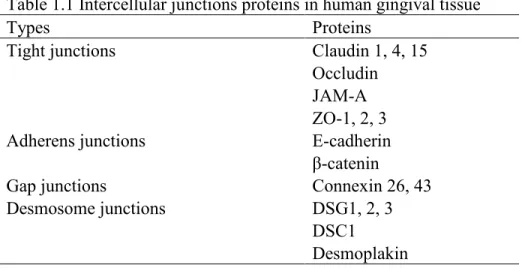
Table 1.1 Intercellular junctions proteins in human gingival tissue……..………20
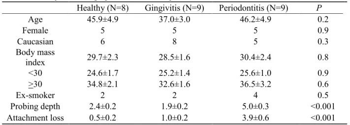
Table 2.1 Subjects demographics and clinical parameters…..………..38
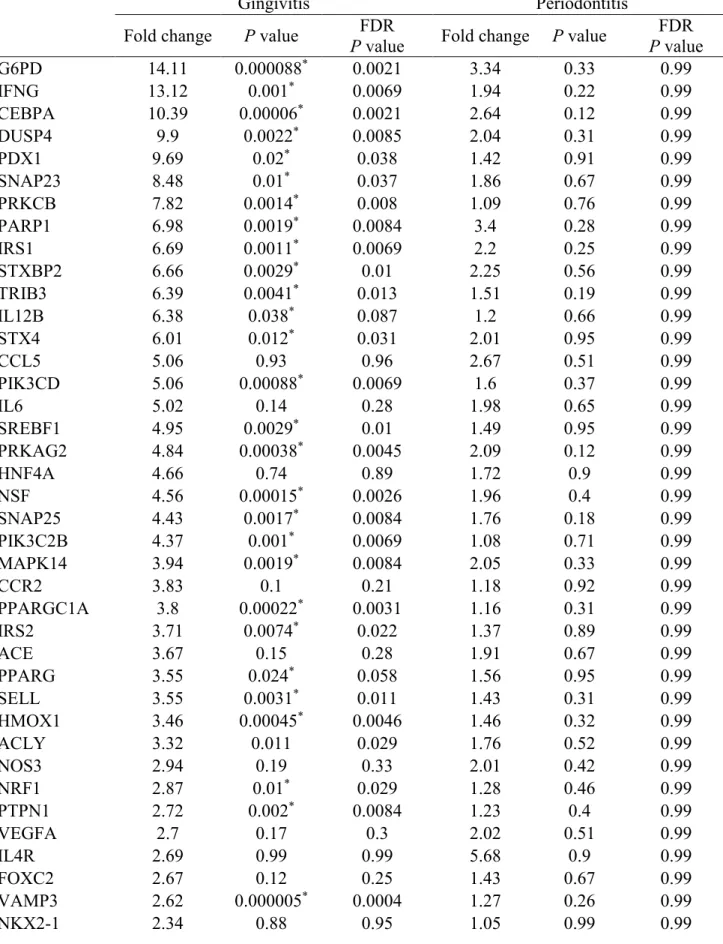
Table 2.2 Gene regulation in gingivitis and periodontitis subjects………39
Table 2.3 Fold Regulation Adjusted by Age for 4 Genes ……….41
Table 4.1 Oligonucleotides used for bisulfite specific PCR and pyrosequencing …….…...……80
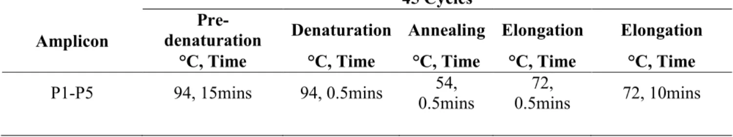
Table 4.2 Bisulfite specific PCR conditions for CpG containing amplicon..………80
Table 4.3 Subjects demographics and clinical parameters……….………….………..81
xi
LIST OF FIGURES
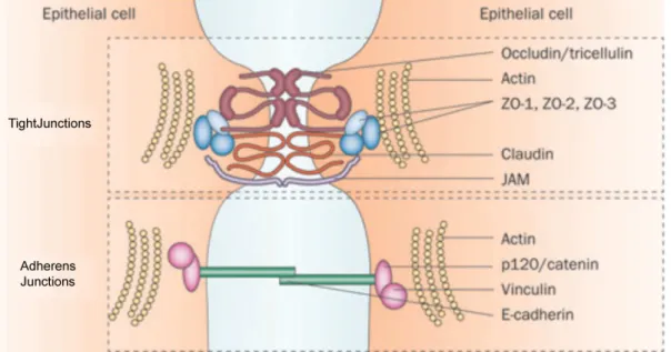
Figure 1.1 Major components of tight junctions and adherens junctions………..21
Figure 1.2 Gap junctions components………...……22
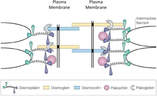
Figure 1.3 Molecular structures of desmosome junctions………...……..23
Figure 2.1 Volcano plot presenting regulation of insulin response genes comparing diseased to healthy subjects……….………..……..42
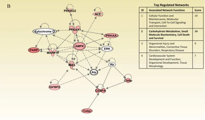
Figure 2.2 Ingenuity pathway analysis comparing diseased to healthy subjects ………….….…44
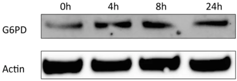
Figure 2.3 G6PD protein increases by E. coli LPS stimulation samples…………...………46
Figure 2.4 Fold change ratio of obesity/non-obese diseased subjects..……….…...…...47
Figure 3.1 Prior exposures of AGEs enhances gingival fibroblast response to LPS…….…...….58
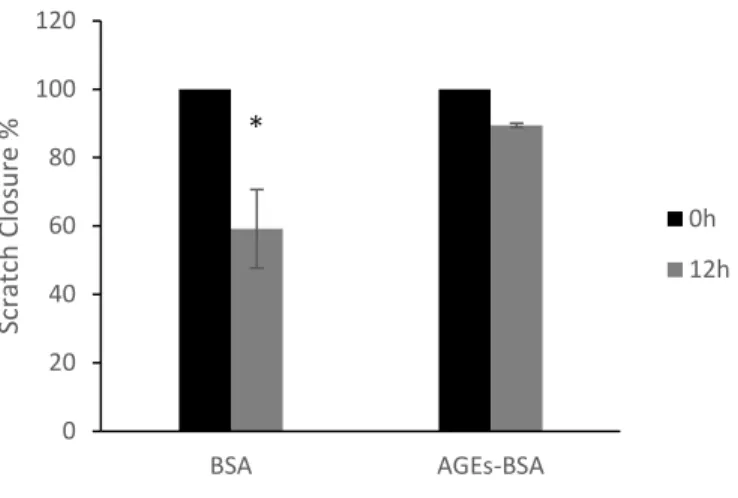
Figure 3.2 AGEs inhibit gingival fibroblast migration.………..…...…....60
Figure 4.1 Genomic sequence of PKP2 promoter fragment……….………...…..83
Figure 4.2 Epithelial PKP2 mRNA transcription is decreased in gingival biopsies from chronic periodontitis ………..……..…...84
Figure 4.3 PKP2 protein is degraded by viable P. gingivalis. ………...…………...85
Figure 4.4 Degradation of PKP2 protein depends on cysteine proteases. ………...…….89
Figure 4.5 PKP2 DNA methylation is associated with periodontal disease. ………...….91
Figure 4.6 PKP2 and DSG1 co-localize under high calcium condition……….………...93
xii
LIST OF ABBREVIATIONS AGEs Advanced glycation end products
AL Attachment loss
aPKC Atypical protein kinase C
ARVC Arrhythmogenic right ventricular cardiomyopathy
BMI Body mass index
BOP Bleeding on probing
BSA Bovine serum albumin
C.r Campylobacter rectus
cAMP Cyclic adenosine monophosphate
CBD Catenin-binding domain
Cdt Cytolethal distending toxin
CEBPA CCAAT/Enhancer Binding Protein (C/EBP), Alpha
cGMP Cyclic guanosine monophosphate
Cox-2 Cyclooxygenase-2
CRB Crumbs-PALS1-PATJ complex
Cx Connexin
DLG Discs large
DMEM Dulbecco’s modified eagle medium
DNM1L Dynamin-like protein
DP Desmoplakin
DSC Desmocollin
xiii
DUSP4 Dual specificity phosphatase 4
ECM Extra cellular matrix
EGFR Epidermal growth factor receptor
FBS Fetus bovine serum
FDR False discover rate
FGF2 Fibroblast growth factor 2
G6PD Glucose-6-phosphate dehydrogenase
GAPDH Glyceraldehyde 3-phosphate dehydrogenase GCSF Granulocyte-colony stimulating factor GEC Gingival epithelial cells
GI Gastrointestinal tract
GLUT Glucose transporters
hBD human β-defensins family
HGEPs Human gingival cells
HGF Human gingival fibroblast
HRP Horseradish peroxidase
IF Intermediate filaments
IFNG Interferon gamma
IL Interleukin
IL6R Interleukin 6 receptor
IP3 Inositol trisphosphate 3
IPA Ingenuity pathway analysis
xiv
IRS Insulin receptor substrate JAM Junctional adhesion molecule
JMD Juxtamembrane domain
JNK c-Jun N-terminal Kinase
LEF1 Lymphoid enhancer-binding factor 1
LGL Lethal giant larvae
LIF Leukemia inhibitor factor
LPS Lipopolysaccharides
MAPK Mitogen-activated protein kinase MCP-1 Monocyte chemoattractant protein-1 MDCK Madin-Darby canine kidney epithelial cells
MMPs Matrix metalloproteases
MOI Multiplicity of infection MUPP1 Multi-PDZ domain protein 1
NADPH Nicotinamide adenine dinucleotide phosphate
NF-κB Nuclear factor kappa-light-chain-enhancer of activated B cells
NLRP/NALP NLR family pyrin domain containing
NLRs Nod like receptors
Nod Nucleotide-binding oligomerization domain
P.g Porphyromonas gingivalis
PALS 1 Proteins associated with Lin Seven 1 PAR Partitioning-defective protein
xv
PBS Phosphate buffered saline
PCR Polymerase chain reaction
PD Probing depth
PG Plakoglobin
PGE2 Prostaglandin E2
PKP Plakophilin
PRRs Pattern recognition receptors
PTGS2 Prostaglandin-endoperoxide synthase 2
PV Pemphigus vulgaris
RAGE Receptor for advanced glycation end products
rRNA Ribosome RNA
STX4 Syntaxin 4
STXBP2 Syntaxin binding protein 2 T2DM Type 2 diabetes mellitus
TCF T cell factor
TER Transepithelial electrical resistance
TLRs Toll-like receptors
TNF-α Tumor necrosis factor alpha
Wnt Wingless-type MMTV integration site family member
1
CHPATER 1: INTRODUCTION Overview of gingiva
Periodontal disease is a biofilm-initiated inflammatory condition that leads to destructions of periodontal tissue, which includes gingiva, cementum, periodontal ligament, and alveolar bone. In the oral cavity where over 700 species of microorganisms reside, the outermost layer of periodontal tissue-gingiva is exposed to constant microbial challenges (Hajishengallis, 2015). When dysbiosis occurs, the gingival-biofilm interface becomes disrupted which eventually leads to clinical symptoms of periodontal disease.
Based on distinct anatomic characteristics, three types of gingiva are defined: marginal gingiva, attached gingiva, and interdental gingiva (Newman et al., 2012). Marginal gingiva is the free end of the gingiva, encompassing the teeth like a collar. It forms the soft tissue wall for the (Newman et al., 2012). Attached gingiva is the soft tissue area between marginal gingiva and alveolar mucosa. Cementum on the cervical side of the root and ridge of alveolar bones can be found underneath the attached gingiva. Attached gingiva also provides support for the gingival tissues to withstand mechanical forces during mastication, speaking, and tooth brushing
(Newman et al., 2012). Interdental gingiva refers to the gingiva that fills the interdental space (Bath-Balogh et al., 2006). The shape and size of interdental gingiva depend on the contact area of the adjacent teeth. Losing interdental gingiva may cause esthetic failure, food retention, and potentially cause periodontal disease.
2
has four layers of cornieum, granulosum, spinosum and basal (Newman et al., 2012). There are three distinctive types of gingival epithelium, namely sulcular epithelium, junctional epithelium, and oral epithelium (Hatakeyama et al., 2006). Sulcular epithelium lines the gingival sulcus and interacts with bacteria intensively. Junctional epithelium, being the base of the gingival sulcus, is responsible for gingival attachment to the teeth. During the progression of periodontal disease, junctional epithelium detaches from the tooth surface, which results in clinical attachment loss (Bosshardt and Lang, 2005). Both sulcular and junctional epithelium are non-keratinized epithelium. The keratinized oral epithelium is the outer layer of the gingival epithelium ranging from marginal to attached gingiva. The main function of gingival epithelium is to form a barrier to defend against mechanical, chemical and microbial intruders.
Gingival epithelial functions
Gingival epithelium provides an early line of defense against bacteria in the gingival sulcus during periodontal disease-associated bacterial disturbance (Schroeder and Listgarten, 1997). Most surfaces and cavities of human organs are lined with epithelium to act as physical and chemical barriers (Peterson and Artis, 2014). Therefore, much of our knowledge of epithelial functions is learned from organs like skin, intestine, urinary bladder, and eyes. An intact
epithelial sheet relies on the three characteristics: 1) epithelial cell-to-cell adhesion; 2) epithelial homeostasis: 3) mucosal inflammation.
Epithelial cell-to-cell adhesion
cell-to-3
cell adhesions. These junctional structures not only contribute to sealing the epithelium but also exert a variety of biological functions. Four types of intercellular junctions are commonly identified as tight junctions, adherens junctions, gap junctions, and desmosome junctions (Alberts, 2008). In Figure 1.1-1.3, major components of intercellular junctions are presented (Durham and Garrett, 2009; Green and Gaudry, 2000; Neunlist et al., 2013)
Tight junctions
Tight junctions reside on the most apical side of the epithelium and have complex protein compositions. To date, at least 40 tight junctions-associated proteins have been discovered, but only three major transmembrane proteins are well characterized, which are claudin, occludin, and junctional adhesion molecule (JAM) (Gonzalez-Mariscal et al., 2003; Schneeberger and Lynch, 2004; Yamazaki et al., 2008).
Occludin is the first identified transmembrane component of tight junctions with a
transmembrane domain that passes through the membrane four times (Furuse et al., 1993). Within occludin, there are one N-terminus and one C-terminus facing inside and two extracellular loop domains facing outside of the cell. The long C-terminus functions as the docking sites for tight junctions-associated proteins (eg. ZO-1, ZO-2, ZO-3) (Furuse et al., 1994; Haskins et al., 1998; Itoh et al., 1999). Zonula occludens (ZO) are critical scaffolding proteins that bridge the transmembrane proteins and actin cytoskeletons. ZO also has a dual role in regulating cell growth (Bauer et al., 2010). Overexpression of occludin appears to increase numbers of tight junctions strands, with accordingly increased transepithelial electrical resistance (TER) activity, which is an indicator for tight junctions activities (McCarthy et al., 1996).
4
not essential for tight junctions (Saitou et al., 2000; Schulzke et al., 2005). One piece of evidence is that occludin deficient mice are still viable and maintain an intact barrier in the GI tract as well as the urinary bladder. These findings have suggested that there might be alternative proteins for occludin (Schulzke et al., 2005). In endothelial cells from non-neuronal tissues, occludin is apparently missing even though the tight junctions complex persists (Hirase et al., 1997).
Additionally, occludin-deficient embryonic stem cells bear proper tight junctions (Saitou et al., 1998). These findings have suggested that occludin mainly associates with tight junctions in a regulatory fashion (Nusrat et al., 2005).
Claudin has a structure similar to that of occludin with a tetraspan transmembrane domain and a relatively shorter N-terminus and C-terminus facing cytosolically (Furuse et al., 1998; Morita et al., 1999). The C-terminus of claudin not only binds to ZO-1, ZO-2, ZO-3, but also binds to multi-PDZ domain protein 1 (MUPP1) and PALS-1 associated tight junctions protein (PATJ) (Jeansonne et al., 2003; Poliak et al., 2002). Since its discovery in 1998, claudin has become an integral transmembrane component of tight junctions (Furuse et al., 1998). When claudin is introduced into fibroblast that originally lacks tight junctions, fibroblast has newly formed tight junctions-like strands and starts aggregating. Claudin-1 deficient mice demonstrated a more severe phenotype than occludin deficient mice with much compromised epidermal
barriers (Furuse et al., 2002). The interaction between ZO and claudin is also critical for tight junctions. A lack of ZO-1 significantly delays the assembly of tight junctions, suggesting that actin cytoskeletons are required to maintain the characteristics of tight junctions (McNeil et al., 2006).
5
containing PDZ binding motifs that bind to ZO-1 and MUPP-1 (Hamazaki et al., 2002). JAM participates in regulating epithelial barriers. One study showed that antibodies against JAM had no effect on changing the confluency of epithelial cells monolayers, but inhibited the
reconstruction of tight junctions after being disrupted (Liu et al., 2000). Although inhibiting JAM in endothelial cells improves transendothelial migration of neutrophils, dampening JAM does not affect the transepithelial migration of neutrophils (Zemans et al., 2009).
Adherens junctions
Adherent junctions are positioned basally to tight junctions in epithelium and their molecular structures are relatively simple, comprising epithelial cadherin (E-cadherin), p120 catenin, β-catenin, and α-catenin (van Roy and Berx, 2008). E-cadherin belongs to a family of cadherin proteins that are calcium-dependent. E-cadherin consists of an extracellular domain, a transmembrane domain, and a cytoplasmic domain. The extracellular domain forms bonds between adjacent cells, while the cytoplasmic tail binds to p120 catenin and β-catenin to exert biological functions (van Roy and Berx, 2008). Similar to other transmembrane proteins, E-cadherin is assembled in the Golgi apparatus and later translocated to the plasma membrane. To engage in the dynamics of adherens junctions, membrane-bound E-cadherin experiences active turnovers through endocytosis and degradation (Bryant and Stow, 2004).
p120 catenin binds to the juxtamembrane domain (JMD) of E-cadherin, which contributes to stabilizing adherens junctions complex at the cell membrane (Reynolds et al., 1992). No
6
regulating E-cadherin expression (Ireton et al., 2002). Additionally, the interactions between p120 and microtubules assist in shifting roles of adherens junctions from cell adhesiveness to cell movement (Yanagisawa et al., 2004). β-catenin binds to E-cadherin as well, but only through the catenin-binding domain (CBD). Unlike p120, β-catenin is required for E-cadherin trafficking to the plasma membrane. The association between β-catenin and E-cadherin is essential for the transportation of cadherin from endoplasmic reticulum to the membrane, and protects E-cadherin from being degraded. More importantly, β-catenin interacts with α-catenin, which subsequently couples with actin filaments and some other cytoskeleton-associated molecules, including vinculin, afadin, formin, and ZO-1 (Kobielak and Fuchs, 2004; Watabe-Uchida et al., 1998). Theses interactions provide more pieces of evidence to prove that adherens junctions are closely associated with cell movement.
In addition to its function as part of adherens junctions, β-catenin belongs to Wnt signaling, which controls many basic physiological processes like cell proliferation, cell polarity, and tissue homeostasis (Eastman and Grosschedl, 1999). When Wnt receptors are activated, β-catenin proteins start accumulating in the cytosol and translocating to the nucleus. Inside the nucleus, β-catenin acts as a co-activator for the transcription factors family TCF/LEF, which controls the transcription of various target genes (Eastman and Grosschedl, 1999). In summary, adherens junctions play important roles in cell adhesion, cell motility, and linking the intercellular junctions to the intracellular signaling pathways.
7
channel-like structure of gap junctions. This organization creates a space (~2-4 nm) between the cells and this where the name of “Gap” is introduced (Maeda et al., 2009). Based on whether six connexins are from the same or different connexons, there are hetermeric or homomeric
connexon channels. Connexins have distinct expression patterns among the cell or tissue types. For instance, Cx26 is highly expressed in the skin, cochlea, liver, and placenta, while Cx46 and Cx50 are predominantly found in retina cells. Moreover, Cx43 is considered to be the major gap junctions protein in cardiomyocytes (Evans and Martin, 2002). In gingival epithelium, Cx43 is abundantly expressed, while Cx32 is clearly missing and Cx26 is moderately expressed (Ye et al., 2000).
Due to an unique architecture, gap junctions allow diffusions of ions (K+ and Ca2+), second messengers (cAMP, cGMP, and IP3), and small metabolites between cells, all of which
participate in relaying electrical and biochemical signals across the body (Mese et al., 2007). In addition to the cellular communications, connexins bind with ZO-1, calmodulin, and tubulin to participate in cell movement (Barker et al., 2002; Giepmans et al., 2001; Peracchia et al., 2000). Cx43 is a functional target for Wnt signaling, which links gap junctions with adherens junctions (van der Heyden et al., 1998).
Desmosome junctions
Desmosome junctions are widely expressed at all layer of epithelium except for stratum cornieum (Getsios et al., 2004). Two cadherin proteins-desmoglein (DSG, 1-4) and desmocollin (DSC, 1-3) constitute for the extracellular components of desmosomes; three intracellular
8
DSG and DSC are both glycoproteins that form strong adhesiveness between the opposing plasma membranes through their extracellular domains, in a calcium dependent manner. The interactions can be either homophilic (DSC-DSC, DSG-DSG) or heterophilic (DSG-DSC) (Chitaev and Troyanovsky, 1997; Syed et al., 2002). The role of DSG is widely studied in an autoimmune disease-pemphigus vulgaris (PV), which is caused by aberrant productions of immunoglobulins against DSG1 and DSG3 (Hashimoto et al., 1995). The phenotype of PV is characterized by basal keratinocytes dissociation in the epidermis and oral mucosa due to loss of desmosomes. Targeted disruptions of Dsg3 in mice led to the dissociations of basal cells in the oral mucosa, which mimicked the phenotype of PV (Koch et al., 1997). Functions of DSG1 are further described in bullous impetigo, in which exfoliative toxins secreted by Staphylococcus aureus specifically degrade DSG1, causing keratinocyte dissociations (Amagai et al., 2000). In addition to DSG, DSC1 is also a target in PV, as Dsc1 deficient mice display loss of cell-to-cell adhesions in the epidermis with defective barrier functions (Chidgey et al., 2001).
Plakoglobin is also called γ-catenin due to its homology to the catenin members. Similar to β-catenin, plakoglobin localizes with E-cadherin as well, but with much stronger binding affinities (Chitaev et al., 1996). Mice lacking plakoglobin have defective heart and skins as embryos (Bierkamp et al., 1996). Plakophilins are also important desmosome molecules, which will be discussed in a separate session. Desmoplakin is the most abundant desmosome molecule (Mueller and Franke, 1983). However, it does not bind to cadherins directly but rather bind to plakoglobin and plakophilin through its N-terminus binding sites. The C-terminus of
9
desmsomes in the epidermis, desmoplakin-null keratinocytes have fewer desmosomes formed in vitro (Mueller and Franke, 1983). Actin organizations and sealing properties during the epithelial sheet formation are largely potentiated in Desmoplakin-null keratinoctyes (Mueller and Franke, 1983). Therefore, ample evidence has suggested that desmoplakin is required for the assembly of functional desmosomes. In the absence of desmosome molecules, mice experience early embryo stalls due to malfunctions of the epidermis, neuroepithelium, heart and blood vessels, suggesting critical roles of desmosomes.
In summary, intercellular junctions play crucial roles in cell-to-cell adhesion. Additionally, these molecules exert certain fundamental physiological activities. Much evidence has suggested that all four junctional complexes tend to work together by interacting with each other directly and indirectly.
Epithelial homeostasis
Epithelial homeostasis represents the basic biological features of epithelial cells,
10
plasma membranes. In the lateral domain, junctional molecules are predominantly expressed. At the basal domain, integrin and hemidesmosome are uniquely expressed to adhere epithelium to ECM. The underlying control over the apical basalateral polarity has been widely studied with three complexes being identified: 1) PAR3-PAR6-aPKC complex; 2) Crumbs-PALS1-PATJ complex (CRB); 3) LGL-Scribble-DLG complex (Martin-Belmonte et al., 2001). Planar cell polarity refers to the polarization of cells within the plane of the cell sheet. When cells divide perpendicularly to the plane, it gives rise to stratified epithelium; when cells divide parallel to the plane, it expands the epithelial sheet (Rodriguez-Boulan and Macara, 2014).
Cell migration is an integral part of epithelial homeostasis, playing a central role in tissue remodeling and disease development. Upon receiving biological or mechanical cues, cells spread on the attachment sheet and move to a designated area. Migratory activities largely rely on the cytoskeleton structures like actin, microtubules, and intermediate filaments. Both cell-to-cell and cell-to-ECM adhesions affect cell migration (Cuvelier et al., 2007; Vicente-Manzanares et al., 2005). In a large-scale gene search, Simpson KJ et al has reported three major signaling nodes (β-catenin, β-integrin, actin) are involved in migration (Simpson et al., 2008). However, enhanced cell adhesion does not necessarily increase migration. In the same study, genes that accelerated migration may also inhibit adhesion, suggesting a negative correlation. In this end, cell adhesion and cell migration interact in two directions.
Cell death refers to the cessation of cellular functions, which counteracts to cell proliferation. Highly proliferative epithelial cells require active cell death to maintain the epithelial
11
epithelial cell death (Brozovic et al., 2006; Kang et al., 2012; Stathopoulou et al., 2009; Tsuda et al., 2010).
Mucosal inflammation
Although the role of epithelium in defending microorganisms has been empirically focused on its physical barrier capability, its immune aspects are also of great significance. Epithelial cells display several pattern recognition receptors (PRRs) that are either membrane-bound or cytoplasmic distributed (Peterson and Artis, 2014). Upon activations, PRRs promote a pyramid of signaling events to produce pro-inflammatory cytokines and chemokines. Pro-inflammatory cytokines like interleukin-1 beta (IL-1β), interleukin-6 (IL-6), and tumor necrosis factor alpha (TNF-α) promote inflammations to restrict infections. However, a prolonged inflammatory status brings damages to the tissue. Epithelial cells secrete a wide range of chemokines via PRRs to attract leukocytes to the local lesions to diminish bacterial or viral intrusions. Another immune feature for epithelial cells lies is to produce anti-microbial peptides, the expression of which also depends on PRRs (Nakatsuji and Gallo, 2012).
For epithelial cells, two major PRRs are toll-like receptors (TLRs) and nucleotide-binding oligomerization domain (Nod) like receptors (NLRs) (Janeway and Medzhitov, 2002; Meylan et al., 2006). TLRs interact with bacterial components such as lipopolysaccharides (LPS),
lipoproteins, flagellin, unmethylated CpG DNA, and etc. NLRs are cytoplasmic receptors that recognize bacterial peptidoglycan, bacterial toxins, and other pathogenic structures of
12
gingivalis produce high levels of IL-1β. Gingival epithelial cells that are challenged by A. actinomycetemcomitans induce high levels of IL-8 (Stathopoulou et al., 2010). F. nucleatum increase more IL-6 productions in gingival epithelial cells than commensal bacteria-S. gordini (Hasegawa et al., 2007). Via TLR2, P.gingivalis induce monocyte chemoattractant protein-1 (MCP-1) and IL-8, both of which are chemokines that promote neutrophils to migrate and exert phagocytosis at the site of infections (Hasegawa et al., 2007).
The best-characterized antimicrobial peptides in gingival epithelial cells are members of human β-defensins family (hBD). hBD-1 is constitutively expressed in gingival epithelial cells, while hBD-2 and hBD-3 are stimulation-dependent (Gursoy and Kononen, 2012;
Krisanaprakornkit et al., 1998). hBDs are differentially regulated in gingival epithelial cells by oral bacteria regardless whether they are pathogenic or commensal (Gursoy and Kononen, 2012). Gingival epithelial cells markedly produce hBD in response to the challenge of F. nucleatum and TLR2 and NLRP2 are involved in this regulation (Ji et al., 2009).
Overview of Plakophilins
Plakophilins are a group of armadillo family members (PKP1, PKP2, PKP3, and p0071) that are widely expressed in many tissues and organs. PKPs are located in both cytosol and nucleus. In addition to being structural scaffolds to increase desmosome plaques, PKPs regulate the intracellular signalings as well (Bass-Zubek 2009).
PKP1 is the first identified and smallest PKPs member (Heid et al., 1994). PKP1 expression is increased from basal to granular layers in stratified epithelium, suggesting its roles in
keratinocyte differentiation (McMillan et al., 2003). PKP1 assists in forming desmosome
13
Bornslaeger 2001). Patients with autosomal recessive defects of PKP1 gene have ectodermal dysplasia and skin fragility. In these patients, deregulated desmosome sizes and numbers can be found in the epidermis (McMillan JR, 2003).
PKP2 is the best-characterized member of PKPs. Rare mutations of PKP2 gene in humans cause arrhythmogenic right ventricular cardiomyopathy (ARVC), which is an inherited heart disease that is characterized by a sudden myocardium breakdown (van Tintelen et al., 2006). PKP2 works in building cadherin-PKP-DP links in the desmosome assembly. A loss of PKP2 results in failure of DP to accumulate at the cell border. Protein kinanse C alpha (PKCα) is the major functional molecule in this process. PKP2 recruits PKCα to form a complex with DP. Subsequently, PKCα phosphorylates DP, modulates its interactions with IF, and guides DP assembly into the cell border (Bass-Zubek et al., 2008). Since PKP2 is the major PKPs member in cardiomyocytes, impaired PKP2 cannot be compensated by other PKPs, resulting in dampened desmosomes. Grossmann KS et al found that PKP2 null mice exhibited an embryonic lethality due to defects of heart morphogenesis, indicating PKP2 also plays important roles in cardiac development (Grossmann et al., 2004).
Functions of PKP2 are not limited to being structural molecule of desmosomes. PKP2 is highly expressed in the basal layer of epithelium, an area that is particularly active for cell renewal. Studies have shown that PKP2 induces the phosphorylation of epidermal growth factor receptor (EGFR) to promote cell proliferation (Arimoto et al., 2014; Kazlauskas, 2014).
14
associates with RNA polymerase III in the nucleus (Mertens et al., 2001). Since pol III regulates
tRNA, rRNA 5S, and some small RNAs, PKP2 may present post-translational activities.
Unlike PKP1 and 2, PKP3 is not expressed in the nucleus and mice lacking PKP3 present relatively mild phenotypes (Sklyarova et al., 2008). Current evidence associates PKP3 with cancer development. PKP3 binds to dynamin-like protein (DNM1L) to act in vesicle trafficking within the secretory pathway in non-small cell lung carcinomas (Furukawa et al., 2005). By associating with RNA-binding proteins, PKP3 is involved in protein translation and RNA metabolisms responding to environmental stressors (Hofmann et al., 2006). p0071 is referred as PKP4 but its structure is more closed to p120 catenin. p0071 has been shown to regulate Rho signaling during cytokinesis (Wolf et al., 2006). In one study that shows the staining pattern for all PKP members in primary oropharyngeal tumors, only p0071 is associated with tumor grade, clinical parameters, and patient survivals (Papagerakis et al., 2003).
It is noteworthy that even though PKPs have been described in various diseases, the role of PKP2 in periodontal disease remains unknown.
Alterations of junctional molecules in periodontal disease
Gingival epithelium is constantly exposed to microorganisms in the form of gingival plaque. To date, various studies have addressed the disturbance of junctional molecules under
periodontitis and the underlying mechanisms.
15
and JAM-A (Choi et al., 2014; Ye et al., 2014). Fujita et al challenged rats with LPS in the
gingival sulcus for 8 weeks and found that claudin-1 was significantly decreased in the junctional epithelium (Fujita et al., 2012). Numerous studies have also shown that E-cadherin is
significantly reduced in periodontitis gingival biopsies than healthy controls (Arun et al., 2010; Loo et al., 2010; Nagarakanti et al., 2007). Connexin 26 and 43 are reduced in the pathological lining epithelium of periodontal pockets, indicating the alterations of gap junctions (Ye et al., 2000). In addition, DSG1 and DSC1 mRNA are down-regulated in periodontitis gingival biopsies, indicating the alteration of desmosomes (Abe et al., 2011).
While tissue data provides convincing evidence linking dampened junctional molecules to periodontitis, results from in vitro studies do not always have the same trend. Epithelial bound E-cadherin can be degraded by P. gingivalis, which is mainly due to proteases produced by
gingipain (Katz et al., 2000; Katz et al., 2002). However, cytolethal distending toxin (Cdt) from A. actinomycetemcomitans increases cytoplasmic distribution of E-cadherin and overall
expression of β-catenin in both gingival tissue explants and gingival epithelial cells (Damek-Poprawa et al., 2013). Proteases from P. gingivalis degrade occludin protein in epithelial cells, accompanied by decreased TER activities (Katz et al., 2002). Collectively, bacterial products regulate junctional molecules productions, leading to altered epithelial homeostasis.
Gingival fibroblast functions
Human gingival fibroblast (HGF) is major resident cell in gingival connective tissue. One major function of HGF is to exert immune responses. This function is mainly through activations of PRRs. In addition to TLRs and NLRs, another important PRR in HGF is receptor for
pro-16
inflammatory cytokines (eg. IL-1 and IL-6), chemokines (eg. IL-8), and enzymes (eg. Cox-2, MMPs) under the circumstances of periodontitis or bacterial challenges (Morton and Dongari-Bagtzoglou, 2001; Takada et al., 1991; Tamura et al., 1992; Yu et al., 2012). Although immune cells like monocytes and macrophages take a major responsibility in producing cytokines, both of them develop LPS tolerance quickly (Mages et al., 2007). LPS tolerance allows pathogens to escape from immune surveillance during sustained bacterial exposures and elongated
inflammatory conditions. HGFs appear not to display LPS tolerance, which indicates that HGF may compensate for limiting bacterial effects when monocytes and macrophages are
antisensitized (Ara et al., 2009).
HGF is also responsible for synthesis and degradation of ECM, a group of molecules that constitutes for the backbones of the gingival architecture. HGF produces ECM and at the same time secrets matrix metalloproteases (MMPs) to degrade ECM. Therefore, HGF is the key cell in ECM remodeling within gingival tissues. Collagen is the major ECM in gingiva and the
predominant type is type I collagen (Newman et al., 2012). HGF secrets a large amount of fibronectin as well, which is a glycoprotein that mediates the adhesions between fibroblast and ECM and fibroblast and surrounding cells. Over 20 MMPs have been identified in gingiva and many of them have distinct roles in development of periodontal disease: MMP1 and MMP8 are collagenases that degrade type I, II, III, and X collagens; MMP2 and MMP9 degrade gelatins and elastin; MMP3 and MMP10 degrade pro-collagen and fibronectin (Reynolds et al., 1994).
17
acquired from both mice and human gingiva demonstrates phenotypes of induced pluripotent stem (iPS) cells (Egusa et al., 2010; Wada et al., 2011).
Alterations of gingival fibroblast in periodontal disease
A good understanding about alterations of gingival fibroblast in periodontal disease sheds lights on the pathogenesis of periodontal disease. Whole or components of pathogens
unanimously enhance productions of pro-inflammatory cytokines in HGF (Agarwal et al., 1995; Imatani et al., 2001). Topical additions of IL-1β and TNF-α result in increased transcription of IL-6, IL-11, and leukemia inhibitor factor (LIF) in HGF (Palmqvist et al., 2008). Both LPS and IL-1 induce prostaglandin E2 (PGE2) in HGF through increasing cyclooxygenase 2 (Cox-2) (Okamura et al., 1999; Richards and Rutherford, 1988). PGE2 is a well-known vasodilator engaging in increasing the vascular permeability and promoting inflammation that leads to periodontium destructions (Offenbacher et al., 1993).
HGF presents altered ECM regulation during periodontal disease as well. Periodontitis connective tissues have decreased collagen fibers and MMPs when compared to healthy tissues (Seguier et al., 2001). Periodontal pathogens might be likely reasons since they make proteases that potently degrade ECM (Bachrach et al., 2004; Zhou and Windsor, 2006).
Not many studies investigate the wound healing potential of gingival fibroblast under healthy or diseased conditions. Smoking has long been regarded as a risk factor for periodontitis and impaired wound healing. Nicotine inhibits HGF migration via Rac signaling pathways (Fang Y 2005).
18
All human cells need to obtain energy in order to act and survive. Fat, protein, and glucose constitute for the major energy sources. Glucose uptake and utilization are predominantly affected by insulin. Glucose transporters (GLUT 1-4) and related insulin signaling molecules modulate glucose uptake. Inside cells, insulin regulates glucose utilization through a series of enzymatic reactions. Improper glucose metabolism leads to severe consequences and the most profound consequence is diabetes.
Diabetes is a hyperglycemic condition that affects approximately 30 millions of Americans (Centers for Disease and Prevention, 2014). As the most common form of diabetes, type 2 diabetes is characterized by insulin resistance, which refers to failures of tissues to react to a normal level of insulin. When unnecessary large amounts of glucose deposit in the tissues and organs, physiological damages occur with consequential complications. Periodontal disease is one of the diabetic complications and these two diseases share a two-way relationship (Mealey et al., 2006). Systemic inflammation is considered to be the common links between insulin
resistance and periodontal disease. Major pro-inflammatory mediators are elevated in gingivitis and periodontitis subjects systematically (Page, 1991). A lot of these mediators are casually linked to altered glucose utilization and metabolism (Shoelson et al., 2006). TNF-α, IL-6, IL-1β, and increased level of oxidative stress are all known to cause insulin resistance by disrupting c-Jun N-terminal Kinase (JNK) and insulin signaling pathways (Evans et al., 2005; Hotamisligil et al., 1993; Lee et al., 2003).
19
20
Table 1.1 Intercellular junctions proteins in human gingival tissue
Types Proteins
Tight junctions Claudin 1, 4, 15 Occludin JAM-A ZO-1, 2, 3 Adherens junctions E-cadherin
β-catenin
Gap junctions Connexin 26, 43
Desmosome junctions DSG1, 2, 3 DSC1
21
22 Figure 1.2 Gap junction components.
23
Figure 1.3 Molecular structures of desmosome junctions. Adapted from (Green and Gaudry, 2000)
Plasma Membrane
24
CHAPTER 2: INSULIN RESPONSE GENE EXPRESSION IN PERIODONTAL DISEASE-MODULATION OF OBESITY
Ning Yu†#, Silvana P. Barros*†, Shaoping Zhang†, Kevin L. Moss†, Sherrill T. Phillips†, Steven Offenbacher*†
* Department of Periodontology, School of Dentistry, University of North Carolina at Chapel hill, Chapel Hill, NC, USA
† Center for Oral and Systemic Diseases, University of North Carolina at Chapel Hill, Chapel Hill, NC, USA
# Oral Biology Ph.D Curriculum, School of Dentistry, University of North Carolina at Chapel Hill, Chapel Hill, NC. USA
Abstract
Background: Bacterial infections are known to alter glucose metabolism within tissues via mechanisms of inflammation. We conducted this study to examine whether insulin response genes are differentially expressed in gingival tissues comparing samples from experimental gingivitis and periodontitis subjects to those from healthy individuals.
Methods: Total RNA was extracted from gingival biopsies from twenty-six participants
25
quantification, we confirmed the up-regulation of the key gene using lipopolysaccharides (LPS) stimulated primary gingival epithelial cells by western-blot.
Results: The mRNA expression patterns of genes that are associated with insulin response and glucose metabolism are markedly different in experimental gingivitis subjects compared to healthy controls. 32 genes are up-regulated significantly by at least 2-fold adjusted for FDR (P<0.05). Periodontitis subjects show similar but attenuated changes in gene expression patterns and no genes meet the significance criteria. IPA demonstrates significant activation of
carbohydrate metabolism network in experimental gingivitis, but not in periodontitis. Glucose-6-phosphate dehydrogenase (G6PD) protein increases in response to LPS stimulation in primary
26 Introduction
Periodontal disease is a biofilm-initiated inflammatory condition that affects the tooth supporting apparatus. Two major periodontal diseases, gingivitis and periodontitis respectively affect 75% and 47% of the adult population in the United States (Albandar et al., 1999; Eke et al., 2012). The pathogenesis of periodontal disease has been described in many aspects, but little is known regarding periodontal tissue glucose metabolism and insulin responsiveness.
27
block lipoprotein lipase production, which may as a result increase glucose utilization (Kim et al., 2001).
Gingivitis and periodontitis are both know to be associated with dramatic elevations in inflammatory cytokines and chemokines (Page, 1991). However, whether gingival inflammation, comparing acute and chronic gingival inflammation, affects glucose metabolism and insulin response gene expression locally is still unknown. Therefore, we conducted this study to begin to explore whether genes associated with glucose metabolism and insulin response were
28
Material and Methods Study Participant Selection and Gingival Tissue Biopsies
Twenty-six participants, aged 18 to 64, provided informed consent and were recruited in the dental clinics from School of Dentistry at University of North Carolina at Chapel Hill. All procedures were approved by Institutional Review Board (IRB) of the University of North Carolina at Chapel Hill. Participants were sequentially enrolled with either periodontally healthy, experimental gingivitis or chronic periodontitis with the following exclusion criteria: 1. No use of antibiotics within one month before the screening examinations; 2. No signs of systemic diseases with any oral manifestation at the time of the enrollment; 3. No self-reporting treatment for systemic diseases three months prior to the collections of gingival biopsies. Besides obtaining demographic information and periodontal clinical parameters, examiners also recorded all
participants’ weight and height to compute Body Mass Index (BMI).
8 periodontally healthy participants were either healthy volunteers or patients who were undergoing crown-lengthening procedures and they exhibited ≤4mm probing depth (PD), no signs of bleeding on probing (BOP). 9 chronic periodontitis patients representing chronic
inflammation were enrolled with exhibitions of ≥5mm of PD, presence of BOP and radiographic demonstration of bone loss. 9 experimental gingivitis patients representing acute inflammation were acquired from a 3-week stent-induced biofilm overgrowth procedure (Offenbacher et al., 2009). A gingival biopsy was obtained from each subject at the site of marginal gingiva.
29
RNA Isolation and Assays for Insulin Response Genes Expression Profile
Total RNA was isolated from gingival biopsies using RNeasy Mini kit (Qiagen). 600ng of the RNA were reverse transcribed to cDNA using RT2 First Strand Kits (Qiagen). To examine genes of interests, we used a diabetes-pathway focused RT2 Profiler PCR array (PAHS-023) in a 7500 Sequence Detection System (ABI Prism, Applied Biosystems). The mRNA expression levels were normalized using Glyceraldehyde 3-phosphate dehydrogenase (GAPDH) as the housekeeping gene. We performed the delta-delta Ct method to calculate fold-changes of regulated genes. The cut-off Ct value for qPCR was set at 35 and all genes within this threshold were included in the analysis. The identified genes were then mapped to known molecular networks in IPA to obtain statistical significance estimates for pathways and networks ranked by Fisher Exact scores.
Cell culture, LPS stimulation, and Western blot
30 Statistical Analysis
31 Results Participants
The demographic information and periodontal parameters of the participants are shown in Table 2.1. No statistically significant differences in age, gender, race, BMI, and smoking history were found among participants with either healthy or diseased gingival tissues. As expected, clinical parameters such as probing depth and attachment loss exhibited higher readings in chronic periodontitis subjects compared to healthy and gingivitis participants.
Gene Expression Patterns in Gingivitis and Periodontitis Subjects.
We compared gene expression patterns between healthy, experimental gingivitis, and periodontitis gingival biopsies. The volcano plot showing the log2 (fold change) plotted versus the –log10 (p value) for the 76 genes that were within the Ct value threshold can be seen in Figure 2.1. Genes that were up-regulated by at least 2-fold are illustrated in Figure 2.1 as red dots with the clustering of genes within the upper right quadrant for statistically significant genes. Only a few genes were down-regulated as indicated as green dots. In Figure 2.1A which
compared gingivitis to healthy subjects, 45 genes were up-regulated >2-fold and 32 of them were at p<0.05 after FDR adjusted t-test. In this comparison, 5 genes were down-regulated >2-fold but none of them reached statistical significance (p>0.05). In Figure 2.1B, periodontitis group
showed similar directional but much attenuated changes in gene expression with 15 genes being up-regulated >2-fold. None of them reached statistical significance by FDR. Only 1 gene was found down-regulated >2-fold but did not reach statistical significance as well (p>0.05). In Table 2.2, we reported the complete expression data for each individual gene in gingivitis and
32
chose the top 4 regulated genes in overall comparisons (G6PD, IFNG, CEBPA, DUSP4) and analyzed their fold regulation differences adjusted by age. In Table 2.3, age-adjusted fold change for these genes did not differ much from the unadjusted fold change. When comparing gingivitis to healthy subjects, the age-adjusted fold change differences were even slightly increased. For instance, adjusted G6PD fold regulation was increased by 11%. This gene expression profile suggests that genes that are associated with glucose metabolism and insulin responsiveness are more highly expressed in acute gingival inflammation, but not significantly up-regulated in chronic inflammation.
Molecular Networks Analysis in Gingivitis and Periodontitis Subjects
33
G6PD protein expression increases in response to LPS stimulation
To confirm that the elevated mRNA expression data seen in experimental gingivitis likely is mirrored by increased protein expression we created an in vitro acute inflammatory by
stimulating HGEPs with E. coli LPS and detected G6PD protein expression by Western blot. G6PD was selected as a marker because it was the top up-regulate mRNA species identified (Table 2.2). As demonstrated in Figure 2.3, G6PD protein expression increased in response to LPS stimulation starting from 4 hours of stimulation, suggesting that inflammatory stimuli likely induce G6PD on both mRNA and protein levels.
The Modulating Roles of Obesity
To address the potential role of obesity in modulating insulin responses, we divided all three groups of subjects to obese and non-obese subgroups based on BMI. After normalizing against non-obese healthy subjects, fold change ratio of obese/non-obese diseases subjects are presented in Figure 2.4. It appears that majority of the fold change differences between obese and non-obese subjects are within 2-fold differences. In gingivitis group, more genes are
34 Discussion
This study suggests that during the induction of experimental gingivitis, the acute gingival inflammatory response is locally activating glucose metabolism and insulin response genes, but in chronic inflammation there has been a re-establishment of a relative balance in insulin/glucose metabolism within the local gingival tissues.
The experimental gingivitis group has an overall markedly up-regulated gene profile for these glucose/insulin response genes. As can be seen in Table 2, the top 4 up-regulated genes in gingivitis biopsies are G6PD, IFNG, CEBPA, and DUSP4. Both tissue mRNA data and gingival epithelial cells protein data suggest that gingival inflammation affects G6PD expression (Table 2.2 and Figure 2.3). The augmentation in G6PD is likely a response to attenuate oxidative stress by generating NADPH, rather than participate in the pentose or carbohydrate driven pathway for 5-carbon sugars. The increased INFG mRNA is a clear sign for inductions of gingival
inflammation since interferon-γ is an activator for both innate and adaptive immunity. Wong N et al demonstrated that restricting interferon-γ in mice improved their glucose metabolism (Wong et al., 2011). There was also a significant increase in CEBPA, which encodes for CCAAT
35
However, a previous study found that DUSP4 protein expression in gingival epithelial cells was decreased by periodontal pathogens, which was in contrast with our data showing a significant up-regulation of DUSPS mRNA in gingivitis (Hasegawa et al., 2007). One limitation of our study is that we did not have adequate biopsy samples to confirm protein expression levels, as our emphasis was on transcriptomic analysis.
In addition, the expression of key insulin signaling molecules, namely IRS1, IRS2, SLC2A4 (GLUT4), STX4, STXBP2 and PIK3R1 are all enhanced in experimental gingivitis. This suggests that locally acute inflammation up-regulates glucose metabolism by increasing local cellular uptake and glucose utilization. The IPA analysis as shown in Figure 2.2 confirms that pathway associated with carbohydrate metabolism is activated by acute inflammation. However, chronic inflammation associated with periodontitis was not as capable as acute inflammation in
regulating glucose/insulin response genes as shown in Table 2.2. This suggests that in chronic inflammation there is some adaptation in the localized tissue response to create a more
homeostatic glycolic local environment. Importantly, we had no naturally occurring gingivitis samples to compare in this study. However, we might expect them to have similar
insulin/glucose profiles as periodontitis, if the naturally occurring gingivitis represents a chronic inflammatory processe. The finding of altered metabolism during acute induction might suggest that acute episodes of increased inflammation superimposed upon an existing chronic gingivitis or periodontitis may also be associated with enhanced insulin resistance and glucose utilization of the local periodontal tissues, but that will require experimental confirmation.
In gingival tissues, glucose metabolism may be important not only for tissue metabolism but also affect microenvironment that supports the growth of periodontal pathogens. We
36
the extension of the supragingival adherent plaque that contains saccharolytic bacteria into the subgingival pocket provides an important “feeder bed” to provide nutrients of intermediate metabolism including lactate and formate to support the metabolism of the assacharolytic organisms.
We conducted a secondary analysis to consider the potential effects of obesity, because obesity affects both insulin responsiveness and periodontal disease (Franchini et al., 2011; Pischon et al., 2007). In Figure 2.4, certain genes are differentially regulated by obesity and this trend is more apparent in gingivitis group. More subjects are needed to study the modulatory roles of obesity. Studies have shown that obesity interacts with local gingival inflammation. Mizutani K et al showed that obese mice had selective insulin resistance in gingival tissues (Mizutani et al., 2014). In gingiva, specific miRNAs are differentially expressed in obese
periodontitis compared to non-obese periodontitis subjects, suggesting that obesity may regulate genes post-transcriptionally (Perri et al., 2012).
37
limiting the generalizability of the findings, and we did not design the study to enable protein analysis. Although this is a pilot study with relatively few subjects (26 subjects), it demonstrates the utility of pathway analysis, which allows each molecular observation to contribute to the overall significance of networks.
38
Table 2.1 Subjects demographics and clinical parameters
Healthy (N=8) Gingivitis (N=9) Periodontitis (N=9) P
Age 45.9±4.9 37.0±3.0 46.2±4.9 0.2
Female 5 5 5 0.9
Caucasian 6 8 5 0.3
Body mass
index 29.7±2.3 28.5±1.6 30.4±2.4 0.8
<30 24.6±1.7 25.2±1.4 25.6±1.0 0.9
>30 34.8±2.1 32.6±1.6 36.5±3.2 0.6
Ex-smoker 2 2 4 0.5
39
Table 2.2 Gene regulation in gingivitis and periodontitis subjects
Gingivitis Periodontitis
Fold change P value
FDR
P value Fold change P value
FDR P value
G6PD 14.11 0.000088* 0.0021 3.34 0.33 0.99
IFNG 13.12 0.001* 0.0069 1.94 0.22 0.99
CEBPA 10.39 0.00006* 0.0021 2.64 0.12 0.99
DUSP4 9.9 0.0022* 0.0085 2.04 0.31 0.99
PDX1 9.69 0.02* 0.038 1.42 0.91 0.99
SNAP23 8.48 0.01* 0.037 1.86 0.67 0.99
PRKCB 7.82 0.0014* 0.008 1.09 0.76 0.99
PARP1 6.98 0.0019* 0.0084 3.4 0.28 0.99
IRS1 6.69 0.0011* 0.0069 2.2 0.25 0.99
STXBP2 6.66 0.0029* 0.01 2.25 0.56 0.99
TRIB3 6.39 0.0041* 0.013 1.51 0.19 0.99
IL12B 6.38 0.038* 0.087 1.2 0.66 0.99
STX4 6.01 0.012* 0.031 2.01 0.95 0.99
CCL5 5.06 0.93 0.96 2.67 0.51 0.99
PIK3CD 5.06 0.00088* 0.0069 1.6 0.37 0.99
IL6 5.02 0.14 0.28 1.98 0.65 0.99
SREBF1 4.95 0.0029* 0.01 1.49 0.95 0.99
PRKAG2 4.84 0.00038* 0.0045 2.09 0.12 0.99
HNF4A 4.66 0.74 0.89 1.72 0.9 0.99
NSF 4.56 0.00015* 0.0026 1.96 0.4 0.99
SNAP25 4.43 0.0017* 0.0084 1.76 0.18 0.99
PIK3C2B 4.37 0.001* 0.0069 1.08 0.71 0.99
MAPK14 3.94 0.0019* 0.0084 2.05 0.33 0.99
CCR2 3.83 0.1 0.21 1.18 0.92 0.99
PPARGC1A 3.8 0.00022* 0.0031 1.16 0.31 0.99
IRS2 3.71 0.0074* 0.022 1.37 0.89 0.99
ACE 3.67 0.15 0.28 1.91 0.67 0.99
PPARG 3.55 0.024* 0.058 1.56 0.95 0.99
SELL 3.55 0.0031* 0.011 1.43 0.31 0.99
HMOX1 3.46 0.00045* 0.0046 1.46 0.32 0.99
ACLY 3.32 0.011 0.029 1.76 0.52 0.99
NOS3 2.94 0.19 0.33 2.01 0.42 0.99
NRF1 2.87 0.01* 0.029 1.28 0.46 0.99
PTPN1 2.72 0.002* 0.0084 1.23 0.4 0.99
VEGFA 2.7 0.17 0.3 2.02 0.51 0.99
IL4R 2.69 0.99 0.99 5.68 0.9 0.99
FOXC2 2.67 0.12 0.25 1.43 0.67 0.99
VAMP3 2.62 0.000005* 0.0004 1.27 0.26 0.99
40
ME1 2.33 0.01* 0.018 0.96 0.71 0.99
ICAM1 2.15 0.32 0.49 0.9 0.47 0.99
FOXP3 2.1 0.83 0.95 1.4 0.91 0.99
CEACAM1 1.85 0.21 0.34 0.91 0.48 0.99
INS 1.65 0.91 0.95 0.91 0.7 0.99
GSK3B 1.65 0.0019* 0.0084 0.89 0.47 0.99
PPARGC1B 1.61 0.59 0.79 1 0.61 0.99
SLC2A4 1.54 0.48 0.68 0.73 0.71 0.99
FBP1 1.41 0.9 0.95 2.02 0.36 0.99
IGFBP5 1.38 0.66 0.82 1.73 0.54 0.99
MAPK8 1.31 0.18 0.31 0.97 0.86 0.99
CTLA4 1.31 0.57 0.78 1.26 0.82 0.99
TNFRSF1A 1.29 0.99 0.99 0.88 0.57 0.99
PRKAA1 1.27 0.66 0.82 2.27 0.69 0.99
VAPA 1.26 0.08 0.18 0.93 0.4 0.99
PPARA 1.25 0.61 0.81 1.04 0.91 0.99
RAB4A 1.21 0.65 0.82 1.81 0.8 0.99
AGT 1.05 0.16 0.29 0.65 0.21 0.99
INPPL1 0.96 0.85 0.95 0.85 0.59 0.99
INSR 0.87 0.85 0.95 1.08 0.64 0.99
IKBKB 0.87 0.42 0.62 1.28 0.52 0.99
FOXG1 0.87 0.29 0.46 0.47 0.16 0.99
ENPP1 0.78 0.91 0.95 1.05 0.87 0.99
NFKB1 0.68 0.09 0.2 0.81 0.34 0.99
TNF 0.65 0.16 0.29 0.87 0.83 0.99
IL10 0.62 0.75 0.89 1.17 0.9 0.99
PIK3R1 0.6 0.47 0.67 0.59 0.35 0.99
IDE 0.5 0.39 0.59 1.75 0.73 0.99
AKT2 0.45 0.5 0.69 0.97 0.89 0.99
CD28 0.42 0.2 0.33 0.54 0.22 0.99
STXBP1 0.28 0.77 0.9 0.77 0.98 0.99
TGFB1 0.23 0.68 0.83 0.66 0.99 0.99
41
Table 2.3 Fold Regulation Adjusted by Age for 4 Genes
G6PD IFNG CEBPA DUSP4
Crude FR
Adjusted FR
Crude FR
Adjusted FR
Crude FR
Adjusted FR
Crude FR
Adjusted FR
Healthy 2.54 2.34 2.42 2.10 1.70 1.53 1.69 1.65
43
45
46
48
49
CHAPTER 3: EFFECTS OF ADVANCED GLYCATION END PRODUCTS ON GINGIVAL FIBROBLAST MIGRATION AND GENE REGULATION
Ning Yu1,3, Silvana P. Barros2,3, Steven Offenbacher2,3
Curriculum of Oral Biology1, Department of Periodontology2, Center for Oral and Systemic Diseases3,
School of Dentistry, University of North Carolina at Chapel Hill, Chapel Hill, NC, USA
Abstract
Background: The presence and accumulation of advanced glycation end products (AGEs) in gingiva has been evident in chronic periodontitis patients, which is even more drastic among those who have diabetes. We conducted this study to examine whether transcription of key regulatory genes in gingival wound healing and migration were affected by AGEs in gingival fibroblast.
Methods: AGEs were prepared in vitro using bovine serum albumin (BSA) as a carrier. Human gingival fibroblast (HGF-1) was pretreated with AGEs-BSA (1ug/ml) for 24h, then stimulated with AGEs-BSA (1ug/ml) with E.coli LPS (0, 100, 1000ng/ml) for 24h. Total RNA was
50
fibroblast growth factor 2 (FGF2) genes. Scratch assay and transwell assay were conducted to examine the migration activity of gingival fibroblast affected by AGEs. HGF-1 that was pretreated and stimulated with BSA served as controls.
Results: Compared to BSA treated cells, HGF-1 that was pretreated and stimulated by AGEs-BSA and LPS showed significantly increased transcription of PTGS2, MMP1 genes, but not FGF2 gene. AGEs-BSA treated cells displayed a delayed migration as proved by the scratch assay and transwell assay.
51 Introduction
Periodontal disease and diabetes mellitus especially type 2 diabetes (T2DM) share a bidirectional relationship (Mealey et al., 2006). Periodontal disease adds a burden of systemic inflammation by inducing pro-inflammatory cytokines including TNF-a, IL-6, and IL-1β (Page, 1991). These cytokines are known to activate insulin resistance, which occurs at the beginning state for T2DM and a later state for T1DM. Periodontal disease may lead to severe consequences in the diabetic population. In a study conducted among Pima Indians with T2DM, severe
periodontitis significantly increased the mortality rate from diabetic nephropathy and ischemic heart disease (Saremi et al., 2005). Treating periodontal disease, on the other hand, improves the glycemic controls for diabetic patients (Teeuw et al., 2010). From the perspective of how
diabetes affects periodontal disease, many mechanisms have been proposed and one possible mechanism is the influence of advanced glycation end products (AGEs).
AGEs are a heterogeneous group of non-enzymatically formed proteins from having a sustained contact with sugars. For diabetic subjects, their tissues and organs have higher AGEs accumulations than those of non-diabetic subjects. Receptor for AGEs (RAGE) has been found to interact with AGEs and triggers the downstream NF-κB pathway (Schmidt et al., 1994). As a result, inflammation and reactive oxygen species are elevated, which subsequently change the physiology of the tissues. Accumulations of AGEs are evident in gingival tissues. AGEs
aggregate more potently in the gingival epithelium and the underlying connective tissues among diabetic subjects compared to their non-diabetic counterparts (Abbass et al., 2012; Zizzi et al., 2013).
52
2001). Chang PC et al found that anti-AGEs agents facilitated the palatal gingival wound healing by reducing inflammation (Chang et al., 2014). During the gingival wound healing process, connective tissue has a unique role in tissue remodeling. Gingival fibroblast, as the most
abundant resident cells in the connective tissue, is a reservoir for inflammatory cytokines (eg. IL-1β, TNFα, PGE2), effector molecules (eg. MMPs, TIMPs), and growth factors (eg. FGF, CTNF). All these molecules actively participate in the tissue remodeling process. Fu Y et al found that AGEs induced MMP1 productions in human gingival fibroblast through activating NF-κB pathway, which further indicated the importance of AGEs on periodontal disease. However, whether AGEs affect anabolic growth factors and whether AGEs inhibit gingival fibroblast migration in vitro remain unknown.
The objective of this study is to investigate effects of AGEs on gingival fibroblast migration and gene regulation. We hypothesized that AGEs inhibited gingival fibroblast migration.
53
Material and Methods Preparation of AGEs-BSA compounds
AGEs-BSA compounds were prepared by incubating BSA at a 1mM concentration with 1M glucose, 1.5mM phenylmethanesulfonyl fluoride, and 1% penicillin-streptomycin in phosphate-buffered saline (PBS) solutions (ph=7.4), at 37°C for 10 weeks. Same compositions without glucose were incubated simultaneously as BSA controls. Both AGEs-BSA and BSA solutions were dialyzed against PBS using 20K MWCO dialysis cassettes, at 4°C overnight. We then used Bradford protein assay (Sigma) to quantify for the concentration of BSA in both groups. BSA and AGEs-BSA were diluted to a concentration of 1mg/ml and stored at -80°C until use. Cell culture and in vitro stimulations
HGF-1 was purchased from ATCC (CRL-2014) and maintained in DMEM low glucose medium (Gibco) supplemented with 10% fetal bovine serum (FBS, Gibco) and 1% penicillin-streptomycin (Invitrogen). We seeded HGF-1 in 6 well plates at a density of 2x10⁵ and changed the medium every other day until AGEs challenges began. AGEs-BSA or BSA compounds were added to HGF-1 culture for 24h. The medium was then switched to E. coli LPS (0, 100ng/ml, 1000ng/ml, strain 55 O:H) containing medium in the continuing presence of AGEs-BSA or BSA for 24h. At the end, cells were harvested for RNA isolation.
RNA isolation and qRT-PCR
Total RNA was isolated from HGF-1 cells using RNeasy Mini kit. RNA quality and quantity were measured by nanodrop. 500ng total RNA was reverse transcribed to cDNA. Gene
54
ABI expression (Hs00153133_m1, Hs00899658_m1, Hs00266645_m1). The mRNA expression levels were normalized using 18s ribosome RNA as the housekeeping gene.
Migration Assay
In the transwell migration assay, HGF-1 cells were undergoing BSA or AGEs-BSA treatment in the absence of serum overnight. 2x10⁴ cells were added to the upper well of the transwell inserts (Millipore, 6.5mm2). Inserts were then immersed into medium containing BSA or AGEs-BSA (10, 100, 1000ng/ml) in the presence of 30% FBS (chemo-attractant) in a 24 well plate. Following 12h of migration, inserts were removed from the culture plate and fixed with 4% paraformaldehyde at room temperature for 10mins, followed by 1XPBS washing 3 times. We applied a cotton swap to clean the remaining cells apically. The last step was to stain the nuclei using DAPI solution (D1306, Invitrogen) for 5mins, avoiding lights. Nuclei staining were read under a fluorescent microscopy (EVOS). For each insert, 5 fields (four corners and a central spot from a cross) were chosen to count for the average cell numbers. In the scratch assay, 2x10⁵ HGF-1 cells were seeded into 6-well plates until they formed a monolayer. After overnight serum starvation, cell monolayers were scraped with a sterile 200ul pipette tip in the midline. We immediately aspirated the culture medium, rinsed the cells with 1XPBS 3 times, and added medium containing either BSA or AGEs-BSA. Both medium were supplemented with 30% FBS. After 12h of migration, cell images were captured from an inverted phase contrast microcopy. Two scratch wounds were made for each treatment and two independent experiments were performed. Image J software was used to quantify for the wound closure percentage. Statistical Analysis